non-coding flavivirus rna displays rnai suppressor activity in insect and mammalian cells
TRANSCRIPT

Noncoding Flavivirus RNA Displays RNA Interference SuppressorActivity in Insect and Mammalian Cells
Esther Schnettler,a,b,c Mark G. Sterken,a Jason Y. Leung,a Stefan W. Metz,a Corinne Geertsema,a Rob W. Goldbach,a Just M. Vlak,a
Alain Kohl,b,c Alexander A. Khromykh,d and Gorben P. Pijlmana
Laboratory of Virology, Wageningen University, Wageningen, Netherlandsa; MRC–University of Glasgow Centre for Virus Research 8, Glasgow, Scotland, United Kingdomb;The Roslin Institute and Royal (Dick) School of Veterinary Studies, University of Edinburgh, Easter Bush, Scotland, United Kingdomc; and Australian Infectious DiseaseResearch Centre, School of Chemistry and Molecular Biosciences, The University of Queensland, St. Lucia, Brisbane, Australiad
West Nile virus (WNV) and dengue virus (DENV) are highly pathogenic, mosquito-borne flaviviruses (family Flaviviridae) thatcause severe disease and death in humans. WNV and DENV actively replicate in mosquitoes and human hosts and thus encoun-ter different host immune responses. RNA interference (RNAi) is the predominant antiviral response against invading RNA vi-ruses in insects and plants. As a countermeasure, plant and insect RNA viruses encode RNA silencing suppressor (RSS) proteinsto block the generation/activity of small interfering RNA (siRNA). Enhanced flavivirus replication in mosquitoes depleted forRNAi factors suggests an important biological role for RNAi in restricting virus replication, but it has remained unclear whetheror not flaviviruses counteract RNAi via expression of an RSS. First, we established that flaviviral RNA replication suppressedsiRNA-induced gene silencing in WNV and DENV replicon-expressing cells. Next, we showed that none of the WNV encodedproteins displayed RSS activity in mammalian and insect cells and in plants by using robust RNAi suppressor assays. In contrast,we found that the 3=-untranslated region-derived RNA molecule known as subgenomic flavivirus RNA (sfRNA) efficiently sup-pressed siRNA- and miRNA-induced RNAi pathways in both mammalian and insect cells. We also showed that WNV sfRNA in-hibits in vitro cleavage of double-stranded RNA by Dicer. The results of the present study suggest a novel role for sfRNA, i.e., as anucleic acid-based regulator of RNAi pathways, a strategy that may be conserved among flaviviruses.
Arthropod-borne (arbo)viruses form a diverse group of medi-cally important viruses, many of which are emerging patho-
gens (72). Arboviruses take a unique position within the viro-sphere by displaying active replication in both vertebrate hosts(humans, other mammals, and birds) and invertebrate vectors(e.g., mosquitoes, ticks, midges, and sandflies) (41). Upon infec-tion of mosquitoes, the virus persistently replicates in multipletissues of the insect, and high virus titers accumulate in the salivaryglands by the end of this so-called extrinsic incubation time, typ-ically 1 to 2 weeks. Since the virus needs the vector for successfulinfection of the vertebrate hosts to complete the transmission cy-cle, evolutionary pressure has likely caused the virus to be onlymildly or nonpathogenic to the arthropod host (17). Nonetheless,to perpetuate the viral life cycle involving invertebrate vector andvertebrate host, the virus must be sufficiently equipped to crossthe initial midgut infection barrier in the vector and must be ableto disseminate within the arthropod to eventually accumulateprogeny virus in the salivary glands.
Mosquitoes and other arthropods have an array of mecha-nisms to fight microbial and viral infections. RNA-induced genesilencing or RNA interference (RNAi) is the key component of theinsect innate immune system to limit a diverse range of RNAviruses, including flaviviruses (6, 55), while the Toll, IMD, andJAK-STAT pathways also contribute to control flavivirus infec-tion in mosquitoes (17). As a countermeasure, insect-specific vi-ruses have been demonstrated to suppress this antiviral RNAi re-sponse by producing specialized proteins that obstruct one ormore of the key RNAi components. Well-studied examples are theFlock House virus (FHV) B2 viral RNA silencing suppressor (RSS)(37), the Cricket paralysis virus (CrPV) 1A RSS (44, 71), and therelated Drosophila C virus (DCV) 1A protein (65). For FHV, it wasshown that suppression of antiviral RNAi by expression of a viral
RSS was crucial for establishing efficient viral replication and vi-rion production (31).
Until now, no viral RSS have conclusively been identified inarboviral genomes. For dengue virus (DENV), it was suggestedfrom preliminary experiments that none of the DENV matureviral proteins could suppress RNAi (32). More recently, it washypothesized that arboviruses may not even need a RSS, since theysubject themselves to antiviral RNAi and replicate at lower levelsto establish persistent infection of the insect host (64). While itremains to be seen whether this is true for all arboviruses withoutexception, persistent virus infection of the arthropod—the hall-mark of arbovirus replication— does not inevitably mean that thevirus does not display RSS activity. For example, the insect-spe-cific viruses DCV, CrPV, and FHV all encode strong RSSs in theirgenomes (44, 65, 71), and yet all of these viruses can persistentlyinfect their insect hosts. Conversely, it can be hypothesized thatpersistently infecting arboviruses may encode RSSs, for example, toallow sufficient levels of viral replication in vector insects, especially inview of the high potency of the antiviral RNAi response (6).
West Nile virus (WNV) and DENV are highly pathogenic,mosquito-borne viruses (family Flaviviridae, genus Flavivirus)that cause severe disease and death in humans (34). Flavivirusvirions contain a single copy of the viral genome, which encodes a
Received 4 May 2012 Accepted 24 September 2012
Published ahead of print 3 October 2012
Address correspondence to Gorben P. Pijlman, [email protected], orAlexander A. Khromykh, [email protected].
Copyright © 2012, American Society for Microbiology. All Rights Reserved.
doi:10.1128/JVI.01104-12
13486 jvi.asm.org Journal of Virology p. 13486–13500 December 2012 Volume 86 Number 24
Dow
nloa
ded
from
http
s://j
ourn
als.
asm
.org
/jour
nal/j
vi o
n 25
Nov
embe
r 20
21 b
y 12
2.14
8.13
4.13
.
polyprotein that is proteolytically cleaved into seven nonstruc-tural proteins (NS1, NS2A, NS2B, NS3, NS4A, NS4B, and NS5)and three structural proteins (C, prM, and E). The positive-stranded RNA genome of �11 kb is flanked by 5= and 3= untrans-lated regions (UTRs), which play essential roles in initiation oftranslation and RNA replication (40). Interestingly, recent reportshave demonstrated that many, most likely all, flaviviruses producea second, subgenomic flavivirus RNA (sfRNA), which accumu-lates to high levels in a diverse range of infected mammalian andinsect cells (33, 52, 56). sfRNA is a positive-sense, noncoding RNAmolecule representing the last 525 nucleotides (nt) (in the case ofWNV) of the viral genomic RNA that is generated as the result ofincomplete degradation of genomic RNA by the cellular exoribo-nuclease XRN1 in cytoplasmic processing bodies (PBs) (19, 52,60). PBs are cytoplasmic granules functional in mRNA degrada-tion, mRNA surveillance and translational repression. The RNAimachinery is also concentrated in PBs (16). It has been shown thatsfRNA production is required for viral pathogenicity in a mam-malian animal model, although its exact mode of action remainsto be uncovered (52). Recent studies suggest that sfRNA plays arole in inhibiting the alpha/beta interferon (IFN-�/�) response inmice (59) and also may serve as the main source of WNV-encodedmiRNA in mosquito cells (25).
According to the arbovirus definition, flaviviruses actively rep-licate in both the invertebrate vector and the vertebrate host andthus encounter an array of different host immune responses, in-cluding RNAi during virus replication in mosquitoes (17). By de-pleting crucial insect RNAi factors it became clear that RNAi isvery efficient in limiting flavivirus replication in insects (10, 55).However, it remains to be uncovered how exactly flavivirusesevade and/or suppress the antiviral RNAi response.
Here we have studied the interaction between WNV and RNAiin different model systems. We have screened the various WNVproducts, including viral proteins and sfRNA, for activity as RNAisuppressor in different RNAi suppressor assays. We show thatsfRNA but not viral proteins display RNAi suppressor activity ininsect and mammalian cells. Finally, we provide evidence usingDENV that this novel role of sfRNA is characteristic for flavivi-ruses.
MATERIALS AND METHODSPlasmids. The mammalian expression plasmids pGL3 and pRL-CMV(Promega), the short hairpin-encoding plasmid Firefly luciferase (pShh1-Ff1) (48), or the nonspecific pEF-MBP, pEF-MBP-NS3, were as previ-ously described (57). The 3=UTRs of WNV and DENV and MBP-HDVrwere cloned into the mammalian expression vector pcDNA-DEST40 (In-vitrogen) downstream of the cytomegalovirus (CMV) promoter usingGateway technology. The pAdvantage construct expressing adenovirusVA RNAI/II was purchased from Promega. The miRNA-based sensorconstructs pCMV-Fluc-miRNA30-AP, pCMV-Fluc-random, pCMV-hsa-miRNA30, and pCMV-hsa-miRNA21 have been described previ-ously (77). The selectable WNV and DENV replicons have been previ-ously described (35, 53, 54). The insect expression vectors encodingMBP-HDVr, the DENV 3=UTR, and the WNV 3=UTR either fused toMBP or alone were cloned behind the baculovirus OpIE2 promoter intothe pIB-GW (58) vector using Gateway technology. The expression vec-tors encoding either MBP or Cymbidium ringspot virus (CymRSV) P19have been described previously, as well as the inducible insect miRNA-based sensor constructs, pMT-FF-3=UTR, pMT-pri-dme-miRNA1, andpMT-pri-dme-miRNA-12 (58) and the inducible Firefly and Renilla lu-ciferase constructs (65), all expressed via the Drosophila metallothioneingene promoter. Insect expression vectors encoding short hairpin RNA
were constructed by annealing previously described DNA oligonucleo-tides (69) either against Firefly luciferase or enhanced green fluorescentprotein (eGFP) and cloning these as KpnI-XbaI fragments in pMT-B (In-vitrogen) behind the Drosophila metallothionein gene promoter. For theRNA silencing experiments in U4.4 cells, Firefly and Renilla luciferaseconstructs were used, which have previously been described (46) and areexpressed via the OpIE2 or AcIE1 promoters, respectively.
To check the functionality of the WNV miRNA sensor constructs(described in reference 25 and now recloned in pGL3 [Promega] forexpression in mammalian cells), pSuper plasmids expressing smallinterfering RNA (siRNA) from the H1 RNA polymerase III promoterwere constructed (8). The following complementary oligonucleotideswith (partial) restriction sites (indicated in boldface) and complemen-tary regions (underlined) were designed according to methods pub-lished online by OligoEngine and were cloned in the BglII and HindIII sites ofpSuper: pSUPER-A1A2-F, GATCCCCGCTCTGCACAACCAGCCACACGGCACTTCAAGAGAGTGCCGTGTGGCTGGTTGTGCAGAGCTTTTTA; pSUPER-A1A2-R, AGCTTAAAAAGCTCTGCACAACCAGCCACACGGCACTCTCTTGAAGTGCCGTGTGGCTGGTTGTGCAGAGCGGG; pSUPER-C1C2-F, GATCCCCGACAATGGTGGCTGGTGGTGCGAGAATTCAAGAGATTCTCGCACCACCAGCCACCATTGTCTTTTTA; and pSUPER-C1C2-R, AGCTTAAAAAGACAATGGTGGCTGGTGGTGCGAGAATCTCTTGAATTCTCGCACCACCAGCCACCATTGTCGGG. Plasmid pSuper-A1A2 expresses a short hairpin consisting ofA1A2, a short loop region and the reverse complement sequence A1A2rc.Upon expression in mammalian cells, this plasmid expresses siRNA thatwill silence transcripts containing both A1A2 or A1A2rc sequences. Sim-ilarly, pSuper-C1C2 targets C1C2 or C1C2rc containing transcripts.
Cell culture and transfection. African green monkey kidney Vero andbaby hamster kidney BHK-21 cells were grown as a monolayer in Dul-becco modified Eagle medium (Gibco/BRL) supplemented with 10% fetalcalf serum (FCS; Gibco), streptomycin (100 �g/ml), and penicillin (100U/ml) at 37°C and 5% CO2. To reach a confluence of 60 to 70% at the timeof transfection, the cells were trypsinized 24 h pretransfection and seededin 24-well plates at a concentration of 1.1 � 105 cells per well. Transfec-tions were performed using JetPei according to the manufacturer’s in-structions. To create stable mammalian replicon cell lines, Vero cells weretransfected with capped, in vitro-transcribed replicon RNA, using Lipo-fectamine2000 (Invitrogen) according to the manufacturer’s instructions.At 24 h posttransfection (hpt), transfected cells were selected with puro-mycin (WNV replicon [36, 53]) or G418 (DENV replicon [54]). For thetransient RNA silencing suppressor assays using short hairpin constructsas inducer molecules, (replicon) cells were transfected with luciferase ex-pression plasmids, i.e., 100 ng of Firefly luciferase (pGL3; Promega), 2 ngof Renilla luciferase (pRL-CMV; Promega), and 4 ng of short hairpinencoding plasmids, either nonspecific or Firefly luciferase specific(pShh1-Ff1 [48]). In the case of transient expression of the RNA silencingsuppressor, the cells were also cotransfected with the corresponding ex-pression plasmid (MBP, MBP-NS3, MBP-NS3mutant, MBP-HDVr,WNV sfRNA, or DENV sfRNA).
The miRNA-based sensor experiments were performed in a similarway as previously described for the shRNA-induced silencing experi-ments. Briefly, cells were seeded in 24-well plates and cotransfected with200 ng of sensor construct (pCMV-FF-miRNA30-AP or pCMV-FF-ran-dom [77]), 2 ng of Renilla luciferase plasmid (pRL-CMV; Promega), and10 ng of pri-miRNA expression plasmid vectors expressing either humanpri-hsa-miRNA21 or pri-hsa-miRNA30 (77). All cells were lysed at 48 hpt,and the luciferase activity was determined using a dual luciferase reporterassay (15).
Drosophila melanogaster Schneider-2 (S2) cells were grown in Sch-neider’s medium (Invitrogen) supplemented with 10% heat-inactivatedFCS (Gibco) at 28°C. The cells were seeded 24 h pretransfection in a96-well plate at a concentration of 5 � 104 cells per well. Transfectionswere performed using Cellfectin II (Invitrogen) according to the manu-facturer’s instructions. For the shRNA suppressor assays, the S2 cells were
RNAi Suppressor Activity of Noncoding Flavivirus RNA
December 2012 Volume 86 Number 24 jvi.asm.org 13487
Dow
nloa
ded
from
http
s://j
ourn
als.
asm
.org
/jour
nal/j
vi o
n 25
Nov
embe
r 20
21 b
y 12
2.14
8.13
4.13
.

cotransfected with luciferase-expressing plasmids (15 ng of pMT-Flucand 6 ng of pMT-Rluc) and 50 ng of short hairpin-expressing vectors,either Fluc specific or nonspecific. The cells were also cotransfected with100 ng of the RNA silencing suppressor expressing plasmid (MBP, RiceHoja Blanca virus [RHBV] NS3, or WNV sfRNA). For the miRNA-basedsuppressor assays, the S2 cells were cotransfected with 100 ng (unlessstated otherwise) of RNA silencing suppressor expression plasmid (MBP,Carnation Italian ringspot virus [CIRV] P19, or WNV sfRNA), 12.5 ng ofpMT-Fluc-3=UTR, 3 ng of pMT-Rluc, and 2.5 ng of pMT-miRNA (eitherpri-dme-miRNA1 or pri-dme-miRNA12). Expression of the inducibleconstructs was induced 48 hpt by 5 �M CuSO4 and assayed 24 h postin-duction.
Aedes albopictus U4.4 and Aedes pseudoscutellaris Ap61 mosquito cellswere grown in Leibovitz L-15 medium (Invitrogen) supplemented with10% heatinactivated FCS (Gibco), 2% tryptose phosphate broth (Sigma),and 1% nonessential amino acids (Invitrogen) at 28°C. Then, 1.5 � 105
cells were seeded per well in a 24-well plate. Transfections were performed24 h after seeding using Lipofectamine 2000 (Invitrogen) according to themanufacturer’s instructions. For the double-stranded RNA (dsRNA)-in-duced silencing assay, 8 ng of pAcIE1-Renilla, 50 ng of pIZ-Firefly, and500 ng of the different pIB-vectors (MBP, MBP-HDVr, 3=UTR WNV, or3=UTR DENV) were cotransfected in U4.4 cells. Silencing was inducedafter 24 h by transfection of 1.8 ng of dsRNA, using either Firefly lu-ciferase-specific (dsFluc) or off-target (ds-scrambled) constructs. Thecells were lysed 24 h after dsRNA transfection, and the luciferase expres-sion was determined using a dual luciferase reporter assay (15).
The siRNA-induced silencing assay was performed as previously de-scribed (57). Briefly, Vero cells were seeded and transfected 24 h later withconstructs expressing the sfRNA constructs, tombusvirus P19, or MBP-HDVr as a negative control. At 24 hpt, and the cells were cotransfectedwith specific (siFluc) or nonspecific siRNA (si-scrambled) molecules andconstructs expressing Firefly luciferase and Renilla luciferase. The cellswere lysed, and the luciferase expression was determined 48 h after thesecond transfection.
SFV replicons. SFV replicons were constructed as follows. The WNV3=UTR-HDVr was PCR amplified from the existing insect expressionplasmid (pIB-3=UTR WNV-HDVr) to introduce BamHI and XmaI sitesfor cloning behind the subgenomic 26S promoter of SFV-3H-Rluc repli-con, resulting in SFV-3H-Rluc-3=UTR WNV. The SFV1-3H-Rluc repli-con was generated by replacement of the BglII-SpeI restriction fragmentfrom pSFV4-3H-Rluc (28) with the corresponding fragment from pSFV1.SFV-3H-Rluc-ECMV IRES was constructed in a similar way using BamHIand XmaI sites. SFV-3H-Rluc replicons were linearized by SpeI and invitro transcribed by using a SP6 MEGAscript kit (Ambion) in the presenceof cap analogue according to the manufacturer’s protocol. In vitro-tran-scribed RNA was transfected in U4.4 cells 24 h after seeding in a 24-wellplate (1.5 � 105 cells/well). Luciferase expression was measured at 24 hptusing a Renilla luciferase assay.
Statistics. The relative luciferase expression (RL) was calculated asRLi � IFluc,i/IRluc,i, where I is the measured intensity and i is the sample. Tocancel out construct-specific effects, values under treatment (for example,cotransfection with shFluc) were normalized against the same constructthat was treated with a negative control (in this example, sh-scrambled).Thus, NRLx � RLi,treated/RLi,negative control.
Experiments were performed in duplicate or in triplicate and repeatedindependently at least twice. The independent experiments were averagedas follows:
NRL—
� �x
n NRLx
n
where x is the xth experiment and n is the total number of experiments.The statistics were calculated using custom written scripts in R (www.r-project.org). In the case of pairwise testing a two-sample independent ttest was performed, as provided by R. Multiple testing was done by apply-ing Tukey’s honestly significant difference test (Tukey’s HSD; also known
as Tukey’s range test), and the q-value was calculated and compared to theindexed q-value in the studentized range distribution available in R. Sig-nificant differences (P � 0.05) are indicated in the figures by asterisks.
dsRNA production and electromobility shift assays (EMSAs). Ra-dioactively labeled 114-nt dsRNA, siRNA, and miRNA molecules wereproduced as previously described (58). Briefly, the 114-nt dsRNA wasgenerated by T7 RNA polymerase (Promega) transcription from an eGFPPCR product in the presence of [�-32P]CTP (Perkin-Elmer), followed byincubation at 70°C for 10 min and cooling down slowly. After treatmentwith DNase I and RNase A, dsRNA molecules were loaded onto an 8%PAGE– 0.5� Tris-borate-EDTA (TBE) native gel and gel purified. siRNAand miRNA molecules were produced by end labeling of the GFP siRNAguide strand or the ath-miRNA171a strand using [�-32P]ATP (Perkin-Elmer) and T4 polynucleotide kinase, followed by gel purification (22).The binding reaction was performed as previously described (24, 58). Atotal of 0.5 nM radiolabeled RNA was incubated for 20 min at roomtemperature with �2 �g of total protein from the cell extract. Complexeswere separated on a native 0.5� TBE acrylamide gel, either a 5% gel in thecase of 114-nt dsRNA or an 8% gel for small dsRNAs, and then visualizedby exposure to a phosphorimager screen overnight.
Ago1/2 depletion in insect cells. Templates for dsRNA production ofDrosophila melanogaster Ago1 and Ago2 were produced by PCR usingprimers with T7 promoter sequences (Ago1 FW, TAATACGACTCACTATAGGGAGAGTTCCG TTACCTGAAGATCACC; Ago1 RV, TAATACGACTCACTATAGGGAGAAGAAGAGTCGAGTGTGATGGC; Ago2 FW,TAATACGACTCACTATAGGGAGATACTATGGTGAAGAACGGGTCG; and Ago2 RV, TAATACGACTCACTATAGGGAGAGAACATGTCCTCAATCTCCTCC). PCR products were gel purified and used fordsRNA production with the RNAi MEGAscript kit (Ambion). Experi-ments investigating the effect of Ago1 or Ago2 knockdown on the miR1sensor constructs in S2 cells were basically performed as previously de-scribed (58). S2 cells were mixed with 200 ng of dsRNA of either Ago1,Ago2, or eGFP (unspecific) during seeding. Transfections were performedusing Lipofectamine 2000 (Invitrogen) as described by the manufacturer.Knockdown efficiencies were checked by reverse transcription-PCR (RT-PCR) using the above primers and Western blot analysis using antibodiesagainst Ago1/2 (Abcam, ab5070 and ab5072).
In vitro Dicer assay. dsRNA of 700 bp of the GFP gene was transcribedin vitro as described previously (58) using T7 RNA polymerase (Invitro-gen). dsRNA was digested into siRNA by recombinant human Dicer(Genlantis), either in the absence or in the presence of decreasingamounts of in vitro-transcribed WNV sfRNA (52). Reactions productswere loaded on an RNase-free agarose gel and stained with ethidium bro-mide.
Immunofluorescence and Beta-galactosidase staining. WNVrepcells expressing �-galactosidase were visualized using X-Gal (5-bromo-4-chloro-3-indolyl-�-D-galactopyranoside) staining. After being washedwith phosphate-buffered saline (PBS), the cells were fixed for 10 min with4% paraformaldehyde, followed by washing with PBS and the addition ofX-Gal solution (53). The cells were incubated at 37°C, and blue cells werevisualized by microscopy. Maintenance of viral replication in DENVrepcells was determined by immunofluorescence using a J2 monoclonal an-tibody that specifically recognizes long dsRNA and a rhodamine anti-mouse secondary antibody (Molecular Probes). Detection was performedas previously described (18).
RESULTSWNV RNA replication impairs RNA silencing in mammaliancells. To investigate whether West Nile virus (WNV) RNA repli-cation had any effect on the efficiency of RNAi in mammaliancells, shRNA-induced silencing assays were performed in eitherwild-type Vero cells or Vero cells stably expressing a WNV repli-con. Stable WNV replicon cells were established by transfectingVero cells with in vitro-transcribed WNV replicon RNA express-ing �-galactosidase as the reporter protein (Fig. 1A), followed by
Schnettler et al.
13488 jvi.asm.org Journal of Virology
Dow
nloa
ded
from
http
s://j
ourn
als.
asm
.org
/jour
nal/j
vi o
n 25
Nov
embe
r 20
21 b
y 12
2.14
8.13
4.13
.

puromycin selection (36, 53). X-Gal staining of puromycin-resis-tant WNV replicon cells, with nontransfected Vero cells as thenegative control, confirmed a successful selection procedure (Fig.1B). Vero cells with or without WNV replicon were transfectedwith plasmids encoding Firefly luciferase as the reporter, Renillaluciferase as internal transfection control, and a plasmid express-ing either a shRNA against firefly luciferase (shFluc) or off-target(sh-scrambled) (57). The sequence-specific silencing of Fluc byshFluc was significantly less efficient in WNV replicon cells com-pared to silencing in normal Vero cells, with 22% versus 10%relative luciferase activity, respectively (Fig. 1C). These resultsshow that WNV RNA replication has a negative effect on the effi-ciency of shRNA-induced RNA silencing in mammalian cells.
WNV nonstructural proteins do not suppress RNAi in mam-malian cells or in plants. Interference with the (antiviral) RNAsilencing machinery is a common feature of plant and insect vi-ruses, which encode specialized proteins called RNA silencingsuppressors (RSSs). Even in some mammalian viruses, proteinshave been reported with apparent RSS activity, e.g., NS1 of influ-enza A virus or Ebola virus VP35 (9, 21, 63). The majority of theseRSSs act by binding to dsRNA, which are key molecules in themammalian IFN response but also in the RNA silencing pathways(13).
In order to find an explanation for the observed reduced effi-ciency of RNAi in flavivirus replicon cells (Fig. 1B), we examinedwhether any of the WNV products, i.e., nonstructural proteins
and/or sfRNA, could act in a fashion similar to other RSS proteinsby binding dsRNA molecules.
First, in an EMSA, cell extracts of Vero or Vero-WNVrep cellswere mixed with either radiolabeled siRNA or long 114-nt dsRNAmolecules and loaded onto native acrylamide gels (Fig. 2). Noretardation could be observed for any of the investigated extracts,in contrast to the positive controls, RHBV-NS3 (binding exclu-sively siRNA [24]) (Fig. 2A, left) and influenza virus NS1 (bindinglong dsRNA [70]) (Fig. 2A, right). To rule out putative cell line-specific differences, WNV replicon cell lines were also establishedin mosquito (Ap61) and rodent (BHK-21) cells, and cell extractswere tested in a similar EMSA (Fig. 2B). Again, no retardation ofsi/miRNA (Fig. 2B, left) or long dsRNA (Fig. 2B, right) could beobserved for any of the investigated extracts. Together, these re-sults suggest that none of the nonstructural proteins of WNV wasable to efficiently bind small or longer dsRNA molecules in thisexperimental setup.
Other studies suggested that none of the mature DENV pro-teins showed activity as RSSs (32). To investigate whether similarresults could be obtained with WNV proteins, we cloned them andtested for RSS activity in reporter-based plant (Fig. 2C) and mam-malian (Fig. 2D) RNAi suppressor assays (9, 57). The WNV capsidprotein was also cloned and tested, since only a truncated version(20 amino acids) of capsid is expressed from the WNV replicon inFig. 1 (27).
Clearly, none of the WNV nonstructural (NS) proteins nor thecapsid (C) protein could suppress reporter gene silencing in plants(Fig. 2C) or mammalian cells (Fig. 2D), in contrast to the positivecontrols, TSWV-NSs and RHBV-NS3, which are potent RSSs usedin plant and mammalian cells, respectively (57, 58). In both assays,the expression of WNV NS1, NS3, and NS5 was confirmed byWestern blot analysis (data not shown). There is a possibility thatthe expression levels of WNV products were not high enough todisplay RSS activity; however, the positive controls in the plantand mammalian RNAi suppressor assays (TSWV-NSs andRHBV-NS3, respectively) were produced from the same expres-sion vectors and worked efficiently (Fig. 2C and D). Overall, theseresults suggest that the observed interference of WNV repliconRNA replication with shRNA-induced RNAi is independent ofWNV-encoded viral proteins.
Noncoding flavivirus RNA of WNV suppresses shRNA-in-duced silencing in mammalian cells. Since WNV proteins did notsuppress RNAi, the question remained which WNV products pro-duced from replicon RNA are able to suppress RNAi? Therefore,we also tested in our assays a plasmid producing a noncodingWNV RNA product called sfRNA (52). sfRNA is an abundantlyexpressed, 3=UTR-derived RNA molecule 525 nt in length, with acomplex RNA structure containing many stem-loops (19, 52),and shares some structural similarity to the RSS of adenovirus VARNAI/II (3). A plasmid expressing VA RNA was included in themammalian RNAi suppressor assay as an additional positive con-trol.
In contrast to the result obtained for the WNV nonstructuralproteins, adenovirus VA RNA very efficiently suppressed shRNA-induced silencing in mammalian cells, with 87% relative luciferaseactivity (Fig. 2D). Notably, sfRNA of WNV was also able to sup-press RNAi, with an efficacy (63% relative luciferase activity)similar to that of the positive control RHBV-NS3 (64% relativeluciferase activity) but slightly less efficient than VA RNA (Fig.2D). The remote possibility that the short hepatitis delta virus
FIG 1 WNV RNA replication suppresses shRNA-mediated gene silencing inmammalian cells. (A) Schematic representation of the puromycin-selectableWNV replicon, encoding �-galactosidase (Bgal) as a reporter. UTR, untrans-lated region; PAC, puromycin acetyltransferase; NS, nonstructural protein.(B) WNV replication in Vero cells expressing the WNV replicon (WNVrep)was verified by �-galactosidase detection using X-Gal staining (upper panel).Wild-type Vero cells were stained as a control (lower panel). (C) Suppressionof shRNA-induced silencing in Vero WNVrep cells (gray) or normal Vero cells(black). Cells were cotransfected with Firefly luciferase (Fluc), Renilla lucifer-ase (Rluc), and shRNA, either Fluc-specific (shFluc) or off-target (sh-scram-bled). Normalized relative luciferase expression (Fluc/Rluc) was determined at48 hpt, and the means of four independent experiments performed in dupli-cate are shown with the standard errors. Asterisks indicate significance deter-mined by an independent two-sample Student t test (P � 0.05).
RNAi Suppressor Activity of Noncoding Flavivirus RNA
December 2012 Volume 86 Number 24 jvi.asm.org 13489
Dow
nloa
ded
from
http
s://j
ourn
als.
asm
.org
/jour
nal/j
vi o
n 25
Nov
embe
r 20
21 b
y 12
2.14
8.13
4.13
.

FIG 2 WNV sfRNA inhibits shRNA-induced silencing in mammalian cells. (A and B) Electromobility gel shift analysis performed by incubating cell lysates ofnormal Vero, BHK, Ap61, or Vero WNVrep/DENVrep, BHK WNVrep, Ap61 WNVrep cells with radiolabeled siRNA molecules, ath-miRNA171/miRNA171*duplex miRNA molecules, or 114-nt radiolabeled dsRNA molecules for 20 min at room temperature. RNA-protein complexes were separated on a nativepolyacrylamide gel, dried and exposed overnight to a phosphorimager screen. MBP-NS3 of RHBV, P19 of CymRSV, or influenza virus NS1 were used as positivecontrols. (C) WNV NS1-5 does not suppress RNA silencing in plants. Agrobacterium tumefaciens harboring vectors encoding mGFP were coinfiltrated inNicotiana benthamiana leaves with MBP (negative control), Tomato spotted wilt virus (TSWV) NSs (positive control), or different WNV proteins (C107 [capsid],NS12A, NS2B3, NS4AB, and NS5) constructs, respectively. Green fluorescence under UV light was determined in infiltrated leaves 5 days after agroinfiltration.(D) Suppressor activity of WNV proteins and sfRNA on shRNA-induced silencing in Vero cells. Luciferase activity of cells cotransfected with Firefly luciferase(Fluc), Renilla luciferase (Rluc), a specific (shFluc) or off-target (sh-scrambled) shRNA, and different WNV proteins (C107 [capsid], NS12A, NS2B3, NS4AB,and NS5) or WNV sfRNA (gray bars) was measured at 48 hpt. The NS3 of RHBV and VA RNA were used as positive controls (black bars) and a NS3 mutant(NS3m RHBV) as negative control (white bar). The means of two independent experiments performed in duplicate are shown with the standard errors. Asterisksindicate significance by Tukey’s HSD (P � 0.05). (Inset) Schematic representation of the sfRNA structure. SL, stem-loop; DB, dumbbell; RCS, repeated conservedsequence; CS, conserved sequence. (E) To determine the lack of putative RNAi suppressor activity by the added HDVr sequence, the experiments performed forpanel D were repeated with MBP, MBP-HDVr, WNV-sfRNA, and VA RNA. The means of two independent experiments performed in duplicate are shown withthe standard errors. Asterisks indicate significance determined by Tukey’s HSD (P � 0.05).
13490 jvi.asm.org Journal of Virology
Dow
nloa
ded
from
http
s://j
ourn
als.
asm
.org
/jour
nal/j
vi o
n 25
Nov
embe
r 20
21 b
y 12
2.14
8.13
4.13
.
ribozyme (HDVr) sequence present in WNV replicons and theWNV sfRNA constructs could suppress RNA silencing was ruledout by including a plasmid expressing MBP-HDVr as a control,lacking any RSS activity (Fig. 2E). Taken together, the results showthat WNV sfRNA is able to suppress shRNA-induced RNAi inmammalian cells.
WNV sfRNA is processed by human Dicer in vitro and sup-presses RNAi in a concentration-dependent manner. sfRNA isabundantly expressed during infections of all flaviviruses in cul-tured cells of both vertebrate and invertebrate origin (52). WNVsfRNA represents the last 525 nt of the 3=UTR and is predicted tocontain several stem-loop structures (40, 52) with structural sim-ilarity to VA RNAI/II and even to pre-miRNA structures, both ofwhich are known to be processed by Dicer (4). The highly struc-tured adenovirus VA RNAI/II molecules, expressed at very highconcentrations during adenovirus infection, bind to RNAi pro-teins and thereby oversaturate and block the natural silencing ma-chinery (3).
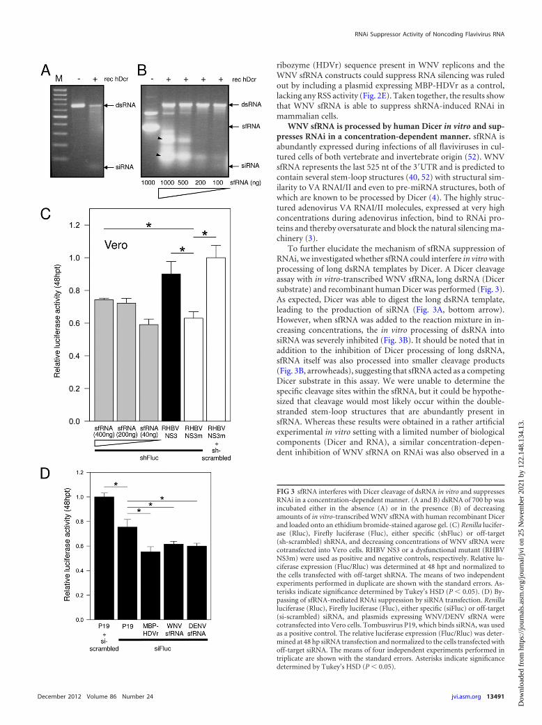
To further elucidate the mechanism of sfRNA suppression ofRNAi, we investigated whether sfRNA could interfere in vitro withprocessing of long dsRNA templates by Dicer. A Dicer cleavageassay with in vitro-transcribed WNV sfRNA, long dsRNA (Dicersubstrate) and recombinant human Dicer was performed (Fig. 3).As expected, Dicer was able to digest the long dsRNA template,leading to the production of siRNA (Fig. 3A, bottom arrow).However, when sfRNA was added to the reaction mixture in in-creasing concentrations, the in vitro processing of dsRNA intosiRNA was severely inhibited (Fig. 3B). It should be noted that inaddition to the inhibition of Dicer processing of long dsRNA,sfRNA itself was also processed into smaller cleavage products(Fig. 3B, arrowheads), suggesting that sfRNA acted as a competingDicer substrate in this assay. We were unable to determine thespecific cleavage sites within the sfRNA, but it could be hypothe-sized that cleavage would most likely occur within the double-stranded stem-loop structures that are abundantly present insfRNA. Whereas these results were obtained in a rather artificialexperimental in vitro setting with a limited number of biologicalcomponents (Dicer and RNA), a similar concentration-depen-dent inhibition of WNV sfRNA on RNAi was also observed in a
FIG 3 sfRNA interferes with Dicer cleavage of dsRNA in vitro and suppressesRNAi in a concentration-dependent manner. (A and B) dsRNA of 700 bp wasincubated either in the absence (A) or in the presence (B) of decreasingamounts of in vitro-transcribed WNV sfRNA with human recombinant Dicerand loaded onto an ethidium bromide-stained agarose gel. (C) Renilla lucifer-ase (Rluc), Firefly luciferase (Fluc), either specific (shFluc) or off-target(sh-scrambled) shRNA, and decreasing concentrations of WNV sfRNA werecotransfected into Vero cells. RHBV NS3 or a dysfunctional mutant (RHBVNS3m) were used as positive and negative controls, respectively. Relative lu-ciferase expression (Fluc/Rluc) was determined at 48 hpt and normalized tothe cells transfected with off-target shRNA. The means of two independentexperiments performed in duplicate are shown with the standard errors. As-terisks indicate significance determined by Tukey’s HSD (P � 0.05). (D) By-passing of sfRNA-mediated RNAi suppression by siRNA transfection. Renillaluciferase (Rluc), Firefly luciferase (Fluc), either specific (siFluc) or off-target(si-scrambled) siRNA, and plasmids expressing WNV/DENV sfRNA werecotransfected into Vero cells. Tombusvirus P19, which binds siRNA, was usedas a positive control. The relative luciferase expression (Fluc/Rluc) was deter-mined at 48 hp siRNA transfection and normalized to the cells transfected withoff-target siRNA. The means of four independent experiments performed intriplicate are shown with the standard errors. Asterisks indicate significancedetermined by Tukey’s HSD (P � 0.05).
RNAi Suppressor Activity of Noncoding Flavivirus RNA
December 2012 Volume 86 Number 24 jvi.asm.org 13491
Dow
nloa
ded
from
http
s://j
ourn
als.
asm
.org
/jour
nal/j
vi o
n 25
Nov
embe
r 20
21 b
y 12
2.14
8.13
4.13
.

shRNA-induced silencing suppressor assay in mammalian cells(Fig. 3C).
Interference with Dicer processing suggests that sfRNA acts asRSS upstream of RISC. In this scenario, transfected siRNA shouldbe capable of bypassing RSS activity, since it is directly loaded intoRISC independent of Dicer. We therefore examined whether ornot sfRNA could suppress siRNA-mediated RNAi. Vero cells weretransfected with luciferase reporter plasmids and with a sfRNA-expressing plasmid. Next, the cells were transfected with Fluc-specific siRNA. Fluc activity was measured 48 h after siRNA trans-fection. The results showed that P19, a strong RSS with highaffinity to siRNA, but not the sfRNAs of WNV and DENV couldsuppress siRNA-induced RNAi (Fig. 3D).
Taken together, the results suggest that WNV sfRNA, as waspreviously reported for VA RNAI/II (3), can interfere in a concen-tration-dependent manner with Dicer processing of dsRNA invitro and of shRNA-induced RNAi pathway in vivo. Bypassing ofthis interference with transfected siRNA suggests that the inhibi-tory effect of sfRNA on RNAi activity is upstream of RISC.
WNV RNA replication interferes with the endogenousmiRNA pathway in mammalian cells. The siRNA pathway in in-sects and plants has clearly been demonstrated to be involved inantiviral responses, but the role of the siRNA pathway in an anti-viral response in mammals is the subject of debate (12). At thesame time, the presence and importance of the miRNA pathway inmammalian gene regulation is well accepted (2). In contrast toplants and insects, mammals code for only one Dicer enzyme, andno clear distinction exists between siRNA and miRNA pathways.Interference of an RSS with mammalian Dicer is therefore ex-pected to negatively affect RNAi induced by both siRNA andmiRNA molecules.
To investigate whether sfRNA indeed would also interfere withthe endogenous miRNA pathway in mammalian cells, a previ-ously described Firefly luciferase-based miRNA reporter assay wasperformed using a Fluc sensor construct harboring multiple hu-man miRNA-30 (hsa-miR30) target sites in the 3=UTR of the FlucmRNA (76). This sensor construct, Fluc-miR30-AP, is silenced byendogenously produced miR30 in Vero cells. As expected,cotransfection of the Fluc-miR30-AP sensor construct and Renillaluciferase as an internal transfection control resulted in decreasedFluc expression (10% relative luciferase activity) compared to cellstransfected in a similar way with a sensor construct harboringrandomized miRNA target sites (Fig. 4A). However, in Vero-WNVrep cells, a significant suppression of miR30-induced silenc-ing (26% in comparison to 10% relative luciferase activity innormal Vero cells) was observed (Fig. 4A). To demonstrate thatthe observed gene silencing via miR30 was truly sequence specific, theexperiment was repeated in a background of overexpression ofthe homologous hsa-miR30 and a heterologous hsa-miRNA-21(miR21). Again, cotransfection of the Fluc-miR30-AP sensor con-struct in Vero cells, together with a plasmid overexpressing hsa-miR30 (76), led to silencing (28% relative luciferase activity) of theFluc signal. The observed silencing was sequence specific, because incells transfected with a plasmid expressing hsa-miR21 (76) no silenc-ing was observed (Fig. 4B). In Vero-WNVrep cells, a significant sup-pression of miR30-induced silencing (82% compared to 28% relativeluciferase activity in normal Vero cells) was observed (Fig. 4B). Inconjunction with the experiments shown earlier, the results pre-sented here demonstrate that WNV RNA replication suppresses bothsiRNA- and miRNA-induced RNAi in mammalian cells.
RSS activity of sfRNA is not restricted to the 3=SL. In mos-quito cells, we have recently shown that the 3=SL (Fig. 2D, inset) in
FIG 4 Suppression of the mammalian miRNA pathway in WNV replicon cells. (A) Vero cells either expressing the WNV replicon (Vero-WNV [s]) or lackingthe replicon (Vero wild type [�]) were transfected with expression vectors encoding pCMV-luc-miR30-AP or pCMV-luc-random alone or in combination witheither a specific miRNA (hsa-miRNA30) or off-target (hsa-miRNA21). The results shown are the means of two independent experiments performed in duplicate.Asterisks indicate significance determined by independent two-sample Student t tests (P � 0.05). (B) The luciferase expression was measured 2 days posttrans-fection, and the relative luciferase expression (Fluc/Rluc) was determined. The luciferase level measured with nonspecific has-miRNA21 was set at 1.0. The resultsshown are representative of three independent experiments performed in duplicate. Asterisks indicate significance determined by an independent two-sampleStudent t test (P � 0.05).
Schnettler et al.
13492 jvi.asm.org Journal of Virology
Dow
nloa
ded
from
http
s://j
ourn
als.
asm
.org
/jour
nal/j
vi o
n 25
Nov
embe
r 20
21 b
y 12
2.14
8.13
4.13
.

the 3=UTR (and thus sfRNA) of WNV is a precursor for a viralmiRNA (25) capable of silencing a miRNA reporter construct. Toobtain further insight into the mechanism of sfRNA-mediatedRNAi suppression, we asked the question whether putativemiRNA production from the sfRNA 3=SL was correlated with theobserved Dicer inhibition and RSS activity in both mammalianand insect cells. The miRNA reporter plasmids contain a CMVpromoter for expression in mammalian cells (Fig. 5A) and encodea Fluc gene with a 3=UTR containing 3=SL-derived miRNA targetsequences called A1A2rc and C1C2rc, which are reverse comple-mented to the sequences in the 3=SL (Fig. 5B). First, to verify thefunctionality of the pGL3-based miRNA sensor constructs, theywere cotransfected with pSuper plasmids expressing siRNA spe-cifically targeting either side of the 3=SL (A1A2 or C1C2) (Fig. 5C).This experiment indicated that the A1A2 and C1C2 repeat regionscloned in both orientations in pGL3 in the 3=UTR of Fluc wereefficiently targeted (90% silencing of Fluc) by complementarysiRNA generated from pSuper plasmids, indicating that the sensorconstructs were functional. Next, sfRNA was cotransfected alongwith the miRNA reporter plasmids (Fig. 5D). The result showedthat neither the A1A2rc nor the C1C2rc reporter transcript wassignificantly silenced in the presence of sfRNA. This results sug-
gests that sfRNA is unlikely to be a source of 3=SL-derived miRNAin mammalian cells in contrast to insect cells (25).
Since no viral miRNA derived from the 3=SL could be identi-fied in the miRNA sensor assay, we sought to determine whetherother RNA structures within sfRNA could contribute to the ob-served RSS activity. A number of deletions (from SL-II to CS3,from SL-II to CS1, and 3=SL only) (52) were introduced in sfRNA(Fig. 5E), and plasmids expressing the truncated sfRNA variantswere tested in an RNAi suppressor assay as before (Fig. 5F). Theresults indicated that all sfRNA variants still displayed RSS ac-tivity, including the relatively short variant consisting of onlythe CS1 and 3=SL RNA structures. This result suggested thatmore than a single RNA structure within sfRNA was involvedin RSS activity.
sfRNA suppresses short hairpin RNA-induced silencing ininsect cells. Like many other arboviruses, WNV is transmitted bymosquitoes, actively replicates in mosquito tissues, and eventuallyaccumulates in the salivary glands prior to virus transmission tothe vertebrate host via blood-feeding. A characteristic of mostarbovirus infections is the limited pathology in the infected insectand the establishment of persistent virus replication (17). RNAi isimplicated to be a determining factor for successful arbovirus in-
FIG 5 Mapping of RNAi suppressor activity within WNV sfRNA. (A to D) 3=SL-derived miRNA sensor assay using Firefly luciferase-based sensor constructs. (A)Schematic representation of miRNA sensor constructs for expression in mammalian cells. Reverse complement (rc) of A1A2 and C1C2 tandem repeats areindicated in black and gray, respectively. Sequences used for shRNA cloning into pSuper plasmids are indicated in boldface. Fluc, Firely luciferase; SV40, simianvirus 40 promoter; pA, polyadenylation signal; Xb, XbaI restriction site; Xh, XhoI restriction site. (B) Schematic representation of the WNV 3=SL. A1A2 and C1C2sequences for tandem repeat cloning into miRNA sensor constructs are indicated in black and gray, respectively. (C) Functionality of miRNA sensor constructs.Cells were cotransfected with pRL-TK, pGL3-(sensor constructs), and either pSuper-A1A2 or pSuper-C1C2 or control plasmid. The relative luciferase expression(Firefly/Renilla) was determined at 24 hpt. The means of two independent experiments performed in triplicate are shown with the standard errors. Significancewas tested by an independent two-sample Student t test (P � 0.05). (D) Silencing of miRNA sensor constructs by sfRNA expression in Vero cells. Cells werecotransfected with pRL-TK, pGL3-(sensor constructs), and either pDEST40-sfRNA or control plasmid. The relative luciferase expression (Firefly/Renilla) wasdetermined at 24 hpt. The means of three independent experiments performed in duplicate are shown with the standard errors. Significance was tested by anindependent two-sample Student t test (P � 0.05). (E) Schematic representation of WNV sfRNA with abbreviations as in Fig. 2. The numbers are nucleotidepositions from the 3= terminus of the WNV 3=UTR. Deletions within sfRNA are indicated. (F) Suppressor activity of WNV sfRNA truncations on shRNA-inducedsilencing in Vero cells. The luciferase activity of cells cotransfected with Firefly luciferase (Fluc), Renilla luciferase (Rluc), a specific (shFluc) or off-target(sh-scrambled) shRNA, and WNV sfRNA variants was measured at 48 hpt. The NS3 of RHBV was used as a positive control, and MBP was used as a negativecontrol. The means of two independent experiments performed in duplicate are shown with the standard errors. Asterisks indicate significance determined byTukey’s HSD (P � 0.05).
RNAi Suppressor Activity of Noncoding Flavivirus RNA
December 2012 Volume 86 Number 24 jvi.asm.org 13493
Dow
nloa
ded
from
http
s://j
ourn
als.
asm
.org
/jour
nal/j
vi o
n 25
Nov
embe
r 20
21 b
y 12
2.14
8.13
4.13
.

fection of the mosquito (6). It has been shown that heterologousRSS, e.g., FHV-B2, can enhance alphavirus replication in mosqui-toes (11). To determine whether sfRNA could have a similar effecton alphavirus replication, we engineered sfRNA into a SemlikiForest virus (SFV) replicon and determined the level of replicationin RNAi-competent U4.4 cells. As a control, we included a SFVreplicon expressing another, unrelated, highly structured RNAderived from the encephalomyocarditis virus (EMCV) internalribosome entry site (IRES) (Fig. 6A). Both replicons expressedRenilla luciferase (Rluc) from the nonstructural polyprotein asdescribed previously (28). The results show that SFV replication inmosquito cells is enhanced upon coexpression in cis of sfRNA(Fig. 6B).
To determine the effect of WNV sfRNA on the siRNA pathwayin insect cells, a dsRNA-mediated silencing assay using luciferasereporters was developed for mosquito cells to allow easy quantifi-cation. To this end, Aedes albopictus U4.4 cells were cotransfectedwith plasmids encoding Fluc, Rluc (internal transfection control)and either MBP or WNV sfRNA. At 24 hpt, silencing was inducedby transfection of dsRNA, either specifically against Firefly lucif-erase (dsFluc) or off-target (ds-scrambled), and the luciferase ac-tivity was determined at 48 hpt. Cells cotransfected with MBP,Fluc, and dsFluc carrying plasmids showed silencing of Fluc ex-pression (ca. 47% relative luciferase activity), which was not ob-served with ds-scrambled (Fig. 6C). In the case of cotransfectionof WNV-sfRNA with Fluc and dsFluc, no significant reduction ofFluc activity could be detected, indicating that sfRNA very effi-ciently suppressed dsRNA-induced silencing (Fig. 6C).
To ensure that the observed effect of sfRNA was not ananomaly of dsRNA-induced silencing in mosquito U4.4 cells, asimilar experiment was performed in Drosophila melanogasterS2 cells, using a shRNA-mediated silencing assay. In this exper-iment, S2 cells were cotransfected with inducible plasmids en-coding Fluc, Rluc (internal control), and shRNA constructsspecifically targeting Fluc (shFluc) or a control shRNA target-ing GFP (shGFP). Next, Fluc, Rluc, and shRNA expression wasinduced by the addition of CuSO4 to the medium at 24 hpt, andthe Fluc activity was measured at 48 hpt and normalized to theRluc readings (Fig. 6D). Cells cotransfected with MBP, Fluc,and shFluc plasmids showed silencing of Fluc expression (ca.55% relative luciferase activity), which was not observed withthe shGFP-negative control (Fig. 6D). When a plasmid express-ing WNV sfRNA was cotransfected with Fluc and shFluc, how-ever, Fluc silencing was suppressed with am efficiency similarto that of the positive control RHBV-NS3 (Fig. 6D) (24), with81 and 83% relative luciferase activities, respectively. Similar towhat was observed in mammalian cells, the RSS activity ofWNV sfRNA was concentration dependent (Fig. 6D). In con-clusion, these experiments show that WNV sfRNA is able tointerfere with the siRNA-based pathway in insect cells.
sfRNA suppresses the induced miRNA pathway in insectcells. In addition to the antiviral siRNA pathway, insects also havea functional miRNA pathway that has a function in gene regula-tion similar to that seen, for example, in mammals. In contrast tomammals, however, insects have two distinct Dicer enzymes,Dcr-1 and Dcr-2, instead of one, with dedicated functions in themiRNA or siRNA pathway, respectively (1). Although no clearantiviral activity has been attributed to the miRNA pathway ininsects, the question remained as to whether WNV sfRNA, apartfrom its activity in inhibiting the siRNA pathway, could also in-
terfere with the miRNA pathway in insect cells. Several viral RSSproteins have been demonstrated to be able to interfere with boththe siRNA and the miRNA pathways in different organisms(14, 58).
D. melanogaster S2 cells were cotransfected with an inducibleFluc dme-miRNA1-sensor construct (Fluc-3=UTR), vectors en-coding either specific dme-miRNA1 or off-target dme-miRNA12,Rluc (internal transfection control), and either WNV sfRNA,MBP (negative control), or Carnation Italian ringspot virus(CIRV) P19 (positive control). As expected, a decrease in Flucexpression (ca. 48% relative luciferase activity) was observed incells cotransfected with MBP, Fluc-3=UTR, and dme-miRNA1 butnot with the off-target dme-miRNA12 (Fig. 7A). To make surethat the observed Fluc silencing was the result of miRNA1 andthus independent of the siRNA pathway, we checked that themiRNA1-induced silencing was lost in cells treated prior to trans-fection with dsRNA specifically targeting Ago1 but not Ago2 or thenonspecific GFP (Fig. 7B). Successful depletion of Ago1 and Ago2transcripts and proteins was checked by RT-PCR and Westernblotting, respectively (Fig. 7C). Since Ago1 and Ago2 are known tobe predominantly loaded with miRNA and siRNA, respectively,these results confirmed that the miRNA pathway in insect cellswas capable of silencing the Fluc reporter construct harboringdme-miRNA1 target sites.
After the demonstration of sequence-specific silencing bymiRNA1 through the miRNA pathway, the putative suppressoreffect of WNV sfRNA was investigated. A significant and repro-ducible rescue of Fluc expression was observed in the presence ofWNV sfRNA, to a level similar to that observed for CIRV P19 (Fig.7A), suggesting that WNV sfRNA interferes with the miRNApathway in insect cells. Collectively, these results demonstrate thatWNV sfRNA interferes with both the siRNA- and the miRNA-based pathways in insect cells.
DENV also displays RSS activity in mammalian and insectcells. sfRNA is produced by all flaviviruses (52, 60). To determinewhether the observed interference with the RNA silencing path-ways is common among flaviviruses, the same experiments wereconducted with DENV1.
First, Vero cells stably expressing a DENV1 replicon (Vero-DENVrep) were established by transfection of Vero cells withDENV1 replicon RNA harboring an IRESneo cassette (54). Stablereplicon cells were selected with G418. Active DENV repliconRNA replication was confirmed by immunofluorescence using theJ2 anti-dsRNA antibody (Fig. 8A). Vero-DENVrep cells showedless silencing (ca. 28% relative luciferase activity) if transfectedwith shRNA specific against Firefly luciferase in comparison towild-type Vero cells (10% relative luciferase activity) (Fig. 8B).
DENV sfRNA was also tested in the shRNA-mediated si-lencing assays in mammalian (Vero) and RNAi-competentmosquito (U4.4) cells and displayed a similar RSS activity (Fig.8C and D) compared to WNV sfRNA (Fig. 2D and E and Fig.8C). Furthermore, no RNAi suppressor activity of DENV1nonstructural proteins was detected in the plant suppressorassay (data not shown), and no retardation of either small orlong dsRNA was observed in DENV replicon cell extracts (Fig.2A). These results show that the ability of sfRNA to interferewith the RNA silencing is observed for both WNV and DENV,in mammalian as well as in insect cells.
Schnettler et al.
13494 jvi.asm.org Journal of Virology
Dow
nloa
ded
from
http
s://j
ourn
als.
asm
.org
/jour
nal/j
vi o
n 25
Nov
embe
r 20
21 b
y 12
2.14
8.13
4.13
.

DISCUSSION
RNA silencing is known as an important antiviral response ininsects and plants. Until now, all investigated plant and “true”insect viruses have been shown to encode RSS proteins, which are
essential for the establishment of a successful viral infection. Ourresults and those of others (32) show that no proteins with RSSactivity have thus far been identified in flaviviruses, despite the factthat these viruses are successfully infecting their mosquito vector.
FIG 6 WNV sfRNA interferes with siRNA-induced silencing in insect cells. (A) Schematic representation of SFV constructs. The Renilla luciferase (Rluc) gene is insertedupstream of nsP4. sfRNA or the EMCV IRES sequence is inserted downstream of the 26S subgenomic promoter. Schematic RNA structures of sfRNA and the EMCVIRES are shown. (B) Capped, in vitro-transcribed SFV RNA was transfected in Aedes albopictus U4.4 mosquito cells. Rluc expression was measured at 24 hpt. The meansof four independent experiments performed in duplicate are shown with the standard errors. Asterisks indicate significance determined by independent two-sampleStudent t test (P � 0.05). (C) Aedes albopictus U4.4 mosquito cells were cotransfected with Firefly luciferase (Fluc), Renilla luciferase (Rluc), and MBP or WNV sfRNAconstructs. After 24 h, silencing was induced by transfection of either specific (dsFluc) or off-target (ds-scrambled) in vitro-transcribed dsRNA. The luciferase activity wasmeasured at 48 hpt, and the normalized relative luciferase activity (Fluc/Rluc) is shown with the standard errors (means of two independent experiments performed induplicate). Asterisks indicate significance determined by Tukey’s HSD (P � 0.05). (D) Concentration-dependent effect of sfRNA on shRNA-induced silencing deter-mined in D. melanogaster S2 cells by transfection of decreasing concentrations of WNV-sfRNA (140, 50, and 20 ng) construct in concert with Firefly luciferase (Fluc),Renilla luciferase (Rluc), and either specific (shFluc) or off-target (sh-scrambled) shRNA. RHBV NS3 was used as a positive control. The luciferase activity was measuredat 48 hpt, and the normalized relative luciferase activity (Fluc/Rluc) is shown with the standard errors (means of three independent experiments performed in duplicate).Asterisks indicate significance determined by Tukey’s HSD (P � 0.05).
RNAi Suppressor Activity of Noncoding Flavivirus RNA
December 2012 Volume 86 Number 24 jvi.asm.org 13495
Dow
nloa
ded
from
http
s://j
ourn
als.
asm
.org
/jour
nal/j
vi o
n 25
Nov
embe
r 20
21 b
y 12
2.14
8.13
4.13
.

The RNA silencing pathway is the major antiviral response inmosquitoes against flavivirus infection (6), and the knockdown ofthis pathway results in an increase of viral replication in mosqui-toes (55). It has remained unclear to what extent and how flavivi-ruses tweak this antiviral RNAi response for its own benefit. In thepresent study we now show that a noncoding, 3=UTR-derived vi-ral RNA produced by the flaviviruses WNV and DENV displaysRNAi suppressor activity in both insect and mammalian cells.
This noncoding viral RNA is present in the 3=UTR of all flavi-virus genomes and, interestingly, is also abundantly produced asan XRN1-exonuclease-resistant molecule in flavivirus-infectedcells of both insect and mammalian origin (52). This predominantnoncoding RNA molecule was previously named subgenomic fla-vivirus RNA (sfRNA) and is completely identical to the 3=UTRwith the exception that it misses the first �100 nt (52). The flavi-virus 3=UTR and hence also sfRNA share some structural similar-ities with the adenovirus VA RNAI/II molecules, which have beenshown to act as efficient RSS in mammalian cells (3). Previously, itwas shown that sfRNA is important for virus-induced cytopathicand pathogenicity in mammals (52), and more recent studies sug-gested the inhibition of the host innate immune response as one ofthe potential mechanism of its action (59). Our present work sug-gests another potential mechanism for the sfRNA, i.e., as nucleicacid-based RSS both in both mammalian and in insect cells.
We have carried out most of the experiments with the plasmidsexpressing a truncated 3=UTR corresponding to the size of sfRNA(525 nt). Therefore, we cannot conclude at this stage whether ornot the entire 3=UTR present in (the context of) the viral genomehas the same RSS activity as sfRNA. Based on studies on genomecyclization during flavivirus infection, which show that the 3=UTRis base paired to the 5=UTR (26, 29, 67), the genomic viral 3=UTRmay be far less accessible to RNAi factors compared to the abun-dantly produced sfRNA. The flaviviral genomic RNA replicationis localized within endoplasmic reticulum membrane-enclosedvesicle packets (39, 73), whereas sfRNA is localized in cytoplasmicprocessing bodies (52), structures that are enriched in RNAi fac-tors (16, 49). This suggests that sfRNA is more likely to interferewith RNAi than the 3=UTR of genomic viral RNA. Mutations inthe 3=UTR leading to reduced expression of sfRNA (19, 52) alsomodify the secondary structure and likely function of sfRNA. Thismakes it difficult to discriminate whether the 3=UTR itself or thesfRNA, which is derived from it, displays the observed RSS activityin cells with active WNV replication.
Results from in vitro Dicer cleavage experiment suggest thatWNV sfRNA may act as an RSS by substrate competition (Fig. 3),thereby possibly oversaturating Dicer in a concentration-depen-dent manner. Although this experiment is highly artificial andlargely ignores the complexity of factors present in a flavivirus-
FIG 7 WNV sfRNA interferes with Ago1-dependent miRNA-induced silencing in insect cells. (A) Suppression of dme-miRNA1-induced silencing in S2 cells bycotransfection of a pMT-Renilla luciferase (Rluc), pMT-Firefly luciferase (Fluc)-dme-miRNA1 sensor construct, either specific (dme-miRNA1) or off-target(dme-miRNA12) primary miRNA and either MBP, CIRV P19 (as a positive control), or WNV sfRNA (180 or 50 ng). After induction at 48 hpt, the relativeluciferase expression (Firefly/Renilla) was determined at 72 hpt, and the means of three independent experiments in duplicate are shown with the standard errors.Asterisks indicate significance by Tukey’s HSD (P � 0.05). (B) Silencing of the pMT-Firefly luciferase (Fluc)-dme-miRNA1 sensor construct is Ago1 dependent.S2 cells were soaked with 200 ng of dsRNA either Ago1, Ago2, or EGFP (unspecific) during seeding. dme-miRNA1-induced silencing was induced by cotrans-fection of pMT-Renilla luciferase (Rluc), pMT-Firefly luciferase (Fluc)-dme-miRNA1 sensor construct, either specific (dme-miRNA1) or off-target (dme-miRNA12) primary miRNA. The means of three independent experiments in duplicate are shown with the standard errors. Asterisks indicate significancedetermined by an independent two-sample Student t test (P � 0.05). (C) Confirmation of successful Ago1/2 depletion in S2 cells. To check the knockdown levelof Ago1/2, RT-PCR was carried out on total RNA purified from dsRNA-silenced cells. Western blot analysis of Drosophila melanogaster Ago1/2 was performedto confirm successful depletion of Ago1/2 in S2 cells.
Schnettler et al.
13496 jvi.asm.org Journal of Virology
Dow
nloa
ded
from
http
s://j
ourn
als.
asm
.org
/jour
nal/j
vi o
n 25
Nov
embe
r 20
21 b
y 12
2.14
8.13
4.13
.

infected cell, the proposed RNA decoy mechanism is supported bythe observed concentration-dependent RNAi suppressor activityof sfRNA in shRNA-mediated silencing experiments (Fig. 3C and6D), as well as the ability to bypass the RSS activity of sfRNA bytransfection with siRNA (Fig. 3D). Nucleic acid-based decoymechanisms in other viral systems have recently been publishedfor SFV (61) and Cauliflower mosaic virus (CaMV) (7). In SFVinfection, hot spot-derived viral siRNAs have been found to berather inefficient in silencing of viral genomic RNA, thus allowingviral replication while saturating RNAi factors (61). DuringCaMV infection, high concentrations of RNA produced from aviral noncoding region are subsequently processed into siRNAsand incorporated into RISC to presumably act as a decoy (7).Although CaMV already encodes a RSS protein that interfereswith the RNA-dependent RNA polymerase-mediated amplifica-tion of siRNA, the virus may in this case, paradoxically, benefitfrom the massive production of siRNAs derived from its noncod-ing RNA as backup strategy. Thus, abundant production of non-coding or viral siRNA can be advantageous for viral infection.
Although sfRNA itself may not lead to massive production ofdecoy viral siRNA, its strong secondary structure (19, 45, 52) maydecrease overall Dicer activity or render the genomic viral RNAinaccessible for an activated RISC. The suggested RNA decoymechanism by sfRNA probably results in a lower RSS activitycompared to that of most of the known proteinaceous RSS of trueinsect viruses but may still allow sufficient levels of flaviviral rep-lication needed for successful virus transmission by the vectormosquitoes. Recent research has shown that the expression of
protein RSS (derived from true insect viruses) by alphavirusesleads to the rapid death of infected mosquitoes (11, 43). Similarly,we now show that expression in cis of sfRNA also enhances SFVreplication in RNAi-competent mosquito cells (Fig. 6B). In lightof these results, it is clear that a delicate balance exists betweenarbovirus replication and vector survival. Although all arbovirusesfor which this has been analyzed induce RNAi responses, it nowappears that inhibition or evasion does take place (61), and it islikely that the scale of inhibition or evasion is a key factor. Ourpresent results do not allow us to directly compare RNAi suppres-sor activity of sfRNA to that of the protein RSS, but future workwill focus on addressing this important question. It is worth not-ing, however, that the level of sfRNA produced from plasmidDNA transfection in most of our RNAi suppressor assays is lowerthan the sfRNA level produced in virus-infected or replicon-transfected cells (52).
Despite the fact that all flaviviruses produce sfRNA (52) andthus might suppress RNAi, flavivirus replication can still be inhib-ited by endogenous miRNA when miRNA target sites are artifi-cially engineered in the viral RNA (23, 30, 51). This suggests thatsfRNA does not fully block RNAi, which is in agreement withthe lower RSS activity of sfRNA in comparison to, for example,VA RNA.
In addition to the RSS activity of sfRNA on the antiviral siRNApathway in insect cells, we observed similar suppressor activity ofsfRNA on the insect miRNA pathway. In line with this result,others have recently shown that WNV infection of Culex mosqui-toes alters the expression levels of a subset of host miRNAs (62). In
FIG 8 Suppression of RNA silencing in mammalian and mosquito cells by DENV sfRNA. (A) Viral RNA replication in Vero cells stably expressing the DENV1replicon was verified by immunofluorescence using a primary J2 anti-dsRNA antibody and a corresponding rhodamine-labeled secondary antibody (upperpanel). Wild-type Vero cells were stained as a control (lower panel). (B) The effect of DENV RNA replication on shRNA-induced silencing was investigated innormal Vero cells (black) and in Vero cells stably expressing the DENV replicon (gray). The cells were cotransfected with Firefly luciferase (Fluc), Renillaluciferase (Rluc), and shRNA, either Fluc-specific (shFluc) or off-target (sh-scrambled). The normalized relative luciferase expression (Fluc/Rluc) was deter-mined at 48 hpt. The means of three independent experiments performed in duplicate are shown with the standard errors. Asterisks indicate significancedetermined by an independent two-sample Student t test (P � 0.05). (C) RNA silencing suppression by the DENV sfRNA was investigated in Vero cells bycotransfection of pFluc, Rluc, either specific (shFluc) or off-target (sh-scrambled) shRNA, and expression constructs of MBP, MBP-HDVr, DENV, sfRNA, oradenoviral VA RNA. The luciferase expression was measured at 48 hpt. The means of two independent experiments performed in duplicate are shown with thestandard errors. Asterisks indicate significance by Tukey’s HSD (P � 0.05). (D) Effect of DENV sfRNA was determined by cotransfection of Aedes albopictus-derived U4.4 cells with Firefly luciferase (Fluc), Renilla luciferase (Rluc) and MBP, DENV sfRNA, or WNV sfRNA (as positive control) constructs. After 24 h,silencing was induced by transfection of either specific (dsFluc) or off-target (ds-scrambled) in vitro-transcribed dsRNA. The luciferase activity was determined48 h after primary transfection and normalized to the cells transfected with off-target dsRNA. The means of three independent experiments performed induplicate are shown with the standard errors. Asterisks indicate significance determined by Tukey’s HSD (P � 0.05).
RNAi Suppressor Activity of Noncoding Flavivirus RNA
December 2012 Volume 86 Number 24 jvi.asm.org 13497
Dow
nloa
ded
from
http
s://j
ourn
als.
asm
.org
/jour
nal/j
vi o
n 25
Nov
embe
r 20
21 b
y 12
2.14
8.13
4.13
.
contrast, Aedes albopictus C7/10 mosquito cells harboring a WNVreplicon did not display these changes in endogenous miRNA ex-pression (62). However, the relative levels of viral RNA replica-tion, which normally correlate with sfRNA expression levels, ofpersistent WNV replicon in C7/10 versus virus infection of Culexmosquitoes was not shown. It remains therefore possible thatsfRNA levels in C7/10 were too low to observe an effect on themiRNA profile or that defects in RNAi factors in C7/10 cells (42)play a determining role.
Dual activity of RSS proteins on siRNA- and miRNA-relatedpathways has been observed previously for plant-infecting virusessuch as Tomato spotted wilt virus (58) and tombusvirus (66), aswell as the adenoviral VA RNA (38), but their functional impor-tance during viral replication is still unclear. In insects no suchmechanisms have been reported yet, but some results suggest theimportance of other antiviral responses in addition to the siRNApathway. For example, a negative effect on viral replication ofWNV lacking sfRNA has been observed in Dicer-2-deficientC6/36 mosquito cells (52). This could due to the activity ofDicer-1 (miRNA-related) or a Dicer-independent RNA silencingpathway involving piRNAs (42, 68) or to another antiviral re-sponse independent of RNAi (17).
Herpes-, adeno-, baculo-, and ascoviruses produce virus-en-coded miRNAs targeting either viral RNA or host genes involvedin cell proliferation, DNA repair, and RNA regulation (5, 20).Adenoviral VA RNAs are processed by Dicer to viral miRNAs thatare loaded into Argonaute, which can result in its saturation. Inlight of the similarities between the secondary structures of ade-novirus VA RNA and the WNV sfRNA, it may be possible that theinteraction of sfRNA with the miRNA pathway is related to theprocessing of a virus-encoded miRNA. Indeed, sfRNA was re-cently shown to be the main source of 3=SL-derived viral miRNAin WNV-infected insect cells (25). It is therefore possible that thefunctionality of sfRNA as Dicer substrate may determine its RSSactivity, but our results with miRNA sensors show that this viralmiRNA is not (efficiently) produced in mammalian cells (Fig.5D). In addition, sfRNA variant lacking the 3=SL still did displaysome RSS activity (Fig. 5F), suggesting that production of 3=SL-derived viral miRNA is likely not required for sfRNA to interferewith the RNAi machinery. Thus, despite the similarities with VARNA, which is very efficiently loaded into RISC as viral miRNA(75), sfRNA displays RSS activity in the cytoplasm without beingfully processed. To fully understand the biological importance ofRNAi suppression by sfRNA in mammalian cells, more elaboratestudies (e.g., on the effect of the depletion of Dicer or other RNAicomponents) on flavivirus replication would be needed.
There is some evidence that might suggest the involvement ofmiRNA in antiviral RNAi in mammalian cells (for a review, seereference 47) and possible links between the miRNA pathway andthe IFN response (50, 74). A dual function of a viral product inboth of these pathways has been reported for adenovirus VA RNA,which is able to interact with both the IFN response and the RNAiin mammalian cells. We previously reported that sfRNA was re-quired for virus-induced pathogenicity in mammals and hypoth-esized a putative function in the antiviral IFN pathway (52). In-deed, recent data show that sfRNA contributes to viral evasion ofthe type I IFN-mediated antiviral response (59). It is thereforetempting to speculate that the observed effects of sfRNA on theRNAi pathways and on modulation of the IFN response in mam-
malian cells are linked, although further studies are required tosupport this supposition.
In conclusion, our findings show that sfRNAs of WNV andDENV can interfere with two distinct arms of the RNAi path-way—siRNA and miRNA mediated—in both insect and mamma-lian cells. Future experiments will elucidate whether this novelsfRNA function holds for all flaviviruses and will hopefully revealthe complete picture describing the different functional roles offlavivirus noncoding RNA in the innate immune responses ofmosquitoes and vertebrate hosts.
ACKNOWLEDGMENTS
E.S. is supported by the Netherlands Organization for Scientific ResearchNWO (Rubicon Fellowship 825.10.021). We acknowledge the graduateschool “Production Ecology and Resource Conservation” of WageningenUniversity for offering a visiting scientist grant to J.Y.L. to conduct part ofthese studies during a 6-month period at the Laboratory of Virology inWageningen. We acknowledge the UK Medical Research Council and aBBSRC Roslin Institute Strategic Programme grant for funding for A.K.
We thank Andres Merits for generating the SFV constructs, Ronaldvan Rij for Drosophila melanogaster dsRNA templates and primer se-quences, Byron Martina for the Aedes pseudoscutellaris Ap61 mosquitocells, Pei-Yong Shi for providing the DENV1 replicon construct, and RoyHall and Paul Young for providing WNV antisera.
REFERENCES1. Aliyari R, Ding SW. 2009. RNA-based viral immunity initiated by the
Dicer family of host immune receptors. Immunol. Rev. 227:176 –188.2. Ambros V. 2004. The functions of animal microRNAs. Nature 431:350 –
355.3. Andersson MG, et al. 2005. Suppression of RNA interference by adeno-
virus virus-associated RNA. J. Virol. 79:9556 –9565.4. Aparicio O, Razquin N, Zaratiegui M, Narvaiza I, Fortes P. 2006.
Adenovirus virus-associated RNA is processed to functional interferingRNAs involved in virus production. J. Virol. 80:1376 –1384.
5. Asgari S. 2011. Role of microRNAs in insect host-microorganism inter-actions. Front. Physiol. 2:48.
6. Blair CD. 2011. Mosquito RNAi is the major innate immune pathwaycontrolling arbovirus infection and transmission. Future Microbiol.6:265–277.
7. Blevins T, et al. 2011. Massive production of small RNAs from a non-coding region of Cauliflower mosaic virus in plant defense and viral coun-ter-defense. Nucleic Acids Res. 39:5003–5014.
8. Brummelkamp TR, Bernards R, Agami R. 2002. A system for stableexpression of short interfering RNAs in mammalian cells. Science 296:550 –553.
9. Bucher E, Hemmes H, de Haan P, Goldbach R, Prins M. 2004. Theinfluenza A virus NS1 protein binds small interfering RNAs and sup-presses RNA silencing in plants. J. Gen. Virol. 85:983–991.
10. Chotkowski HL, et al. 2008. West Nile virus infection of Drosophilamelanogaster induces a protective RNAi response. Virology 377:197–206.
11. Cirimotich CM, Scott JC, Phillips AT, Geiss BJ, Olson KE. 2009.Suppression of RNA interference increases alphavirus replication and vi-rus-associated mortality in Aedes aegypti mosquitoes. BMC Microbiol.9:49. doi:10.1186/1471-2180-9-49.
12. Cullen BR. 2006. Is RNA interference involved in intrinsic antiviral im-munity in mammals? Nat. Immunol. 7:563–567.
13. Ding SW. 2010. RNA-based antiviral immunity. Nat. Rev. Immunol.10:632– 644.
14. Dunoyer P, Lecellier CH, Parizotto EA, Himber C, Voinnet O. 2004.Probing the microRNA and small interfering RNA pathways with virus-encoded suppressors of RNA silencing. Plant Cell 16:1235–1250.
15. Dyer BW, Ferrer FA, Klinedinst DK, Rodriguez R. 2000. A noncom-mercial dual luciferase enzyme assay system for reporter gene analysis.Anal. Biochem. 282:158 –161.
16. Eulalio A, Behm-Ansmant I, Schweizer D, Izaurralde E. 2007. P-bodyformation is a consequence, not the cause, of RNA-mediated gene silenc-ing. Mol. Cell. Biol. 27:3970 –3981.
Schnettler et al.
13498 jvi.asm.org Journal of Virology
Dow
nloa
ded
from
http
s://j
ourn
als.
asm
.org
/jour
nal/j
vi o
n 25
Nov
embe
r 20
21 b
y 12
2.14
8.13
4.13
.
17. Fragkoudis R, Attarzadeh-Yazdi G, Nash AA, Fazakerley JK, Kohl A.2009. Advances in dissecting mosquito innate immune responses to arbo-virus infection. J. Gen. Virol. 90:2061–2072.
18. Fros JJ, et al. 2010. Chikungunya virus nonstructural protein 2 inhibitstype I/II interferon-stimulated JAK-STAT signaling. J. Virol. 84:10877–10887.
19. Funk A, et al. 2010. RNA structures required for production of sub-genomic flavivirus RNA. J. Virol. 84:11407–11417.
20. Grundhoff A, Sullivan CS. 2011. Virus-encoded microRNAs. Virology411:325–343.
21. Haasnoot J, et al. 2007. The Ebola virus VP35 protein is a suppressor ofRNA silencing. PLoS Pathog. 3:e86. doi:10.1371/journal.ppat.0030086.
22. Haley B, Tang G, Zamore PD. 2003. In vitro analysis of RNA interferencein Drosophila melanogaster. Methods 30:330 –336.
23. Heiss BL, Maximova OA, Pletnev AG. 2011. Insertion of microRNAtargets into the flavivirus genome alters its highly neurovirulent pheno-type. J. Virol. 85:1464 –1472.
24. Hemmes H, Lakatos L, Goldbach R, Burgyan J, Prins M. 2007. The NS3protein of Rice Hoja Blanca tenuivirus suppresses RNA silencing in plantand insect hosts by efficiently binding both siRNAs and miRNAs. RNA13:1079 –1089.
25. Hussain M, et al. 2011. West Nile virus encodes a microRNA-like smallRNA in the 3= untranslated region which up-regulates GATA4 mRNA andfacilitates virus replication in mosquito cells. Nucleic Acids Res. 40:2210 –2223.
26. Khromykh AA, Meka H, Guyatt KJ, Westaway EG. 2001. Essential role ofcyclization sequences in flavivirus RNA replication. J. Virol. 75:6719–6728.
27. Khromykh AA, Westaway EG. 1997. Subgenomic replicons of the flavi-virus Kunjin: construction and applications. J. Virol. 71:1497–1505.
28. Kiiver K, et al. 2008. Properties of nonstructural protein 1 of SemlikiForest virus and its interference with virus replication. J. Gen. Virol. 89:1457–1466.
29. Kofler RM, Hoenninger VM, Thurner C, Mandl CW. 2006. Functionalanalysis of the tick-borne encephalitis virus cyclization elements indicatesmajor differences between mosquito-borne and tick-borne flaviviruses. J.Virol. 80:4099 – 4113.
30. Lee TC, et al. 2010. Utilizing liver-specific microRNA-122 to modulatereplication of dengue virus replicon. Biochem. Biophys. Res. Commun.396:596 – 601.
31. Li H, Li WX, Ding SW. 2002. Induction and suppression of RNA silenc-ing by an animal virus. Science 296:1319 –1321.
32. Li HW, Ding SW. 2005. Antiviral silencing in animals. FEBS Lett. 579:5965–5973.
33. Lin KC, Chang HL, Chang RY. 2004. Accumulation of a 3=-terminalgenome fragment in Japanese encephalitis virus-infected mammalian andmosquito cells. J. Virol. 78:5133–5138.
34. Lindenbach BD, Thiel HJ, Rice CM. 2007. Flaviviridae: the viruses andtheir replication, p 1101–1152. In Knipe DM, Howley PM (ed), Fieldsvirology, 5th ed. Lippincott-Raven Publishers, Philadelphia, PA.
35. Liu WJ, Chen HB, Wang XJ, Huang H, Khromykh AA. 2004. Analysisof adaptive mutations in Kunjin virus replicon RNA reveals a novel rolefor the flavivirus nonstructural protein NS2A in inhibition of beta inter-feron promoter-driven transcription. J. Virol. 78:12225–12235.
36. Liu WJ, Sedlak PL, Kondratieva N, Khromykh AA. 2002. Complemen-tation analysis of the flavivirus Kunjin NS3 and NS5 proteins defines theminimal regions essential for formation of a replication complex andshows a requirement of NS3 in cis for virus assembly. J. Virol. 76:10766 –10775.
37. Lu R, et al. 2005. Animal virus replication and RNAi-mediated antiviralsilencing in Caenorhabditis elegans. Nature 436:1040 –1043.
38. Lu S, Cullen BR. 2004. Adenovirus VA1 noncoding RNA can inhibit smallinterfering RNA and microRNA biogenesis. J. Virol. 78:12868–12876.
39. Mackenzie J. 2005. Wrapping things up about virus RNA replication.Traffic 6:967–977.
40. Markoff L. 2003. 5=- and 3=-noncoding regions in flavivirus RNA. Adv.Virus. Res. 59:177–228.
41. Metz SW, Pijlman GP. 2011. Arbovirus vaccines; opportunities for thebaculovirus-insect cell expression system. J. Invertebr. Pathol.107(Suppl):S16 –S30.
42. Morazzani EM, Wiley MR, Murreddu MG, Adelman ZN, Myles KM.2012. Production of virus-derived ping-pong-dependent piRNA-likesmall RNAs in the mosquito soma. PLoS Pathog. 8:e1002470. doi:10.1371/journal.ppat.1002470.
43. Myles KM, Wiley MR, Morazzani EM, Adelman ZN. 2008. Alphavirus-derived small RNAs modulate pathogenesis in disease vector mosquitoes.Proc. Natl. Acad. Sci. U. S. A. 105:19938 –19943.
44. Nayak A, et al. 2010. Cricket paralysis virus antagonizes Argonaute 2 tomodulate antiviral defense in Drosophila. Nat. Struct. Mol. Biol. 17:547–554.
45. Olsthoorn RC, Bol JF. 2001. Sequence comparison and secondary struc-ture analysis of the 3= noncoding region of flavivirus genomes revealsmultiple pseudoknots. RNA 7:1370 –1377.
46. Ongus JR, Roode EC, Pleij CW, Vlak JM, van Oers MM. 2006. The 5=non-translated region of Varroa destructor virus 1 (genus Iflavirus): struc-ture prediction and IRES activity in Lymantria dispar cells. J. Gen. Virol.87:3397–3407.
47. Ouellet DL, Provost P. 2010. Current knowledge of microRNAs andnoncoding RNAs in virus-infected cells. Methods Mol. Biol. 623:35– 65.
48. Paddison PJ, Caudy AA, Hannon GJ. 2002. Stable suppression of geneexpression by RNAi in mammalian cells. Proc. Natl. Acad. Sci. U. S. A.99:1443–1448.
49. Parker R, Sheth U. 2007. P bodies and the control of mRNA translationand degradation. Mol. Cell 25:635– 646.
50. Pedersen IM, et al. 2007. Interferon modulation of cellular microRNAs asan antiviral mechanism. Nature 449:919 –922.
51. Pham AM, Langlois RA, TenOever BR. 2012. Replication in cells ofhematopoietic origin is necessary for dengue virus dissemination. PLoSPathog. 8:e1002465. doi:10.1371/journal.ppat.1002465.
52. Pijlman GP, et al. 2008. A highly structured, nuclease-resistant, noncod-ing RNA produced by flaviviruses is required for pathogenicity. Cell HostMicrobe 4:579 –591.
53. Pijlman GP, Kondratieva N, Khromykh AA. 2006. Translation of the flavi-virus Kunjin NS3 gene in cis but not its RNA sequence or secondary structureis essential for efficient RNA packaging. J. Virol. 80:11255–11264.
54. Puig-Basagoiti F, et al. 2006. Triaryl pyrazoline compound inhibits fla-vivirus RNA replication. Antimicrob. Agents Chemother. 50:1320 –1329.
55. Sanchez-Vargas I, et al. 2009. Dengue virus type 2 infections of Aedesaegypti are modulated by the mosquito’s RNA interference pathway. PLoSPathog. 5:e1000299. doi:10.1371/journal.ppat.1000299.
56. Scherbik SV, Paranjape JM, Stockman BM, Silverman RH, Brinton MA.2006. RNase L plays a role in the antiviral response to West Nile virus. J.Virol. 80:2987–2999.
57. Schnettler E, Hemmes H, Goldbach R, Prins M. 2008. The NS3 proteinof Rice Hoja Blanca virus suppresses RNA silencing in mammalian cells. J.Gen. Virol. 89:336 –340.
58. Schnettler E, et al. 2010. Diverging affinity of tospovirus RNA silencingsuppressor proteins, NSs, for various RNA duplex molecules. J. Virol.84:11542–11554.
59. Schuessler A, et al. 2012. West Nile virus non-coding subgenomic RNAcontributes to viral evasion of type I interferon-mediated antiviral re-sponse. J. Virol. 86:5708 –5718.
60. Silva PA, Pereira CF, Dalebout TJ, Spaan WJ, Bredenbeek PJ. 2010. AnRNA pseudoknot is required for production of yellow fever virus sub-genomic RNA by the host nuclease XRN1. J. Virol. 84:11395–11406.
61. Siu RW, et al. 2011. Antiviral RNA interference responses induced bySemliki Forest virus infection of mosquito cells: characterization, origin,and frequency-dependent functions of virus-derived small interferingRNAs. J. Virol. 85:2907–2917.
62. Skalsky RL, Vanlandingham DL, Scholle F, Higgs S, Cullen BR. 2010.Identification of microRNAs expressed in two mosquito vectors, Aedesalbopictus and Culex quinquefasciatus. BMC Genomics 11:119. doi:10.1186/1471-2164-11-119.
63. Soldan SS, Plassmeyer ML, Matukonis MK, Gonzalez-Scarano F. 2005.La Crosse virus nonstructural protein NSs counteracts the effects of shortinterfering RNA. J. Virol. 79:234 –244.
64. Umbach JL, Cullen BR. 2009. The role of RNAi and microRNAs in animalvirus replication and antiviral immunity. Genes Dev. 23:1151–1164.
65. van Rij RP, et al. 2006. The RNA silencing endonuclease Argonaute 2mediates specific antiviral immunity in Drosophila melanogaster. GenesDev. 20:2985–2995.
66. Varallyay E, Valoczi A, Agyi A, Burgyan J, Havelda Z. 2010. Plantvirus-mediated induction of miR168 is associated with repression ofARGONAUTE1 accumulation. EMBO J. 29:3507–3519.
67. Villordo SM, Gamarnik AV. 2009. Genome cyclization as strategy forflavivirus RNA replication. Virus Res. 139:230 –239.
68. Vodovar N, et al. 2012. Arbovirus-derived piRNAs exhibit a ping-pong
RNAi Suppressor Activity of Noncoding Flavivirus RNA
December 2012 Volume 86 Number 24 jvi.asm.org 13499
Dow
nloa
ded
from
http
s://j
ourn
als.
asm
.org
/jour
nal/j
vi o
n 25
Nov
embe
r 20
21 b
y 12
2.14
8.13
4.13
.

signature in mosquito cells. PLoS One 7:e30861. doi:10.1371/journal.pone.0030861.
69. Wakiyama M, Matsumoto T, Yokoyama S. 2005. Drosophila U6 promoter-driven short hairpin RNAs effectively induce RNA interference in Schneider 2cells. Biochem. Biophys. Res. Commun. 331:1163–1170.
70. Wang W, et al. 1999. RNA binding by the novel helical domain of theinfluenza virus NS1 protein requires its dimer structure and a small num-ber of specific basic amino acids. RNA 5:195–205.
71. Wang XH, et al. 2006. RNA interference directs innate immunity againstviruses in adult Drosophila. Science 312:452– 454.
72. Weaver SC, Reisen WK. 2010. Present and future arboviral threats. An-tivir. Res. 85:328 –345.
73. Westaway EG, Mackenzie JM, Kenney MT, Jones MK, Khromykh AA.1997. Ultrastructure of Kunjin virus-infected cells: colocalization of NS1
and NS3 with double-stranded RNA, and of NS2B with NS3, in virus-induced membrane structures. J. Virol. 71:6650 – 6661.
74. Witwer KW, Sisk JM, Gama L, Clements JE. 2010. MicroRNA regulationof IFN-� protein expression: rapid and sensitive modulation of the innateimmune response. J. Immunol. 184:2369 –2376.
75. Xu N, Segerman B, Zhou X, Akusjarvi G. 2007. Adenovirus virus-associated RNAII-derived small RNAs are efficiently incorporated into theRNA-induced silencing complex and associate with polyribosomes. J. Vi-rol. 81:10540 –10549.
76. Zeng Y, Cullen BR. 2003. Sequence requirements for micro RNA pro-cessing and function in human cells. RNA 9:112–123.
77. Zeng Y, Yi R, Cullen BR. 2003. microRNAs and small interfering RNAscan inhibit mRNA expression by similar mechanisms. Proc. Natl. Acad.Sci. U. S. A. 100:9779 –9784.
Schnettler et al.
13500 jvi.asm.org Journal of Virology
Dow
nloa
ded
from
http
s://j
ourn
als.
asm
.org
/jour
nal/j
vi o
n 25
Nov
embe
r 20
21 b
y 12
2.14
8.13
4.13
.