rita, a novel modulator of notch signalling, acts via nuclear export...
TRANSCRIPT

RITA, a novel modulator of Notch signalling,acts via nuclear export of RBP-J
Stephan Armin Wacker1,11, CristobalAlvarado2,11, Gotz von Wichert2,Uwe Knippschild3, Jorg Wiedenmann4,Karen Clau�5, Gerd Ulrich Nienhaus6,7,Horst Hameister8, Bernd Baumann9,Tilman Borggrefe10, Walter Knochel1
and Franz Oswald2,*1Institute of Biochemistry, University of Ulm, Ulm, Germany,2Department of Internal Medicine I, University of Ulm, Ulm, Germany,3Department of General, Visceral and Transplantation Surgery,University of Ulm, Ulm, Germany, 4School of Ocean and Earth Science,National Oceanography Centre, University of Southampton,Southampton, UK, 5Institute of Biophysics, University of Ulm, Ulm,Germany, 6Institute of Applied Physics and Centre for FunctionalNanostructures, Karlsruhe Institute of Technology, Karlsruhe, Germany,7Department of Physics, University of Illinois at Urbana-Champaign,Urbana, IL, USA, 8Institute of Human Genetics, University of Ulm, Ulm,Germany, 9Institute of Physiological Chemistry, University of Ulm, Ulm,Germany and 10Department of Cellular and Molecular Immunology,Max-Planck-Institute of Immunobiology, Freiburg, Germany
The evolutionarily conserved Notch signal transduction
pathway regulates fundamental cellular processes during
embryonic development and in the adult. Ligand binding
induces presenilin-dependent cleavage of the receptor and a
subsequent nuclear translocation of the Notch intracellular
domain (NICD). In the nucleus, NICD binds to the recombi-
nation signal sequence-binding protein J (RBP-J)/CBF-1
transcription factor to induce expression of Notch target
genes. Here, we report the identification and functional
characterization of RBP-J interacting and tubulin associated
(RITA) (C12ORF52) as a novel RBP-J/CBF-1-interacting pro-
tein. RITA is a highly conserved 36 kDa protein that, most
interestingly, binds to tubulin in the cytoplasm and shuttles
rapidly between cytoplasm and nucleus. This shuttling RITA
exports RBP-J/CBF-1 from the nucleus. Functionally, we
show that RITA can reverse a Notch-induced loss of primary
neurogenesis in Xenopus laevis. Furthermore, RITA is able
to downregulate Notch-mediated transcription. Thus, we
propose that RITA acts as a negative modulator of the
Notch signalling pathway, controlling the level of nuclear
RBP-J/CBF-1, where its amounts are limiting.
The EMBO Journal (2011) 30, 43–56. doi:10.1038/
emboj.2010.289; Published online 23 November 2010
Subject Categories: signal transduction; development
Keywords: neurogenesis; notch; nucleo-cytoplasmic
shuttling; RBP-J; Xenopus
Introduction
The Notch signalling pathway is a highly conserved key
player in the regulation of fundamental cellular processes,
including stem cell maintenance, control of cell differentia-
tion, and proliferation (Artavanis-Tsakonas et al, 1999; Kopan
and Ilagan, 2009). Aberrant Notch signalling occurs in a
variety of human disorders (reviewed in Miele et al, 2006;
Koch and Radtke, 2007; Roy et al, 2007). Notch signalling
appears to be a short-range communication that is activated
via direct cell-to-cell contacts. Membrane-associated ligands
(Delta, Jagged (Serrate in Drosophila melanogaster)) have
been identified that upon binding induce proteolytic clea-
vage, resulting in the release of the Notch intracellular
domain (NICD). NICD subsequently translocates to the
nucleus and activates transcription of Notch target genes.
NICD does not bind to DNA by itself but interacts with the
DNA-binding protein recombination signal sequence-binding
protein Jk (RBP-J), also called CSL (CBF-1, Su(H), LAG1))
(Tamura et al, 1995).
In the absence of NICD in the nucleus, RBP-J inhibits
transcription of Notch target genes by recruiting repressor
complexes. A number of components of the repressor com-
plexes have been identified so far (Borggrefe and Oswald,
2009), including Mint/SHARP (Oswald et al, 2002; Kuroda
et al, 2003), CtBP (Morel et al, 2001; Oswald et al, 2005), ETO
(Salat et al, 2008), HDAC activity (Kao et al, 1998), and H3K4
demethylase activity (Moshkin et al, 2009; Liefke et al, 2010).
When NICD is present in the nucleus, RBP-J-corepressor
complexes disassemble. NICD and RBP-J form the core of a
transcriptional activator complex that enables transcription
of Notch target genes. The transcriptional activation includes
amongst others the function of the Mastermind-like coacti-
vator as well as HAT activity (Wu et al, 2000; Oswald et al,
2001; Wallberg et al, 2002; Wilson and Kovall, 2006).
Subsequent NICD phosphorylation, ubiquitination and pro-
teosomal degradation leads to a rapid termination of the
Notch signal (Fryer et al, 2004).
Notch signalling can be modulated on different levels of
this pathway. It mainly takes place at the level of the Notch
receptor and its ligands. Mechanisms include temporally and
spatially restricted expression of ligand and receptor, ligand
endocytosis and trafficking, availability and state of the
receptor on the cell surface, and proteolytic events that finally
result in the release of NICD (Bray, 2006; Fortini, 2009).
However, the transcription factor RBP-J may represent a
second regulatory bottleneck of Notch signalling, as it is the
crucial component for recognition of the DNA target sequences
in both, the repressor complex and activator complexes.
Here, we report for the first time a potential mechanism
modulating the Notch signalling pathway on the level of the
RBP-J transcription factor. We identify and functionally char-
acterize RBP-J interacting and tubulin associated (RITA)
(C12ORF52) as a novel RBP-J-interacting protein. RITA is a
highly conserved, 36 kDa protein that has no significantReceived: 23 August 2010; accepted: 26 October 2010; publishedonline: 23 November 2010
*Corresponding author. Department of Internal Medicine I, University ofUlm, Albert-Einstein-Allee 23, Ulm 89081, Germany.Tel.: þ 49 731 500 44544; Fax: þ 49 731 500 44502;E-mail: [email protected] authors contributed equally to this work
The EMBO Journal (2011) 30, 43–56 | & 2011 European Molecular Biology Organization | All Rights Reserved 0261-4189/11
www.embojournal.org
&2011 European Molecular Biology Organization The EMBO Journal VOL 30 | NO 1 | 2011
EMBO
THE
EMBOJOURNAL
THE
EMBOJOURNAL
43
homologies to any other protein. We identify a tubulin-
interaction domain, a functional nuclear localization signal
(NLS), a nuclear export signal (NES), and the RBP-J-interac-
tion domain. On a functional level, RITA interferes with
Notch- and RBP-J-mediated transcription. It is subject to
rapid nucleo-cytoplasmic shuttling and, most importantly,
mediates the nuclear export of RBP-J to tubulin fibres. In
Xenopus laevis RITA counteracts the transcriptional activa-
tion of Notch target genes and the resulting loss of primary
neurogenesis induced by dominant active Notch-1. This
observation points to a novel regulatory function on the
Notch signalling pathway in vivo: RITA induces nuclear
export of RBP-J and thereby may function as a negative
modulator of an activated Notch signalling pathway via the
regulated shuttling of RBP-J.
Results
In order to identify RBP-J-interacting proteins, we performed
a yeast two-hybrid screen on a human embryonic brain
library, using the human splice variant RBP-2N as a bait.
We identified a 618-bp open reading frame fused to the Gal4
activation domain. BLAST searches revealed an identity to
the not yet characterized hypothetical protein C12ORF52
(FLJ14827, NM_0328489). We named this novel 269 amino
acid (aa) protein RITA (Supplementary Figure S1).
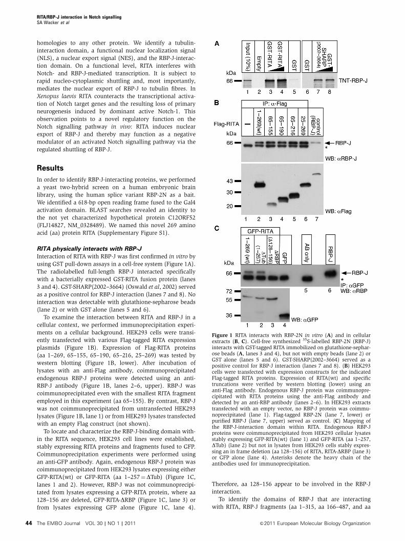
RITA physically interacts with RBP-J
Interaction of RITA with RBP-J was first confirmed in vitro by
using GST pull-down assays in a cell-free system (Figure 1A).
The radiolabelled full-length RBP-J interacted specifically
with a bacterially expressed GST-RITA fusion protein (lanes
3 and 4). GST-SHARP(2002–3664) (Oswald et al, 2002) served
as a positive control for RBP-J interaction (lanes 7 and 8). No
interaction was detectable with glutathione-sepharose beads
(lane 2) or with GST alone (lanes 5 and 6).
To examine the interaction between RITA and RBP-J in a
cellular context, we performed immunoprecipitation experi-
ments on a cellular background. HEK293 cells were transi-
ently transfected with various Flag-tagged RITA expression
plasmids (Figure 1B). Expression of Flag-RITA proteins
(aa 1–269, 65–155, 65–190, 65–216, 25–269) was tested by
western blotting (Figure 1B, lower). After incubation of
lysates with an anti-Flag antibody, coimmunoprecipitated
endogenous RBP-J proteins were detected using an anti-
RBP-J antibody (Figure 1B, lanes 2–6, upper). RBP-J was
coimmunoprecipitated even with the smallest RITA fragment
employed in this experiment (aa 65–155). By contrast, RBP-J
was not coimmunoprecipitated from untransfected HEK293
lysates (Figure 1B, lane 1) or from HEK293 lysates transfected
with an empty Flag construct (not shown).
To locate and characterize the RBP-J-binding domain with-
in the RITA sequence, HEK293 cell lines were established,
stably expressing RITA proteins and fragments fused to GFP.
Coimmunoprecipitation experiments were performed using
an anti-GFP antibody. Again, endogenous RBP-J protein was
coimmunoprecipitated from HEK293 lysates expressing either
GFP-RITA(wt) or GFP-RITA (aa 1–257¼DTub) (Figure 1C,
lanes 1 and 2). However, RBP-J was not coimmunoprecipi-
tated from lysates expressing a GFP-RITA protein, where aa
128–156 are deleted, GFP-RITA-DRBP (Figure 1C, lane 3) or
from lysates expressing GFP alone (Figure 1C, lane 4).
Therefore, aa 128–156 appear to be involved in the RBP-J
interaction.
To identify the domains of RBP-J that are interacting
with RITA, RBP-J fragments (aa 1–315, aa 166–487, and aa
Figure 1 RITA interacts with RBP-2N in vitro (A) and in cellularextracts (B, C). Cell-free synthesized 35S-labelled RBP-2N (RBP-J)interacts with GST-tagged RITA immobilized on glutathione-sephar-ose beads (A, lanes 3 and 4), but not with empty beads (lane 2) orGST alone (lanes 5 and 6). GST-SHARP(2002–3664) served as apositive control for RBP-J interaction (lanes 7 and 8). (B) HEK293cells were transfected with expression constructs for the indicatedFlag-tagged RITA proteins. Expression of RITA(wt) and specifictruncations were verified by western blotting (lower) using ananti-Flag antibody. Endogenous RBP-J protein was coimmunopre-cipitated with RITA proteins using the anti-Flag antibody anddetected by an anti-RBP antibody (lanes 2–6). In HEK293 extractstransfected with an empty vector, no RBP-J protein was coimmu-noprecipitated (lane 1). Flag-tagged RBP-2N (lane 7, lower) orpurified RBP-J (lane 7, upper) served as control. (C) Mapping ofthe RBP-J-interaction domain within RITA. Endogenous RBP-Jproteins were coimmunoprecipitated from HEK293 cellular lysatesstably expressing GFP-RITA(wt) (lane 1) and GFP-RITA (aa 1–257,DTub) (lane 2) but not in lysates from HEK293 cells stably expres-sing an in frame deletion (aa 128–156) of RITA, RITA-DRBP (lane 3)or GFP alone (lane 4). Asterisks denote the heavy chain of theantibodies used for immunoprecipitation.
RITA/RBP-J interaction in Notch signallingSA Wacker et al
The EMBO Journal VOL 30 | NO 1 | 2011 &2011 European Molecular Biology Organization44

166–334) and full-length RBP-J (aa 1–487) were synthesized
in a cell-free system and examined in GST pull-down assays
(Supplementary Figure S2) using GST-mNICD and GST-RITA
as baits. Both, NICD and RITA strongly interact with RBP-J
(aa 166–487; Supplementary Figure S2C and D, lane 3) and
RBP-J (aa 166–334; Supplementary Figure S2C and D, lane 4).
The later fragment represents the Beta-Trefoil-Domain (BTD)
of RBP-J (Kovall, 2008). We conclude that NICD and RITA
bind to the BTD of RBP-J.
RITA interferes with Notch-1 and RBP-VP16-mediated
transcription
To study the effects of RITA on the transcriptional regulation
of a Notch target gene, luciferase experiments were
performed in HeLa cells using an HES1-specific reporter
construct together with expression plasmids for mNotch-
1DE, RBP-VP16, RITA, and RITA proteins defective in RBP-J
interaction (Figure 2). As shown previously (Oswald et al,
2005; Salat et al, 2008), expression of the dominant active
form of mNotch-1 (Notch-1DE) strongly activated the HES1
luciferase reporter. Additional expression of increasing
amounts of RITA gradually impaired transcriptional activity
from the reporter. This appeared to be specific, as expression
of RITA proteins lacking the RBP-J-interaction domain (RITA
156–269 and RITA-DRBP) had no effect on Notch-1DE-
mediated transcriptional activation (Figure 2A). Similar
effects on HES1 promoter activity were observed with a
transcriptional activator RBP-J protein (RBP-VP16). Again,
transcriptional activation was lost after coexpression of RITA,
but not after coexpression of interaction-defective RITA pro-
teins (Figure 2B). Similar results were obtained, when a
luciferase construct with 12 RBP-J-binding sites in front of a
minimal promoter (pGa981/6) was used (data not shown).
RITA is a tubulin-binding protein
We studied the subcellular localization of RITA by fluores-
cence microscopy on HeLa cells transfected with a construct
coding for RITA fused to GFP. Surprisingly, the RITA-GFP
fusion protein showed a fibrillar localization in interphase
cells (Figure 3Aa). Costaining of GFP-RITA-transfected cells
with an anti-a-tubulin antibody showed clear regions of
colocalization in interphase cells (Figure 3Ab and c) as well
as in dividing cells (Figure 3Ad, e, and f), suggesting that
RITA associates with tubulin. Similar structures were
detected with immunofluorescence assays using Flag-tagged
RITA constructs (data not shown). Upon treatment of trans-
fected cells with nocodazole, which interferes with the poly-
merization of microtubules, the fibrillar staining disappeared
within 2 h (Supplementary Figure S3A; Supplementary Movie
S1). During mitosis, GFP-RITA highlighted the spindle
apparatus (Figure 3Ad). In dividing HEK293 cells stably
expressing GFP-RITA, labelling of the microtubule organizing
centre and the mitotic spindle apparatus was observed
(Supplementary Figure S3B; Supplementary Movie S2).
A similar localization of GFP-RITA was found in dissociated
embryonic cells from X. laevis (Supplementary Figure S3C).
GST pull-down assays were performed using purified bovine
a-/b-tubulin as well as lysates from HEK293 cells (Figure 3B).
GST-RITA interacted with bovine tubulin as confirmed by
western blotting using the anti-a-tubulin antibody (Figure 3B,
right, lane 1). In lysates from HEK293 cells, both endogenous
a-tubulin and endogenous RBP-J protein were pulled down
with GST-RITA(wt) (Figure 3B, right, lane 2). RBP-J and
tubulin interact with different domains of RITA, as the GST-
RITA(DTub) lost tubulin-binding capacity, but still interacted
with RBP-J (Figure 3B, right, lane 5). In contrast GST-
RITA(156–269) failed to bind RBP-J, but still interacted with
tubulin (Figure 3B, right, lane 7). Neither a-tubulin nor RBP-J
interacted with GSTalone (Figure 3B, right, lanes 3, 4, and 6).
The region of RITA that confers the tubulin interaction was
confirmed by fluorescence microscopy after transfection of
GFP-RITA deletions into HeLa cells. The C-terminal truncated
RITA (1–257, DTub) (Figure 3C, middle panel) showed diffuse
localization mainly in the cytoplasm, whereas an N-terminal
truncated RITA (156–269; Figure 3C, right panel) showed a
tubulin-like localization similar to full-length RITA (1–269;
Figure 2 RITA interferes with Notch- (A) and RBP-VP16- (B) mediated transcriptional activation of the HES1 promoter. The reporter constructHES1-Luc was transfected into HeLa cells alone (1mg) or together with 50 ng mNotch-1DE expression plasmid (A) or 50 ng RBP-VP16expression plasmid (B) and increasing amounts of expression plasmid (50 and 100 ng) for RITA(wt) or for the RBP-J-interaction-defective RITA(156–269) and RITA-DRBP. Mean values and s.d. (error bars) based on at least four independent experiments are shown. � denotes absence of;þ denotes presence of.
RITA/RBP-J interaction in Notch signallingSA Wacker et al
&2011 European Molecular Biology Organization The EMBO Journal VOL 30 | NO 1 | 2011 45

Figure 3C, left panel). Colocalization of tubulin with either
GFP-RITA(wt) or GFP-RITA (156–269) were shown using an
anti-tubulin antibody (Supplementary Figure S3D). These
results demonstrate that (i) RITA localizes at tubulin struc-
tures, (ii) it interacts directly with tubulin, and (iii) tubulin
and RBP-J interaction occur at separate domains of RITA.
RITA localizes at centrosomes
In fluorescence microscopy images of RITA, we noticed
specific punctate structures in the cells, possibly centro-
somes. Thus, we probed for colocalization of RITA with
different centrosomal markers (Figure 4). Indeed, the
GFP-RITA spots (Figure 4A, upper, left) colocalized with
Figure 3 RITA is a tubulin-binding protein. (A) RITA fused to GFP colocalizes with endogenous tubulin in interphase cells (a–c) and highlightsthe spindle apparatus during mitosis (d–f). HeLa cells were transiently transfected with an expression plasmid for GFP-RITA. At 24 h aftertransfection, cells were fixed and immunostained using an anti-tubulin antibody. The subcellular localization of GFP-RITA and tubulin wasdetermined by fluorescence microscopy. Magnification � 630. (B) RITA interacts with tubulin and with RBP-J with different binding domains.(Left panel) The anti-tubulin antibody used in GST pull-down experiments recognizes the bovine a-/b-tubulin as shown by western blotting.(Right panel) Bovine a-tubulin interacts with GST-RITA (lane 1) but not with GST alone (lane 3). Endogenous a-tubulin together withendogenous RBP-J from HEK293 cell lysates is pulled down with GST-RITA (lane 2) but not with GSTalone (lanes 4 and 6). GST-RITA(DTub, aa1–257) is defective in tubulin binding (lane 5) and GST-RITA (aa 156–269) is defective in RBP-J binding (lane 7). (C) The C-terminus of RITA(aa 156–269) is necessary for tubulin interaction. HeLa cells were transiently transfected with expression plasmids for the indicated RITAproteins fused to GFP. At 24 h after transfection, the living cells were analysed by fluorescence microscopy; scale bars, 5 mm; a, anti; � denotesabsence of; þ denotes presence of.
RITA/RBP-J interaction in Notch signallingSA Wacker et al
The EMBO Journal VOL 30 | NO 1 | 2011 &2011 European Molecular Biology Organization46

antibody-labelled pericentrin (Figure 4A, lower, right).
Whereas the pericentrin antibody labelled an extended region
around the centrosomes (Figure 4B, middle), RITA could only
be detected in two clearly distinct substructures (Figure 4B,
left), suggesting that RITA localizes directly at the centrioles.
A 3D reconstruction of pericentrin/RITA localization at the
centrosomal region is shown in Supplementary Movie S3.
Centrosomal localization of RITA was also shown using an
antibody against the NIMA-related protein kinase Nek2,
which is known to localize at centrosomes (Fry et al, 1998)
(Figure 4C), and in live HeLa cells transfected with GFP-RITA
and g-tubulin fused to the red fluorescent protein mRuby
(Figure 4D).
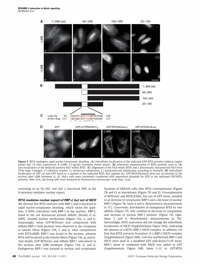
RITA is subject of rapid nucleo-cytoplasmic shuttling
We observed that some of the RITA deletion mutants loca-
lized in the nucleus, suggesting that RITA may undergo
nuclear import and export processes. Indeed, GFP-RITA
expressing cells treated with the nuclear export inhibitor
leptomycin B (LMB), displayed GFP-RITA localization in the
nucleus within 60 min (Figure 5Ae). We obtained similar
results using HEK293 cells stably expressing GFP-RITA
(Supplementary Movie S4). Nuclear accumulation was inde-
pendent of tubulin binding, because RITA(DTub), which only
shows diffuse localization in the cytoplasm (compare
Figure 3C, middle panel, and Supplementary Figure S3D,
middle panel), also accumulated in the nucleus after LMB
treatment (Supplementary Movie S5). In contrast, an
N-terminally truncated form of RITA (aa 25–269) binds to
microtubules but shows enhanced nuclear localization even
without LMB treatment (Supplementary Movie S6). We
searched for an NLS within RITA, using the four deletion
constructs shown in Figure 5B. RITA (aa 85–269) shows
nuclear accumulation (Figure 5Ab), which is enhanced only
marginally after LMB treatment (Figure 5Af). In contrast,
RITA (aa 105–269) shows tubulin association (Figure 5Ac)
and, remarkably, fails to accumulate in the nucleus after LMB
treatment (Figure 5Ag). An N-terminally and C-terminally
truncated RITA (aa 25–155) fails to bind to tubulin and
locates to the nucleus even without LMB treatment (Figure
5Ad and h). From these experiments, we conclude that the
region between aa 85 and 105 may contain a putative NLS.
Alignment with a canonical NLS from SV40 large T-antigen
(TAg) revealed a similarity of basic amino acids within this
region (Figure 5C). Cells transfected with a C-terminal fusion
of the identified peptide sequence (L T P R K K N K Y) in
tandem to GFP showed nuclear staining (Figure 5Db). In
addition, the RKK to AAA substitution in GFP-RITA(NLSmut)
did not show nuclear accumulation after LMB treatment
(compare Figure 5Dc and d). These data clearly prove the
presence of a functional NLS.
A putative NES was identified within the N-terminal region
of RITA proteins from different species (including X. laevis
and human) by the netNES 1.1 software (la Cour et al, 2004)
(Figure 6A). The presence of the NES was verified by
site-directed mutagenesis of RITA from both species.
Substitution of critical amino acids (asterisks in Figure 6A,
lower) to alanine resulted in nuclear accumulation of
GFP-xlRITA(NESmut) (Figure 6B, middle panel) and GFP-
hsRITA(NESmut) (Figure 6B, right panel) without LMB treat-
ment. Consequently, (i) RITA is a nucleo-cytoplasmic shuttle
protein, (ii) nuclear import of RITA is mediated by an NLS
Figure 4 RITA localizes at centrosomes. (A, B) Subcellular localization of RITA and pericentrin (arrows) analysed by epifluorescence (A) orconfocal microscopy (B). (C) Subcellular localization of RITA and Nek2 (arrows). HeLa cells were transiently transfected with an expressionplasmid for GFP-RITA. At 24 h after transfection, cells were fixed and immunostained. Cells were counterstained with DAPI (A, C) to show thenucleus. (D) Colocalization of RITA and g-tubulin. HeLa cells were transiently cotransfected with GFP-RITA and mRuby-g-tubulin. After 24 h,living cells were analysed by fluorescence microscopy; scale bars, 5 mm.
RITA/RBP-J interaction in Notch signallingSA Wacker et al
&2011 European Molecular Biology Organization The EMBO Journal VOL 30 | NO 1 | 2011 47
consisting of aa 92–100, and (iii) a functional NES at the
N-terminus mediates nuclear export.
RITA mediates nuclear export of RBP-J, but not of NICD
We showed that RITA interacts with RBP-J and is involved in
rapid nucleo-cytoplasmic shuttling, which raises the ques-
tion, if RITA colocalizes with RBP-J in the nucleus. RBP-J,
fused to the red fluorescent protein mRuby (Kredel et al,
2009), showed nuclear localization (Figure 7Aa, e, and i).
Surprisingly, when GFP-RITA(wt) was coexpressed with
mRuby-RBP-J, both proteins were observed in the cytoplasm
at tubulin fibres (Figure 7Ab, f, and j). After coexpression
with RITA-DRBP, RBP-J was found in the nucleus, whereas
RITA still localized at the tubulin fibres (Figure 7Ac, g, and k).
And finally, GFP-RITA(wt) and mRuby-RBP-J colocalized in
the nucleus after LMB treatment (Figure 7Ad, h, and l).
Endogenous RBP-J was assayed in nuclear and cytoplasmic
fractions of HEK293 cells after RITA overexpression (Figure
7B and C) or knockdown (Figure 7D and E). Overexpression
of RITA(wt) and RITA(DTub), but not of GFP alone, resulted
in an increase of cytoplasmic RBP-J and a decrease of nuclear
RBP-J (Figure 7B, lanes 4 and 6, densitometric measurements
in 7C). Conversely, knockdown of endogenous RITA by two
siRNAs (Figure 7D, left) resulted in decrease of cytoplasmic
and increase of nuclear RBP-J proteins (Figure 7D, right,
lanes 5 and 6, densitometric measurements in 7E).
Interestingly, RITA expression did not change the subcellular
localization of NICD (Supplementary Figure S4A), indicating
the absence of a RITA–RBP-J–NICD complex. In addition, we
find that RITA prevents formation of a RBP-J/NICD complex
(Supplementary Figure S4B). Cell-free synthesized RBP-J and
NICD were used in a modified GST pull-down-Co-IP assay.
RBP-J alone or combined with NICD was added to GST
(Supplementary Figure S4B, lanes 1–3) or GST-RITA
Figure 5 RITA undergoes rapid nucleo-cytoplasmic shuttling. (A) Subcellular localization of the indicated GFP-RITA proteins without (upperpanel) and 1 h after Leptomycin B (LMB; 2.5 ng/ml) treatment (lower panel). (B) Schematic representation of RITA proteins used in (A)and localization of the deduced putative NLS within RITA. (C) Alignment of the NLS within RITA and a functionally characterized NLS fromSV40 large T-antigen. (*) identical residue, (:) conserved substitution, (.) semiconserved substitution according to clustalW. (D) Subcellularlocalization of GFP (a) and GFP fused to a tandem of the indicated RITA NLS peptide (b). GFP-RITA(NLSmut) does not accumulate in thenucleus after LMB treatment (c, d). HeLa cells were transiently transfected with expression plasmids for GFP or the indicated GFP-RITAproteins. After 24 h, the living cells were analysed by fluorescence microscopy; scale bars, 5mm.
RITA/RBP-J interaction in Notch signallingSA Wacker et al
The EMBO Journal VOL 30 | NO 1 | 2011 &2011 European Molecular Biology Organization48

(Supplementary Figure S4B, lanes 4–6). RBP-J was only
coprecipitated with NICD in the absence of GST-RITA
(Supplementary Figure S4B, lanes 3 and 7). These results
clearly indicate that RITA modulates the nuclear export of
RBP-J and protects RBP-J from NICD loading.
RITA expression reverses Notch-induced loss of primary
neurogenesis in Xenopus laevis
A search for homologous sequences in other species yielded
RITA proteins in many different organisms, representing a
broad range of the metazoan phylum and including the
placozoan Trichoplax adhaerens (Supplementary Figure
S5A). Tubulin association as well as physical interaction of
Trichoplax RITA (taRITA) and RBP-J (taRBP) are conserved
(Supplementary Figure S5B and C). We did not find RITA
protein sequences in D. melanogaster by BLAST searches.
However, Drosophila Su(H), the homologue of RBP-J, inter-
acts with human RITA in GST pull-down assays (Supple-
mentary Figure S5D), suggesting that a functional homologue
of RITA also exists in flies. Expression analysis in mouse
embryos and adult tissue indicates a ubiquitous expression
of RITA, although the levels vary in different tissues
(Supplementary Figure S6A and B). In embryos of the am-
phibian X. laevis, maternal expression of RITA is detected.
Zygotic expression levels are low during gastrula stages and
then increase during neurula stages (Supplementary Figure
S6C). Analysis of the spatial expression pattern by quantita-
tive PCR on embryonic fragments (Supplementary Figure
S6D) and by whole-mount in situ hybridization at neurula
and tadpole stages (Supplementary Figure S6E) indicate a
ubiquitous spatial expression. The temporal expression
pattern suggests that RITA may be a component of the
Notch signalling pathway during neurogenesis. To test this
hypothesis, we first verified that the RBP-J/RITA interaction
is conserved for the X. laevis proteins (Supplementary Figure
S7A and B). Subsequently, we analysed how manipulations
of RITA, as a putative novel modulator of the Notch signal
transduction pathway, affects neurogenesis and organogen-
esis in Xenopus embryos. Numbers for the following
experiments are listed in Supplementary Table S3.
We pursued a loss-of-function approach. Antisense
morpholino oligonucleotides against the start of the RITA
Figure 6 Identification of a nuclear export signal (NES) within RITA. (A, upper) Putative NES sequences within xlRITA (left panel) and hsRITA(right panel) were identified by the netNES 1.1 software. (A, lower) Putative NES sequences (red) and positions within the RITA proteins. Themarked amino acids (asterisks) are mutated to alanine in the RITA(NESmut) constructs. (B) Mutation of NES sequences within RITA proteinsfrom Xenopus laevis (middle panel) and human (right panel) leads to nuclear accumulation without LMB treatment. Wild-type RITA fromXenopus laevis (left panel) served as a control. HeLa cells were transiently transfected with the indicated GFP-RITA constructs. After 24 h, livingcells were analysed by fluorescence microscopy; scale bar, 5mm.
RITA/RBP-J interaction in Notch signallingSA Wacker et al
&2011 European Molecular Biology Organization The EMBO Journal VOL 30 | NO 1 | 2011 49

transcript (MOa) efficiently inhibited translation of RITA(wt),
but not of a Flag-tagged RITA(wt) in vitro (Supplementary
Figure S7C). In vivo, a clear reduction of the neuronal marker
N-tubulin was observed at neurula stage 15 after morpholino
injection, but not in controls without morpholinos
(Figure 8A, upper). Changing the morpholino sequence in
five positions (MOb), thereby reducing its affinity to RITA
mRNA, abolished this effect. Moreover, a standard control
Figure 7 Nucleo-cytoplasmic shuttling of RITA together with RBP-J. (A) Colocalization of RITA and RBP-J. RBP-J-mRuby is found in thenucleus (a, e, i). Coexpression of RITA directs RBP-J out of the nucleus to tubulin fibres (b, f, j). After coexpression of RITA-DRBP (defective forRBP-J interaction), RBP-J remains in the nucleus (c, g, k). LMB treatment leads to colocalization of RITA and RBP-J in the nucleus (d, h, l).HeLa cells were transiently cotransfected with GFP-RITA or GFP-RITA-DRBP together with RBP-J-mRuby. After 24 h, living cells were analysedby fluorescence microscopy. Where indicated, cells were treated with leptomycin B (LMB; 2.5 ng/ml) 1 h prior to imaging, scale bar¼ 5 mm.(B–E) Changes of nuclear versus cytoplasmic RBP-J after overexpression (B, C) or knockdown (D, E) of RITA. HEK293 cells were transientlytransfected with the indicated GFP-RITA constructs (B, lanes 1, 3, 4, and 6) or GFP alone (B, lanes 2 and 5). At 24 h after transfection, cellularlysates were fractionated. (D, E) HEK293 cells were transiently transfected with RITA siRNAs (D, right panel, lanes 2, 3, 5, and 6). At 48 h aftertransfection, cellular lysates were fractionated. Cytoplasmic and nuclear fractions were analysed by western blotting. Knockdown of RITA wasevaluated on mRNA level (D, left panel) by qPCR. Densitometric analysis in (C, E) was performed with the ImageJ software. Signal intensity inthe control experiment was set to 1. Mean and standard error from three experiments are shown.
RITA/RBP-J interaction in Notch signallingSA Wacker et al
The EMBO Journal VOL 30 | NO 1 | 2011 &2011 European Molecular Biology Organization50

morpholino (ctMO) had no influence on the N-tubulin ex-
pression (Figure 8, upper). None of these injections resulted
in gastrulation defects (data not shown). At the late tadpole
stage 45, several malformations were noticed in MOa-injected
embryos. The most obvious effects were smaller eyes, reduced
elements of the jaw (Meckel’s cartilage, palatoquadrate,
Figure 8 Functional characterization of RITA during development of Xenopus laevis. (A) Loss-of-function experiments. Injection of anantisense Morpholino oligonucleotide (MOa) results in reduction of N-tubulin expression at stage 15 (S15 N-Tub), compared to not injectedembryos (ni). Injection of either the mutated Morpholino (MOb) or the standard control Morpholino (ctrl MO) did not result in this reduction.At stage 45 (S45), MOa-injected embryos show reduction of the jaw (mc, Meckel’s cartilage), reduced branchial baskets (bb), smaller eyes (ey),and a deformed intestine (in). MOb and ctrl MO-injected embryos develop as not injected controls. (B) Gain-of-function experiments.Increasing doses of RITA(wt) mRNA (100, 200, 300 pg per embryo) resulted in gastrulation defects as indicated by the open blastopore(arrowheads) and in ectopic activation of N-tubulin at stage 15. Phenotypes at stage 45 show extensive malformations at higher doses,correlated to the gastrulation defects. (C) Rescue of the MO phenotype with low doses of MO-insensitive Flag-RITA(wt) mRNA. The knockdownof N-tubulin activation by the MO approach at stage 15 (MOa, compared to not injected control ni) is compensated by coinjection with the Flag-RITA(wt) mRNA (MOaþRITA(wt)), even at mRNA doses that solely do not have effects on N-tubulin expression (RITA(wt)). Correspondingly,the MOa effect on stage 45 embryos is restored by coinjection of Flag-RITA(wt) mRNA. (D) Rescue of a Notch-induced neurogenesis phenotypeby RITA. A knockdown of N-tubulin at stage 15 by injection of the mNotch-1DE mRNA (N1DE), compared to not injected control (ni), isrestored by coinjection of RITA mRNA (N1DEþRITA(wt)) at mRNA doses that solely do not have a significant effect on N-tubulin expression(RITA(wt)). Stage 45 phenotypes are rescued as well. (E) Influence of different MO-insensitive RITA constructs on the effects of MOa injections.The inhibition of N-tubulin expression at stage 15 by the MOa is compensated by coinjection with RITA(wt). This effect is absent in both, theconstruct deficient of RBP-J binding (þRITA(DRBP)) and the NLS-mutated construct (þRITA(NLSmut)), but it is mimicked by the tubulinbinding deficient RITA(DTub). (F) Influence of different RITA constructs on the effects of Notch-1DE. The inhibition of N-tubulin expression byNotch-1DE (N1DE) is antagonized by coinjection with RITA(wt). Again, this is mimicked by RITA(DTub), but neither by RITA(DRBP) nor byRITA(NLSmut). (G) RITA(wt) restores missexpression of Notch target genes after injection of the constitutive active mNotch-1DE. Upregulationof xEsr-1, xEsr-2, xEsr-7, xEsr-10, and downregulation of N-tubulin after Notch-1DE injection (N1DE) was rescued by coinjection with a lowdose of RITA(wt) (N1DEþRITA(wt)) that solely did not show effects (RITA(wt)). Mean values and standard error of the mean (error bars)based on three experiments are shown. Numbers of injected embryos and phenotypes for (A–D) are listed in Supplementary Table S3.(H) Schematic drawing of identified domains within RITA. Numbers (of amino acids) indicate positions in respect of human RITA. NES nuclearexport signal, RCR1 and RCR2 RITA conserved repeats, NLS nuclear localization signal, RBP-binding domain, Tubulin-binding domain.
RITA/RBP-J interaction in Notch signallingSA Wacker et al
&2011 European Molecular Biology Organization The EMBO Journal VOL 30 | NO 1 | 2011 51

ceratohyale), reduced branchial arches, and a deformation of
the intestine (Figure 8A, lower). Oedemas were observed
regularly, axial structures were not significantly affected
(not shown). Effects of MOb injections were much milder,
showing an almost normal phenotype, ctMO-injected
embryos looked like not injected controls (Figure 8A).
A gain-of-function approach by injecting RITA(wt) mRNA
showed dose-dependent responses (Figure 8B). RNA doses of
o100 pg per embryo resulted in normal N-tubulin patterns at
stage 15 and a normal phenotype at stage 45. Doses 4100 pg
induced gastrulation defects and an apparent increase of
N-tubulin expression (Figure 8B, upper). Identical results
were obtained with mRNA encoding the Flag-tagged form
of RITA(wt) (not shown). Stage 45 tadpoles assembled de-
fects of many organ systems, including eyes, CNS, head and
axial skeleton, and intestine (Figure 8B, lower). A dose
4300 pg RITA mRNA per embryo was lethal before the end
of neurulation (not shown).
The specificity of the RITA knockdown phenotypes was
further supported by an experiment attempting to override
the MOa effect by injection of an MO-insensitive mRNA,
using a Flag-tagged RITA(wt) construct. As the MOa se-
quence is not close to the start of translation of this construct,
the inhibition is inefficient (Supplementary Figure S7C).
A low dose of Flag-tagged RITA mRNA (40 pg per embryo)
did not have any visible effect, if injected solely. However,
this dose was entirely sufficient to compensate the effects of
MOa injections, as shown by a normal N-tubulin expression
at stage 15 and normal phenotypes at stage 45 (Figure 8C).
Activation of the Notch signalling pathway by the Delta
ligand results in a process called lateral inhibition during
neurogenesis (Chitnis, 1995; Chitnis et al, 1995). In line with
this process, injection of mRNA encoding a dominant active
form of Notch-1 (Notch-1DE) resulted in a reduced number of
primary neurons (Oswald et al, 2002). We tested the func-
tional role of RITA as a negative modulator of Notch signal-
ling by performing coinjection experiments using Notch-1DE
together with RITA(wt). Importantly, coinjections with fairly
low doses of RITA(wt) mRNA (100 pg per embryo) were
indeed efficient to antagonize the effects of the constitutive
active Notch-1DE on N-tubulin expression at stage 15 and
phenotypes at stage 45 (Figure 8D). To further investigate the
function of RITA as a negative modulator of the Notch
signalling pathway, mRNA synthesized from different MO-
insensitive flag-tagged constructs was injected into Xenopus
embryos in combination with either MOa or in combination
with the constitutive active Notch-1DE (N1DE). As shown
above, RITA(DTub) is defective in tubulin binding,
RITA(DRBP) is defective in RBP-J binding, and in
RITA(NLSmut) nuclear localization is disabled. Whereas
RITA(DRBP) and the RITA(NLSmut) mRNAs lost the ability
to restore the MOa effects on N-tubulin expression,
RITA(DTub) mRNA mimicked the rescue effect of RITA(wt)
(Figure 8E). Therefore, restoration depends on nuclear
localization and on RBP-J binding of RITA, but not on its
ability to interact with tubulin. Consistently, in coinjection
experiments with Notch-1DE, antagonistic effects were
obtained from RITA(DTub) and from RITA(wt).
RITA(DRBP) and the RITA(NLSmut) failed to restore the
N-tubulin expression, confirming the necessity of both,
nuclear localization and RBP-J interaction of RITA
(Figure 8F). In addition, quantitative PCR analysis revealed
that the transcriptional activation of several Notch targets
(xEsr-1, xEsr-2, xEsr-7, xEsr-10, xHesR1) by dominant-acti-
vated Notch-1DE as well as the resulting downregulation of
N-tubulin was reversed after coinjection of RITA(wt)
(Figure 8G).
In summary, the RITA-specific gain-of-function and loss-of-
function experiments in X. laevis show strong effects on
Notch targets during embryogenesis. Most importantly, in
agreement with reporter-gene assays in cell culture, RITA acts
as a negative modulator of the Notch signalling pathway. This
function of RITA depends on both, nuclear localization and
RBP-J-interaction domains, but not on the ability to bind to
tubulin.
Discussion
In this study we present the novel, highly conserved RBP-J-
interacting protein RITA. Identified domains are depicted in
Figure 8H. We show that besides interaction with RBP-J, RITA
interacts with microtubules and locates at the centrosomes.
On a molecular level, RITA undergoes a rapid nucleo-cyto-
plasmic shuttle process and transports RBP-J out of the
nucleus to tubulin fibres. We propose that it thereby limits
the amount of nuclear RBP-J. On a functional level, RITA
affects Notch-mediated transcription and primary neurogen-
esis. From our data, we postulate that RITA acts as a negative
modulator of Notch signalling, representing a novel regula-
tory mechanism on the level of DNA binding.
RITA–RBP-J interaction
Experiments with deletion constructs demonstrate that the
central part of RITA is necessary for RBP-J interaction
(Figure 1B). RITA interacts with the BTD of RBP-J
(Supplementary Figure S2). We do not have evidence that
RITA forms a ternary complex with RBP-J and NICD.
In contrast to the colocalization of RITA and RBP-J
(Figure 7), NICD remains localized in the nucleus, when
RITA is accumulated at the tubulin fibres (Supplementary
Figure S4A). Notch proteins form a high-affinity RBP-J-inter-
action module with their RBP-J association module (RAM)
domain. A highly conserved W-X-P motif within the RAM
domain contributes to the interaction with the RBP-J BTD
(Tamura et al, 1995). Interestingly, we also find a W-X-P motif
within the RBP-J-interaction domain of RITA proteins from
various species (Supplementary Figure S5A). This W-X-P
motif within RITA may contribute to RBP-J interaction and
thereby compete against the NICD binding to RBP-J
(Supplementary Figure S4B).
Identification of specific domains within RITA
Initially, we could not identify conserved domains within
RITA using bioinformatics tools. Microscopic and biochem-
ical analysis revealed that RITA interacts with tubulin. The
C-terminus turned out to be essential for tubulin binding, as a
deletion of the last 12 aa resulted in a diffuse cytoplasmic
localization (Figure 3). This is further supported by the fact
that a RITA protein N-terminally fused to EGFP lost its
tubulin-binding capability (not shown), and the high con-
servation of the last amino acids within different species (see
Supplementary Figure S5A). A carboxy-terminal fusion of the
last 14 aa of human RITA to EGFP did not bind to tubulin (not
shown), suggesting that the C-terminus of RITA is necessary,
RITA/RBP-J interaction in Notch signallingSA Wacker et al
The EMBO Journal VOL 30 | NO 1 | 2011 &2011 European Molecular Biology Organization52

but not sufficient for the tubulin interaction. However, aa
156–269 of human RITA are sufficient for tubulin binding
(see Figure 3C), indicating that additional sequences mediate
tubulin binding. RITA also localizes at the centrosomes
(Figure 4). The region responsible for this interaction appears
to differ from the one that confers binding to a-/b-tubulin, as
a carboxy-terminally truncated RITA protein (aa 1–257) still
localizes at the centrosomes (Supplementary Movie S1). The
biological function of RITA at the centrosomes is not yet
clarified. A recent study has shown a role of centrosome
inheritance during asymmetric cell divisions in the develop-
ing brain, maintaining neural progenitor cells in the neocor-
tex (Wang et al, 2009). One could speculate, if RITA has a role
in this context.
RITA rapidly accumulates in the nucleus after inhibition of
nuclear export by LMB. This behaviour indicates that the
subcellular localization of RITA depends on nuclear import
and export processes. We identified a functional NLS together
with a functional NES within RITA, mediating nucleo-cyto-
plasmic shuttling (Figures 5 and 6). In addition, nuclear
transport of RITA is an essential prerequisite for its function
in X. laevis primary neurogenesis (Figure 8E and F). Similarly
to human RITA, the RITA proteins from X. laevis and
T. adhaerens accumulate in the nucleus after LMB treatment
of transfected cells (not shown).
How is the nuclear–cytoplasmic shuttle process of RITA
regulated?
It has been shown previously, that RBP-J/CSL proteins can be
detected in the nucleus as well as in the cytoplasm and their
subcellular distribution changes in defined physiological
contexts (Sakai et al, 1995; Zhou and Hayward, 2001; Krejci
and Bray, 2007). Here, we provide clear evidence that RITA
mediates this redistribution. Most signalling pathways make
use of limiting their central transcription factors in the
nucleus to activate, modulate, or fine-tune their transcrip-
tional output. Regulation of subcellular localization of RBP-J
by RITA may depend on phosphorylation/dephosphorylation
of RITA. Besides the C-terminal sequences, the NLS, and the
W-X-P motif, two highly conserved, almost identical amino
acid stretches (RITA Conserved Repeat 1 and 2 (RCR1 and
RCR2 in Figure 8H and Supplementary Figure S5A)) can be
exclusively identified within RITA proteins. RCR1 and RCR2
show the consensus motif S-Y-X-D-E-(S/T)-L-F-G, which
could represent specific protein–protein interaction motifs
or target sites for posttranslational modifications. Indeed,
RITA is highly phosphorylated (not shown). A database
search for putative kinases, using the Minimotif Miner
(MnM 2.0) software (Balla et al, 2006), revealed target sites
for several serine/threonine kinases. Therefore, yet not
specified kinases and phosphatases may mediate RITA action
in regulating the subcellular localization of RBP-J.
Consequences of limiting the amount of nuclear RBP-J
by RITA
RBP-J is a key element in the regulation of transcription by
Notch signalling. After nuclear translocation, NICD interacts
with RBP-J and recruits a coactivator complex to activate
transcription of Notch target genes. The transcriptional
response to NICD may be controlled by the amount of RBP-
J in the nucleus. In addition, NICD was reported to interact
with nuclear components of additional signal transduction
pathways. Especially crosstalk between Notch signalling,
NF-kB signalling, and TGFb signalling have been described
(Kluppel and Wrana, 2005; Osipo et al, 2008; Poellinger and
Lendahl, 2008). Given the fact, that after signal activation
NICD is found at very low concentrations in the nucleus, the
abundance of its major interaction partner RBP-J might be
critical for functional association of NICD with additional
nuclear factors. Therefore, regulation of the subcellular
distribution of RBP-J by RITA and, as a consequence, its
concentration in the nucleus, might be critical for the cross-
talk of Notch signalling with other pathways. In this regard, it
will be interesting to see differential expression of RITA
during development and disease and to find correlations to
possible changes in Notch target gene expression.
RITA modulates early organogenesis in Xenopus laevis
The Notch signalling pathway affects many different devel-
opmental processes, most prominently the effect on primary
neurogenesis (Chitnis et al, 1995; Wettstein et al, 1997).
Moreover, effects on germ layer formation (Abe et al, 2005;
Contakos et al, 2005), somitogenesis (Jen et al, 1997; Davis
et al, 2001), development of heart (Rones et al, 2000; Miazga
and McLaughlin, 2009), pronephros (McLaughlin et al,
2000), head structures and eyes (Onuma et al, 2002; Ogino
et al, 2008), and axial structures (Lopez et al, 2003) have
been described in X. laevis and other organisms. A loss-of-
function approach, using a morpholino against RITA, and
RITA gain-of-function experiments resulted in defects that are
in correspondence with Notch overexpression phenotypes.
An initial analysis in mouse shows highest levels of RITA
expression in several of the organs that are affected by Notch
pathway manipulations, for example brain, heart, muscle,
kidney, and intestine (Supplementary Figure S6A and B).
In accordance with a function of RITA as regulator of Notch
signalling are the rescue experiments. A RITA mRNA coin-
jection in Xenopus embryos restored all observed effects of a
constitutive active Notch construct Notch-1DE, including
restoration of the N-tubulin expression, phenotypical effects,
and changes in the expression levels of known downstream
targets of the Notch signalling pathway.
These results and overlapping expression patterns of RITA
and Notch strengthen the paradigm that RITA acts as a
general negative modulator of an activated Notch signalling
pathway. RITA may do so by interacting with RBP-J, regulat-
ing its cytoplasmic/nuclear localization and consequently
modulating the Notch-dependent transcriptional response.
This general molecular mechanism, that is the regulated
targeting of a transcription factor in or out of the nucleus
by posttranslational modifications or a specific inhibitory
protein, is now for the first time indicated within the Notch
signalling pathway.
Materials and methods
OligonucleotidesA list of all oligonucleotides used for plasmid construction and real-time PCR and the morpholino sequences, used for a specificknockdown in X. laevis, are included in Supplementary data.
Yeast two-hybrid screeningYeast two-hybrid screening was carried out with the MATCHMAKERsystem (Clontech) according to the manufacturer’s instructions asdescribed previously (Oswald et al, 2002).
RITA/RBP-J interaction in Notch signallingSA Wacker et al
&2011 European Molecular Biology Organization The EMBO Journal VOL 30 | NO 1 | 2011 53

PlasmidsThe vectors pcDNA3-Flag-RBP-2N, CMV-RBP-VP16, HES1-LUC,pcDNA3-Flag-1-SHARP(RBPID), pcDNA3mNotch-1DE, plasmid forexpression of GST-mNotch-1-IC (GST-mNICD) were described inOswald et al (2002). The expression plasmids for mNotch-1-ICmRuby and mNotch-1-IC(DRBP/DEP)-mRuby were made by ex-changing the fluorescent tag in the corresponding GFP constructs(Oswald et al, 2001). Constructs for bacterial expression of SHARP-specific GST-fusion proteins (SHARP-RBPID, SHARP-2002–3663)were described in Oswald et al (2005). The expression constructpcDNA3-RBP-2N-mRuby was described in Salat et al (2008). Theexpression plasmid for X. laevis RBP-J, pCS2-xlSu(H), was a kindgift from Ying Cao, University of Ulm. The expression plasmid forg-Tubulin fused to mRuby, was kindly provided by MW Davidson(The Florida State University, Tallahassee, FL). Expression plasmidsfor the RITA proteins (human, X. laevis, and T. adhaerens) weremade by PCR. For further details see Supplementary data.
Cell culture and preparation of cell extractsHEK293 cells were transfected with DNA using the calciumphosphate coprecipitation method (Promega). HEK293 were trans-fected with siRNA using the GeneEraserTM siRNA transfection rea-gent (Stratagene). HeLa cells were transfected using the Nanofectine(PAA) transfection reagent. All transfections were performedaccording to the manufacturer’s instructions. The cell linesHEK293 (ATCC CRL 1573) and HeLa (ATCC CCL 2) were grown inDulbecco’s modified Eagle’s medium (DMEM, Gibco) supplemen-ted with 10% fetal calf serum, penicillin, and streptomycin. TheHEK293 cell lines stably expressing GFP and GFP-RITA fusionproteins (HEK293RITA 1–269(wt), HEK293RITA 1–257, HEK293RITA DRBP,HEK293RITA 25–269, HEK293EGFP) were propagated with 300mg/mlNeomycin (G418, PAA) within the medium.
Coimmunoprecipitation and western blottingWhole-cell lysates for western blotting and immunoprecipitationexperiments were prepared as described previously (Salat et al,2008). Nuclear and cytoplasmic fractions were prepared usingstandard procedures as described in Supplementary data. IPexperiments were carried out using whole-cell extracts either fromHEK293 cells stably expressing GFP-RITA fusion proteins orHEK293 cells 24 h after transfection with Flag-RITA expressingconstructs as described in Supplementary data.
In vitro protein translationProteins were synthesized in the presence of [35S] methionine usinga reticulocyte lysate-coupled transcription/translation system(Promega). Translation and labelling quality were monitored bySDS–PAGE.
GST pull-down assayGST-protein and the GST-fusion proteins were expressed inEscherichia coli strain BL21 (Stratagene) and stored as wholebacterial lysates at �801C. Approximately 1 mg of GST-protein andGST-fusion protein were immobilized with glutathione-sepharosebeads (Amersham) and incubated together with the in vitrotranslated proteins under rotation at 41C for 1 h. Beads werewashed four times with 600 ml buffer A (40 mM HEPES (pH 7.5),5 mM MgCl2, 0.2 mM EDTA, 0.5% Nonidet P40 (NP-40), and100 mM KCl) and four times with 600 ml buffer B (equivalent tobuffer A, but containing 300 mM KCl). After the washing steps, thebeads were resuspended in SDS–PAGE loading buffer and theproteins were separated by SDS–PAGE. The gels were dried andexposed to X-ray films.
Luciferase assayHeLa cells (5�104) were transfected in 24-well plates with 1mg ofreporter plasmid DNA together with various amounts of expression
plasmid. Luciferase activity was determined from at least threeindependent transfections with 20ml of cleared lysate in an LB 9501luminometer (Berthold) using the luciferase assay system fromPromega.
Quantitative RNA analysisTotal RNA was isolated from different mouse tissues, ES cells andX. laevis embryos as previously described (Oswald et al, 2002). HalfXenopus embryos were cut with eyebrow knives after removal ofthe perivitelline membrane using forceps. Expression levels ofmRNA were quantified using real-time PCR (TaqMan, PE AppliedBiosystems). For PCR, cDNAs were reverse transcribed from 2mg oftotal RNA. The PCR reaction (denaturation 951C for 2 min followedby 40 cycles of 951C for 15 s and 601C for 1 min) was performedusing Sybr Green PCR Core Reagents (PE Applied Biosystems) andprimer combinations are listed in Supplementary data.
Injection of mRNA and morpholinosEmbryos were staged according to Nieuwkoop and Faber (1956).In vitro fertilization, embryo culture, and microinjections werecarried out as previously described (Wacker et al, 2000). For detailsabout injection of mRNAs and MOs see Supplementary data.Numbers for the injection experiments and distribution of attainedphenotypes are listed in Supplementary Table S3.
Detection of gene expression by in situ hybridizationWhole-mount in situ hybridization was performed as previouslydescribed (Harland, 1991), except that the RNAse step was omitted.RITA digoxigenin-labelled probes were generated from pcDNA3-RITA(xl) as a template (sense T7/Xba, antisense SP6/EcoRI). Anantisense digoxigenin-labelled probe was used for N-tubulin(Marcus et al, 1998).
Fluorescence microscopyFor immunofluorescence imaging, HeLa cells were prepared asdescribed in Supplementary data. The following antibodies andantisera were used: anti-a-Tubulin, mouse monoclonal IgG, (T9026,Sigma), secondary antibody, Cy3TM-coupled sheep anti-mouse IgG(C2181, Sigma), anti-Pericentrin (Martin-Subero et al, 2003),secondary antibody, Alexa-Fluor-568-coupled goat anti-rabbit IgG(A11011, Invitrogen). Production of the anti-NEK-2 antiserum isdescribed in Supplementary data. The secondary antibody wasA11011 (Invitrogen).
Supplementary dataSupplementary data are available at The EMBO Journal Online(http://www.embojournal.org).
Acknowledgements
We thank Y Cao and B Schierwater for providing us with reagents.We thank B Korte, N Heymann, R Rittelman, and S Schirmer forexcellent technical assistance. This study was supported by theDeutsche Forschungsgemeinschaft (DFG, SFB 497/B9 and SFB 518/A18 to FO, SFB 497/A1 to WK, Wi1990/2-1 to JW). GUN acknowl-edges support by the State of Baden-Wurttemberg and the DFG(CFN, Ni 291/9 and SFB 497/C10).
Authors’ contributions: FO and SW wrote the manuscript; FO andWK conceived and designed the experiments; FO, SW, CA, GW, UK,KC, HH, and BB performed the experiments; and FO, SW, CA, JW,GUN, and TB analysed the data.
Conflict of interest
The authors declare that they have no conflict of interest.
References
Abe T, Furue M, Kondow A, Matsuzaki K, Asashima M (2005)Notch signaling modulates the nuclear localization of carboxy-
terminal-phosphorylated smad2 and controls the competence ofectodermal cells for activin A. Mech Dev 122: 671–680
RITA/RBP-J interaction in Notch signallingSA Wacker et al
The EMBO Journal VOL 30 | NO 1 | 2011 &2011 European Molecular Biology Organization54

Artavanis-Tsakonas S, Rand MD, Lake RJ (1999) Notch signaling:cell fate control and signal integration in development. Science284: 770–776
Balla S, Thapar V, Verma S, Luong T, Faghri T, Huang CH,Rajasekaran S, del Campo JJ, Shinn JH, Mohler WA,Maciejewski MW, Gryk MR, Piccirillo B, Schiller SR, SchillerMR (2006) Minimotif Miner: a tool for investigating proteinfunction. Nat Methods 3: 175–177
Borggrefe T, Oswald F (2009) The Notch signaling pathway:transcriptional regulation at Notch target genes. Cell Mol LifeSci 66: 1631–1646
Bray SJ (2006) Notch signalling: a simple pathway becomes com-plex. Nat Rev Mol Cell Biol 7: 678–689
Chitnis A, Henrique D, Lewis J, Ish-Horowicz D, Kintner C (1995)Primary neurogenesis in Xenopus embryos regulated by ahomologue of the Drosophila neurogenic gene Delta. Nature375: 761–766
Chitnis AB (1995) The role of Notch in lateral inhibition and cellfate specification. Mol Cell Neurosci 6: 311–321
Contakos SP, Gaydos CM, Pfeil EC, McLaughlin KA (2005)Subdividing the embryo: a role for Notch signaling during germlayer patterning in Xenopus laevis. Dev Biol 288: 294–307
Davis RL, Turner DL, Evans LM, Kirschner MW (2001) Moleculartargets of vertebrate segmentation: two mechanisms controlsegmental expression of Xenopus hairy2 during somiteformation. Dev Cell 1: 553–565
Fortini ME (2009) Notch signaling: the core pathway and itsposttranslational regulation. Dev Cell 16: 633–647
Fry AM, Meraldi P, Nigg EA (1998) A centrosomal function for thehuman Nek2 protein kinase, a member of the NIMA family of cellcycle regulators. EMBO J 17: 470–481
Fryer CJ, White JB, Jones KA (2004) Mastermind recruitsCycC:CDK8 to phosphorylate the Notch ICD and coordinateactivation with turnover. Mol Cell 16: 509–520
Harland RM (1991) In situ hybridization: an improved whole-mountmethod for Xenopus embryos. Methods Cell Biol 36: 685–695
Jen WC, Wettstein D, Turner D, Chitnis A, Kintner C (1997)The Notch ligand, X-Delta-2, mediates segmentation of theparaxial mesoderm in Xenopus embryos. Development 124:1169–1178
Kao HY, Ordentlich P, Koyano-Nakagawa N, Tang Z, Downes M,Kintner CR, Evans RM, Kadesch T (1998) A histone deacetylasecorepressor complex regulates the Notch signal transductionpathway. Genes Dev 12: 2269–2277
Kluppel M, Wrana JL (2005) Turning it up a Notch: cross-talkbetween TGF beta and Notch signaling. Bioessays 27: 115–118
Koch U, Radtke F (2007) Notch and cancer: a double-edged sword.Cell Mol Life Sci 64: 2746–2762
Kopan R, Ilagan MX (2009) The canonical Notch signaling pathway:unfolding the activation mechanism. Cell 137: 216–233
Kovall RA (2008) More complicated than it looks: assembly ofNotch pathway transcription complexes. Oncogene 27: 5099–5109
Kredel S, Oswald F, Nienhaus K, Deuschle K, Rocker C, Wolff M,Heilker R, Nienhaus GU, Wiedenmann J (2009) mRuby, a brightmonomeric red fluorescent protein for labeling of subcellularstructures. PLoS One 4: e4391
Krejci A, Bray S (2007) Notch activation stimulates transient andselective binding of Su(H)/CSL to target enhancers. Genes Dev 21:1322–1327
Kuroda K, Han H, Tani S, Tanigaki K, Tun T, Furukawa T, TaniguchiY, Kurooka H, Hamada Y, Toyokuni S, Honjo T (2003) Regulationof marginal zone B cell development by MINT, a suppressor ofNotch/RBP-J signaling pathway. Immunity 18: 301–312
la Cour T, Kiemer L, Molgaard A, Gupta R, Skriver K, Brunak S(2004) Analysis and prediction of leucine-rich nuclear exportsignals. Protein Eng Des Sel 17: 527–536
Liefke R, Oswald F, Alvarado C, Ferres-Marco D, Mittler G,Rodriguez P, Dominguez M, Borggrefe T (2010) Histone demethy-lase KDM5A is an integral part of the core Notch-RBP-J repressorcomplex. Genes Dev 24: 590–601
Lopez SL, Paganelli AR, Siri MV, Ocana OH, Franco PG, Carrasco AE(2003) Notch activates sonic hedgehog and both are involved inthe specification of dorsal midline cell-fates in Xenopus.Development 130: 2225–2238
Marcus EA, Kintner C, Harris W (1998) The role of GSK3beta inregulating neuronal differentiation in Xenopus laevis. Mol CellNeurosci 12: 269–280
Martin-Subero JI, Knippschild U, Harder L, Barth TF, Riemke J,Grohmann S, Gesk S, Hoppner J, Moller P, Parwaresch RM,Siebert R (2003) Segmental chromosomal aberrations and centro-some amplifications: pathogenetic mechanisms in Hodgkin andReed-Sternberg cells of classical Hodgkin’s lymphoma? Leukemia17: 2214–2219
McLaughlin KA, Rones MS, Mercola M (2000) Notch regulates cellfate in the developing pronephros. Dev Biol 227: 567–580
Miazga CM, McLaughlin KA (2009) Coordinating the timing ofcardiac precursor development during gastrulation: a new rolefor Notch signaling. Dev Biol 333: 285–296
Miele L, Golde T, Osborne B (2006) Notch signaling in cancer. CurrMol Med 6: 905–918
Morel V, Lecourtois M, Massiani O, Maier D, Preiss A, SchweisguthF (2001) Transcriptional repression by suppressor of hairlessinvolves the binding of a hairless-dCtBP complex in Drosophila.Curr Biol 11: 789–792
Moshkin YM, Kan TW, Goodfellow H, Bezstarosti K, Maeda RK,Pilyugin M, Karch F, Bray SJ, Demmers JA, Verrijzer CP (2009)Histone chaperones ASF1 and NAP1 differentially modulateremoval of active histone marks by LID-RPD3 complexes duringNOTCH silencing. Mol Cell 35: 782–793
Nieuwkoop PD, Faber J (1956) Normal Table of Xenopus laevis(Daudin). Amsterdam: North Holland Publishing Company
Ogino H, Fisher M, Grainger RM (2008) Convergence of a head-fieldselector Otx2 and Notch signaling: a mechanism for lens speci-fication. Development 135: 249–258
Onuma Y, Takahashi S, Asashima M, Kurata S, Gehring WJ (2002)Conservation of Pax 6 function and upstream activation by Notchsignaling in eye development of frogs and flies. Proc Natl Acad SciUSA 99: 2020–2025
Osipo C, Golde TE, Osborne BA, Miele LA (2008) Off the beatenpathway: the complex cross talk between Notch and NF-kappaB.Lab Invest 88: 11–17
Oswald F, Kostezka U, Astrahantseff K, Bourteele S, Dillinger K,Zechner U, Ludwig L, Wilda M, Hameister H, Knochel W,Liptay S, Schmid RM (2002) SHARP is a novel componentof the Notch/RBP-Jkappa signalling pathway. EMBO J 21:5417–5426
Oswald F, Tauber B, Dobner T, Bourteele S, Kostezka U, Adler G,Liptay S, Schmid RM (2001) p300 acts as a transcriptionalcoactivator for mammalian Notch-1. Mol Cell Biol 21:7761–7774
Oswald F, Winkler M, Cao Y, Astrahantseff K, Bourteele S, KnochelW, Borggrefe T (2005) RBP-Jkappa/SHARP recruits CtIP/CtBPcorepressors to silence Notch target genes. Mol Cell Biol 25:10379–10390
Poellinger L, Lendahl U (2008) Modulating Notch signaling bypathway-intrinsic and pathway-extrinsic mechanisms. CurrOpin Genet Dev 18: 449–454
Rones MS, McLaughlin KA, Raffin M, Mercola M (2000) Serrate andNotch specify cell fates in the heart field by suppressing cardio-myogenesis. Development 127: 3865–3876
Roy M, Pear WS, Aster JC (2007) The multifaceted role of Notch incancer. Curr Opin Genet Dev 17: 52–59
Sakai T, Furukawa T, Iwanari H, Oka C, Nakano T, Kawaichi M,Honjo T (1995) Loss of immunostaining of the RBP-J kappatranscription factor upon F9 cell differentiation induced byretinoic acid. J Biochem 118: 621–628
Salat D, Liefke R, Wiedenmann J, Borggrefe T, Oswald F (2008)ETO, but not leukemogenic fusion protein AML1/ETO, augmentsRBP-Jkappa/SHARP-mediated repression of notch target genes.Mol Cell Biol 28: 3502–3512
Tamura K, Taniguchi Y, Minoguchi S, Sakai T, Tun T, Furukawa T,Honjo T (1995) Physical interaction between a novel domain ofthe receptor Notch and the transcription factor RBP-J kappa/Su(H). Curr Biol 5: 1416–1423
Wacker S, Grimm K, Joos T, Winklbauer R (2000) Development andcontrol of tissue separation at gastrulation in Xenopus. Dev Biol224: 428–439
Wallberg AE, Pedersen K, Lendahl U, Roeder RG (2002) p300 andPCAF act cooperatively to mediate transcriptional activation fromchromatin templates by notch intracellular domains in vitro. MolCell Biol 22: 7812–7819
Wang X, Tsai JW, Imai JH, Lian WN, Vallee RB, Shi SH (2009)Asymmetric centrosome inheritance maintains neural progenitorsin the neocortex. Nature 461: 947–955
RITA/RBP-J interaction in Notch signallingSA Wacker et al
&2011 European Molecular Biology Organization The EMBO Journal VOL 30 | NO 1 | 2011 55

Wettstein DA, Turner DL, Kintner C (1997) The Xenopus homologof Drosophila suppressor of hairless mediates Notchsignaling during primary neurogenesis. Development 124:693–702
Wilson JJ, Kovall RA (2006) Crystal structure of the CSL-Notch-Mastermind ternary complex bound to DNA. Cell 124:985–996
Wu L, Aster JC, Blacklow SC, Lake R, Artavanis-Tsakonas S, GriffinJD (2000) MAML1, a human homologue of Drosophila master-mind, is a transcriptional co-activator for NOTCH receptors. NatGenet 26: 484–489
Zhou S, Hayward SD (2001) Nuclear localization of CBF1 isregulated by interactions with the SMRT corepressor complex.Mol Cell Biol 21: 6222–6232
RITA/RBP-J interaction in Notch signallingSA Wacker et al
The EMBO Journal VOL 30 | NO 1 | 2011 &2011 European Molecular Biology Organization56