ringing the bell for cancer patients - … · ringing the bell for cancer patients ... in 2009 a...
TRANSCRIPT

RINGING THE
BELL FOR
CANCER PATIENTS
R I V E R S I D E WA LT E R
R E E D C A N C E R P R O G R A M
2010
ANNUAL REV IEW

Riverside Walter Reed Cancer Program
Riverside Walter Reed Hospital has been treating cancer patients on the Middle Peninsula since 2004. Treatment resources include Surgery, Medical Oncology, Radiation Oncology and Supportive
Care measures.
Radiation Therapy is delivered via an External Beam Linear Accelerator. IMRT is also available providing a more precise focusing of the beam, minimizing the damage to surrounding tissues. Radiation oncologists consult National Comprehensive Cancer Guidelines (NCCN) guidelines when developing treatment plans.
Medical Oncology provides nine treatment chairs and one treatment bed for chemotherapy outpatients. All Medical Oncologists at the Riverside Cancer Infusion Center consult the National Comprehensive Cancer Network (NCCN) treatment guidelines when making recommendations. In addition to providing custom chemotherapy regimens, patients may also undergo diagnostic procedures such as bone marrow biopsies if needed.
Community Outreach is an essential part of the Walter Reed Cancer Program. In 2010 the American Cancer Society Look Good Feel Better Program was established at Walter Reed and is holding monthly meetings. This free program offers woman an opportunity to improve their self esteem. Monthly cervical cancer screenings are offered at the Gloucester-Mathews Free clinic as well. Cancer Services Community Outreach continues to be an active participant at many other local events and festivals providing information and awareness to the public.
Hospice is offered when treatment is no longer an option for a patient. A full range of services are made available to the patient and their family. Patients can maintain a close working relationship with their PCP and also utilize the service of the hospice medical director, if the need should arise. The patient also has the benefit of the hospice’s expertise in palliative care. By acknowledging the physical and emotional needs of the patient, family and caregivers, hospice is dedicated to providing information to assist in maintaining the highest possible levels of functioning and to promote comfort in the patient’s place of residence. Hospice strives to improve the quality of life for the terminally ill and their loved ones.
Review of 2009 Accessions
The Riverside Cancer Registry began collecting and reporting data for Riverside Walter Reed Hospital in 2007. Since then the registry has documented information on over 800 patients. A 90% follow-up is to be maintained for patients diagnosed and/or treated for cancer at our facility. To date the cancer registry has achieved a follow-up rate of over 96%.
In 2009 a total of 241 new cases were identified. Of those, 211 (87.5%) were analytic (diagnosed and /or treated at RWRH). The top five sites for RWRH are Breast, Prostate, Lung, Colorectal and Bladder. The analytic growth trend for these sites is shown below in Figure 1. These top 5 sites account for 63% of the total analytic caseload seen at RWRH in 2009. From 2007 to 2009 the total case load for RWRH has grown from 199 patients in 2007 to 241 patients in 2009, a 21% growth over 2 years.
The Cancer Registry serves as a repository of information for physicians and administrators to utilize when investigating equipment purchase, treatment decisions, staffing issues, and overall program needs.
Cancer Conferences provide an opportunity for physicians to prospectively review cases with the multidisciplinary team. In addition to the monthly general cancer conferences, physicians on the middle peninsula can remotely attend the weekly Breast Cancer Conference. Case conferences serve as important education offerings for the physicians and other members of the healthcare team. In 2009 there were 20 patients presented at Cancer Conference, 100% prospective discussion. Multidisciplinary representation was at each conference.
Jennifer L. Brown, BS Cancer Registry Supervisor
2 0 0 9 S TAT I S T I C S1

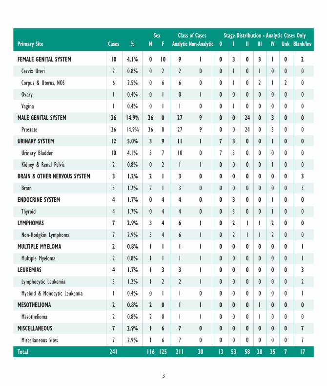
Sex ClassofCases StageDistribution-AnalyticCasesOnlyPrimarySite Cases % M F AnalyticNon-Analytic 0 I II III IV Unk Blank/Inv
ORALCAVITY&PHARYNX 10 4.1% 7 3 10 0 0 4 0 2 1 3 0
Tongue 1 0.4% 1 0 1 0 0 0 0 1 0 0 0
SalivaryGlands 2 0.8% 2 0 2 0 0 1 0 0 0 1 0
FloorofMouth 1 0.4% 1 0 1 0 0 0 0 1 0 0 0
Nasopharynx 1 0.4% 0 1 1 0 0 0 0 1 0 0 0
Tonsil 4 1.7% 2 2 4 0 0 2 0 0 1 1 0
Oropharynx 1 0.4% 1 0 1 0 0 0 0 0 0 1 0
DIGESTIVESYSTEM 42 17.4% 23 19 37 5 0 10 9 6 8 3 1
Esophagus 1 0.4% 0 1 1 0 0 0 0 0 1 0 0
Stomach 2 0.8% 2 0 2 0 0 1 0 1 0 0 0
ColonExcludingRectum 19 7.9% 11 8 16 3 0 4 5 2 5 0 0
Rectum&Rectosigmoid 10 4.1% 2 8 8 2 0 4 1 1 0 1 1
Anus,AnalCanal&Anorectum 2 0.8% 2 0 2 0 0 0 1 0 0 1 0
Liver&IntrahepaticBileDuct 2 0.8% 2 0 2 0 0 1 0 0 0 1 0
Pancreas 6 2.5% 4 2 6 0 0 0 2 2 2 0 0
RESPIRATORYSYSTEM 40 16.6% 29 11 36 4 1 9 4 7 15 0 0
NasalCavity,MiddleEar&AccessorySinuses 2 0.8% 1 1 2 0 0 0 0 0 2 0 0
Larynx 10 4.1% 10 0 10 0 1 5 2 2 0 0 0
Lung&Bronchus 28 11.6% 18 10 24 4 0 4 2 5 13 0 0
SOFTTISSUE 1 0.4% 1 0 1 0 0 0 1 0 0 0 0
SoftTissue(includingHeart) 1 0.6% 1 0 1 0 0 0 1 0 0 0 0
SKINEXCLUDINGBASAL&SQUAMOUS 9 3.7% 7 2 7 2 0 1 2 2 1 1 0
Melanoma–Skin 7 2.9% 5 2 6 1 0 1 2 1 1 1 0
OtherNonepithelialSkin 2 0.8% 2 0 1 1 0 0 0 1 0 0 0
BREAST 52 21.6% 0 52 48 4 5 18 17 6 2 0 0
Breast 52 21.6% 0 52 48 4 5 18 17 6 2 0 0
2
Sex ClassofCases StageDistribution-AnalyticCasesOnlyPrimarySite Cases % M F AnalyticNon-Analytic 0 I II III IV Unk Blank/Inv
FEMALEGENITALSYSTEM 10 4.1% 0 10 9 1 0 3 0 3 1 0 2
CervixUteri 2 0.8% 0 2 2 0 0 1 0 1 0 0 0
Corpus&Uterus,NOS 6 2.5% 0 6 6 0 0 1 0 2 1 2 0
Ovary 1 0.4% 0 1 0 1 0 0 0 0 0 0 0
Vagina 1 0.4% 0 1 1 0 0 1 0 0 0 0 0
MALEGENITALSYSTEM 36 14.9% 36 0 27 9 0 0 24 0 3 0 0
Prostate 36 14.9% 36 0 27 9 0 0 24 0 3 0 0
URINARYSYSTEM 12 5.0% 3 9 11 1 7 3 0 0 1 0 0
UrinaryBladder 10 4.1% 3 7 10 0 7 3 0 0 0 0 0
Kidney&RenalPelvis 2 0.8% 0 2 1 1 0 0 0 0 1 0 0
BRAIN&OTHERNERVOUSSYSTEM 3 1.2% 2 1 3 0 0 0 0 0 0 0 3
Brain 3 1.2% 2 1 3 0 0 0 0 0 0 0 3
ENDOCRINESYSTEM 4 1.7% 0 4 4 0 0 3 0 0 1 0 0
Thyroid 4 1.7% 0 4 4 0 0 3 0 0 1 0 0
LYMPHOMAS 7 2.9% 3 4 6 1 0 2 1 1 2 0 0
Non-HodgkinLymphoma 7 2.9% 3 4 6 1 0 2 1 1 2 0 0
MULTIPLEMYELOMA 2 0.8% 1 1 1 1 0 0 0 0 0 0 1
MultipleMyeloma 2 0.8% 1 1 1 1 0 0 0 0 0 0 1
LEUKEMIAS 4 1.7% 1 3 3 1 0 0 0 0 0 0 3
LymphocyticLeukemia 3 1.2% 1 2 2 1 0 0 0 0 0 0 2
Myeloid&MonocyticLeukemia 1 0.4% 0 1 1 0 0 0 0 0 0 0 1
MESOTHELIOMA 2 0.8% 2 0 1 1 0 0 0 1 0 0 0
Mesothelioma 2 0.8% 2 0 1 1 0 0 0 1 0 0 0
MISCELLANEOUS 7 2.9% 1 6 7 0 0 0 0 0 0 0 7
MiscellaneousSites 7 2.9% 1 6 7 0 0 0 0 0 0 0 7
Total 241 116 125 211 30 13 53 58 28 35 7 17
3

Cancer Program Annual Report 2010
The Riverside Walter Reed Hospital (RWRH) cancer program officially began in March 2004 with the opening of the Cancer Center. The Center is a freestanding facility on the Walter Reed campus. It includes space for radiation oncology and medical oncology. Radiation oncology includes a Siemens dual energy linear accelerator capable of treating patients with IMRT and CT simulator as well as physician, physics, management and clerical office space. There is also space for a nursing office and examination rooms as well as spacious patient waiting area. The radiation oncology center is a accredited by the ACR/ASTRO program, the premier accreditation body in the U.S.
Medical Oncology includes a dedicated pharmacy, physician and nursing office space along with multiple infusion stations including a private infusion room. Both oncology practices have grown steadily since their opening. Patients served at the center come from a wide area of rural Eastern Virginia including the Middle Peninsula and Northern Neck.
In 2007 RWRH established a tumor registry and active cancer committee. Processes to seek accreditation by the American College of Surgeons Commission on Cancer are currently underway.
Future plans look to expand the medical oncology floor space to allow for more treatment space as well as numerous adjunctive services currently offered in the Newport News and Williamsburg offices. In addition, increasing cooperation between the RWRH Cancer Center and Riverside Tappahannock Hospital remains a goal for the Riverside Walter Reed cancer program.
James Wassum, MDRadiation Oncology
Specialist
4
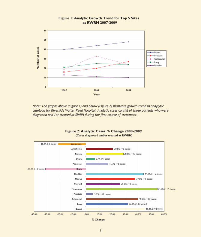
Figure 1: Analytic Growth Trend for Top 5 Sites at RWRH 2007-2009
0
10
20
30
40
50
60
2007 2008 2009
Year
Nu
mb
er o
f C
ases
BreastProstateColorectalLungBladder
Note: The graphs above (Figure 1) and below (Figure 2) illustrate growth trend in analytic caseload for Riverside Walter Reed
45.2% (+98
32.1% (+ 62 cases)
40.0% (+28 cases)
5.2% (+12 cases)
54.8% (+17 cases)
28.6% (+10 cases)
20.5% (+8 cases)
-21.4% (-3 cases)
6.7% (+1 case)
16.7% (+3 cases)
44.1% (+15 cases)
37.5% (+9 cases)
-31.3% (-10 cases)
25.8% (+8 cases)
-40.0% -30.0% -20.0% -10.0% 0.0% 10.0% 20.0% 30.0% 40.0% 50.0% 60.0%
Breast
Lung
Colorectal
Prostate
Melanoma
Thyroid
Uterus
Bladder
Brain
Pancreas
Ovary
Kidney
Lymphoma
% Change
Figure 2: Analytic Cases: % Change 2008-2009 (Cases diagnosed and/or treated at RWRH)
cases)
Leukemias
Note: The graphs above (Figure 1) and below (Figure 2) illustrate growth trend in anaylytic caseload for Riverside Walter Reed Hospital. Analytic cases consist of those patients who were diagnosed and /or treated at RWRH during the first course of treatment.
5

Urothelial carcinoma is cancer of the lining of the bladder, ureter, or renal pelvis. It is most commonly discovered in the bladder and histologically appears as transitional cell carcinoma in the majority of cases. In the past year doctors discovered 70,000 new cases and there were around 15,000 deaths from this disease. Men are affected three times more commonly than women. Smoking carries a fourfold increased risk. People who work with industrial cleansers, machinery, dyes, inks, paints, and hairdressing supplies may be at risk. People who have needed radiation for pelvic cancers have an increased risk of bladder cancer. Exposure to Cyclophosphamide is yet another agent that is a risk factor. There is not strong evidence for a familial tendency to develop urothelial cancer.
The classic presentation is an OLDER MAN who SMOKES presenting with GROSS PAINLESS HEMATURIA. Often the patient will complain of urinary urgency, frequency, and occasionally dysuria. Alternatively, the patient may have no symptoms and is identified solely by microscopic hematuria on a routine urinalysis. Sometimes bladder cancer is incidentally noticed on a CAT scan done for other reasons – reports might indicate bladder wall thickening if not an intravesical lesion.
The urologic work-up of microscopic or gross hematuria includes a CT UROGRAM to check the upper urinary tracts for cancer, stones, or obstruction. It is important to note that a CAT scan may miss a small urothelial tumor in the bladder. This is the reason that we must also perform CYSTOSCOPY. With the newest flexible endoscopes, this can be easily done in the office. Urinary cytology is helpful when positive. There are several office based tumor screening tests that look for proteins made by these cancers (e.g., urinary NMP, BTA stat). These are rapidly performed, office based tests that suffer somewhat by false positives from active infection or stones. Like urinary cytology, they are helpful but are not yet accurate enough to replace cystoscopy. The Urovision FISH test (fluorescence and in situ hybridization) is a urinary test that looks at a molecular level for cancer. It has been criticized for being too sensitive a test. It may provide an “anticipatory positive” - ie, suggestion of a “cancer to come” even though the urologist cannot find it by conventional radiologic or endoscopic means.
Bladder Cancer at Riverside Walter Reed Hospital
Roger E. Schultz, MDHampton Roads
Urology
Figure 3: 2007-2009 Analytic Bladder Cancers by Gender - RWRH Only
910
3
41
7
0
2
4
6
8
10
12
14
2007 2008 2009
Year
# o
f C
ases
FemaleMale
Figure 3 illustrates the distribution of cases among gender from 2007 to 2009. Generally there is a higher ratio of males to females; however in 2009 more females were diagnosed with bladder cancer at RWRH than males.
6
The cystoscopic appearance of a classic papillary transitional cell cancer is a true “Aunt Minnie.” Once you’ve seen her, you always remember the appearance. Less commonly, bladder cancers may appear as nodules, or raised, reddened areas that are difficult to diagnose. ALL bladder lesions require histologic confirmation. The urologist will schedule an examination under anesthesia (pelvic and rectal bimamual exam to palpate for tumor fixation. He then performs a transurethral resection of the bladder tumor (TURBT). This is done with regional or general anesthesia. In the last few years, TURBT is usually followed by the intravesical instillation of MITOMYCIN. Papillary tumors often break up when resected, and there is data to show that immediate instillation of Mitomycin after TURBT can reduce tumor “seeding” of other areas of the bladder lining, thereby reducing recurrence rates. After TURBT, the urologist places a urethral catheter and instills 20-40cc of Mitomycin in solution into the bladder. It is drained one hour later in the recovery area.
The prognosis for recurrence is higher when there are large tumors and/or multifocal tumors. The overall prognosis is more ominous if the tumor is nodular, as opposed to papillary, and broad based. The pathologist can tell us the histologic tumor type (typically transitional cell carcinoma, less commonly squamous carcinoma or adenocarcinoma), the tumor grade, and the depth of tumor invasion. Newer molecular markers may soon be widely available to stratify those at high risk for progressive disease. Fortunately, the majority of tumors are superficial and easily resectable lesions. About 50-70% are stage Ta (at the epithelial surface), 20-30% are T1 (invades lamina propria), 20-25% are T2 (muscle invasive). Thankfully, few are widely metastatic at presentation.
Bladder cancers can recur, particularly if there are large and/or multifocal at first presentation. After TURBT patients are begun on a routine cystoscopic surveillance program. Selected circumstances may require the use of intravesical agents to reduce recurrent rates – especially patients with T1 tumors, CIS, or frequent recurrences. The most common agent employed is BCG, a weakened tuberculosis strain that is instilled in the bladder via catheterization once weekly for 6 weeks (“Induction therapy”). Patient must retain the BCG for 2 hours if possible, then eliminate it. The immune response can be amplified by repeat exposure to intravesical BCG (“Maintenance dosing”). They will receive another 3 weeks of intravesical BCG every 6 months out to about three years. Some people cannot tolerate the dysuria, urgency, and frequency that BCG may cause. Symptoms may be ameliorated by reducing the BCG concentration. Alternative intravesical agents include Alpha 1 Interferon, Mitomycin, Valrubucin, and Gemticibine.
Muscle invasive bladder cancer (T2) usually requires open surgery. When it involves the dome of the bladder, it may be amenable to partial cystectomy with wide margins of resection. In most cases, it will require removing the bladder/prostate/regional lymph nodes in men and an anterior exenteration in women. The classic urinary diversion is a uretero-ileal
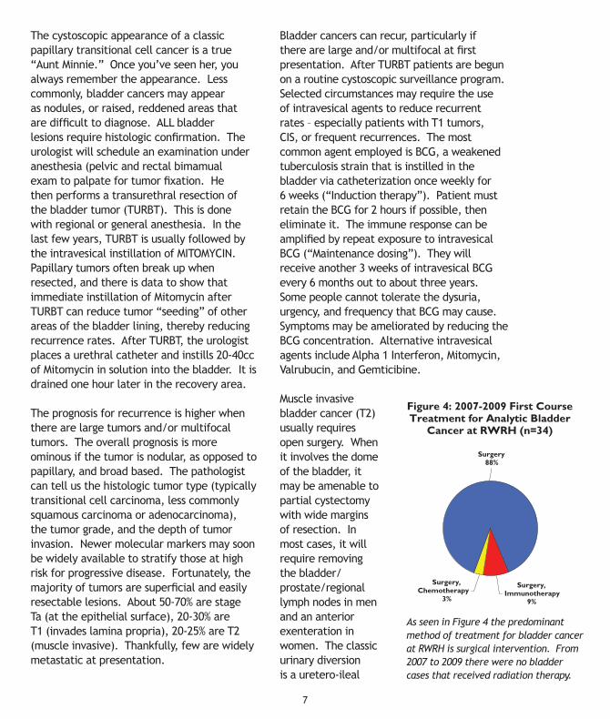
Figure 4: 2007-2009 First Course Treatment for Analytic Bladder
Cancer at RWRH (n=34)
Surgery, Chemotherapy
3%
Surgery, Immunotherapy
9%
Surgery 88%
As seen in Figure 4 the predominant method of treatment for bladder cancer at RWRH is surgical intervention. From 2007 to 2009 there were no bladder cases that received radiation therapy.
7

conduit (Bricker conduit). Ureters cannot be brought up to the abdominal wall to drain because they will stenose. A short segment of ileum is interposed between the ureters and the abdominal is wall because the ileal stoma is less apt to obstruct. An ostomy appliance can be applied to the UROSTOMY to collect urine.
In the last 15 years urologists have been offering continent urinary diversions to avoid the need for an external appliance. These diversions involve the creation of a neobladder, a reservoir instead of a conduit. The neobladder is made from bowel (large, small, or both) that is reconfigured to become more spherical. It may be attached to the native urethra so that the patient can void spontaneously. This is called an orthotopic neobladder. Alternatively, the patient may require a urinary pouch (eg, Koch pouch, Indina Pouch) that is accessed by self catheterization through a small, flush stoma on
the abdominal wall. The tiny stoma is easily concealed and the patient is dry between catheterizations. Continent diversions avoid the inconvenience of a collection bag on the abdomen, but they are constructed at a big cost – much longer surgery and recovery, the risk of anastomotic leaks, loss of bowel to create a neobladder (potential dumping syndrome, B12 malabsorption, metabolic acidosis). Some orthotopic neobladders will leak at night during deep sleep.
Patients who are too old or too ill for radical cystectomy may be treated by a combination of deep TURBT and chemotherapy/radiation. Metastatic bladder cancer is treated with chemotherapy and/or radiation. Urologists must also remain involved because of recurrent urinary bleeding from the principal tumor that may require re-resection or diversionary ureteral stents. The prognosis with advanced disease is poor – most patients live less than two years.
RWRH (n=13)
Figure 5: 2007 Comparison of Stage at Diagnosis Bladder Cancer - RWRH vs. NCDB
30.8%
0.0% 0.0%
44.5%
10.9%
4.6%6.7%
23.0%
7.7%
38.5%
13.3%
20.1%
0.0%
5.0%
10.0%
15.0%
20.0%
25.0%
30.0%
35.0%
40.0%
45.0%
50.0%
Stage 0 Stage I Stage II Stage III Stage IV Unk
Stage at Diagnosis
% o
f A
nal
ytic
Cas
es
NCDB (n=44449)
The above graph is a comparison of stage at diagnosis for bladder cases seen at RWRH in 2007 to those recorded in the 2007 National Cancer Data Base.
8

At first glance one may conclude that patients at RWMC have higher stages of bladder cancer at presentation than the national trend (Figure 5). My opinion is that we have seen too few cases to draw any firm conclusions. I would like to offer several personal observations about this population of patients:
Patients often delay their own diagnosis – papillary tumors may bleed on Monday and stop bleeding on Tuesday. Patients get a false sense of security once the bleeding stops. Others may have noted months of
Nancy McKinney, MD, Chair Medical Oncology Melvin Schursky, MD General Surgery Cancer Liaison Physician James Wassum, MD Radiation Oncology Ronald Haggerty, MD Hospitalist Warren Helwig, MD Pathology Val Curran, MD Radiology David Schengber, MD Radiology Elizabeth Martin VP Riverside Tappahannock/Walter Reed Hospital Keith Gregory Service Line Administrator, Oncology Paula Burcher Administrative Director, Radiology Beverly Voglewede Director, Radiation Oncology Services Patricia Emerson Nurse Manager, Outpatient Infusion Center Joe Hughes Director, Performance Improvement Sue Moffitt Nurse Manager, Riverside Walter Reed Hospice Suzanne Riley Director, Radiology Angie Healy Director, Public Relations Fran Holcomb Cancer Education/Outreach Nurse Jennifer Brown Cancer Registry Supervisor Carol Richards Cancer Registrar
2010 ON
CO
LOG
Y
For additional information regarding Riverside Cancer Services, please call (800) 520-7006.
For comments or questions regarding this Annual Report or the Cancer Registry, please call (757) 594-3054.
CO
MM
ITT
EE MEM
BER
S
episodic painless hematuria before they tell a physician, possibly out of ignorance or fear. We could do a better job of educating people about bladder cancer symptoms and continue efforts to stop people from smoking.
Doctors occasionally delay the diagnosis of bladder cancer in women. They may treat women with hematuria for presumed cystitis, when in fact, their urine cultures show no growth. If a patient presents with urinary bleeding and a negative culture, she should be referred for urologic evaluation.

7544 Medical DriveGloucester, Virginia 23061(804) 693-4900http://www.riversideonline.com/rwrh/rmpcc.cfm