ring enhancing lesions in a patient with...
TRANSCRIPT
Jonathan Waks, MSIIIGillian Lieberman, MD
Ring Enhancing Lesions in a Patient with AIDS
Jonathan Waks, Harvard Medical School Year IIIGillian Lieberman, MD
March 2007
2
Jonathan Waks, MSIIIGillian Lieberman, MD
Agenda
• Background: CNS complications of AIDS• Patient presentation• Menu of radiologic tests • Differential diagnosis: ring enhancing lesions• Differentiating CNS lesions• Summary
3
Jonathan Waks, MSIIIGillian Lieberman, MD
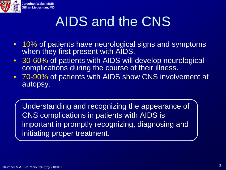
AIDS and the CNS• 10% of patients have neurological signs and symptoms
when they first present with AIDS.• 30-60% of patients with AIDS will develop neurological
complications during the course of their illness.• 70-90% of patients with AIDS show CNS involvement at
autopsy.
Thurnher MM. Eur Radiol 1997;7(7):1091-7
Understanding and recognizing the appearance of CNS complications in patients with AIDS is important in promptly recognizing, diagnosing and initiating proper treatment.
4
Jonathan Waks, MSIIIGillian Lieberman, MD
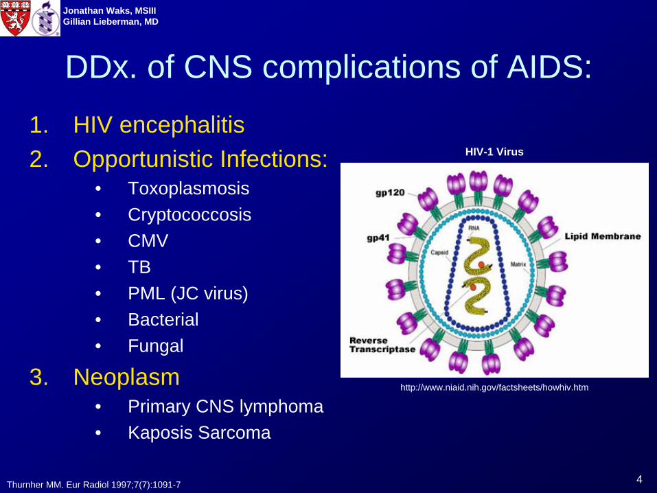
DDx. of CNS complications of AIDS:
1. HIV encephalitis2. Opportunistic Infections:
• Toxoplasmosis• Cryptococcosis• CMV• TB• PML (JC virus)• Bacterial• Fungal
3. Neoplasm• Primary CNS lymphoma• Kaposis Sarcoma
Thurnher MM. Eur Radiol 1997;7(7):1091-7
http://www.niaid.nih.gov/factsheets/howhiv.htm
HIV-1 Virus
5
Jonathan Waks, MSIIIGillian Lieberman, MD
Index Patient – “JL”• 49 year old man with AIDS (last CD4=17, on
HAART) who presented to an OSH for unsteady gait, lower extremity weakness, headache, vomiting, dysarthria and seizures.
• PE:– Temp: 102.4ºF– Multiple CN deficits
• Head CT showed multiple ring enhancing lesions• Started on broad spectrum antibiotics with coverage
for toxoplasma.• No improvement Brain biopsy: non diagnostic• Transferred to BIDMC for further management.
6
Jonathan Waks, MSIIIGillian Lieberman, MD
Before we discuss the imaging that was obtained when JL arrived at BIDMC, let’s first review the radiologic modalities that can be used to evaluate the CNS in a patient with AIDS.
7
Jonathan Waks, MSIIIGillian Lieberman, MD
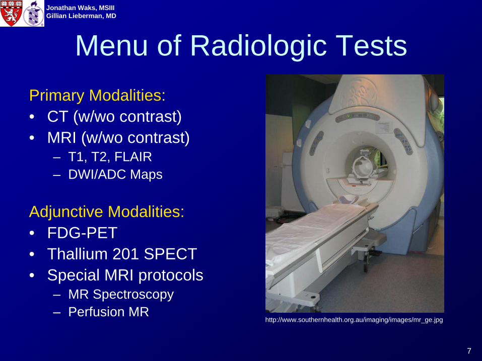
Menu of Radiologic TestsPrimary Modalities:• CT (w/wo contrast)• MRI (w/wo contrast)
– T1, T2, FLAIR– DWI/ADC Maps
Adjunctive Modalities:• FDG-PET• Thallium 201 SPECT• Special MRI protocols
– MR Spectroscopy– Perfusion MR
http://www.southernhealth.org.au/imaging/images/mr_ge.jpg
8
Jonathan Waks, MSIIIGillian Lieberman, MD
Menu of Radiologic TestsPrimary Modalities:• CT (w/wo contrast)• MRI (w/wo contrast)
– T1, T2, FLAIR– DWI/ADC Maps
Adjunctive Modalities:• FDG-PET• Thallium 201 SPECT• Special MRI protocols
– MR Spectroscopy– Perfusion MR
These adjunctive modalities are not used in the routine imaging or evaluation of CNS lesions in patients with AIDS. They are primarily used when the identity of a lesion is in question and additional non-invasive imaging would potentially alter treatment. PET and SPECT scanning are used most frequently. MR spectroscopy and perfusion MR are not routinely used and will not be discussed in this presentation.
Adjunctive imaging modalities will be discussed later on in the presentation
9
Jonathan Waks, MSIIIGillian Lieberman, MD
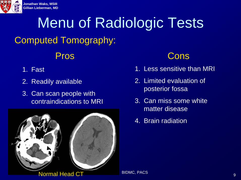
Menu of Radiologic Tests
1. Fast
2. Readily available
3. Can scan people with contraindications to MRI
1. Less sensitive than MRI
2. Limited evaluation of posterior fossa
3. Can miss some white matter disease
4. Brain radiation
BIDMC, PACS
Computed Tomography:
Pros Cons
Normal Head CT
10
Jonathan Waks, MSIIIGillian Lieberman, MD
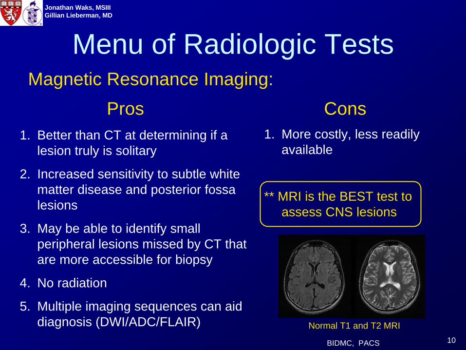
Menu of Radiologic TestsMagnetic Resonance Imaging:
Pros Cons1. Better than CT at determining if a
lesion truly is solitary
2. Increased sensitivity to subtle white matter disease and posterior fossa lesions
3. May be able to identify small peripheral lesions missed by CT that are more accessible for biopsy
4. No radiation
5. Multiple imaging sequences can aid diagnosis (DWI/ADC/FLAIR)
1. More costly, less readily available
** MRI is the BEST test to assess CNS lesions
BIDMC, PACS
Normal T1 and T2 MRI
11
Jonathan Waks, MSIIIGillian Lieberman, MD
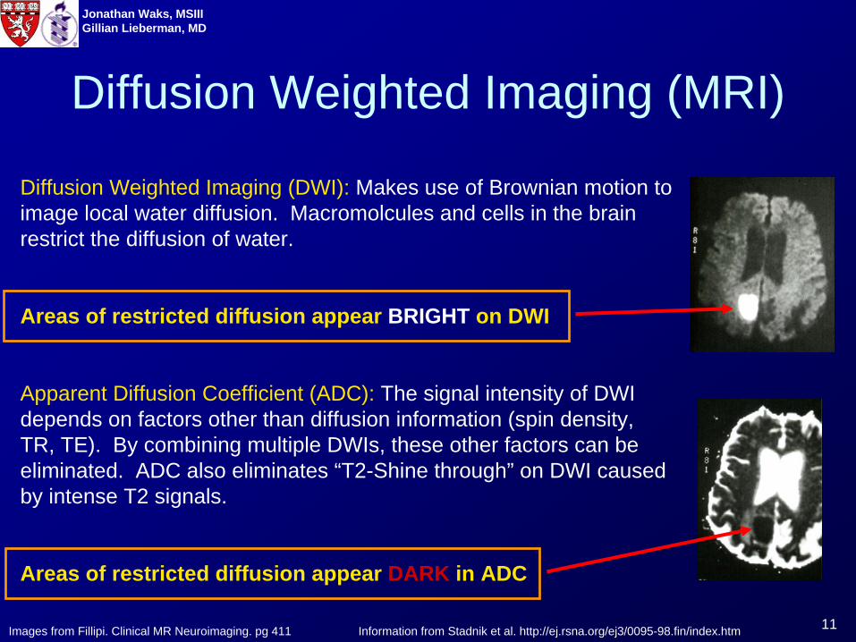
Diffusion Weighted Imaging (MRI)
Diffusion Weighted Imaging (DWI): Makes use of Brownian motion to image local water diffusion. Macromolcules and cells in the brain restrict the diffusion of water.
Areas of restricted diffusion appear BRIGHT on DWI
Apparent Diffusion Coefficient (ADC): The signal intensity of DWI depends on factors other than diffusion information (spin density, TR, TE). By combining multiple DWIs, these other factors can be eliminated. ADC also eliminates “T2-Shine through” on DWI caused by intense T2 signals.
Areas of restricted diffusion appear DARK in ADC
Images from Fillipi. Clinical MR Neuroimaging. pg 411 Information from Stadnik et al. http://ej.rsna.org/ej3/0095-98.fin/index.htm
12
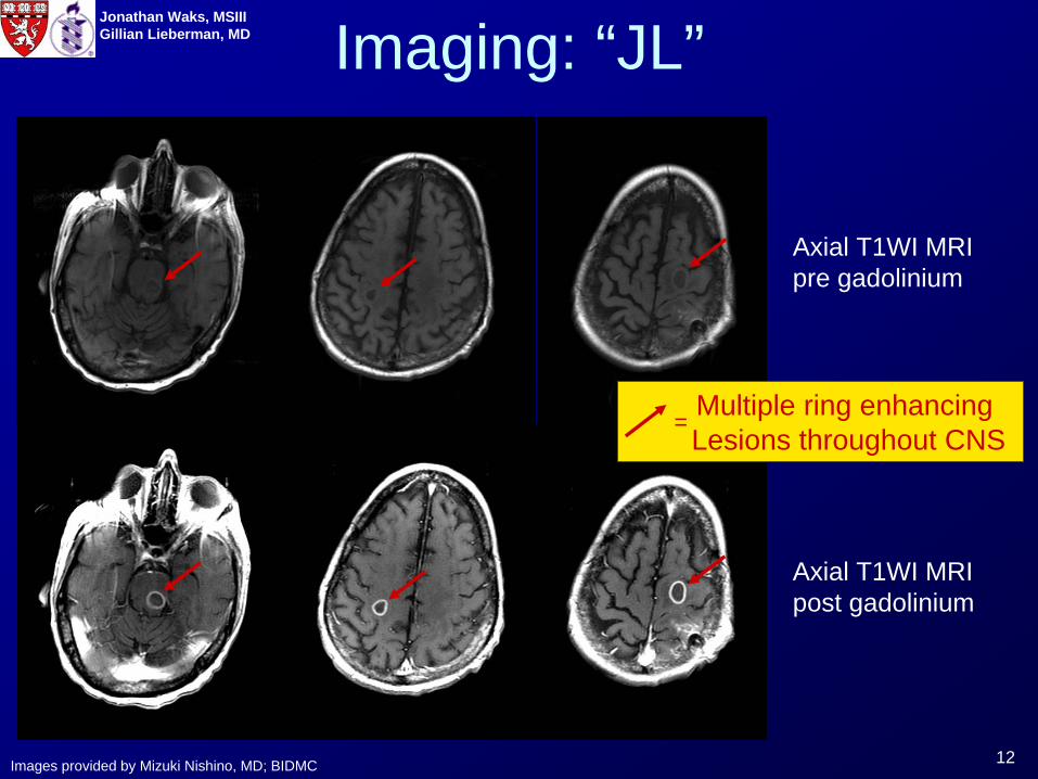
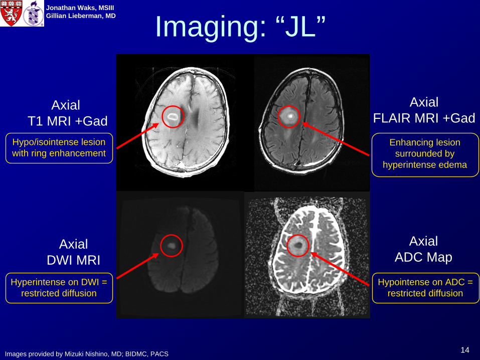
Jonathan Waks, MSIIIGillian Lieberman, MD Imaging: “JL”
Axial T1WI MRI post gadolinium
Images provided by Mizuki Nishino, MD; BIDMC
Axial T1WI MRIpre gadolinium
Multiple ring enhancing Lesions throughout CNS
=
13
Jonathan Waks, MSIIIGillian Lieberman, MD
Looking at additional MRI sequences allows us to better characterize the center of the lesion and surrounding tissue.
14
Jonathan Waks, MSIIIGillian Lieberman, MD Imaging: “JL”
AxialDWI MRI
Axial T1 MRI +Gad
AxialFLAIR MRI +Gad
AxialADC Map
Images provided by Mizuki Nishino, MD; BIDMC, PACS
Hyperintense on DWI = restricted diffusion
Hypo/isointense lesion with ring enhancement
Enhancing lesion surrounded by
hyperintense edema
Hypointense on ADC = restricted diffusion
15
Jonathan Waks, MSIIIGillian Lieberman, MD
There is a well defined differential diagnosis for ring enhancing lesions in the CNS:
16
Jonathan Waks, MSIIIGillian Lieberman, MD
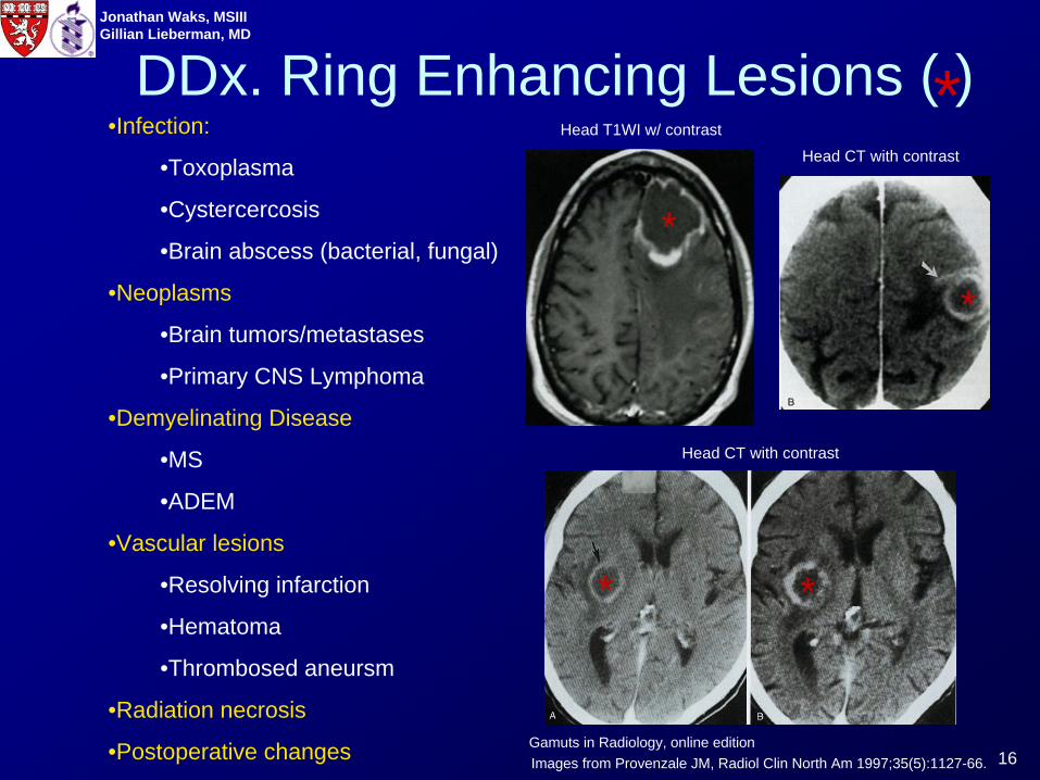
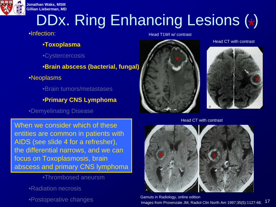
DDx. Ring Enhancing Lesions ( )•Infection:
•Toxoplasma
•Cystercercosis
•Brain abscess (bacterial, fungal)
•Neoplasms
•Brain tumors/metastases
•Primary CNS Lymphoma
•Demyelinating Disease
•MS
•ADEM
•Vascular lesions
•Resolving infarction
•Hematoma
•Thrombosed aneursm
•Radiation necrosis
•Postoperative changes Gamuts in Radiology, online editionImages from Provenzale JM, Radiol Clin North Am 1997;35(5):1127-66.
*
*
*
**
Head CT with contrastHead T1WI w/ contrast
Head CT with contrast
17
Jonathan Waks, MSIIIGillian Lieberman, MD
•Infection:
•Toxoplasma
•Cystercercosis
•Brain abscess (bacterial, fungal)
•Neoplasms
•Brain tumors/metastases
•Primary CNS Lymphoma
•Demyelinating Disease
•MS
•ADEM
•Vascular lesions
•Resolving infarction
•Hematoma
•Thrombosed aneursm
•Radiation necrosis
•Postoperative changes Gamuts in Radiology, online editionImages from Provenzale JM, Radiol Clin North Am 1997;35(5):1127-66.
When we consider which of these entities are common in patients with AIDS (see slide 4 for a refresher), the differential narrows, and we can focus on Toxoplasmosis, brain abscess and primary CNS lymphoma
DDx. Ring Enhancing Lesions ( )*
*
*
**
Head CT with contrastHead T1WI w/ contrast
Head CT with contrast
18
Jonathan Waks, MSIIIGillian Lieberman, MD
We will now explore the narrowed differential in more detail.
1. Toxoplasmosis
2. Primary CNS Lymphoma
3. Bacterial brain abscess
19
Jonathan Waks, MSIIIGillian Lieberman, MD
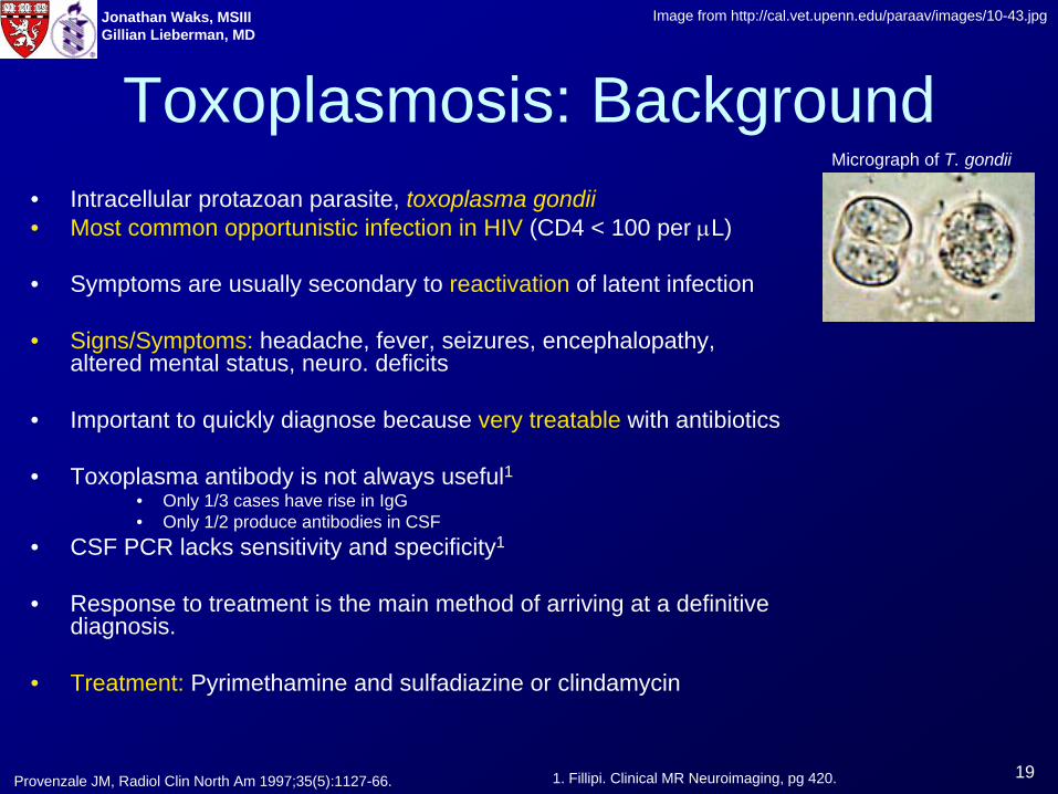
Toxoplasmosis: Background• Intracellular protazoan parasite, toxoplasma gondii• Most common opportunistic infection in HIV (CD4 < 100 per μL)
• Symptoms are usually secondary to reactivation of latent infection
• Signs/Symptoms: headache, fever, seizures, encephalopathy, altered mental status, neuro. deficits
• Important to quickly diagnose because very treatable with antibiotics
• Toxoplasma antibody is not always useful1• Only 1/3 cases have rise in IgG• Only 1/2 produce antibodies in CSF
• CSF PCR lacks sensitivity and specificity1
• Response to treatment is the main method of arriving at a definitive diagnosis.
• Treatment: Pyrimethamine and sulfadiazine or clindamycin
Image from http://cal.vet.upenn.edu/paraav/images/10-43.jpg
1. Fillipi. Clinical MR Neuroimaging, pg 420.
Micrograph of T. gondii
Provenzale JM, Radiol Clin North Am 1997;35(5):1127-66.
20
Jonathan Waks, MSIIIGillian Lieberman, MD
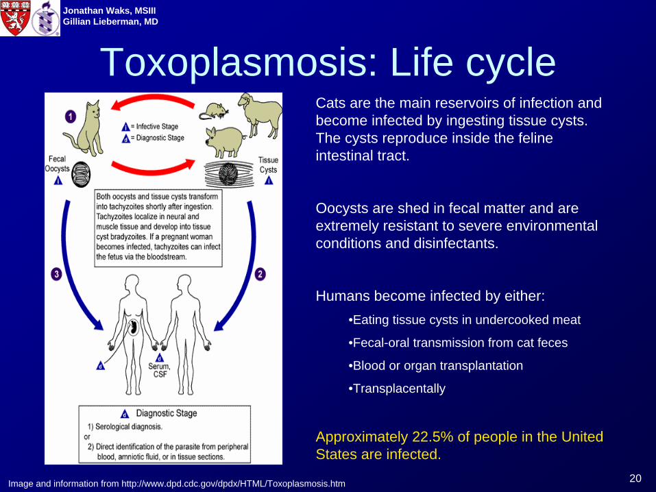
Toxoplasmosis: Life cycle
Image and information from http://www.dpd.cdc.gov/dpdx/HTML/Toxoplasmosis.htm
Cats are the main reservoirs of infection and become infected by ingesting tissue cysts. The cysts reproduce inside the feline intestinal tract.
Oocysts are shed in fecal matter and are extremely resistant to severe environmental conditions and disinfectants.
Humans become infected by either:•Eating tissue cysts in undercooked meat
•Fecal-oral transmission from cat feces
•Blood or organ transplantation
•Transplacentally
Approximately 22.5% of people in the United States are infected.
21
Jonathan Waks, MSIIIGillian Lieberman, MD
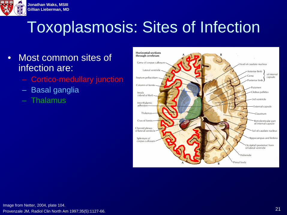
Toxoplasmosis: Sites of Infection
• Most common sites of infection are:– Cortico-medullary junction– Basal ganglia– Thalamus
Image from Netter, 2004, plate 104.Provenzale JM, Radiol Clin North Am 1997;35(5):1127-66.
22
Jonathan Waks, MSIIIGillian Lieberman, MD
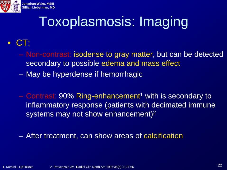
• CT:– Non-contrast: isodense to gray matter, but can be detected
secondary to possible edema and mass effect– May be hyperdense if hemorrhagic
– Contrast: 90% Ring-enhancement1 with is secondary to inflammatory response (patients with decimated immune systems may not show enhancement)2
– After treatment, can show areas of calcification
Toxoplasmosis: Imaging
1. Koralnik, UpToDate 2. Provenzale JM, Radiol Clin North Am 1997;35(5):1127-66.
23
Jonathan Waks, MSIIIGillian Lieberman, MD
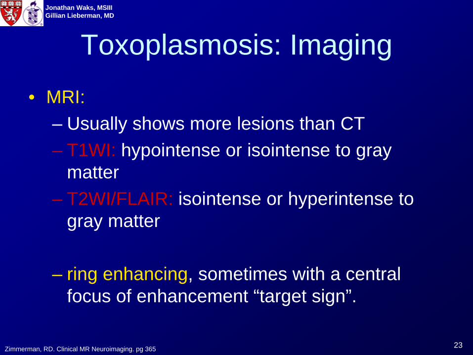
• MRI:– Usually shows more lesions than CT– T1WI: hypointense or isointense to gray
matter– T2WI/FLAIR: isointense or hyperintense to
gray matter
– ring enhancing, sometimes with a central focus of enhancement “target sign”.
Toxoplasmosis: Imaging
Zimmerman, RD. Clinical MR Neuroimaging. pg 365

24
Jonathan Waks, MSIIIGillian Lieberman, MD
Toxoplasmosis Lesions
T2 MRI, non-contrast T1 MRI w/ contrast
Images from Thurnher MM. Eur Radiol 1997;7(7):1091-7
Head CT w/ contrast
Below are 2 patients with CNS lesions that were subsequently shown to be toxoplasmosis.
Patient 1Patient 2
Hypodense, ring enhancing lesion and surrounding edema Hyperintense,
enhancing lesionHypointense, ring enhancing lesion
25
Jonathan Waks, MSIIIGillian Lieberman, MD
Primary CNS Lymphoma: Background• Most common AIDS related neoplasm (2-5% patients)• After Toxoplasmosis, is second most common cerebral mass
lesion in AIDS patients.• Almost always of B-cell, Non-Hodgkins type• Likely related to EBV • Presenting symptoms: neurological deficits, encephalopathy,
seizure (similar to toxoplasmosis)• Median survival < 1 year
• Treatment: Radiation and corticosteroids
Provenzale JM, Radiol Clin North Am 1997;35(5):1127-66.
26
Jonathan Waks, MSIIIGillian Lieberman, MD
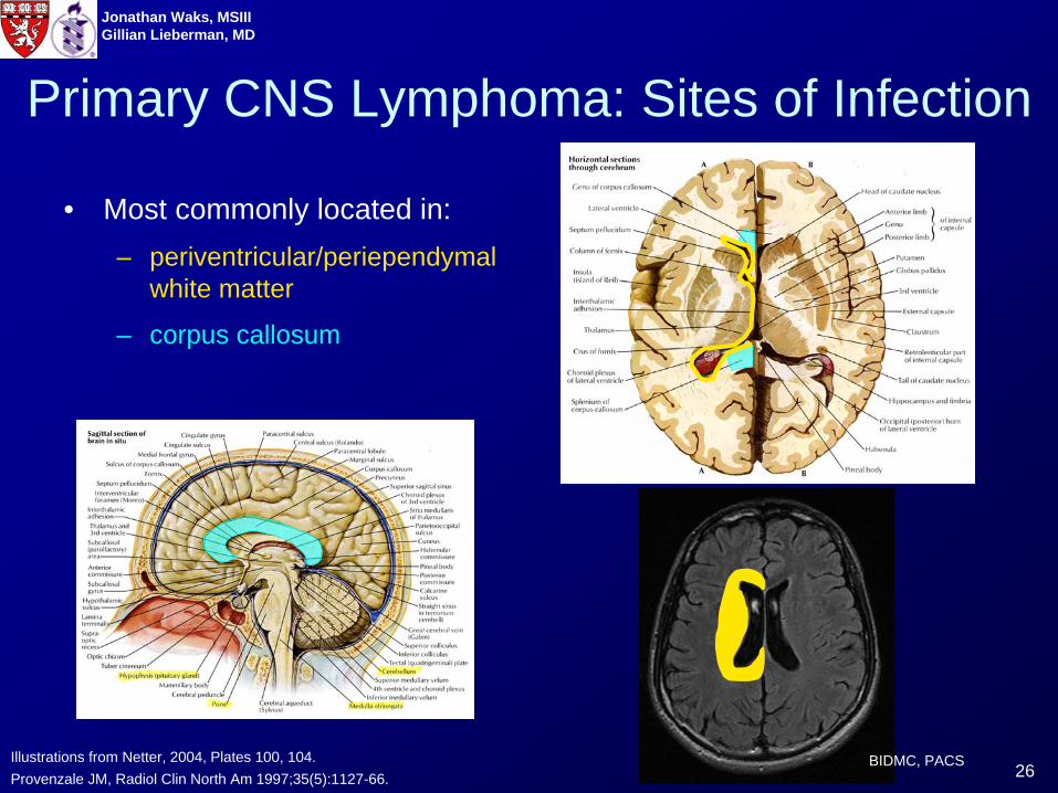
Primary CNS Lymphoma: Sites of Infection
• Most commonly located in:– periventricular/periependymal
white matter
– corpus callosum
Provenzale JM, Radiol Clin North Am 1997;35(5):1127-66.Illustrations from Netter, 2004, Plates 100, 104. BIDMC, PACS
27
Jonathan Waks, MSIIIGillian Lieberman, MD
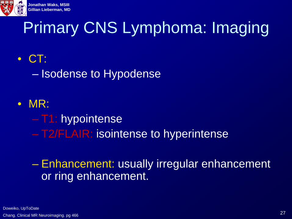
Primary CNS Lymphoma: Imaging
• CT:– Isodense to Hypodense
• MR:– T1: hypointense– T2/FLAIR: isointense to hyperintense
– Enhancement: usually irregular enhancement or ring enhancement.
Chang. Clinical MR Neuroimaging. pg 466
Doweiko, UpToDate
28
Jonathan Waks, MSIIIGillian Lieberman, MD
Primary CNS Lymphoma can have a wide range of appearances:
29
Jonathan Waks, MSIIIGillian Lieberman, MD
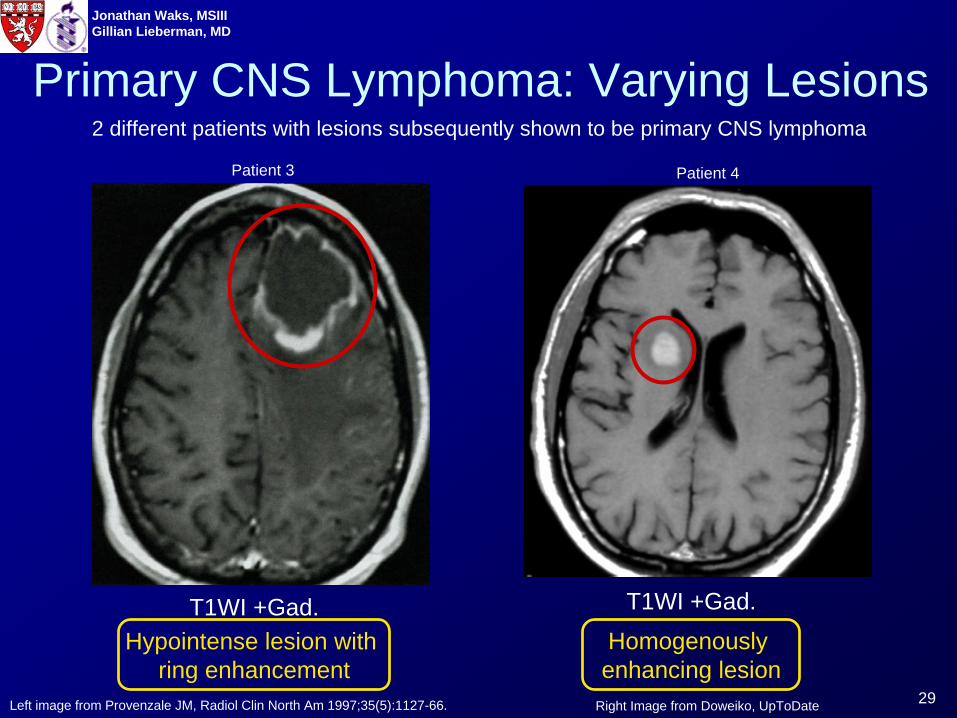
Primary CNS Lymphoma: Varying Lesions
T1WI +Gad.
Right Image from Doweiko, UpToDate
T1WI +Gad.Hypointense lesion with
ring enhancementHomogenously enhancing lesion
Left image from Provenzale JM, Radiol Clin North Am 1997;35(5):1127-66.
2 different patients with lesions subsequently shown to be primary CNS lymphoma
Patient 3 Patient 4
30
Jonathan Waks, MSIIIGillian Lieberman, MD
Bacterial Abscess: Background• Often presents with headache, altered mental
status, nausea, vomiting, seizures, neuro. deficits due to expanding mass.1
• Hypointense on T1, Hyperintense on T21
• Capsule is hypointense on T21
• Ring enhancing with surrounding edema1
• Less common in AIDS patients than toxoplasmosis or primary CNS lymphoma2
– Often associated with bacteremia
1. Fillipi, Clinical MR Neuroimaging. Pg 409
2. Koralnik, UpToDate
31
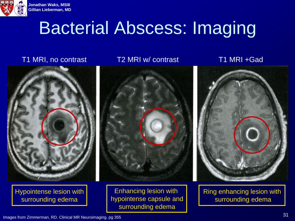
Jonathan Waks, MSIIIGillian Lieberman, MD
T1 MRI, no contrast T2 MRI w/ contrast T1 MRI +Gad
Hypointense lesion withsurrounding edema
Enhancing lesion with hypointense capsule and
surrounding edema
Ring enhancing lesion with surrounding edema
Bacterial Abscess: Imaging
Images from Zimmerman, RD. Clinical MR Neuroimaging. pg 355
32
Jonathan Waks, MSIIIGillian Lieberman, MD
Lymphoma vs. ToxoplasmosisToxoplasmosis and primary CNS lymphoma are the two most common brain lesions in patients with AIDS, but, as has been shown, both can have very similar clinical features and appearance on CT and MRI.
The definitive diagnosis is usually provided by brain biopsy, but biopsy is not a benign procedures and is associated with possible morbidity and mortality. (8.4% morbidity, 2.9% mortality)1
Delay in diagnosis while waiting to see if a patient responds to initial therapy is a significant problem:2
• lesions can rapidly progress.• unnecessary therapies are associated with unnecessary toxicity.• Incorrect initial treatment may result in a biopsy that could have potentially been prevented.
1. Doweiko, UpToDate
2. Fillipi, Clinical MR Neuroimaging. Pg 420
33
Jonathan Waks, MSIIIGillian Lieberman, MD
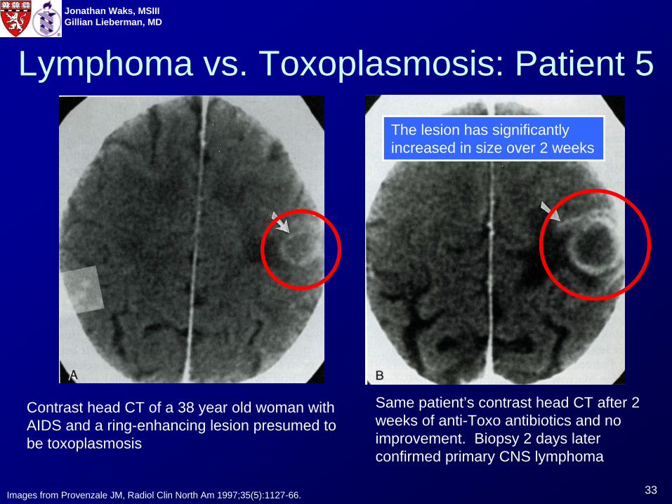
Lymphoma vs. Toxoplasmosis: Patient 5
Contrast head CT of a 38 year old woman with AIDS and a ring-enhancing lesion presumed to be toxoplasmosis
Same patient’s contrast head CT after 2 weeks of anti-Toxo antibiotics and no improvement. Biopsy 2 days later confirmed primary CNS lymphoma
Images from Provenzale JM, Radiol Clin North Am 1997;35(5):1127-66.
The lesion has significantly increased in size over 2 weeks
34
Jonathan Waks, MSIIIGillian Lieberman, MD
The appearance of a CNS lesion may give clues as to the diagnosis:
35
Jonathan Waks, MSIIIGillian Lieberman, MD
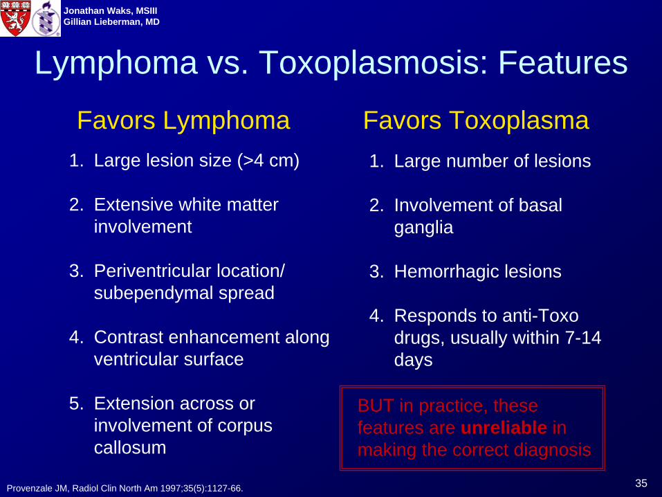
Lymphoma vs. Toxoplasmosis: Features
Favors Lymphoma Favors Toxoplasma1. Large lesion size (>4 cm)
2. Extensive white matter involvement
3. Periventricular location/ subependymal spread
4. Contrast enhancement along ventricular surface
5. Extension across or involvement of corpus callosum
1. Large number of lesions
2. Involvement of basal ganglia
3. Hemorrhagic lesions
4. Responds to anti-Toxo drugs, usually within 7-14 days
BUT in practice, these features are unreliable in making the correct diagnosis
Provenzale JM, Radiol Clin North Am 1997;35(5):1127-66.
36
Jonathan Waks, MSIIIGillian Lieberman, MD
Diffusion Weighted Imaging is one specific application of MRI that has attempted to distinguish ring-enhancing lesions:
37
Jonathan Waks, MSIIIGillian Lieberman, MD
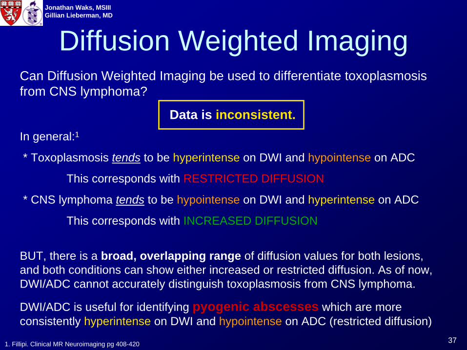
Can Diffusion Weighted Imaging be used to differentiate toxoplasmosis from CNS lymphoma?
Data is inconsistent.In general:1
* Toxoplasmosis tends to be hyperintense on DWI and hypointense on ADC
This corresponds with RESTRICTED DIFFUSION
* CNS lymphoma tends to be hypointense on DWI and hyperintense on ADC
This corresponds with INCREASED DIFFUSION
BUT, there is a broad, overlapping range of diffusion values for both lesions, and both conditions can show either increased or restricted diffusion. As of now, DWI/ADC cannot accurately distinguish toxoplasmosis from CNS lymphoma.
DWI/ADC is useful for identifying pyogenic abscesses which are more consistently hyperintense on DWI and hypointense on ADC (restricted diffusion)
Diffusion Weighted Imaging
1. Fillipi. Clinical MR Neuroimaging pg 408-420
38
Jonathan Waks, MSIIIGillian Lieberman, MD
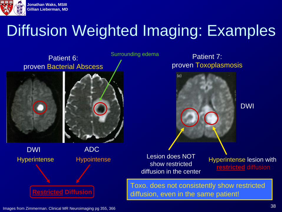
Diffusion Weighted Imaging: Examples
DWI ADC
Patient 6: proven Bacterial Abscess
Patient 7: proven Toxoplasmosis
Lesion does NOT show restricted
diffusion in the center
Hyperintense lesion with restricted diffusion
Images from Zimmerman. Clinical MR Neuroimaging pg 355, 366
DWI
Restricted Diffusion
Hyperintense Hypointense
Surrounding edema
Toxo. does not consistently show restricted diffusion, even in the same patient!
39
Jonathan Waks, MSIIIGillian Lieberman, MD
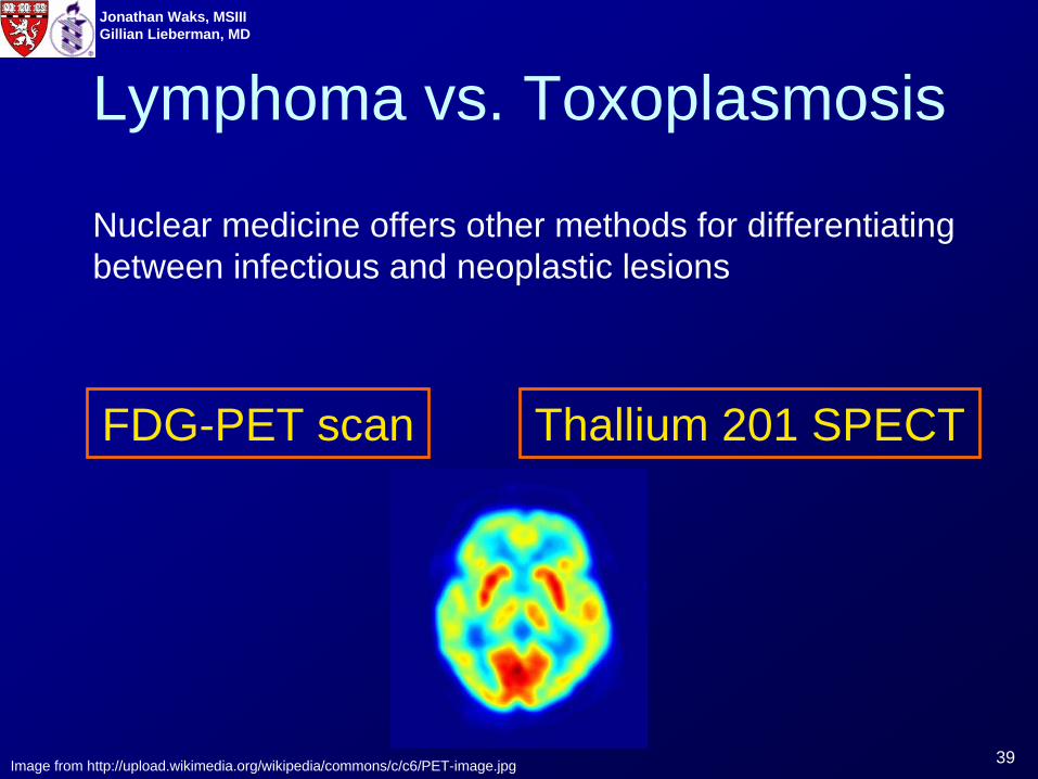
Lymphoma vs. Toxoplasmosis
Nuclear medicine offers other methods for differentiating between infectious and neoplastic lesions
FDG-PET scan Thallium 201 SPECT
Image from http://upload.wikimedia.org/wikipedia/commons/c/c6/PET-image.jpg
40
Jonathan Waks, MSIIIGillian Lieberman, MD
Image from http://www.scq.ubc.ca/?p=474
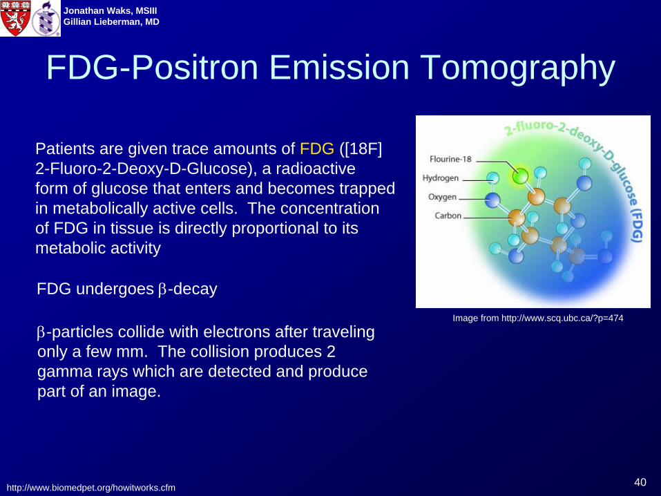
Patients are given trace amounts of FDG ([18F] 2-Fluoro-2-Deoxy-D-Glucose), a radioactive form of glucose that enters and becomes trapped in metabolically active cells. The concentration of FDG in tissue is directly proportional to its metabolic activity
http://www.biomedpet.org/howitworks.cfm
FDG undergoes β-decay
β-particles collide with electrons after traveling only a few mm. The collision produces 2 gamma rays which are detected and produce part of an image.
FDG-Positron Emission Tomography
41
Jonathan Waks, MSIIIGillian Lieberman, MD
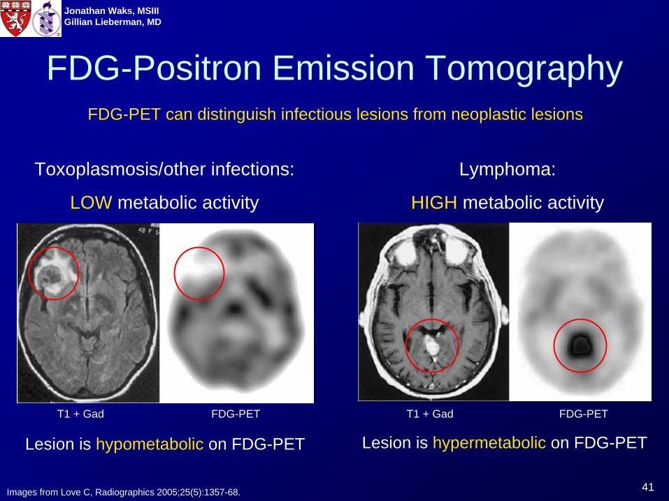
FDG-Positron Emission Tomography
Lymphoma:
HIGH metabolic activity
Toxoplasmosis/other infections:
LOW metabolic activity
Lesion is hypometabolic on FDG-PET Lesion is hypermetabolic on FDG-PET
Images from Love C, Radiographics 2005;25(5):1357-68.
T1 + Gad FDG-PET T1 + Gad FDG-PET
FDG-PET can distinguish infectious lesions from neoplastic lesions
42
Jonathan Waks, MSIIIGillian Lieberman, MD
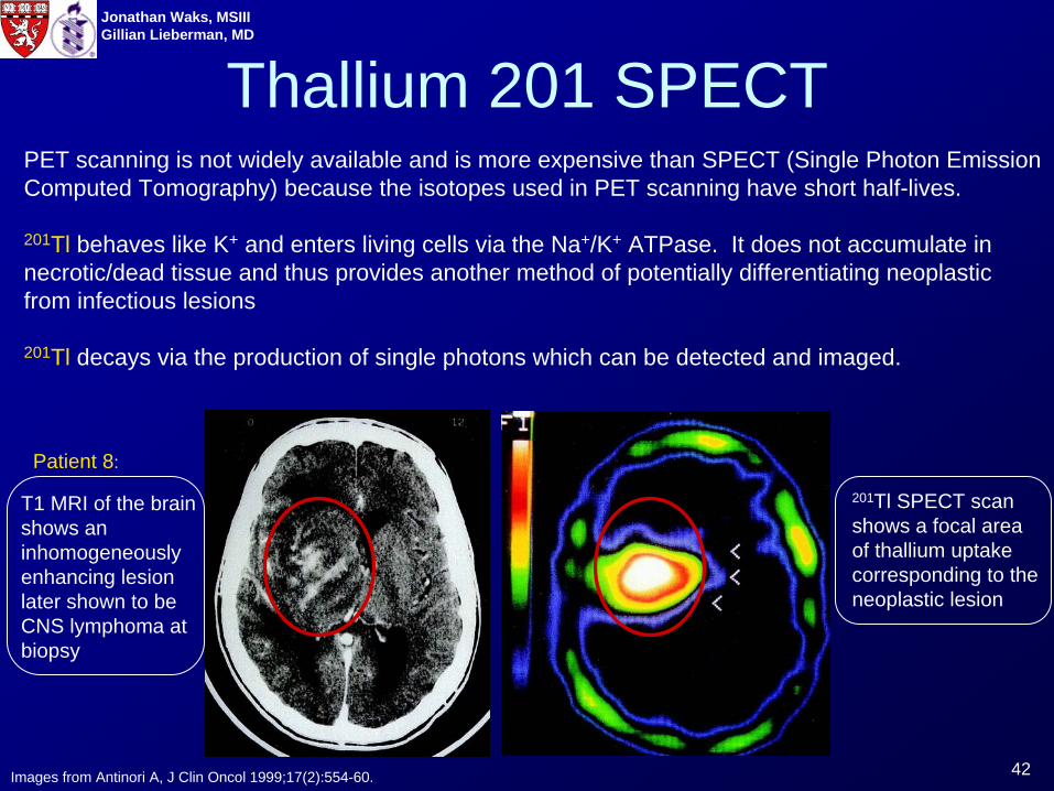
Thallium 201 SPECTPET scanning is not widely available and is more expensive than SPECT (Single Photon Emission Computed Tomography) because the isotopes used in PET scanning have short half-lives.
201Tl behaves like K+ and enters living cells via the Na+/K+ ATPase. It does not accumulate in necrotic/dead tissue and thus provides another method of potentially differentiating neoplastic from infectious lesions
201Tl decays via the production of single photons which can be detected and imaged.
Images from Antinori A, J Clin Oncol 1999;17(2):554-60.
T1 MRI of the brain shows an inhomogeneously enhancing lesion later shown to be CNS lymphoma at biopsy
201Tl SPECT scan shows a focal area of thallium uptake corresponding to the neoplastic lesion
Patient 8:
43
Jonathan Waks, MSIIIGillian Lieberman, MD
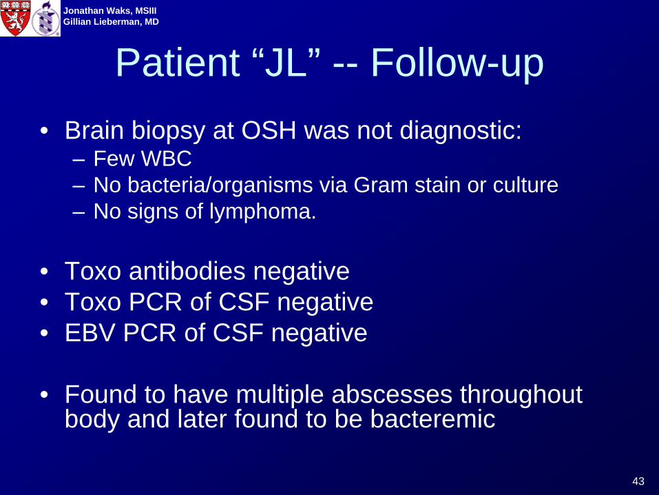
Patient “JL” -- Follow-up• Brain biopsy at OSH was not diagnostic:
– Few WBC– No bacteria/organisms via Gram stain or culture– No signs of lymphoma.
• Toxo antibodies negative• Toxo PCR of CSF negative• EBV PCR of CSF negative
• Found to have multiple abscesses throughout body and later found to be bacteremic
44
Jonathan Waks, MSIIIGillian Lieberman, MD
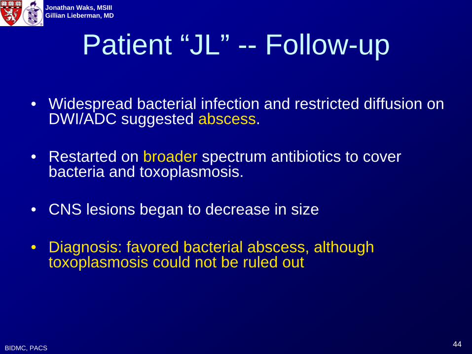
• Widespread bacterial infection and restricted diffusion on DWI/ADC suggested abscess.
• Restarted on broader spectrum antibiotics to cover bacteria and toxoplasmosis.
• CNS lesions began to decrease in size
• Diagnosis: favored bacterial abscess, although toxoplasmosis could not be ruled out
BIDMC, PACS
Patient “JL” -- Follow-up
45
Jonathan Waks, MSIIIGillian Lieberman, MD
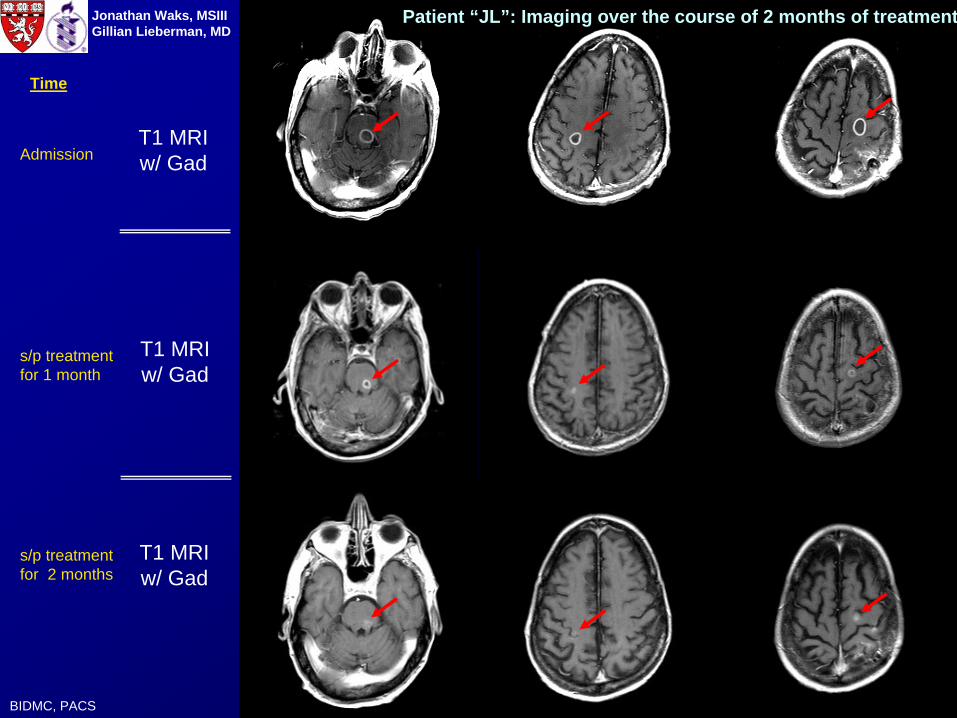
T1 MRIw/ Gad
T1 MRIw/ Gad
T1 MRIw/ Gad
BIDMC, PACS
Patient “JL”: Imaging over the course of 2 months of treatment
Time
Admission
s/p treatment for 1 month
s/p treatment for 2 months
46
Jonathan Waks, MSIIIGillian Lieberman, MD
Summary• CNS complications are extremely common in patients
with AIDS.
• The 2 most common CNS lesions in AIDS patients are toxoplasmosis and primary CNS lymphoma.
• Lesions are often treated empirically, but delay in definitive diagnosis can have significant consequences.
• MRI and nuclear medicine offer non-invasive methods to facilitate the identification of CNS lesions without invasive biopsy
47
Jonathan Waks, MSIIIGillian Lieberman, MD References
1. Antinori A, De Rossi G, Ammassari A, et al. Value of combined approach with thallium-201 single-photon emission computed tomography and Epstein-Barr virus DNA polymerase chain reaction in CSF for the diagnosis of AIDS-related primary CNS lymphoma. J Clin Oncol 1999;17(2):554-60.
2. Chang, Linda and Thomas Ernst. “Physciological MR to evaluate HIV-associated brain disorders.” Clinical MR Neuroimaging. Ed. Jonathan Gillard et al. Cambridge: Cambridge University Press, 2005. 460-478.
3. Doweiko JP, Groopman JE. “AIDS-related lymphomas: Primary central nervous system lymphoma”. UpToDate. www.uptodate.com Accessed 3/14/07
4. Fillipi, Christopher G. “The role of diffusion-weighted imaging in intracranial infection.” Clinical MR Neuroimaging. Ed. Jonathan Gillard et al. Cambridge: Cambridge University Press, 2005. 408-428.
5. “Gamut A-61 Ring-enhancing Lesion on CT or MRI.” Reeder And Felson's Gamuts In Radiology. Ed. Maurice Reeder. New York: Springer, 2003. Accessed electronically.
6. “How does PET work?” PET Imaging Center 2005. http://www.biomedpet.org/howitworks.cfm. Accessed 3/18/07.
7. Koralnik, IJ, “Approach to HIV-Infected patients with central nervous system lesions”. UpToDate. www.uptodate.com Accessed 3/14/07
8. Love, C et al. FDG PET of infection and inflammation. Radiographics. 2005; 25, 5: 1357-1367.
9. Netter, Frank H. Atlas of Human Anatomy, 3rd Edition. Teterboro: Icon Learning Systems, 2004.
10. Provenzale JM, Jinkins JR. Brain and spine imaging findings in AIDS patients. Radiol Clin North Am. 1997 Sep;35(5):1127-66.
11. Stadnik, Tadeusz et al. “Diffusion imaging: from basic physics to practical imaging” 1998 RSNA Scientific Assembly. http://ej.rsna.org/ej3/0095-98.fin/index.htm. Accessed on 3/16/07.
12. Thurnher MM, Thurnher SA, Schindler E. CNS involvement in AIDS: spectrum of CT and MR findings. Eur Radiol 1997;7(7):1091-7.
13. “Toxoplasmosis”. Laboratory Identification of Parasites of Public Health Concern. http://www.dpd.cdc.gov/dpdx/HTML/Toxoplasmosis.htm. Accessed on 3/18/07.
14. Zimmerman, Robert D. “Physiological imaging in infection, inflammation and demyelination: overview.” Clinical MR Neuroimaging. Ed. Jonathan Gillard et al. Cambridge: Cambridge University Press, 2005. 353-379.

48
Jonathan Waks, MSIIIGillian Lieberman, MD
Acknowledgements:
Mizuki Nishino, MD
Gillian Lieberman, MD
Pamela Lepkowski
Larry Barbaras, Webmaster