right ventricular size and function · right ventricular size and function edwin s. tucay, md,...
TRANSCRIPT
RIGHT VENTRICULAR SIZE AND FUNCTION
Edwin S. Tucay, MD, FPCC, FPCC, FPSEPhilippine Society of Echocardiography
Quezon City, Philippines
Echo Mission, BRTTH, Legaspi City, July 1-2, 2016

NO DISCLOSURE
NORMAL HEART = NORMAL FAMILY
Outline
• Need to evaluate the right ventricle• Systematic evaluation of the right ventricle• Right ventricular dimension• Right ventricular systolic function• Recommendation
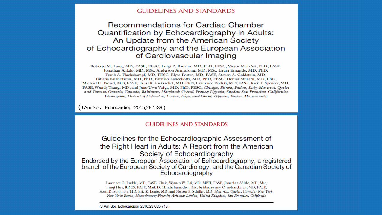
(J Am Soc Echocardiogr 2015;28:1-39.)
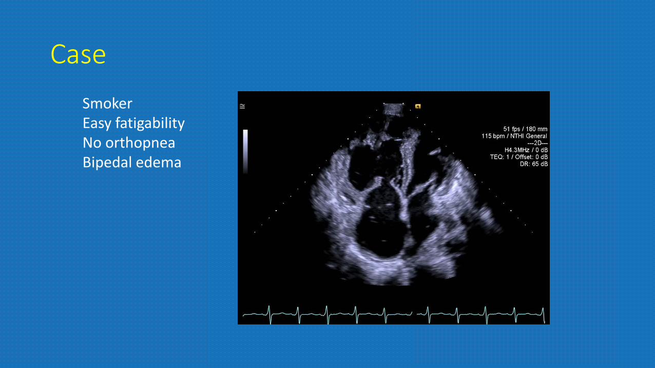
CaseSmoker Easy fatigabilityNo orthopneaBipedal edema
Importance of evaluating the right ventricle
• Role in the clinical outcome of cardiopulmonary disease
• Size and function adversely affected by• left ventricular dysfunction• primary pulmonary hypertension• conditions that affect the tricuspid valve leading to
significant tricuspid regurgitation

Systematic Evaluation of the Right Ventricle• limited due to its complex morphology• comprehensive evaluation:
• right ventricular dimensions• systolic and diastolic function, and RV systolic pressure
• use multiple echo windows: • apical 4-chamber, modified apical 4-chamber, left parasternal long
axis (PLAX) and parasternal short-axis (PSAX), left parasternal RV inflow, and subcostal views.
• 3 D echo imaging continuously improve.
RIGHT VENTRICULAR DIMENSIONS
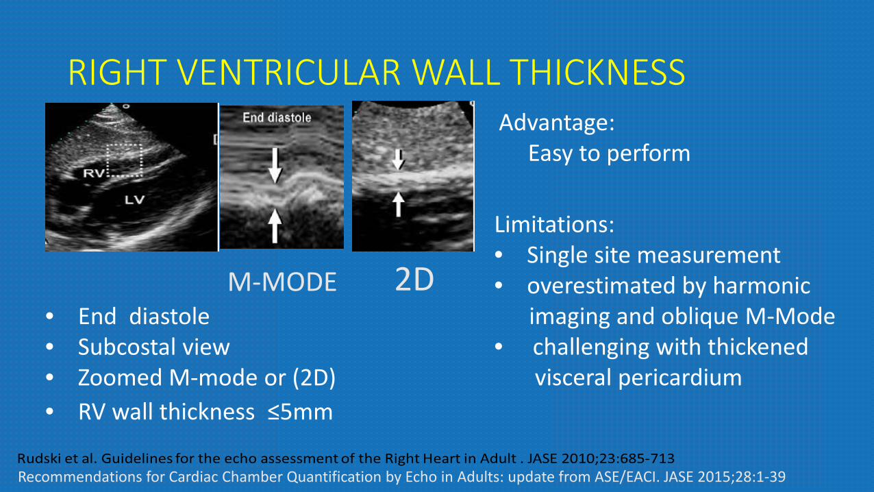
RIGHT VENTRICULAR WALL THICKNESS
• End diastole• Subcostal view• Zoomed M-mode or (2D) • RV wall thickness ≤5mm
2DM-MODE
Recommendations for Cardiac Chamber Quantification by Echo in Adults: update from ASE/EACI. JASE 2015;28:1-39
Advantage: Easy to perform
Limitations:• Single site measurement• overestimated by harmonic
imaging and oblique M-Mode• challenging with thickened
visceral pericardium

RIGHT VENTRICULAR WALL THICKNESS
2DM-MODE
Recommendation:
Abnormal RV wall thickness should be reported in patients suspected of having RV and/or LV dysfunction, using the normal cut off of 5 mm
Recommendations for Cardiac Chamber Quantification by Echo in Adults: update from ASE/EACI. JASE 2015;28:1-39
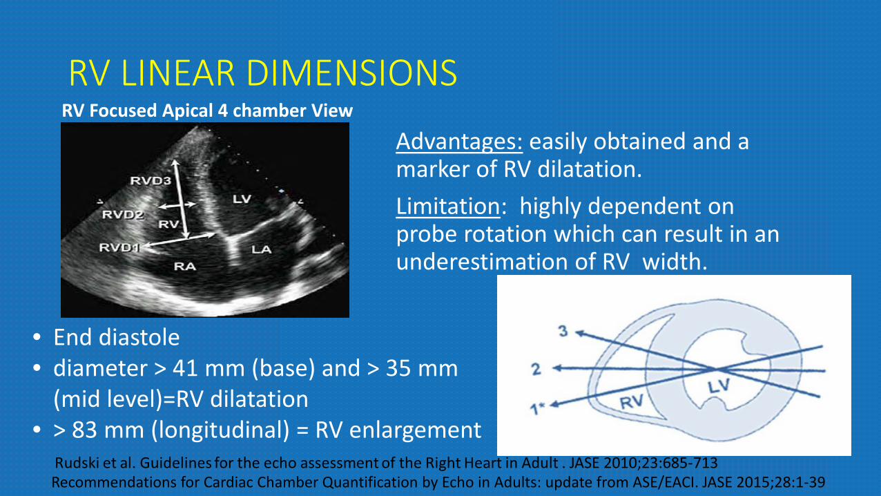
RV LINEAR DIMENSIONSRV Focused Apical 4 chamber View
Advantages: easily obtained and a marker of RV dilatation.Limitation: highly dependent on probe rotation which can result in an underestimation of RV width.
• End diastole• diameter > 41 mm (base) and > 35 mm
(mid level)=RV dilatation• > 83 mm (longitudinal) = RV enlargement
Recommendations for Cardiac Chamber Quantification by Echo in Adults: update from ASE/EACI. JASE 2015;28:1-39
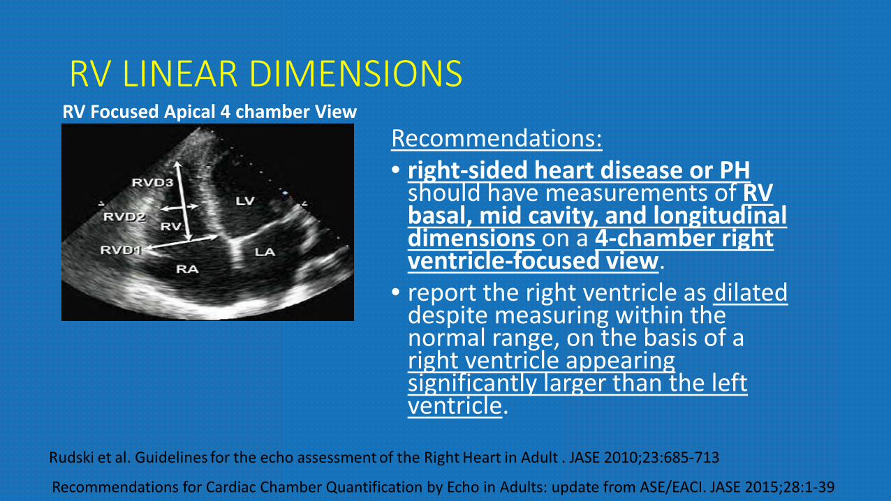
RV LINEAR DIMENSIONSRV Focused Apical 4 chamber View
Recommendations: • right-sided heart disease or PH
should have measurements of RV basal, mid cavity, and longitudinal dimensions on a 4-chamber right ventricle-focused view.
• report the right ventricle as dilateddespite measuring within the normal range, on the basis of a right ventricle appearing significantly larger than the left ventricle.
Recommendations for Cardiac Chamber Quantification by Echo in Adults: update from ASE/EACI. JASE 2015;28:1-39

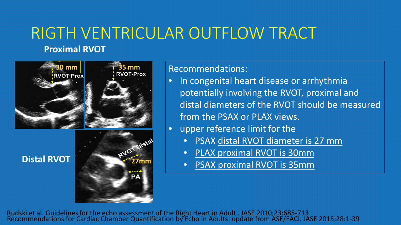
RIGTH VENTRICULAR OUTFLOW TRACTProximal RVOT
Distal RVOT
Advantages: easily obtained from the left PSAX window. Limitation: Limited normative data, window for measurement not yet standardized, wall is often suboptimal.
30 mm
27mm
35 mm
Recommendations for Cardiac Chamber Quantification by Echo in Adults: update from ASE/EACI. JASE 2015;28:1-39
End-diastoleRVOT proximal
PLAX: RV wall to IVS-aortic junctionPSAX: RV wall to Aortic valve
RVOT distalPSAX: just proximal to pulmonic valve
RIGTH VENTRICULAR OUTFLOW TRACTProximal RVOT
Distal RVOT
Recommendations: • In congenital heart disease or arrhythmia
potentially involving the RVOT, proximal and distal diameters of the RVOT should be measured from the PSAX or PLAX views.
• upper reference limit for the • PSAX distal RVOT diameter is 27 mm • PLAX proximal RVOT is 30mm• PSAX proximal RVOT is 35mm
30 mm
27mm
35 mm
Recommendations for Cardiac Chamber Quantification by Echo in Adults: update from ASE/EACI. JASE 2015;28:1-39
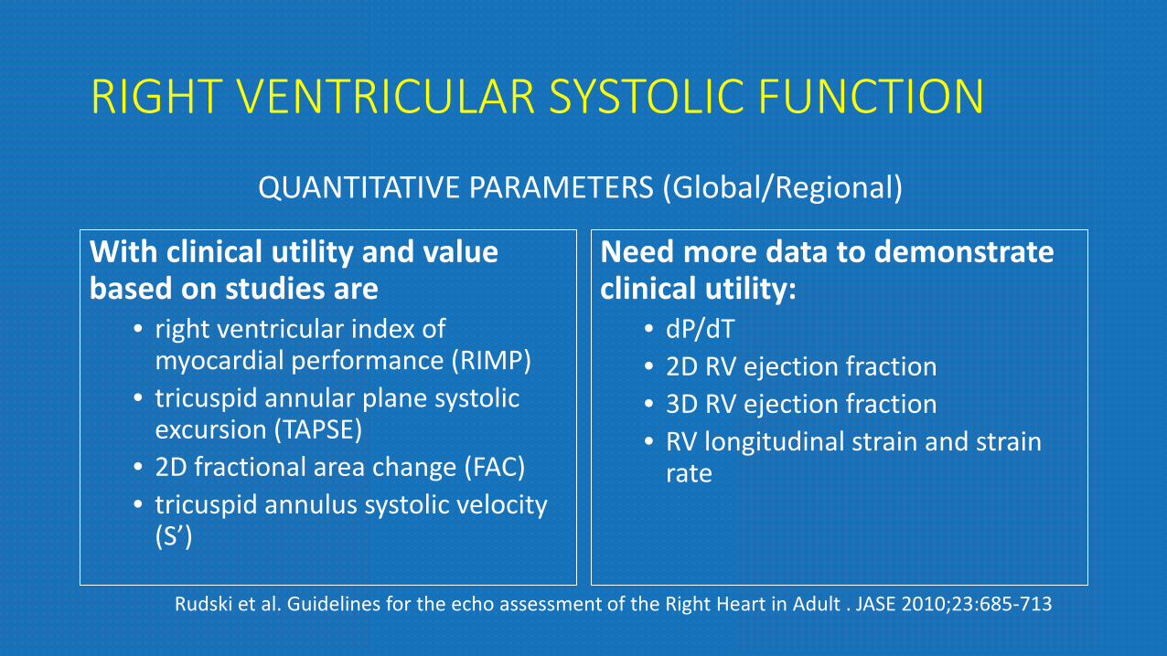
RIGHT VENTRICULAR SYSTOLIC FUNCTION
With clinical utility and value based on studies are
• right ventricular index of myocardial performance (RIMP)
• tricuspid annular plane systolic excursion (TAPSE)
• 2D fractional area change (FAC)• tricuspid annulus systolic velocity
(S’)
Need more data to demonstrate clinical utility:
• dP/dT• 2D RV ejection fraction• 3D RV ejection fraction • RV longitudinal strain and strain
rate
QUANTITATIVE PARAMETERS (Global/Regional)
Rudski et al. Guidelines for the echo assessment of the Right Heart in Adult . JASE 2010;23:685-713
RIGHT VENTRICULAR SYSTOLIC FUNCTION
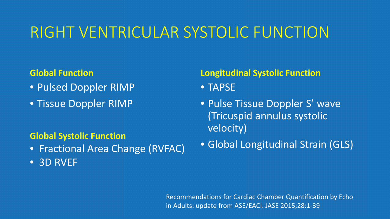
Global Function• Pulsed Doppler RIMP• Tissue Doppler RIMP
Longitudinal Systolic Function• TAPSE• Pulse Tissue Doppler S’ wave
(Tricuspid annulus systolic velocity)
• Global Longitudinal Strain (GLS)Global Systolic Function• Fractional Area Change (RVFAC)• 3D RVEF
Recommendations for Cardiac Chamber Quantification by Echo in Adults: update from ASE/EACI. JASE 2015;28:1-39
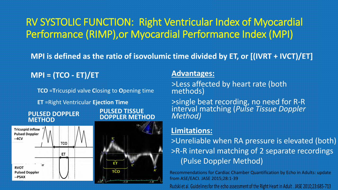
RV SYSTOLIC FUNCTION: Right Ventricular Index of Myocardial Performance (RIMP),or Myocardial Performance Index (MPI)
PULSED DOPPLER METHOD
PULSED TISSUE DOPPLER METHOD
MPI = (TCO - ET)/ET
MPI is defined as the ratio of isovolumic time divided by ET, or [(IVRT + IVCT)/ET]
TCO =Tricuspid valve Closing to Opening time
ET =Right Ventricular Ejection Time
Tricuspid inflowPulsed Doppler--4CV
RVOTPulsed Doppler--PSAX
Advantages:>Less affected by heart rate (both methods)>single beat recording, no need for R-R interval matching (Pulse Tissue Doppler Method)
Recommendations for Cardiac Chamber Quantification by Echo in Adults: update from ASE/EACI. JASE 2015;28:1-39
Limitations:>Unreliable when RA pressure is elevated (both)>R-R interval matching of 2 separate recordings
(Pulse Doppler Method)

RV SYSTOLIC FUNCTION: Right Ventricular Index of Myocardial Performance (RIMP),or Myocardial Performance Index (MPI)
PULSED DOPPLER METHOD
PULSED TISSUE DOPPLER METHOD
Recommendations:• used for initial and serial measurements of
RV function in complement with other quantitative and nonquantitative measures.
• upper reference limit for the right-sided MPI is 0.43 using the pulsed Doppler method and 0.54 using the pulsed tissue Doppler method.
• It should not be used as the sole quantitative method for evaluation of RV function and should not be used with irregular heart rates.
MPI = (TCO - ET)/ET
MPI is defined as the ratio of isovolumic time divided by ET, or [(IVRT + IVCT)/ET]
TCO =Tricuspid valve Closing to Opening timeET =Right Ventricular Ejection Time
Tricuspid inflowPulsed Doppler--4CV
RVOTPulsed Doppler--PSAX
Recommendations for Cardiac Chamber Quantification by Echo in Adults: update from ASE/EACI. JASE 2015;28:1-39
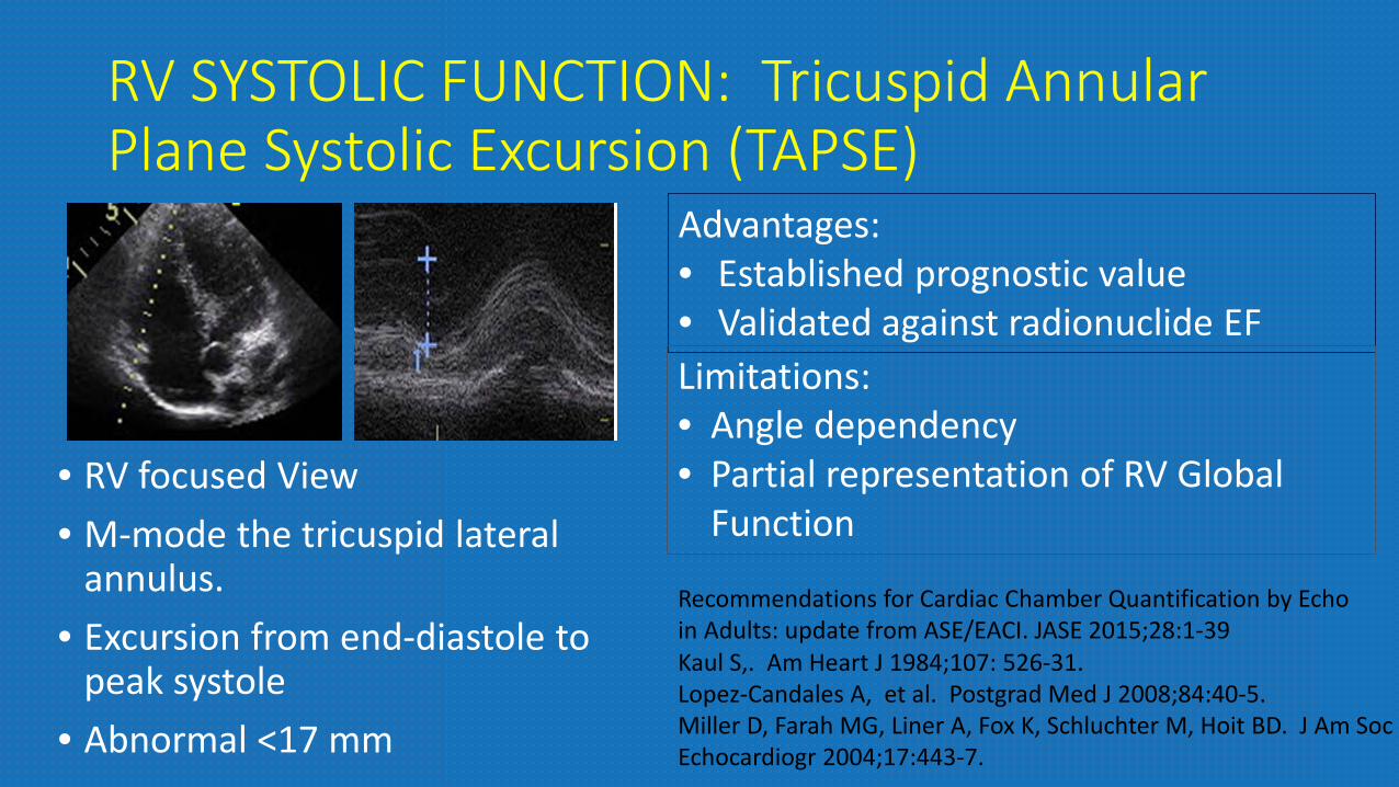
RV SYSTOLIC FUNCTION: Tricuspid Annular Plane Systolic Excursion (TAPSE)
• RV focused View• M-mode the tricuspid lateral
annulus. • Excursion from end-diastole to
peak systole• Abnormal <17 mm
Kaul S,. Am Heart J 1984;107: 526-31.Lopez-Candales A, et al. Postgrad Med J 2008;84:40-5.Miller D, Farah MG, Liner A, Fox K, Schluchter M, Hoit BD. J Am SocEchocardiogr 2004;17:443-7.
Advantages:• Established prognostic value• Validated against radionuclide EFLimitations:• Angle dependency• Partial representation of RV Global
Function
Recommendations for Cardiac Chamber Quantification by Echo in Adults: update from ASE/EACI. JASE 2015;28:1-39

RV SYSTOLIC FUNCTION: Tricuspid Annular Plane Systolic Excursion (TAPSE)
Recommendation: • TAPSE should be used routinely as a simple method of
estimating RV function, with a lower reference value for impaired RV systolic function of 17 mm.
Recommendations for Cardiac Chamber Quantification by Echo in Adults: update from ASE/EACI. JASE 2015;28:1-39
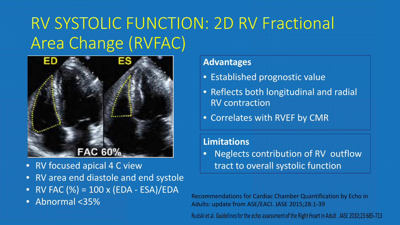
RV SYSTOLIC FUNCTION: 2D RV Fractional Area Change (RVFAC)
Advantages• Established prognostic value• Reflects both longitudinal and radial
RV contraction• Correlates with RVEF by CMR
Limitations• Neglects contribution of RV outflow
tract to overall systolic function• RV focused apical 4 C view• RV area end diastole and end systole• RV FAC (%) = 100 x (EDA - ESA)/EDA• Abnormal <35%
Recommendations for Cardiac Chamber Quantification by Echo in Adults: update from ASE/EACI. JASE 2015;28:1-39
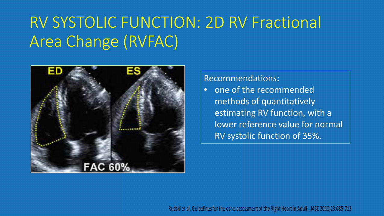
RV SYSTOLIC FUNCTION: 2D RV Fractional Area Change (RVFAC)
Recommendations: • one of the recommended
methods of quantitatively estimating RV function, with a lower reference value for normal RV systolic function of 35%.
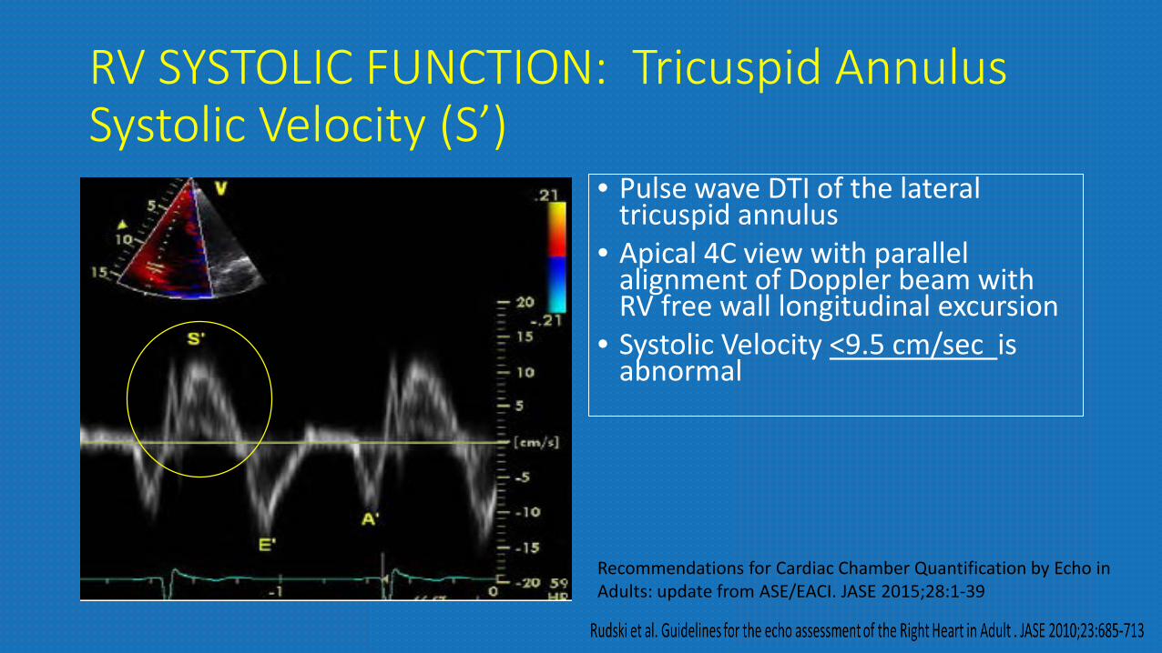
RV SYSTOLIC FUNCTION: Tricuspid Annulus Systolic Velocity (S’)
• Pulse wave DTI of the lateral tricuspid annulus
• Apical 4C view with parallel alignment of Doppler beam with RV free wall longitudinal excursion
• Systolic Velocity <9.5 cm/sec is abnormal
Recommendations for Cardiac Chamber Quantification by Echo in Adults: update from ASE/EACI. JASE 2015;28:1-39

RV SYSTOLIC FUNCTION: Tricuspid Annulus Systolic Velocity (S’)
Advantages• easily measured, reliable and
reproducible. • correlates well with other measures of
global RV systolic function.• Validated against radionuclide EF• Established prognostic value
Limitations• Angle dependent• Not fully representative of RV global
function after thoracotomy, pulmonary thromboendarterectomy or heart transplantation
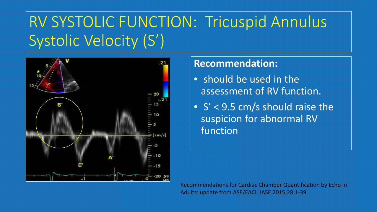
RV SYSTOLIC FUNCTION: Tricuspid Annulus Systolic Velocity (S’)
Recommendation: • should be used in the
assessment of RV function.• S’ < 9.5 cm/s should raise the
suspicion for abnormal RV function
Recommendations for Cardiac Chamber Quantification by Echo in Adults: update from ASE/EACI. JASE 2015;28:1-39
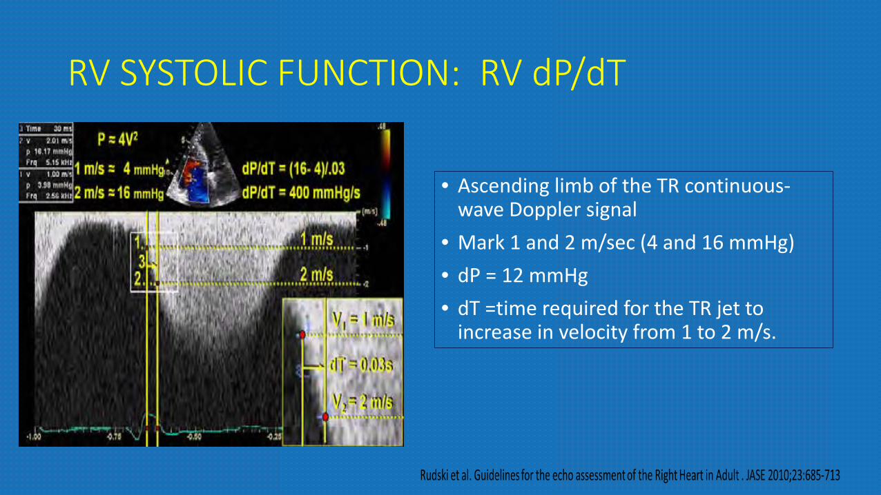
RV SYSTOLIC FUNCTION: RV dP/dT
• Ascending limb of the TR continuous-wave Doppler signal
• Mark 1 and 2 m/sec (4 and 16 mmHg)• dP = 12 mmHg• dT =time required for the TR jet to
increase in velocity from 1 to 2 m/s.

RV SYSTOLIC FUNCTION: RV dP/dT
Recommendations: • RV dP/dt < 400 mm Hg/s is likely abnormal• cannot be recommended for routine use• can be considered in subjects with
suspected RV dysfunction.
• Advangtage Advantage: • simple technique with sound physiologic basis
Limitations:• Lack of data in normal subjects• Load dependent
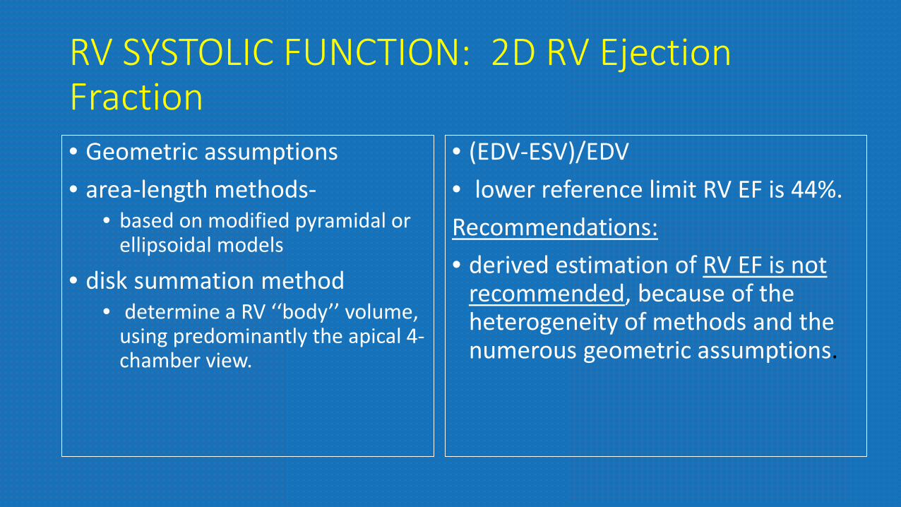
RV SYSTOLIC FUNCTION: 2D RV Ejection Fraction• Geometric assumptions• area-length methods-
• based on modified pyramidal or ellipsoidal models
• disk summation method • determine a RV ‘‘body’’ volume,
using predominantly the apical 4-chamber view.
• (EDV-ESV)/EDV • lower reference limit RV EF is 44%. Recommendations:• derived estimation of RV EF is not
recommended, because of the heterogeneity of methods and the numerous geometric assumptions.
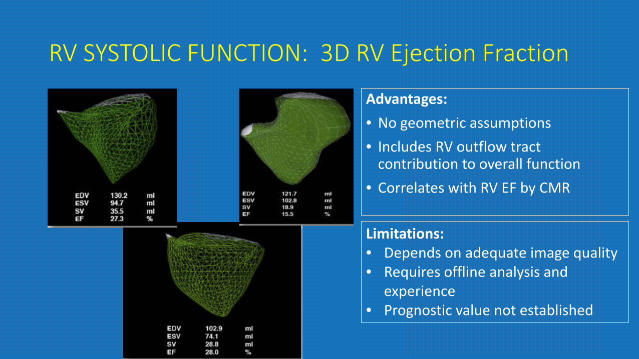
RV SYSTOLIC FUNCTION: 3D RV Ejection Fraction
Advantages: • No geometric assumptions• Includes RV outflow tract
contribution to overall function• Correlates with RV EF by CMR
Limitations:• Depends on adequate image quality• Requires offline analysis and
experience• Prognostic value not established

RV SYSTOLIC FUNCTION: 3D RV Ejection Fraction
Recommendations:• 3D echocardiography RV EF
may be reported.• lower reference limit of 45%• reserve 3D methods for serial
volume and EF determinations.
RV SYSTOLIC FUNCTION: RV Strain and Strain Rate• Strain = percentage change in
myocardial deformation• Strain rate = rate of deformation
of myocardium over time. • Strain rate has been closely
correlated with myocardial contractility in vitro and in vivo
• DTI-derived Strain• Speckle tracking Echo (STE)
derived strain – angle independent
• Global Longitudinal Strain
Jamal F, Bergerot C, Argaud L, Loufouat J, Ovize M. Longitudinal strainquantitates regional right ventricular contractile function. Am J PhysiolHeart Circ Physiol 2003;285:H2842-7.
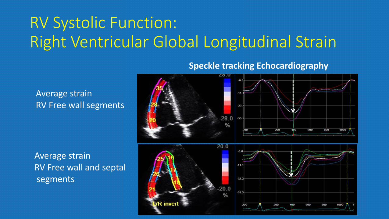
RV Systolic Function:Right Ventricular Global Longitudinal Strain
Average strain RV Free wall segments
Average strain RV Free wall and septalsegments
Speckle tracking Echocardiography

RV SYSTOLIC FUNCTION: RV Global Longitudinal Strain and - 2d Speckle TrackingAdvantages: • relatively angle independent • possesses an improved signal-to-noise ratio. • provide regional function estimates, as well as a more ‘‘global’’
function.Disadvantages: • lack of normative data and need additional validation. • different algorithms in different platforms may result in different
normal ranges.
RV SYSTOLIC FUNCTION: RV Strain and Strain RateRecommendations: • Because of the lack of reproducibility and the paucity of data, this
technique is not recommended for routine clinical use. • No reference limits can be recommended, because of the large
degree of variability.
Recommendation for the evaluation of RV systolic function
• Visual assessment of RV systolic function gives an initial qualitative evaluation of RV systolic function but remains insufficient
• Simple and reproducible methods of assessing RV systolic function should be incorporated into the routine echocardiographic assessment. ( FAC, TAPSE, pulsed tissue Doppler S’, and MPI).
• Combining more than one measure of RV function, such as S’ and MPI, may more reliably distinguish normal from abnormal function.
Miller D, Farah MG, Liner A, Fox K et al. J Am Soc Echocardiogr 2004;17:443-7.Rudski et al. Guidelines for the echo assessment of the Right Heart in Adult . JASE 2010;23:685-713
Recommendation for the evaluation of RV systolic function
• At least one of the above quantitative measures be incorporated into the routine echocardiographic examination and report
• when RV dysfunction is suspected • when the clinical indication for the study relates to a condition that may
affect the right ventricle.
• Techniques such as strain, and strain rate are not currently recommended as routine and are best reserved for specific clinical and research applications.
Miller D, Farah MG, Liner A, Fox K et al. J Am Soc Echocardiogr 2004;17:443-7.Rudski et al. Guidelines for the echo assessment of the Right Heart in Adult . JASE 2010;23:685-713

CaseSmoker Easy fatigabilityNo orthopneaBipedal edema
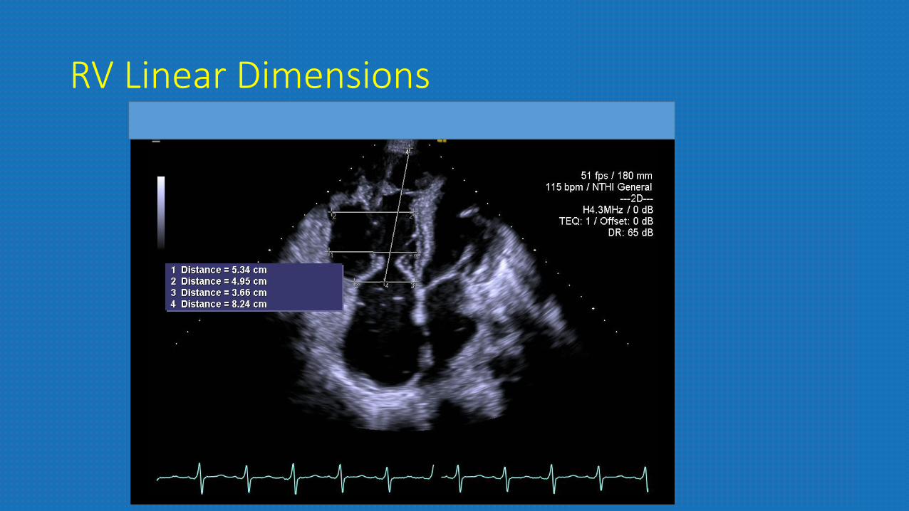
RV Linear Dimensions
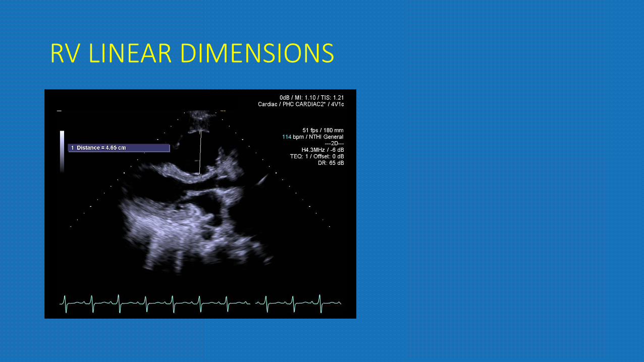
RV LINEAR DIMENSIONS
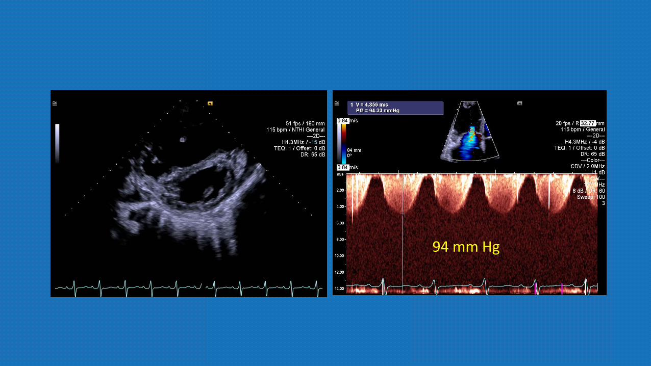
94 mm Hg
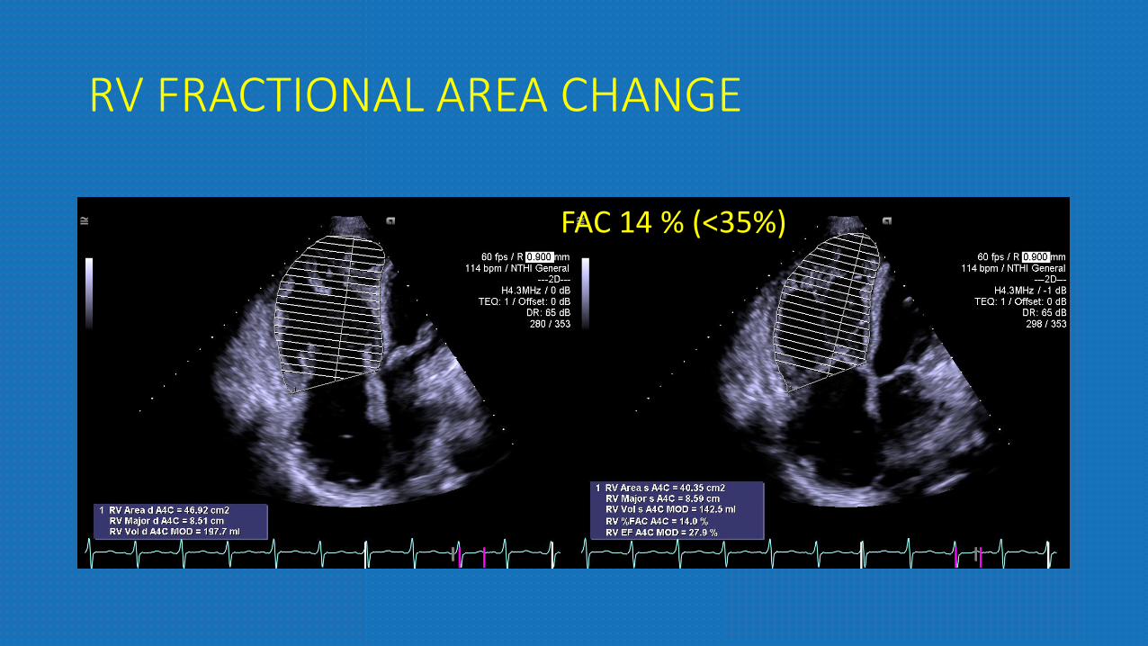
RV FRACTIONAL AREA CHANGE
FAC 14 % (<35%)

TAPSE
14.8 mm (<17 mm)
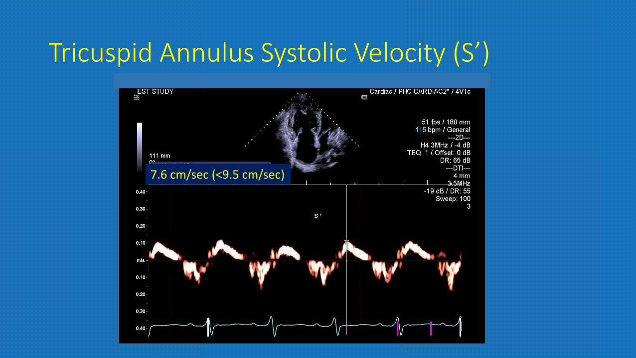
Tricuspid Annulus Systolic Velocity (S’)
0.115 m/s (
7.6 cm/sec (<9.5 cm/sec)
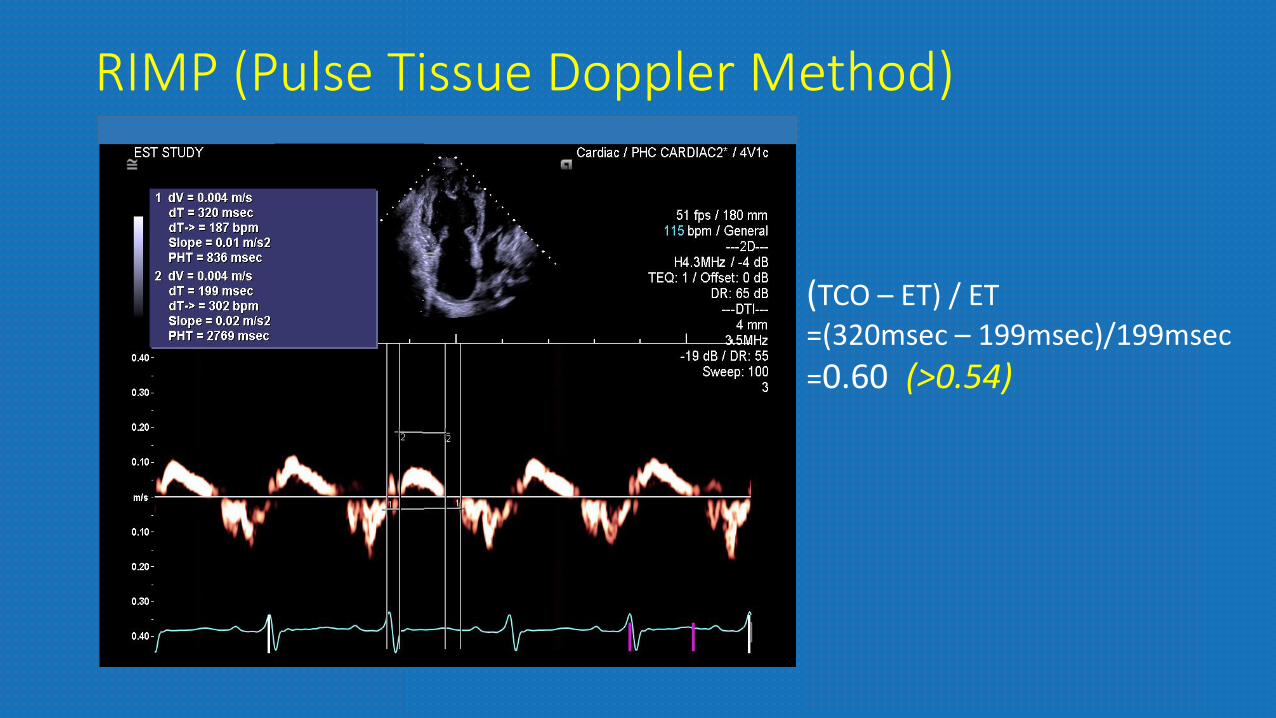
RIMP (Pulse Tissue Doppler Method)
(TCO – ET) / ET=(320msec – 199msec)/199msec=0.60 (>0.54)
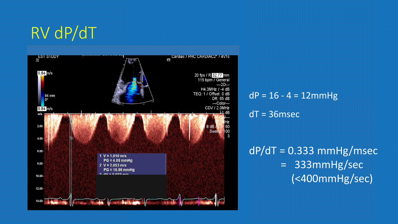
RV dP/dT
dP = 16 - 4 = 12mmHg
dT = 36msec
dP/dT = 0.333 mmHg/msec= 333mmHg/sec
(<400mmHg/sec)
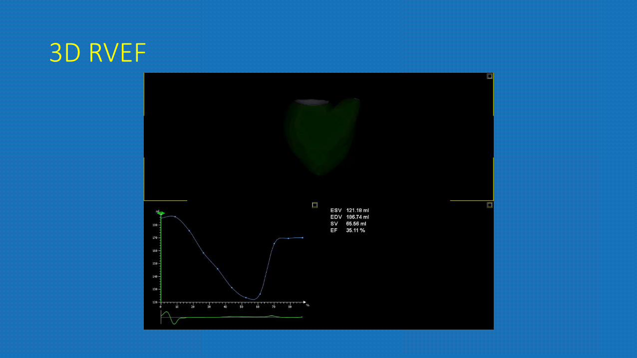
3D RVEF
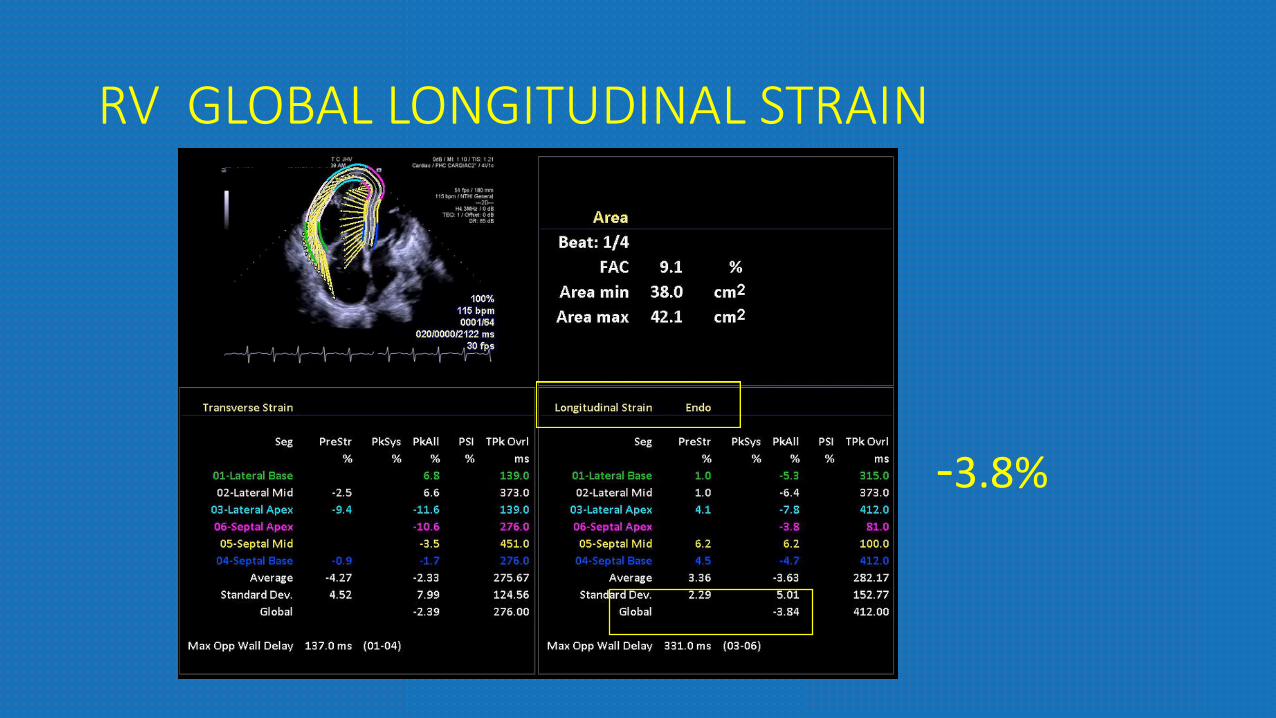
RV GLOBAL LONGITUDINAL STRAIN
-3.8%

CaseSmoker Easy fatigabilityNo orthopneaBipedal edema
DILATED HYPERTROPHIED RVRIGHT VENTRICULAR SYSTOLIC
DYSFUNCTIONSEVERE PULMONARY HYPERTENSION
