idiopathic polymorphic ventricular tachycardia: sebe agn“ase di … · ventricular extrasystole...
TRANSCRIPT
299Copyright © 2017 The Korean Society of Cardiology
Korean Circulation Journal
Review Article
https://doi.org/10.4070/kcj.2016.0303Print ISSN 1738-5520 • On-line ISSN 1738-5555
Idiopathic Polymorphic Ventricular Tachycardia: a “Benign Disease” with a Touch of Bad Luck? Sami Viskin, MDTel Aviv Sourasky Medical Center and Sackler School of Medicine, Tel Aviv University, Tel Aviv, Israel
Ventricular extrasystole originating from the right ventricular outflow tract or the left ventricular outflow tract are the most commonly encountered ventricular arrhythmias recorded in ostensibly healthy individuals with no evidence of heart disease. These ventricular arrhythmias have a distinctive electrocardiographic morphology. The morphology is so distinctive that it is common practice to accept the diagnosis of “idiopathic benign ventricular arrhythmias from the outflow tract” based on this unique morphology when the electrocardiogram during sinus rhythm and the echocardiogram are normal, sometimes removing the need to perform invasive tests in patients. Even if the outflow ventricular extrasystole ultimately triggers sustained ventricular arrhythmia, the resulting ventricular tachycardia (VT) will be a monomorphic VT originating from the outflow tract, which is known to be hemodynamically well tolerated. Thus, idiopathic ventricular arrhythmias originating from outflow tracts are universally considered benign. In 2005, we described a rare form of malignant polymorphic VT resulting in syncope or cardiac arrest. Here, we review the literature on this topic since the emergence of initial descriptions of this intriguing phenomenon. (Korean Circ J 2017;47(3):299-306)
KEY WORDS: Ventricular tachycardia; Sudden cardiac death.
Introduction
Ventricular extrasystole originating from the right ventricular outflow tract (RVOT) or the left ventricular outflow tract (LVOT) is the most commonly encountered ventricular arrhythmia in ostensibly healthy individuals with no evidence of heart disease.1)2) For example, among otherwise healthy athletes with ventricular arrhythmia, the arrhythmia site of origin is the ventricular outflow tract in roughly 70%.3) These ventricular arrhythmias have a characteristic electrocardiographic morphology, with a left bundle
branch block pattern and tall R-waves in the inferior leads of either the normal or right axis (Fig. 1A, C, 2B).4) In fact, this QRS morphology is so distinctive that it is common practice to accept the diagnosis of “idiopathic benign ventricular arrhythmia from the outflow tract” based on this unique morphology when the electrocardiogram (ECG) during sinus rhythm and the echocardiogram are normal, sometimes negating the need for invasive tests.1)5)
A small proportion of patients with an unusually high burden of ventricular extrasystole can developing a reversible deterioration of left ventricular function as manifestation of “tachycardia-induced cardiomyopathy.”6) Still, the vast majority of patients with idiopathic outflow tract arrhythmias have an excellent long-term prognosis. Even if the outflow ventricular extrasystole ultimately triggers sustained ventricular arrhythmia, the resulting ventricular tachycardia (VT) will be a monomorphic VT originating from the outflow tract (Fig. 2A), which is a scenario that is known to be hemodynamically well tolerated.1)5) Thus, the idiopathic ventricular arrhythmias originating from the outflow tract are universally considered benign.1)2)4)7)
In 2005, we described three patients with typical idiopathic RVOT ventricular arrhythmia and no evidence of organic heart disease who unexpectedly developed malignant polymorphic VT resulting in syncope or cardiac arrest.8) In our original cases, the documented
Received: August 16, 2016Revision Received: October 24, 2016Accepted: November 11, 2016Correspondence: Sami Viskin, MD, Department of Cardiology, Tel Aviv Medical Center, Weizman 6, Tel Aviv 64239, IsraelTel: 972-36973311, Fax: 972-36972749E-mail: [email protected]
• The author has no financial conflicts of interest.
This is an Open Access article distributed under the terms of the Creative Commons Attribution Non-Commercial License (http://creativecommons.org/licenses/by-nc/3.0) which permits unrestricted non-commercial use, distribution, and reproduction in any medium, provided the original work is properly cited.

300 Idiopathic Polymorphic RVOT-VT
https://doi.org/10.4070/kcj.2016.0303 www.e-kcj.org
polymorphic VT invariably began with outflow tract ventricular extrasystole with QRS morphology that was indistinguishable from numerous extrasystoles recorded over the years. However, the extrasystoles triggering the polymorphic arrhythmias had a short coupling interval, leading to an R-on-T phenomenon (i.e., an extrasystole “falling” on the descending limb of the T-wave of the preceding sinus beat).8) This short coupling interval, recorded at the time of onset of polymorphic VT, contrasted with the longer coupling intervals recorded in the same patients during typical monomorphic VT episodes (Fig. 1, 2). Taking into account that very short coupling intervals are invariably recorded in highly malignant polymorphic arrhythmias of patients with idiopathic ventricular fibrillation (VF),9) we concluded that this short coupling interval was essential in triggering the malignant VT in our patients. Accordingly, we termed this phenomenon a “short-coupled variant of RVOT-VT.”8) Shortly thereafter, however, the group
of Shimizu in Japan (Noda et al.),10) described a larger series of patients with polymorphic VT also originating from the RVOT in the absence of organic heart disease.10) In this series (which included 16 patients with polymorphic RVOT-VT), the coupling interval was short in some patients, but not in all patients (Fig. 3A). In fact, the mean coupling interval of the extrasystoles triggering polymorphic VT in the patients of that research was not different from the coupling interval initiating monomorphic VT in a group of 85 patients with typical idiopathic benign RVOT VT (see the section on ECG below).10) Thus, the research of Noda et al.10) clarifies that a short coupling interval is not mandatory for triggering this newly recognized entity, now termed “malignant idiopathic polymorphic RVOT VT.”10)11) Here, we review the literature on this topic published since the initial descriptions of this intriguing phenomenon emerged.8)10)
Fig. 1. A female patient with a very long history of palpitations. She fi rst presented in 1985 at the age of 35 years with frequent RVOT extrasystoles, including ventricular bigeminy (A). In 1997 (age 47), she had recurrent syncope with documented polymorphic VT (B). She underwent ICD implantation but declined drug or ablation therapy. Her left ventricular function remains normal despite almost incessant bigeminy (C). 18 years after her initial arrhythmic event, she experienced non-sustained polymorphic VT (ventricular rate approaching 300 beats/min), documented by her ICD (D). She continues to be in good health. Modifi ed from Viskin et al.8) RVOT: right ventricular outfl ow tract, VT: ventricular tachycardia, ICD: implantable cardioverter-defi brillator.
Fig. 2. Typical non-sustained monomorphic VT originated from an extrasystole that clearly began after the end of the T-wave (A). The same patient later developed ventricular extrasystoles of the same morphology, but with a shorter coupling interval. The fi rst extrasystole in the precordial leads began during the descending limb of the T-wave (best appreciated in V5-V6) (B). Ventricular extrasystoles with an even shorter coupling interval (the extrasystoles originated shortly after the peak of the T-wave) triggered non-sustained polymorphic VT. The coupling interval of the extrasystoles initiating polymorphic VT (C) was clearly shorter than the coupling interval preceding monomorphic VT in the same patient (A). Modifi ed from Viskin et al.8) VT : ventricular tachycardia.
A
B
C
D
A
B
C
301Sami Viskin
https://doi.org/10.4070/kcj.2016.0303www.e-kcj.org
Definition
The term “idiopathic polymorphic RVOT VT” should be reserved for patients with frequent and otherwise typical outflow tract ventricular ectopy (see “clinical characteristics” below) who develop rapid polymorphic VT as their only sustained arrhythmia or (not infrequently) in addition to well-tolerated sustained monomorphic VT (Fig. 1, 2).8) This definition recognizes the fact that ECG provides only an approximate estimate of the site of origin of an arrhythmia, and that detailed intracardiac mapping might ultimately localize the site of origin of the arrhythmia to the LVOT or contiguous structures. The term “idiopathic” indicates that organic heart disease has been excluded. However, as discussed in detail elsewhere,12) differentiating a truly idiopathic RVOT VT from RVOT VT related to less-than-overt forms of right ventricular dysplasia is not always straightforward. Finally, this definition assumes that idiopathic polymorphic RVOT VT and idiopathic VF13) are different diseases. Importantly, in 14% of cases of idiopathic VF, the arrhythmia originates in the RVOT,14) and several ECG characteristics differentiate patients with RVOT-related idiopathic VF from the more common form of idiopathic VF originating from Purkinje fibers.14) Specifically, the coupling interval of extrasystoles triggering idiopathic VF from the RVOT, although “short,” is not as short as the ultra-short coupling interval
of typical (Purkinje-related) idiopathic VF9) (355±30 vs. 280±26 msec, p=0.01).14) In addition, RVOT extrasystoles are wider than the Purkinje-related ectopic beats (145±12 vs. 126±18 msec, p=0.04).14)
As opposed to patients with idiopathic VF originating from the RVOT, who usually have cardiac arrest as their presenting symptom (Fig. 4) and who have little-to-no ventricular ectopic activity between arrhythmic events, patients with idiopathic RVOT VT have a long history of palpitations and have frequent RVOT extrasystoles (often with documented episodes of well-tolerated monomorphic VT) before they actually develop polymorphic VT (Fig. 2).8)10)15)
Incidence
In a retrospective series involving 91 patients referred over the course of five years to a single Japanese center for ablation of VT originating from the RVOT in the absence of heart disease, 14 patients (15%) had documented polymorphic VT, including four (4%) with VF.16) Because of recall bias and referral bias, this series probably overestimated the risk of polymorphic VT among patients with RVOT VT. Furthermore, the incidence of polymorphic VT among patients presenting with ventricular extrasystoles (rather than with RVOT VT, as in this series) would be drastically smaller. A different series, involving 130 patients evaluated for ventricular arrhythmia in the absence of organic heart disease, presented a more realistic number, wherein four patients (3%) had documented polymorphic VT originating from the RVOT or cardiac arrest with VF.17) Since our original description of this phenomenon one decade ago, we have not encountered additional patients presenting with idiopathic RVOT extrasystole or RVOT monomorphic VT who have gone on to develop polymorphic VT. Clearly, malignant polymorphic RVOT VT is rare, and the great challenge is to distinguish the small minority of patients with this malignant disease from the majority of patients with benign idiopathic RVOT arrhythmia.
Clinical Characteristics
A typical patient with malignant RVOT VT is an otherwise healthy adult presenting with a long history of palpitations. In different case series, the mean age of patients at the time of presentation with polymorphic VT is 39-45 years.10)16)18) Females predominate, representing 56% to 85% of patients in different series.10)16)18) A family history of sudden death is reported only exceptionally.10)18) In general, patients with polymorphic VT report a history of palpitations beginning 6-10 years prior to the malignant arrhythmic event. In all of these aspects, patients with malignant polymorphic
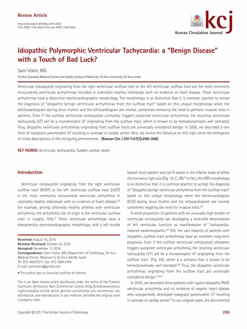
Fig. 3. Examples of polymorphic RVOT VT initiated by extrasystoles without a short coupling interval. (A) Patient #1 in the original series by Noda et al.10) (B) Documentation of RVOT polymorphic VT during an event of spontaneous vagal syncope triggered by venipuncture, as reported by Kataoka et al.28) Both patients had typical RVOT extrasystoles documented by 12-lead ECG. Reproduced from Noda et al.10) and Kataoka et al.28) RVOT: right ventricular outflow tract, VT: ventricular tachycardia, ECG: electrocardiogram.
A
B
302 Idiopathic Polymorphic RVOT-VT
https://doi.org/10.4070/kcj.2016.0303 www.e-kcj.org
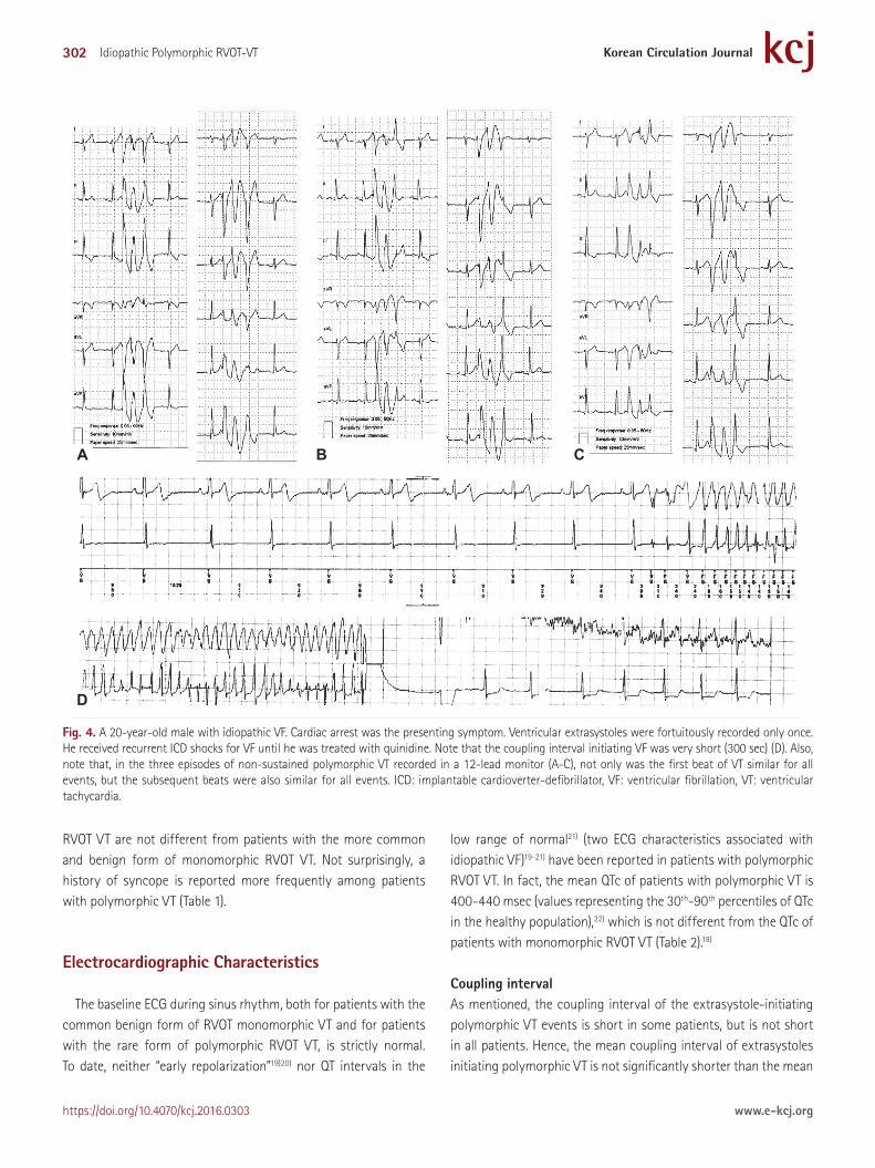
RVOT VT are not different from patients with the more common and benign form of monomorphic RVOT VT. Not surprisingly, a history of syncope is reported more frequently among patients with polymorphic VT (Table 1).
Electrocardiographic Characteristics
The baseline ECG during sinus rhythm, both for patients with the common benign form of RVOT monomorphic VT and for patients with the rare form of polymorphic RVOT VT, is strictly normal. To date, neither “early repolarization”19)20) nor QT intervals in the
low range of normal21) (two ECG characteristics associated with idiopathic VF)19-21) have been reported in patients with polymorphic RVOT VT. In fact, the mean QTc of patients with polymorphic VT is 400-440 msec (values representing the 30th-90th percentiles of QTc in the healthy population),22) which is not different from the QTc of patients with monomorphic RVOT VT (Table 2).18)
Coupling intervalAs mentioned, the coupling interval of the extrasystole-initiating polymorphic VT events is short in some patients, but is not short in all patients. Hence, the mean coupling interval of extrasystoles initiating polymorphic VT is not significantly shorter than the mean
A
D
B C
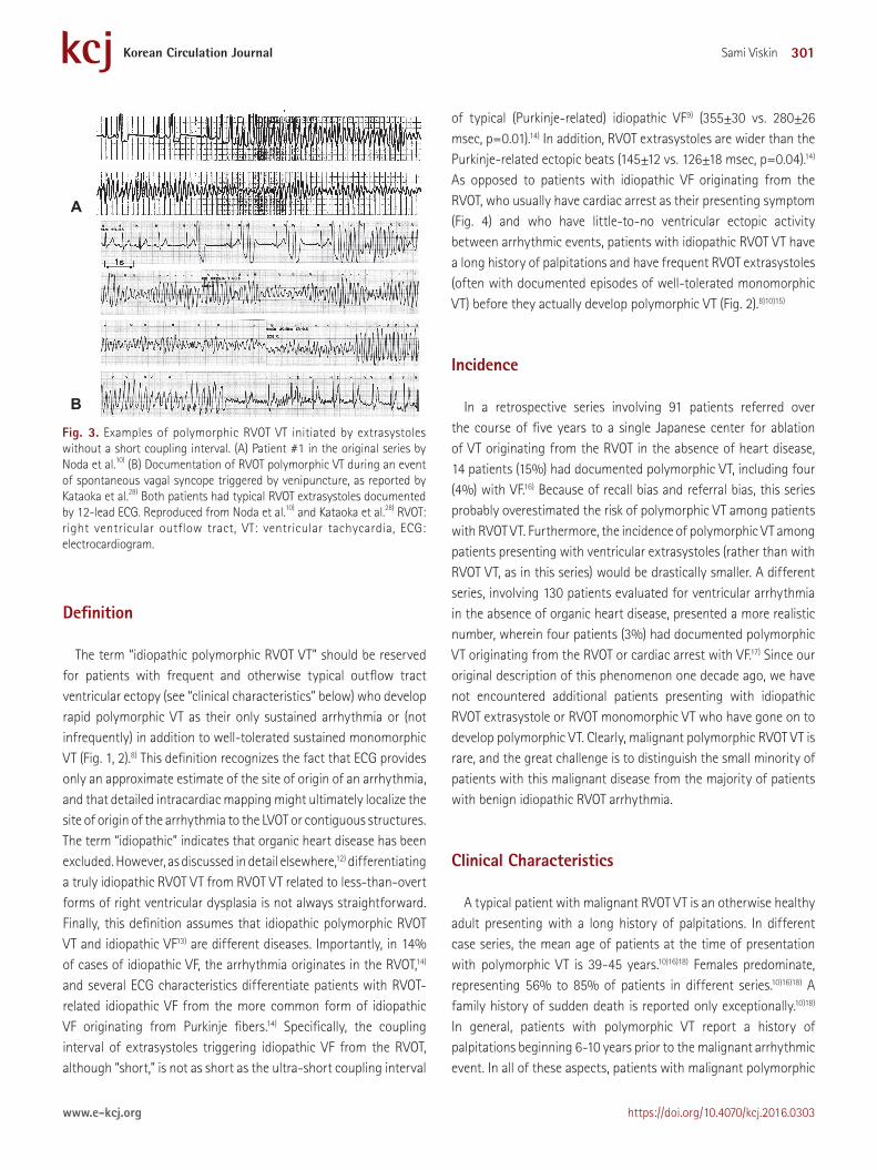
Fig. 4. A 20-year-old male with idiopathic VF. Cardiac arrest was the presenting symptom. Ventricular extrasystoles were fortuitously recorded only once. He received recurrent ICD shocks for VF until he was treated with quinidine. Note that the coupling interval initiating VF was very short (300 sec) (D). Also, note that, in the three episodes of non-sustained polymorphic VT recorded in a 12-lead monitor (A-C), not only was the first beat of VT similar for all events, but the subsequent beats were also similar for all events. ICD: implantable cardioverter-defibrillator, VF: ventricular fibrillation, VT: ventricular tachycardia.
303Sami Viskin
https://doi.org/10.4070/kcj.2016.0303www.e-kcj.org
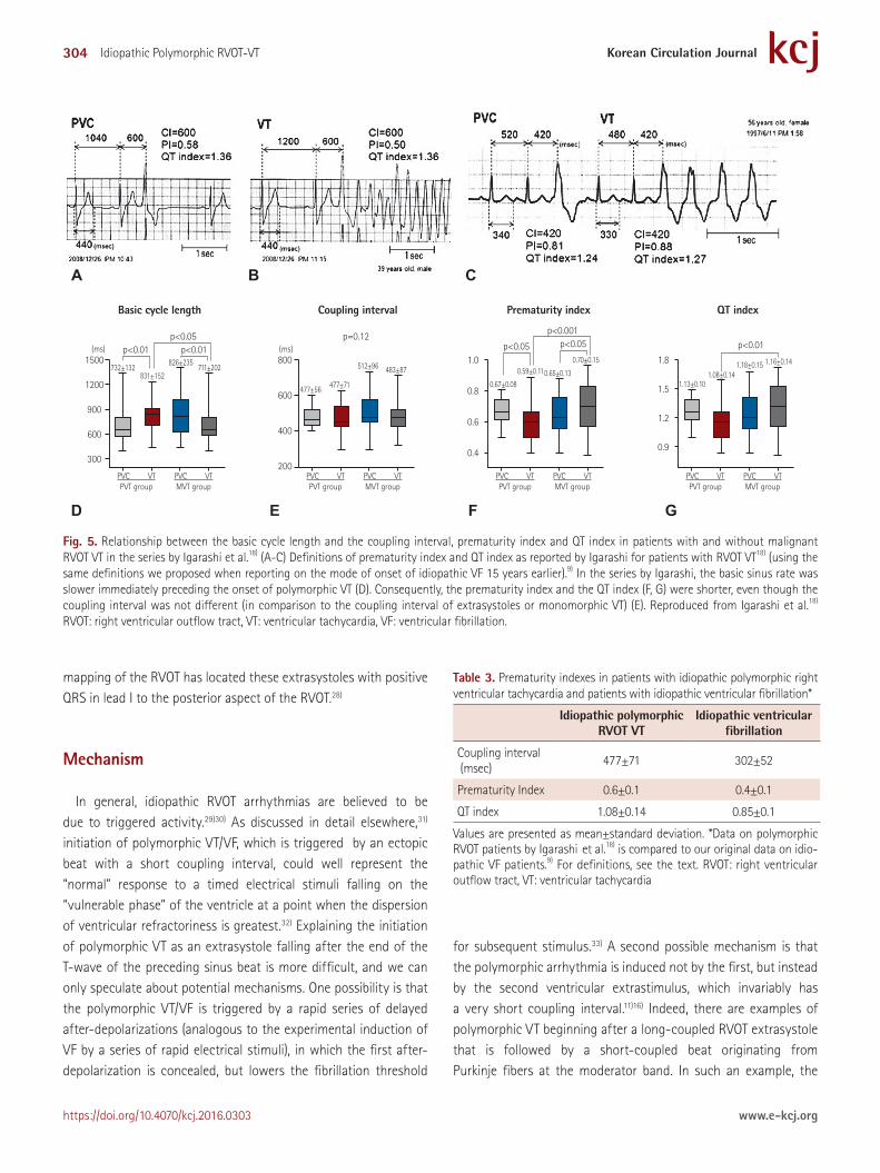
coupling interval of extrasystoles initiating monomorphic VT and is not different from the coupling intervals of extrasystoles that do not trigger arrhythmias (Table 2) (Fig. 5B). Also, RVOT extrasystoles with varying coupling intervals have been associated with an in-creased risk of polymorphic VT.23)
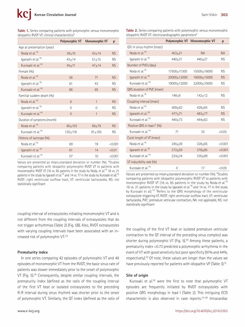
Prematurity indexIn one series comparing 42 episodes of polymorphic VT and 48
episodes of monomorphic VT from the RVOT, the basic sinus rate of patients was slower immediately prior to the onset of polymorphic VT (Fig. 5).18) Consequently, despite similar coupling intervals, the prematurity index (defined as the ratio of the coupling interval of the first VT beat or isolated extrasystoles to the preceding R-R interval during sinus rhythm) was shorter prior to the onset of polymorphic VT. Similarly, the QT index (defined as the ratio of
the coupling of the first VT beat or isolated premature vetricular contraction to the QT interval of the preceding sinus complex) was shorter during polymorphic VT (Fig. 5).18) Among these patients, a prematurity index <0.73 predicted a polymorphic arrhythmia in the event of VT with good sensitivity but poor specificity (91% and 44%, respectively).18) Of note, these values are longer than the values we have previously reported for patients with idiopathic VF (Table 3).9)
Site of originKurosaki et al.16) were the first to note that polymorphic VT
episodes are frequently initiated by RVOT extrasystoles with positive QRS morphology in lead I (Table 2). This intriguing ECG characteristic is also observed in case reports.24-28) Intracardiac
Table 1. Series comparing patients with polymorphic versus monomorphic idiopathic RVOT VT: clinical characteristics*
Polymorphic VT Monomorphic VT p
Age at presentation (year)
Noda et al.10) 39±10 43±14 NS
Igarashi et al.18) 43±14 51±15 NS
Kurosaki et al.16) 45±11 47±14 NS
Female (%)
Noda et al.10) 56 71 NS
Igarashi et al.18) 61 43 NS
Kurosaki et al.16) 86 65 NS
Familial sudden death (%)
Noda et al.10) 6 1 NS
Igarashi et al.18) 0 0 NS
Kurosaki et al.16) 0 1 NS
Duration of symptoms (month)
Noda et al.10) 80±103 69±79 NS
Kurosaki et al.16) 120±118 81±105 NS
History of syncope (%)
Noda et al.10) 69 18 <0.001
Igarashi et al.18) 61 14 <0.01
Kurosaki et al.16) 57 10 <0.001
Values are presented as mean±standard deviation or number (%). *Studies comparing patients with idiopathic polymorphic RVOT VT to patients with monomorphic RVOT VT (16 vs. 85 patients in the study by Noda, et al.10) 18 vs. 21 patients in the study by Igarashi et al.18) and 14 vs. 77 in the study by Kurosaki et al).16) RVOT: right ventricular outflow tract, VT: ventricular tachycardia, NS: not statistically significant
Table 2. Series comparing patients with polymorphic versus monomorphic idiopathic RVOT VT: electrocardiographic parameters*
Polymorphic VT Monomorphic VT p
QTc in sinus rhythm (msec)
Noda et al.10) 403±21 NA NA
Igarashi et al.18) 440±31 440±27 NS
Number of PVCs (day)
Noda et al.10) 17500±11300 15500±16000 NS
Igarashi et al.18) 20000±12000 16000±15000 NS
Kurosaki et al.16) 19000±12000 22000±10000 NS
QRS duration of PVC (msec)
Noda et al.10) 148±8 142±12 NS
Coupling interval (msec)
Noda et al.10) 409±62 428±65 NS
Igarashi et al.18) 477±71 483±77 NS
Kurosaki et al.16) 440±73 444±63 NS
Positive QRS in lead I† (%)
Kurosaki et al.16) 71 35 <0.05
Cycle length of VT (msec)
Noda et al.10) 245±28 328±65 <0.001
Igarashi et al.18) 272±59 378±85 <0.001
Kurosaki et al.16) 224±34 330±69 <0.001
VT inducibility rate (%)
Kurosaki et al.16) 0 17 <0.05
Values are presented as mean±standard deviation or number (%). *Studies comparing patients with idiopathic polymorphic RVOT VT to patients with monomorphic RVOT VT (16 vs. 85 patients in the study by Noda et al.10) 18 vs. 21 patients in the study by Igarashi et al.18) and 14 vs. 77 in the study by Kurosaki et al).16) †Refers to the QRS morphology of the ventricular extrasystole triggering VT. RVOT: right ventricular outflow tract, VT: ventricular tachycardia, PVC: premature vetricular contraction, NA: not applicable, NS: not statistically significant
304 Idiopathic Polymorphic RVOT-VT
https://doi.org/10.4070/kcj.2016.0303 www.e-kcj.org
mapping of the RVOT has located these extrasystoles with positive QRS in lead I to the posterior aspect of the RVOT.28)
Mechanism
In general, idiopathic RVOT arrhythmias are believed to be due to triggered activity.29)30) As discussed in detail elsewhere,31) initiation of polymorphic VT/VF, which is triggered by an ectopic beat with a short coupling interval, could well represent the “normal” response to a timed electrical stimuli falling on the “vulnerable phase” of the ventricle at a point when the dispersion of ventricular refractoriness is greatest.32) Explaining the initiation of polymorphic VT as an extrasystole falling after the end of the T-wave of the preceding sinus beat is more difficult, and we can only speculate about potential mechanisms. One possibility is that the polymorphic VT/VF is triggered by a rapid series of delayed after-depolarizations (analogous to the experimental induction of VF by a series of rapid electrical stimuli), in which the first after-depolarization is concealed, but lowers the fibrillation threshold
for subsequent stimulus.33) A second possible mechanism is that the polymorphic arrhythmia is induced not by the first, but instead by the second ventricular extrastimulus, which invariably has a very short coupling interval.11)16) Indeed, there are examples of polymorphic VT beginning after a long-coupled RVOT extrasystole that is followed by a short-coupled beat originating from Purkinje fibers at the moderator band. In such an example, the
A
D E F G
B C
PVC VTPVT group
1500
1200
900
600
300
PVC VTMVT group
(ms) p<0.01 p<0.01p<0.05
Basic cycle length Coupling interval
PVC VTPVT group
800
600
400
200PVC VTMVT group
(ms)p=0.12
732±132831±152
826±235711±202
477±56 477±71
512±96 483±87
0.67±0.08
0.59±0.110.65±0.13
0.70±0.15
1.13±0.101.08±0.14
1.18±0.15 1.16±0.14
Prematurity index
p<0.05 p<0.05p<0.001
PVC VTPVT group
1.0
0.8
0.6
0.4
PVC VTMVT group
QT index
p<0.01
PVC VTPVT group
1.8
1.5
1.2
0.9
PVC VTMVT group
Fig. 5. Relationship between the basic cycle length and the coupling interval, prematurity index and QT index in patients with and without malignant RVOT VT in the series by Igarashi et al.18) (A-C) Definitions of prematurity index and QT index as reported by Igarashi for patients with RVOT VT18) (using the same definitions we proposed when reporting on the mode of onset of idiopathic VF 15 years earlier).9) In the series by Igarashi, the basic sinus rate was slower immediately preceding the onset of polymorphic VT (D). Consequently, the prematurity index and the QT index (F, G) were shorter, even though the coupling interval was not different (in comparison to the coupling interval of extrasystoles or monomorphic VT) (E). Reproduced from Igarashi et al.18) RVOT: right ventricular outflow tract, VT: ventricular tachycardia, VF: ventricular fibrillation.
Table 3. Prematurity indexes in patients with idiopathic polymorphic right ventricular tachycardia and patients with idiopathic ventricular fibrillation*
Idiopathic polymorphic RVOT VT
Idiopathic ventricular fibrillation
Coupling interval (msec)
477±71 302±52
Prematurity Index 0.6±0.1 0.4±0.1
QT index 1.08±0.14 0.85±0.1
Values are presented as mean±standard deviation. *Data on polymorphic RVOT patients by Igarashi et al.18) is compared to our original data on idio-pathic VF patients.9) For definitions, see the text. RVOT: right ventricular outflow tract, VT: ventricular tachycardia
305Sami Viskin
https://doi.org/10.4070/kcj.2016.0303www.e-kcj.org
second (moderator band) extrasystole is the real trigger of the polymorphic VT (F. E. Marchlinski, personal communication). A third possibility is that the RVOT ectopic activity leading to VF could be a manifestation of modulated parasystole and reflection (a term used to denote a parasystolic pacemaker undergoing slow diastolic depolarization, which has been triggered to fire prematurely by the electrical activity of adjacent tissue).34) The observation that varying coupling intervals correlate with a higher risk of polymorphic VT23) is consistent with this last possibility.
Identification of Patients at Risk
Recognizing the small proportion of patients at risk for potentially lethal polymorphic RVOT VT among the large majority of patients with benign RVOT extrasystoles is a formidable challenge.11) The only clinical characteristic that specifically increases the odds of having polymorphic VT is a history of syncope (Table 1). Certainly, patients with RVOT extrasystoles should be suspected of having polymorphic VT when their clinical history is consistent with a malignant syncope. On the other hand, both benign vagal syncope and benign idiopathic RVOT arrhythmias are common, and it is not unusual to encounter patients who have both—unrelated—medical conditions. It could be argued that patients with RVOT extrasystoles who have syncope that appears to be benign vagal syncope on clinical grounds should be regarded as patients with asymptomatic (i.e., unrelated) extrasystoles. Here is where the challenge becomes extreme,11) because, as reported by Kataoka et al.,28) an otherwise benign episode of spontaneous vagal syncope can unpredictably trigger polymorphic RVOT VT in a prone patient (Fig. 3B). For the asymptomatic patient, documentation of short-coupled extrasystoles probably calls for an aggressive approach with radiofrequency ablation (duly admitting that there is no data about the natural history of short-coupled extrasystoles).
Natural History and Therapy
Patients with documented RVOT polymorphic VT are treated with radiofrequency ablation of any and all ectopic beats that resemble arrhythmia triggers.10)16)18) Whether or not all of these patients should also undergo implantation with an implantable cardioverter-defibrillator (ICD) is an open question, because little is known about the rate of recurrence after successful ablation. Of note, the five-year recurrence of malignant arrhythmias following successful ablation of idiopathic VF is as high as 38%.35) Comparable long-term figures are not available for RVOT polymorphic VT. Similarly,
quinidine is extremely effective for preventing recurrences in idiopathic VF,1)13) but there is no data on the efficacy of quinidine in polymorphic RVOT VT. Non-inducibility of VT at the end of an ablation procedure cannot be used as evidence of success, because patients do not necessarily have inducible VT at baseline (Table 2). Finally, little is known about the natural history of patients with documented idiopathic RVOT polymorphic VT. Interestingly, two of our initial patients remained untreated (except for ICD implantation in both patients).8) One of these patients has remained free of arrhythmias following a cardiac arrest 12 years prior, whereas the other patients has remained asymptomatic, but has documentation of non-sustained polymorphic VT 18 years after her initial arrhythmic event (Fig. 1D). Looking at these patients, we can only wonder if the RVOT polymorphic VT is simply a matter of “bad luck.” That is, can the phenomenon be characterized as an otherwise innocent extrasystole with extremely bad timing (occurring in the vulnerable phase of the cardiac cycle).
References
1. Belhassen B, Viskin S. Idiopathic ventricular tachycardia and
fibrillation. J Cardiovasc Electrophysiol 1993;4:356-68.
2. Lemery R, Brugada P, Bella PD, Dugernier T, van den Dool A, Wellens
HJ. Nonischemic ventricular tachycardia. Clinical course and long-
term follow-up in patients without clinically overt heart disease.
Circulation 1989;79:990-9.
3. Biffi A, Pelliccia A, Verdile L, et al. Long-term clinical significance of
frequent and complex ventricular tachyarrhythmias in trained
athletes. J Am Coll Cardiol 2002;40:446-52.
4. Buxton AE, Waxman HL, Marchlinski FE, Simson MB, Cassidy D,
Josephson ME. Right ventricular tachycardia: clinical and
electrophysiologic characteristics. Circulation 1983;68:917-27.
5. Morady F, Kadish AH, DiCarlo L, et al. Long-term results of catheter
ablation of idiopathic right ventricular tachycardia. Circulation
1990;82:2093-9.
6. Takemoto M, Yoshimura H, Ohba Y, et al. Radiofrequency catheter
ablation of premature ventricular complexes from right ventricular
outflow tract improves left ventricular dilation and clinical status in
patients without structural heart disease. J Am Coll Cardiol
2005;45:1259-65.
7. Gaita F, Giustetto C, Di Donna P, et al. Long-term follow-up of right
ventricular monomorphic extrasystoles. J Am Coll Cardiol
2001;38:364-70.
8. Viskin S, Rosso R, Rogowski O, Belhassen B. The “short-coupled”
variant of right ventricular outflow ventricular tachycardia: a not-
so-benign form of benign ventricular tachycardia? J Cardiovasc

306 Idiopathic Polymorphic RVOT-VT
https://doi.org/10.4070/kcj.2016.0303 www.e-kcj.org
Electrophysiol 2005;16:912-6.
9. Viskin S, Lesh M, Eldar M, et al. Mode of onset of malignant
ventricular arrhythmias in idiopathic ventricular fibrillation. J
Cardiovasc Electrophysiol 1997;8:1115-20.
10. Noda T, Shimizu W, Taguchi A, et al. Malignant entity of idiopathic
ventricular fibrillation and polymorphic ventricular tachycardia
initiated by premature extrasystoles originating from the right
ventricular outflow tract. J Am Coll Cardiol 2005;46:1288-94.
11. Viskin S, Antzelevitch C. The cardiologists’ worst nightmare: sudden
death from “benign” ventricular arrhythmias. J Am Coll Cardiol
2005;46:1295-7.
12. Michowitz Y, Viskin S, Rosso R. Exercise-induced ventricular tachycardia/
ventricular fibrillation in the normal heart: risk stratification and
management. Card Electrophysiol Clin 2016;8:593-600.
13. Viskin S, Belhassen B. Idiopathic ventricular fibrillation. Am Heart J
1990;120:661-71.
14. Haissaguerre M, Shoda M, Jais P, et al. Mapping and ablation of
idiopathic ventricular fibrillation. Circulation 2002;106:962-7.
15. Shimizu W. Arrhythmias originating from the right ventricular
outflow tract: how to distinguish “malignant” from “benign”? Heart
Rhythm 2009;6:1507-11.
16. Kurosaki K, Nogami A, Shirai Y, Kowase S. Positive QRS complex in
lead i as a malignant sign in right ventricular outflow tract
tachycardia: comparison between polymorphic and monomorphic
ventricular tachycardia. Circ J 2013;77:968-74.
17. Valk SD, Dabiri-Abkenari L, Theuns DA, Thornton AS, Res JC, Jordaens L.
Ventricular fibrillation and life-threatening ventricular tachycardia in
the setting of outflow tract arrhythmias--the place of ICD therapy.
Int J Cardiol 2013;165:385-7.
18. Igarashi M, Tada H, Kurosaki K, et al. Electrocardiographic
determinants of the polymorphic QRS morphology in idiopathic right
ventricular outflow tract tachycardia. J Cardiovasc Electrophysiol
2012;23:521-6.
19. Haissaguerre M, Derval N, Sacher F, et al. Sudden cardiac arrest
associated with early repolarization. N Engl J Med 2008;358:2016-23.
20. Rosso R, Kogan E, Belhassen B, et al. J-point elevation in survivors of
primary ventricular fibrillation and matched control subjects
incidence and clinical significance. J Am Coll Cardiol 2008;52:1231-8.
21. Viskin S, Zeltser D, Ish-Shalom M, et al. Is idiopathic ventricular
fibrillation a short QT syndrome? Comparison of QT intervals of
patients with idiopathic ventricular fibrillation and healthy controls.
Heart Rhythm 2004;1:587-91.
22. Ramirez AH, Schildcrout JS, Blakemore DL, et al. Modulators of
normal electrocardiographic intervals identified in a large electronic
medical record. Heart Rhythm 2011;8:271-7.
23. Bradfield JS, Homsi M, Shivkumar K, Miller JM. Coupling interval
variability differentiates ventricular ectopic complexes arising in the
aortic sinus of valsalva and great cardiac vein from other sources:
mechanistic and arrhythmic risk implications. J Am Coll Cardiol
2014;63:2151-8.
24. Uemura T, Yamabe H, Tanaka Y, et al. Catheter ablation of a
polymorphic ventricular tachycardia inducing monofocal premature
ventricular complex. Intern Med 2008;47:1799-802.
25. Kusano KF, Yamamoto M, Emori T, Morita H, Ohe T. Successful
catheter ablation in a patient with polymorphic ventricular
tachycardia. J Cardiovasc Electrophysiol 2000;11:682-5.
26. Takatsuki S, Mitamura H, Ogawa S. Catheter ablation of a monofocal
premature ventricular complex triggering idiopathic ventricular
fibrillation. Heart 2001;86:E3.
27. Ashida K, Kaji Y, Sasaki Y. Abolition of torsade de pointes after
radiofrequency catheter ablation at right ventriuclar outflow tract.
Int J Cardiol 1997;59:171-5.
28. Kataoka M, Takatsuki S, Tanimoto K, Akaishi M, Ogawa S, Mitamura H.
A case of vagally mediated idiopathic ventricular fibrillation. Nat Clin
Pract Cardiovasc Med 2008;5:111-5.
29. Lerman BB, Belardinelli L, West GA, Berne RM, DiMarco JP.
Adenosine-sensitive ventricular tachycardia: evidence suggesting
cyclic AMP-mediated triggered activity. Circulation 1986;74:270-80.
30. Lerman BB, Dong B, Stein KM, Markowitz SM, Linden J, Catanzaro DF.
Right ventricular outflow tract tachycardia due to a somatic cell
mutation in G protein subunitalphai2. J Clin Invest 1998;101:2862-8.
31. Viskin S, Rogowski O. Asymptomatic Brugada syndrome: a cardiac
ticking time-bomb? Europace 2007;9:707-10.
32. Han J, Garcia de Jalon PD, Moe GK. Fibrillation threshold of
premature ventricular responses. Circ Res 1966;18:18-25.
33. Sugimoto T, Schaal SF, Wallace AG. Factors determining vulnerability
to ventricular fibrillation induced by 60-CPS alternating current. Circ
Res 1967;21:601-8.
34. Robles de Medina EO, Delmar M, Sicouri S, Jalife J. Modulated
parasystole as a mechanism of ventricular ectopic activity leading to
ventricular fibrillation. Am J Cardiol 1989;63:1326-32.
35. Knecht S, Sacher F, Wright M, et al. Long-term follow-up of
idiopathic ventricular fibrillation ablation: a multicenter study. J Am
Coll Cardiol 2009;54:522-8.