rheology of soft materialsto materials science and cell biology. recent advances in rheologi-cal...
TRANSCRIPT
Rheology of Soft Materials
Daniel T.N. Chen,1 Qi Wen,2 Paul A. Janmey,2
John C. Crocker,3 and Arjun G. Yodh1,�
1Department of Physics and Astronomy, University of Pennsylvania,
Philadelphia, Pennsylvania 19104; email: [email protected];
2Departments of Physiology, Physics, and Bioengineering, Institute for
Medicine and Engineering, University of Pennsylvania, Philadelphia,
Pennsylvania 19104; email: [email protected];
3Department of Chemical and Biomolecular Engineering,
University of Pennsylvania, Philadelphia, Pennsylvania;
email: [email protected]
Annu. Rev. Condens. Matter Phys. 2010. 1:301–22
First published online as a Review in Advance on
May 24, 2010
The Annual Review of Condensed Matter Physics is
online at conmatphys.annualreviews.org
This article’s doi:
10.1146/annurev-conmatphys-070909-104120
Copyright © 2010 by Annual Reviews.
All rights reserved
1947-5454/10/0810-0301$20.00
�Corresponding author
Key Words
viscoelasticity, nonlinear rheology, microrheology, polymer
network, colloidal glass
Abstract
Research on soft materials, including colloidal suspensions, glasses,
pastes, emulsions, foams, polymer networks, liquid crystals, granu-
lar materials, and cells, has captured the interest of scientists and
engineers in fields ranging from physics and chemical engineering
to materials science and cell biology. Recent advances in rheologi-
cal methods to probe mechanical responses of these complex media
have been instrumental for producing new understanding of soft
matter and for generating novel technological applications. This
review surveys these technical developments and current work in
the field, with partial aim to illustrate open questions for future
research.
301
Ann
u. R
ev. C
onde
ns. M
atte
r Ph
ys. 2
010.
1:30
1-32
2. D
ownl
oade
d fr
om w
ww
.ann
ualr
evie
ws.
org
by U
nive
rsity
of
Penn
sylv
ania
on
11/2
6/10
. For
per
sona
l use
onl
y.

1. INTRODUCTION
Rheology is the study of how materials deform when forces are applied to them. The
foundations of rheology rest on theories describing ideal materials. One example is Robert
Hooke’s description of ideal elastic behavior being a solid wherein the extension of the
material is proportional to the load imposed on it, and which recovers to its initial state
when the load is removed. By contrast, Isaac Newton’s theory describing ideal liquids
states that an irreversible flow will persist as long as a stress is applied, with flow rate
proportional to stress.
Real materials, and especially soft materials, are neither ideal solids nor ideal liquids.
Real soft materials exhibit both elastic and viscous responses and are therefore called
viscoelastic. The internal structures of soft solids and complex fluids composed of colloidal
particles, filamentous polymers, and other supra-molecular arrangements lead to compli-
cated deformations in response to mechanical stress. As a result, the relations between
stress and strain are not simply defined by elastic and viscous constants; rather, they are
functions of time, direction, and extent of deformation. The goal of rheological experi-
ments is to quantify dynamic viscoelasticity over as wide a range of time and deformation
scales as possible, and ultimately to relate these viscoelastic properties to material struc-
ture. Many open questions remain about how to relate structure to viscoelastic response.
This review focuses on recent progress in soft material rheology, with particular orienta-
tion toward new experimental techniques suitable for studies of colloids, glasses, filamen-
tous networks, granular materials, emulsions, and cells.
2. OVERVIEW OF TECHNIQUES
2.1. Rheology
Rheometers generally measure two quantities: stress, the amount of force per unit area
applied to the sample; and strain, the dimensionless degree to which the material deforms.
Material properties, quantified as elastic moduli for solids or viscosity for liquids, are
calculated from the ratio of stress to strain or strain rate, respectively. To characterize fully
the viscoelastic properties of complex soft materials, the relation of stress to strain must
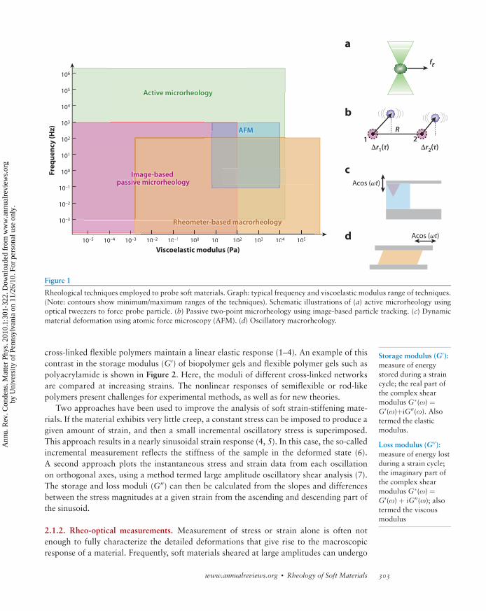
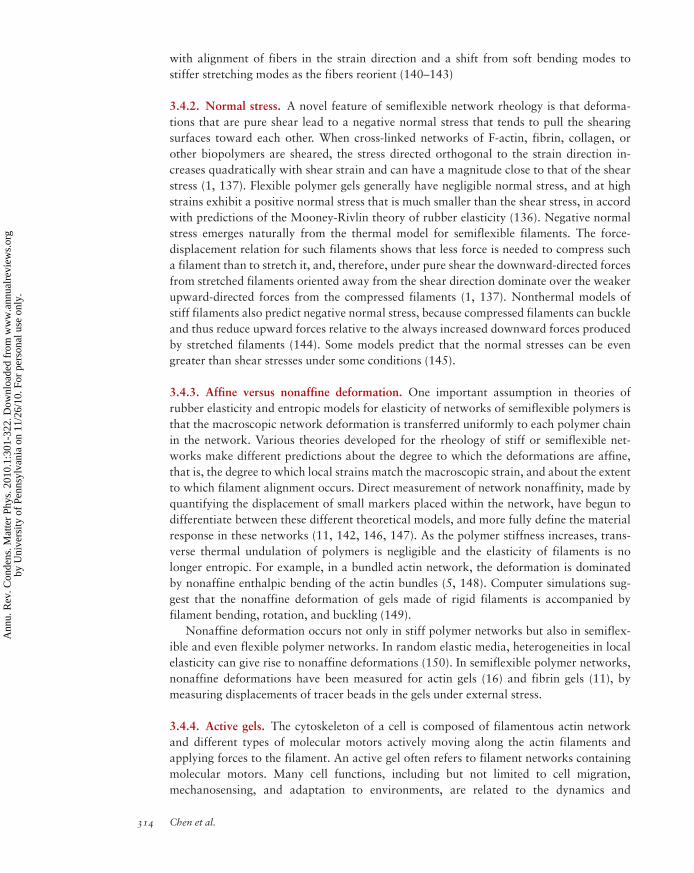
be measured over a wide range of strains, strain rates, and time scales (Figure 1). Recent
advances in rheological methods have been motivated, in part, by attempts to measure
delicate samples with complex time-dependent responses at the micron scale, and
microrheological methods, in particular, have even been extended for use in live cells.
Additional experimental and theoretical progress has been made on systems far from
equilibrium, e.g., systems in which nonthermal sources of energy drive fluctuations and
rheological responses.
2.1.1. Nonlinear measurements. The useful properties of soft materials are often related
to their responses at large strains. This is especially true for biological tissues such as blood
vessels, lung, or muscle that are stretched tens of percent during normal function, and even
more during injury. A common feature of soft tissues is their nonlinear viscoelastic
response, which is evident as a stiffening with increasing strain, termed strain stiffening.
Even networks of purified biopolymers such as actin, fibrin, or collagen exhibit
nonlinear elasticity at strains where rubber-like elastomers or hydrogels made from
Elastic: property of a
material to deform to
a defined extent inresponse to a force and
then return to its
original state when the
force is removed
Stress: force per unit
area; s ¼ F/A; SI unitis N/m2
Viscoelastic: property
of soft materials to
exhibit both elastic
and viscous responsesin a frequency-
dependent manner
Strain: unitless param-
eter quantifying the
extent of deformationafter application of
stress
Viscosity: measure of
resistance of a fluid to
shear stress; Z ¼ s / _g,where _g is the rate ofstrain
Strain stiffening:
rheological response
of a material in which
the stress increaseswith increasing strain,
often used to describe
polymer networks
(Non)-linear elasticity:
Young’s or shear mod-ulus that (does not
change) changes with
strain
302 Chen et al.
Ann
u. R
ev. C
onde
ns. M
atte
r Ph
ys. 2
010.
1:30
1-32
2. D
ownl
oade
d fr
om w
ww
.ann
ualr
evie
ws.
org
by U
nive
rsity
of
Penn
sylv
ania
on
11/2
6/10
. For
per
sona
l use
onl
y.
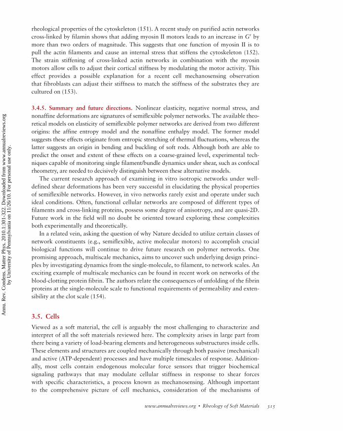
cross-linked flexible polymers maintain a linear elastic response (1–4). An example of this
contrast in the storage modulus (G0) of biopolymer gels and flexible polymer gels such as
polyacrylamide is shown in Figure 2. Here, the moduli of different cross-linked networks
are compared at increasing strains. The nonlinear responses of semiflexible or rod-like
polymers present challenges for experimental methods, as well as for new theories.
Two approaches have been used to improve the analysis of soft strain-stiffening mate-
rials. If the material exhibits very little creep, a constant stress can be imposed to produce a
given amount of strain, and then a small incremental oscillatory stress is superimposed.
This approach results in a nearly sinusoidal strain response (4, 5). In this case, the so-called
incremental measurement reflects the stiffness of the sample in the deformed state (6).
A second approach plots the instantaneous stress and strain data from each oscillation
on orthogonal axes, using a method termed large amplitude oscillatory shear analysis (7).
The storage and loss moduli (G00) can then be calculated from the slopes and differences
between the stress magnitudes at a given strain from the ascending and descending part of
the sinusoid.
2.1.2. Rheo-optical measurements. Measurement of stress or strain alone is often not
enough to fully characterize the detailed deformations that give rise to the macroscopic
response of a material. Frequently, soft materials sheared at large amplitudes can undergo
a
b
c
dViscoelastic modulus (Pa)
10–5 10–4 10–3
10–3
10–2
10–1
100
101
102
104
105
106
103
10–2 10–1 100 101 102 103 104 105
Freq
uenc
y (H
z)
Viscoelastic modulus (Pa)10–2 10–1 100 101 102 103 104 105
AFM
Active microrheology
Image-based passive microrheology
Rheometer-based macrorheology
fE
R21
Δr1(τ) Δr2(τ)
Acos (ωt)
Acos (ωt)
Figure 1
Rheological techniques employed to probe soft materials. Graph: typical frequency and viscoelastic modulus range of techniques.
(Note: contours show minimum/maximum ranges of the techniques). Schematic illustrations of (a) active microrheology usingoptical tweezers to force probe particle. (b) Passive two-point microrheology using image-based particle tracking. (c) Dynamic
material deformation using atomic force microscopy (AFM). (d) Oscillatory macrorheology.
Storage modulus (G0):measure of energy
stored during a straincycle; the real part of
the complex shear
modulusG�(o) ¼G0(o)þiG00(o). Alsotermed the elastic
modulus.
Loss modulus (G00):measure of energy lost
during a strain cycle;the imaginary part of
the complex shear
modulusG�(o) ¼G0(o) þ iG00(o); alsotermed the viscous
modulus
www.annualreviews.org � Rheology of Soft Materials 303
Ann
u. R
ev. C
onde
ns. M
atte
r Ph
ys. 2
010.
1:30
1-32
2. D
ownl
oade
d fr
om w
ww
.ann
ualr
evie
ws.
org
by U
nive
rsity
of
Penn
sylv
ania
on
11/2
6/10
. For
per
sona
l use
onl
y.

structural transitions such as ordering, crystallization, or shear banding, and these transi-
tions confound interpretation in the absence of a model that anticipates their onset. In
shear banding (8–10), for example, the sheared material separates into regions character-
ized by different viscosities. As a result, the same measured stress value can result from two
or more applied strain rates. Experimentally, this complication can be accounted for by
applying shear deformation concurrent with in situ characterization of the material’s
microstructure. A variety of schemes are employed to accomplish this structural character-
ization. Broadly speaking, rheo-optical characterization falls under two categories
depending on whether the information is obtained via real-space imaging or via scattering
in reciprocal space. Real-space techniques involve the use of fluorescence microscopy
(11, 12) or magnetic resonance imaging (13, 14) to image through a shear cell (15) or
rheometer (16). Scattering techniques typically involve measuring changes in 2D scattering
patterns under flow using light, X-rays, or neutrons. The presence of ordering in the
sheared material typically results in the appearance of a distinct peak at the inverse wave-
length of the ordering in the scattering pattern. For more detailed reviews of scattering-
based rheo-optical measurements see References 17 and 18.
Shear banding:
formation of regions
in a materialundergoing shear
strain in which part of
the sample is fluidizedwhile other parts
remain solid
1
100.01 0.1 1
10
100
1000
γ
G, G
' (Pa
)
Polyacrylamide
Actin
Collagen
Fibrin oscillatory
Fibrin steady
VimentinNeurofilaments
Figure 2
Dynamic shear storage moduli measured at different strain amplitudes for a series of cross-linked biopolymer networks. The realpart, G0, of the storage modulus reduces to the shear modulus G at zero frequency. Data shown are G0 (at 10 rad s-1) values for
F-actin, fibrin, collagen, vimentin, and polyacrylamide; and shear modulusG for fibrin and neurofilaments, plotted as a function
of the dimensionless strain g. Strain stiffening behavior is observed in the cross-linked biopolymer networks. Adapted from
Reference 2. Reprinted with permission.
304 Chen et al.
Ann
u. R
ev. C
onde
ns. M
atte
r Ph
ys. 2
010.
1:30
1-32
2. D
ownl
oade
d fr
om w
ww
.ann
ualr
evie
ws.
org
by U
nive
rsity
of
Penn
sylv
ania
on
11/2
6/10
. For
per
sona
l use
onl
y.

2.2. Microrheology
Situations in which traditional rheometers are difficult to use, such as when materials are
available in very low quantities, have spurred the development of microrheological tech-
niques. These techniques, in turn, have augmented the scope of materials that can be
rheologically probed.
2.2.1. Methods and instruments. Microrheology describes a collection of techniques to
quantify the shear moduli of very small samples or objects such as cells. The mechanical
properties of cells were estimated nearly a century ago by watching the motion of magnetic
particles ingested by cells (19, 20). Since then, a number of methods based on particle
tracking have been developed. These techniques can be classified according to whether
they rely on the motion of a probe in response to an applied force (termed active
microrheology) or rather, on the Brownian motion of the material or probe (termed passive
microrheology). In the former case, the principle is the same as a macroscopic rheometer:
An oscillatory stress (or strain) is applied and the resulting strain (or stress) is measured,
and then the complex ratio is computed. Or, alternatively, measurements can be performed
in a step strain (or stress) manner: The creep modulus is computed and then converted to
dynamic shear moduli by standard integral transforms.
Passive microrheology uses the fluctuation-dissipation theorem (FDT) to relate the
spectrum of fluctuating forces in a material proportionally to the loss (viscous) modulus
of the material. By invoking the FDT, one can employ a generalized Stokes-Einstein rela-
tion that relates the Brownian mean-squared displacement (MSD) of the probe particle to
the dynamic shear modulus by simple integral transforms (21, 22). Compared to active
methods, passive methods are generally better suited to softer materials, are straightfor-
ward to calibrate, and can be adapted to a wide range of instruments. All that is required is
a method of measuring particle or probe MSD, such as laser-deflection tracking (22, 23),
optical tweezers (24, 25), diffusing wave spectroscopy (26, 27), dynamic light scattering
(28, 29), image-based tracking (30–32), optical interferometry (33), or atomic force
microscopy (AFM) noise analysis (34).
In contrast, active methods require more complex instrumentation than passive
methods, but they are ultimately more versatile, permitting measurements of harder sam-
ples and nonlinear rheological behavior (35–37). Early work relied on micropipette aspi-
ration (38), later refined to include time-dependent effects (39, 40). The early magnetic
particle research was refined into a quantitative creep method (41) as well as oscillatory
rotational approaches (42), termed magnetic twisting cytometry (MTC) (43, 44). Oscilla-
tory optical tweezers can be used for active measurements (45–47), as well as passive ones.
Much recent work has been performed with AFM (48, 49).
2.2.2. Two-point microrheology. The proper interpretation of all microrheology me-
thods, however, relies on knowing the boundary conditions at the probe/soft material
interface and the shape of the strain field, which can be poorly controlled compared
to a macroscopic rheometer. Pioneering early work in cells examined the motion of
nonmagnetic tracers near driven magnetic particles to map out the deformation field (42,
50) and found significant deviations from continuum behavior. Two-point microrheology
(TPM) (31) uses the correlated motion of two well-separated tracers to measure the
rheological response, with the effect that the measurement becomes insensitive to tracer
FDT: fluctuation-
dissipation theorem
MSD: mean-square
displacement
Shear modulus:
constant describing a
material’s resistance todeformation as given
by the ratio of shear
stress to shear strain,G ¼ s / g
MTC:magnetic twist-ing cytometry
TPM: two-pointmicrorheology
www.annualreviews.org � Rheology of Soft Materials 305
Ann
u. R
ev. C
onde
ns. M
atte
r Ph
ys. 2
010.
1:30
1-32
2. D
ownl
oade
d fr
om w
ww
.ann
ualr
evie
ws.
org
by U
nive
rsity
of
Penn
sylv
ania
on
11/2
6/10
. For
per
sona
l use
onl
y.

boundary conditions (51, 52). This robustness can be turned around to study the nature of
the probes’ boundary conditions with the matrix (24, 32) and even inertial effects (53).
Whereas much early TPM work used an image-based passive approach, it has been
adapted to dynamic light scattering (54) and optical tweezer-based instruments (25, 55).
2.2.3. Force spectroscopy of active materials. Finally, in the interesting case where the
sample contains endogenous force generators, a combination of both passive and active
microrheology data provides a method for quantifying the internal forces. Early
researchers noted that the MSDs of embedded tracers in cells had a functional form incom-
patible with Brownian motion (56, 57); such motions could be modeled as a viscoelastic
material driven by fluctuating stresses having a simple power-law form (56) and were
subsequently confirmed by MTC (58) and two-point measurements in cells (59), with the
latter also showing that two-point techniques do not require FDT. Similar measurements
on non-Brownian stresses were made in bacterial baths (47) and in a cytoskeleton model of
reconstituted biopolymers and motors (60); the latter compares favorably to measurements
of living cells (61). A recent study combining active and passive measurements on the same
cell-adhered probes reported non-Brownian stresses having the same frequency depen-
dence as expected from the FDT, but with an amplitude several times larger (62), which
may be interpreted in terms of an effective granular temperature.
3. SOFT MATERIALS
3.1. Colloidal Suspensions and Glasses
Full understanding of even a monodisperse hard-sphere colloidal suspension’s rheology
requires detailed consideration of the interplay between Brownian, conservative inter-
particle, and hydrodynamic forces. The central quantity of interest for determining colloid
rheology is the spatial distribution of the particles, termed the microstructure, which is set by
the balance of forces in equilibrium. Under imposed flow, the ability of the microstructure to
rearrange to accommodate flow and interparticle forces determines its macroscopic rheolog-
ical response. The many-body, shear-history-dependent nature of the microstructure renders
the prediction of the suspension’s dynamics and rheology highly nontrivial. Driven in part by
their technological importance in many staple industrial products such as paints and food
additives, the study of colloidal suspensions continues. Currently, colloidal suspensions are
also the test bed for ideas in soft matter, e.g., their study is leading to new insights about deep
problems in the field, such as the glass transition (63).
3.1.1. Influence of particle volume fraction. The addition of colloidal particles to a New-
tonian fluid causes the viscosity of the medium to increase. This effect is essentially due to
particles being rigid objects that can move and rotate, but not shear with the suspending
fluid. As such, they exert an additional stress in the form of quadrupolar disturbances on
the fluid. In the very dilute limit (f < .02), where each particle is far enough away from the
other particles (and thus do not interact hydrodynamically), Einstein (64) derived the
relationship between the zero-shear viscosity and volume fraction: � ¼ �0ð1þ 5=2fÞ. As fincreases, the displacement of a particle causes a displacement of the incompressible
suspending fluid, which in turn gives rise to long-range and many-body hydrodynamic
forces that couple neighboring particles. In the dilute limit (f < 0.1), consideration of
pairwise hydrodynamic interactions is sufficient and leads to O(f2) corrections to
(Non)-Newtonian:
class of fluids having
the defining propertythat their viscosity is
(dependent on)
independent of shear
rate.
306 Chen et al.
Ann
u. R
ev. C
onde
ns. M
atte
r Ph
ys. 2
010.
1:30
1-32
2. D
ownl
oade
d fr
om w
ww
.ann
ualr
evie
ws.
org
by U
nive
rsity
of
Penn
sylv
ania
on
11/2
6/10
. For
per
sona
l use
onl
y.

Einstein’s result (65). For a review of more recent work on non-Newtonian rheology in
colloidal suspensions, see Reference 66.
Under the right conditions, colloidal suspensions can crystallize. However, most often
colloidal suspensions undergo a glass transition as particle density is increased to f�0.6.
Near this colloidal glass transition, volume fraction plays a role analogous to temperature
in molecular liquids. Broadly speaking, an increase in the particle volume fraction causes
crowding. Crowding, in turn, leads to dynamical arrest: Thermal energy is insufficient to
drive the configurational rearrangements necessary for equilibration. As a result, the sys-
tem becomes trapped in metastable states, characterized by liquid-like structural order.
Colloidal glasses share many qualitative similarities to granular materials that have led to
the conjecture that they can both be described within a common theoretical framework
known as jamming (67, 68). Accompanying the dynamical arrest are a concomitant diver-
gence of the suspension viscosity and development of a finite yield stress (sy), below which
there is no flow.
The low-shear rheology of noncrystalline, hard-sphere colloidal suspensions of volume
fraction up to f � 0.6 has been extensively studied (69–71). In linear oscillatory measure-
ments (69), it was found that for f � 0.5, the loss modulus (G00) dominates the response
for all frequencies; as f increases, both moduli increase, with the storage modulus
G0 increasing faster than G00 and eventually dominating for lower frequencies. Near
f � 0.58, the low-frequency G0 exhibits a nearly frequency-independent plateau, i.e.,
G0(f) � G0 ¼ f(f)�kBT/a3, where a is the particle radius and f(f) denotes a function of
f (71). This low-frequency behavior is a direct consequence of particles being trapped by
their neighbors and the stress in the suspension being unable to relax on the longest time
scale of the measurement. This linear viscoelasticity is quite generic; most jammed systems
(e.g., entangled polymer solutions, weak polymeric gels, concentrated emulsions) exhibit
similar trends in their respective moduli when densities and shear rates are such that the
relaxation time is longer than the measurement time scale.
Viscometry has been used to measure the divergence of viscosity in noncrystalline hard-
sphere suspensions as the glass transition is approached (70). These measurements, span-
ning several orders of magnitude and taken together with recent measurements of the
structural relaxation time ta from light scattering (72), suggest that the viscosity divergence
occurs at higher density than predictions of mode-coupling theory (MCT) (73). MCT
predicts a power-law divergence of the viscosity at f � 0.58. The current consensus from
experiments appears to be that the viscosity divergence is better captured by a Vogel-
Fulcher-Tammann exponential form � ¼ �0 expðAf=ðf0 � fÞÞ with the critical volume
fraction f0 closer to random close packing frcp ¼ 0.64, in accord with expectations based
on free-volume theory (74).
3.1.2. Influence of shear rate. Shear rate has profound consequences on the rheology of
colloidal suspensions. Shear rate is parameterized by the Peclet number (Pe), which is the
ratio of the applied shear rate _g to the inverse of the Brownian relaxation time D/a2 of the
colloidal suspension: Pe ¼ _g a2/D. Pe is a measure of how far the suspension’s microstruc-
ture is driven from equilibrium. At low Pe, Brownian forces are able to restore shear-
induced perturbations to the equilibrium microstructure on the time scale of the shear
flow. As Pe increases, Brownian motion is insufficient to restore the microstructure and,
as a result, interparticle hydrodynamic forces dominate the suspension’s rheological
response. At Pe � 1, particles become ordered by the flow and can organize into layers
Peclet Number (Pe):
dimensionless ratio of
the applied shear rateto inverse Brownian
relaxation time
quantifying relative
importance ofadvection to diffusion:
Pe ¼ _g a2/D
www.annualreviews.org � Rheology of Soft Materials 307
Ann
u. R
ev. C
onde
ns. M
atte
r Ph
ys. 2
010.
1:30
1-32
2. D
ownl
oade
d fr
om w
ww
.ann
ualr
evie
ws.
org
by U
nive
rsity
of
Penn
sylv
ania
on
11/2
6/10
. For
per
sona
l use
onl
y.
that are able to flow with less resistance, reducing the suspension’s viscosity. This effect,
known as shear thinning, is a generic feature of colloidal suspensions at intermediate Pe.
Shear thinning is defined by the behavior that, at high shear rate, _g, the shear stress s / _gn
with n < 1; therefore, the apparent viscosity � � s= _g decreases as the shear rate increases.This effect covers a wide range of materials that can become Newtonian (n ¼ 1) at low
shear rates or have a yield stress. Empirically, colloidal glasses and most other soft glassy
materials (SGM) have a flow behavior that is well described by the Herschel-Bulkley
relation: s ¼ sy þ C_gn, where sy is the yield stress below which there is no flow and n �1.sy depends on volume fraction and n depends on constitutive properties of the material.
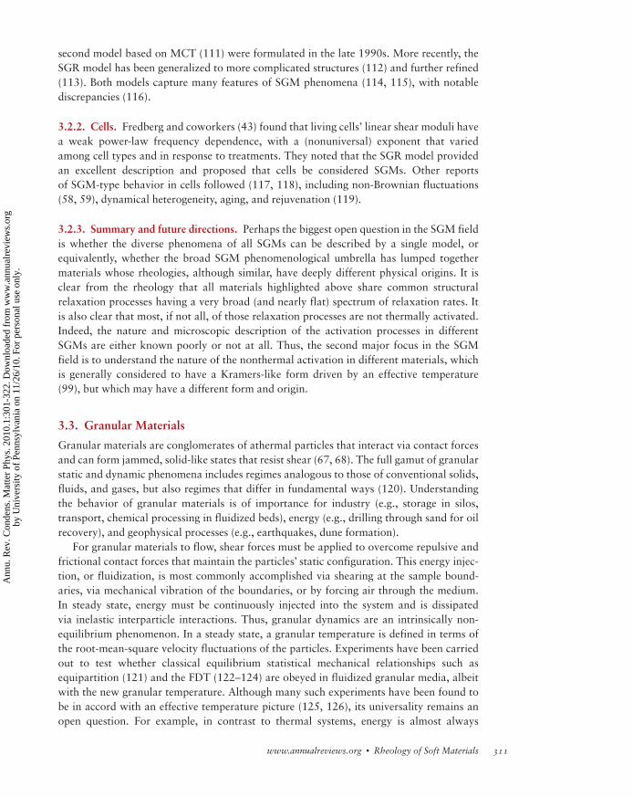
As Pe further increases, hydrodynamic interactions can induce particles to orbit one
another, destabilizing their ordered, layered flow structure. In this case, the increased
number of collisions in the disordered state leads to a regime of shear thickening,
wherein the viscosity dramatically increases. The particles organize into hydroclusters, as
depicted in Figure 3. The shear thickening transition is believed to result from particular
Shear thickening
(thinning):
Non-Newtonianrheological behavior
of a material in which
the stress increases
(decreases) withincreasing shear rate
Shear stress or shear rate
Visc
osit
y
Shear thickeningShear thinningEquilibrium
Figure 3
Relationship between microstructure and viscosity of shear thinning-shear thickening transition in hard-sphere colloidal suspen-
sions. In equilibrium, random collisions among particles (blue spheres) make them naturally resistant to flow. However, as theshear stress or, equivalently, the shear rate increases (increasing yellow gradient fill), particles become organized in the flow,
which lowers their viscosity. At yet higher shear rates, hydrodynamic interactions between particles dominate over stochastic
ones, a change that spawns hydroclusters (orange spheres)—transient fluctuations in particle concentration. The difficulty of
particles flowing around each other in a strong flow leads to a higher rate of energy dissipation and an abrupt increase inviscosity. Based on Figure 2 of Reference 76.
308 Chen et al.
Ann
u. R
ev. C
onde
ns. M
atte
r Ph
ys. 2
010.
1:30
1-32
2. D
ownl
oade
d fr
om w
ww
.ann
ualr
evie
ws.
org
by U
nive
rsity
of
Penn
sylv
ania
on
11/2
6/10
. For
per
sona
l use
onl
y.

hydrodynamic forces known as lubrication forces that arise whenever two surfaces, sepa-
rated by a fluid, move relative to one another in close proximity. Lubrication forces are
short ranged and divergent at contact; thus, they can only be observed at high Pe because
normal Brownian and repulsive interparticle forces conspire to prevent such close
approach. Wagner and coworkers have carefully studied the particle size, surface chem-
istry, concentration, and Pe dependencies for this novel reversible shear thickening effect
(75). More details are found in References 17 and 76. Currently, these materials are being
developed as the basis for soft body armor.
3.1.3. Active particle suspensions. A frontier research area in colloidal suspension rheol-
ogy concerns a new class of soft matter termed active suspensions. Active particle suspen-
sions arise when the motion of constituent particles is driven by nonthermal internal energy
sources. A canonical example of this system is a suspension of swimming microorganisms
in which the particles are actively consuming chemical energy and dissipating it into the
medium through their motion. In such a scenario, the dynamics of the particles are no
longer constrained by the FDT to the medium’s rheological linear response. A recent theory
has predicted that the medium’s viscosity can thus be altered by active forcing (77). The
viscosity can be either enhanced or suppressed, depending on the symmetry of the forcing
mechanism. This prediction is somewhat counter-intuitive, given that passive spherical
particles at low-to-moderate Pe will always increase stress in suspension, resulting in
viscosity enhancement. Recent passive and active microrheological measurements of pas-
sive tracers in bacterial suspensions have observed clear violation of the FDT at low
bacterial densities (f � .001) (47). A continuum theory elucidating the interplay between
active stress, orientational dynamics, and hydrodynamics has been developed and used to
rationalize microrheological measurements of passive tracer dynamics in active suspen-
sions (78). There have been many observations of self-organized collective motion in the
form of jets and vortices in concentrated bacterial suspensions (79, 80), and it is worth-
while to consider what role, if any, this collective-motion-induced ordering plays in mod-
ification of system rheology, i.e., whether such ordering produces viscosity reduction akin
to ordering in sheared colloidal suspensions at moderate Pe. Indeed, recent work has
interpreted the coherence lifetime of vortices as a measure of viscosity reduction in active
bacterial suspensions (81).
3.1.4. Summary and future directions. Many researchers are exploring the structural and
dynamical transitions in colloidal suspensions that accompany the glass transition. Several
such studies have focused on the phenomena of dynamical heterogeneities near the glass
transition wherein groups of particles move in a coordinated fashion with displacements
far exceeding that expected for pure diffusion (82–84). Particles within these dynamically
heterogeneous regions are sometimes qualitatively described as moving in a string-like
manner, and the participation fraction and spatio-temporal extent of these motions often
exhibit scaling near the glass transition critical volume fraction. Relatively unexplored are
the rheological transitions that accompany the glass transition, and even less well under-
stood are the rheological impacts of dynamical heterogeneities, if any, and their possible
connection to theoretical frameworks, such as shear transformation zones (85). Experi-
ments that directly observe correlations between stress relaxation and dynamical heteroge-
neity in a sheared colloidal glass have recently appeared (86, 87). In a related vein, studies
of particle dynamics in aging glasses have been carried out (84, 88). The aim of these
Aging: property of a
material wherein its
measured rheologicalresponse depends on
sample lifetime
www.annualreviews.org � Rheology of Soft Materials 309
Ann
u. R
ev. C
onde
ns. M
atte
r Ph
ys. 2
010.
1:30
1-32
2. D
ownl
oade
d fr
om w
ww
.ann
ualr
evie
ws.
org
by U
nive
rsity
of
Penn
sylv
ania
on
11/2
6/10
. For
per
sona
l use
onl
y.

studies has been to reveal which particle configurations give rise to aging behavior, in
essence linking structure to dynamics. However, rheological studies in which aging glasses
have been simultaneously imaged under weak (perturbative) shear are scarce. Such studies
may unveil the subtle, as-yet-unknown, microscopic mechanisms of aging in glasses. Tech-
nological advances in rheo-optical methods should provide opportunities to achieve a
comprehensive characterization of these phenomena in the near future.
3.2. Soft Glassy Materials
SGM encompass a host of new media including pastes (89), foams (90), emulsions (91, 92),
colloids (69, 93), clay suspensions (94), star polymer gels (95), and multilamellar vesicle
gels (96). These materials share a set of unusual rheological properties (97), whose physical
origin remains mysterious despite considerable research attention. SGM generally appear
as very soft solids, yet flow readily above a yield stress (sy) (98, 99). Rheometric measure-
ments report behavior that can be described by the Herschel-Bulkley relation. Typically,
the exponent n <1, making them shear thinning or pseudoplastic materials. As such, their
flow profiles are often complex (14, 100, 101), displaying wall slip and shear banding
behavior (102, 103).
Linear rheology measurements on these materials show a predominantly elastic
response having an apparent power-law form: G0(o) � ob, with a small exponent 0 < b <
0.3, over a few decades of frequency (69, 91). Moreover, the loss modulus (G00) is anoma-
lously large at low frequencies, when compared to conventional, elastic soft materials:
typically G00(o)/G0(o) � O(0.1). A notable feature of the power-law rheology exponents,
b, is that they vary among different materials and that they can vary continuously (though
typically rather weakly) for a single material type, depending on its formulation (e.g., solid
fraction). Such nonuniversal exponents differ from the behavior seen in most other visco-
elastic materials, such as polymers, whose rheology can be described by physical models
that predict universal exponents having specific numerical values independent of formula-
tion details (104). It has been shown that critical gels (105), polymer gels near the mechan-
ical percolation transition, also show power-law rheology with nonuniversal exponents.
Critical gels are not classified as SGMs however, as their power-law rheology behaviors are
only manifest in a narrow, engineered degree of cross-linking, unlike the robust behavior of
SGMs.
Many SGMs (e.g., pastes and colloidal glasses) display physical aging and rejuvenation
phenomena (106), wherein the measured linear rheology is time dependent, generally
stiffening as a function of sample age, but able to return to early time behavior with modest
shearing (93, 107). Such time dependence definitively indicates that these materials are
nonergodic or glassy—they do not occupy or ever reach a global thermodynamic equilib-
rium state. SGMs that do not display such aging (e.g., foams and some emulsions) typically
have high values of b and have internal rearrangements driven by coarsening and coales-
cence. It is generally supposed that the internal motions place the material in a state of
continuous rejuvenation, creating a nonequilibrium, dynamical steady state. Lastly, still
other SGMs (gels and clay suspensions) display even more complex and diverse aging
behavior (96, 108).
3.2.1. Theoretical models. Early models to describe SGMs, including the soft glassy rhe-
ology (SGR) model (98, 99, 109) based on trap models of the glass transition (110), and a
(Non)-universal
exponents: exponents
characterizingrheological responses
having values that (do
not) depend on details
of sample formulation
Rejuvenation:
property of agingmaterials wherein
rheological response
can be reset to anearlier state via
external shear or inter-
nal rearrangement
processes
310 Chen et al.
Ann
u. R
ev. C
onde
ns. M
atte
r Ph
ys. 2
010.
1:30
1-32
2. D
ownl
oade
d fr
om w
ww
.ann
ualr
evie
ws.
org
by U
nive
rsity
of
Penn
sylv
ania
on
11/2
6/10
. For
per
sona
l use
onl
y.
second model based on MCT (111) were formulated in the late 1990s. More recently, the
SGR model has been generalized to more complicated structures (112) and further refined
(113). Both models capture many features of SGM phenomena (114, 115), with notable
discrepancies (116).
3.2.2. Cells. Fredberg and coworkers (43) found that living cells’ linear shear moduli have
a weak power-law frequency dependence, with a (nonuniversal) exponent that varied
among cell types and in response to treatments. They noted that the SGR model provided
an excellent description and proposed that cells be considered SGMs. Other reports
of SGM-type behavior in cells followed (117, 118), including non-Brownian fluctuations
(58, 59), dynamical heterogeneity, aging, and rejuvenation (119).
3.2.3. Summary and future directions. Perhaps the biggest open question in the SGM field
is whether the diverse phenomena of all SGMs can be described by a single model, or
equivalently, whether the broad SGM phenomenological umbrella has lumped together
materials whose rheologies, although similar, have deeply different physical origins. It is
clear from the rheology that all materials highlighted above share common structural
relaxation processes having a very broad (and nearly flat) spectrum of relaxation rates. It
is also clear that most, if not all, of those relaxation processes are not thermally activated.
Indeed, the nature and microscopic description of the activation processes in different
SGMs are either known poorly or not at all. Thus, the second major focus in the SGM
field is to understand the nature of the nonthermal activation in different materials, which
is generally considered to have a Kramers-like form driven by an effective temperature
(99), but which may have a different form and origin.
3.3. Granular Materials
Granular materials are conglomerates of athermal particles that interact via contact forces
and can form jammed, solid-like states that resist shear (67, 68). The full gamut of granular
static and dynamic phenomena includes regimes analogous to those of conventional solids,
fluids, and gases, but also regimes that differ in fundamental ways (120). Understanding
the behavior of granular materials is of importance for industry (e.g., storage in silos,
transport, chemical processing in fluidized beds), energy (e.g., drilling through sand for oil
recovery), and geophysical processes (e.g., earthquakes, dune formation).
For granular materials to flow, shear forces must be applied to overcome repulsive and
frictional contact forces that maintain the particles’ static configuration. This energy injec-
tion, or fluidization, is most commonly accomplished via shearing at the sample bound-
aries, via mechanical vibration of the boundaries, or by forcing air through the medium.
In steady state, energy must be continuously injected into the system and is dissipated
via inelastic interparticle interactions. Thus, granular dynamics are an intrinsically non-
equilibrium phenomenon. In a steady state, a granular temperature is defined in terms of
the root-mean-square velocity fluctuations of the particles. Experiments have been carried
out to test whether classical equilibrium statistical mechanical relationships such as
equipartition (121) and the FDT (122–124) are obeyed in fluidized granular media, albeit
with the new granular temperature. Although many such experiments have been found to
be in accord with an effective temperature picture (125, 126), its universality remains an
open question. For example, in contrast to thermal systems, energy is almost always
www.annualreviews.org � Rheology of Soft Materials 311
Ann
u. R
ev. C
onde
ns. M
atte
r Ph
ys. 2
010.
1:30
1-32
2. D
ownl
oade
d fr
om w
ww
.ann
ualr
evie
ws.
org
by U
nive
rsity
of
Penn
sylv
ania
on
11/2
6/10
. For
per
sona
l use
onl
y.

injected anisotropically into a granular medium and is transferred heterogeneously down
to the particle scale, due to the interplay of dissipative, gravitational, and boundary effects.
These effects complicate, but also enrich, the dynamics of granular systems, and they must
be carefully considered in the interpretation of experiments geared toward establishing
parallels between granular and thermal systems.
Theories about the rheology of granular materials are challenging, in part due to
the effects of inhomogeneity in force transmission. In contrast to continuous media
such as Newtonian fluids, forces applied to granular media are almost always resisted
inhomogeneously and anisotropically, e.g., by force chains comprised of only a very small
fraction of the total number of particles (127–129). To date, the most striking demonstra-
tion of the existence of force chains has been observed in two-dimensional (2D) granular
packings of disks. In this case, the force chains are observed via compaction of photoelastic
disks that transmit light between crossed polarizers in extinction mode when stressed (130,
131). Qualitatively similar behavior of force chains has been observed in experiments
wherein photoelastic disks were slowly sheared in a 2D Couette geometry (132). Thus,
the motion of an immersed object through a granular medium requires breaking and
reformation of force chains, resulting in temporally intermittent, stick-slip dynamics
(133). The applicability of conventional continuum elastic/fluidic theory to these physical
systems is clearly no longer apparent.
Granular rheology in 3D holds the most relevance for industrial application. Unfortu-
nately, it is often not enough to simply measure stress as a function of shear rate in a
rheometer, as in conventional fluids; additional information from, for example, simulta-
neous imaging of the granular microstructure in both the bulk and near the surfaces is
necessary. To this end, experiments using magnetic resonance imaging (134) and internal
optical imaging using index-matched grains/fluid (135) have been used to visualize internal
granular microstructure under shear in a 3D annular volume. In both such experiments,
shear banding behavior was generally observed wherein particles ordered into layers near
the shearing wall.
3.4. Cross-Linked Networks
Polymer networks are rich in mechanical properties, which can be modulated by varying
the nature of the polymers, cross-linking agents, and the interactions between polymers. In
a cross-linked network, polymers are linked together by permanent bonds that prevent
relative motions of the polymers. The cross-linkers can be covalent bonds between poly-
mers, can be an individual molecule that can bind to multiple filaments, or can be branch
points from a larger fiber in the network. For the most part, the rheological properties of
cross-linked flexible polymers such as rubber are well described by classical theories of
rubber elasticity (136), wherein polymer strands between cross-links are treated as springs
whose elastic response under stretching or compression is derived from the entropic cost of
the polymer strand’s reduced conformational degrees of freedom. Cross-linked networks
are different from entangled networks, which are held together only by physical entangle-
ment between polymers. Most notably, entangled networks possess mechanisms of stress
relaxation that are absent in cross-linked networks. The most important of these mecha-
nisms is reptation, in which stress relaxation occurs via polymer diffusion in a tube formed
by its neighbors (104). Reptation sets a characteristic frequency below which entangled
networks exhibit a linear viscoelastic response that is dominantly liquid-like (G00 > G0).
312 Chen et al.
Ann
u. R
ev. C
onde
ns. M
atte
r Ph
ys. 2
010.
1:30
1-32
2. D
ownl
oade
d fr
om w
ww
.ann
ualr
evie
ws.
org
by U
nive
rsity
of
Penn
sylv
ania
on
11/2
6/10
. For
per
sona
l use
onl
y.

In cross-linked networks, this regime is absent because cross-linking prevents reptation
(and hence stress relaxation), leading to a dominantly elastic (G0 > G00) response. Abovethe reptation frequency, entangled and cross-linked networks are hard to distinguish rheo-
logically. At an intermediate frequency range, often referred to as the rubber plateau, both
networks exhibit a frequency-independent elastic modulus. At frequencies above the rub-
ber plateau, both networks exhibit power-law frequency dependence with exponents char-
acteristic of single polymer responses.
Polymer theory distinguishes three types of filaments according to the ratio between
persistence length (lp) and contour length (lc). The persistence length is defined as the
length scale for the decay of the tangent-tangent correlation along the filament contour,
and it is proportional to the stiffness of the polymer chain. When lc>>lp, the filament is
considered to be soft and exhibit purely entropic elastic response. In the limit of lp>>lc,
filaments are stiff and show no entropic elasticity. Most biopolymers fall between these
two limits—they are semiflexible with lc and lp approximately the same order of magni-
tude. Currently, the properties of semiflexible polymer networks have been intensively
investigated due to their biological relevance. Microscopic properties of the network defor-
mations such as the deformation of individual polymers and the distribution of deforma-
tion fields in the network must be understood to explain the mechanical properties of these
biopolymer networks. Unique properties such as negative normal stress (137) and strain
stiffening at moderate strain amplitudes (2) have been recently observed in these bio-
polymer networks and have spurred the development of theories describing semiflexible
networks.
3.4.1. Nonlinear elasticity of biological polymers. For gels of flexible polymers such as
polyacrylamide, G0 is a constant for strain values up to 100%: The flexible polymer
responds linearly. In contrast, gels of actin, fibrin, and collagen show strain stiffening
behavior at medium strain levels, i.e., the G0 of these gels becomes larger at larger strains.
A small increase (< 20%) in shear stain can lead to tenfold (or more) increase in shear
elastic moduli (2).
The origin of strain stiffening in networks of semiflexible or rod-like polymers has been
addressed by several recent theories. For filaments that are highly elongated but still soft
enough to be deformed by thermal energy, for example, polymers with persistence length
on the order of the network mesh size, nonlinear strain stiffening occurs as a result of the
intrinsically nonlinear force-extension relation of each network strand (2, 138). If the
persistence length of the filament is comparable to the mesh size or distance between
cross-links within the network, then strain stiffening emerges from an entropic model that
considers how thermal fluctuations of semiflexible polymers are constrained as the end-to-
end distance of filament segments between cross-links changes when the sample is
deformed (2, 5, 138, 139). This essentially entropic model accurately predicts the strain-
dependent elasticity of isotropically cross-linked F-actin, intermediate filaments, and fibrin
protofibrils, which have persistence lengths between 0.5 and 10 microns and form net-
works with mesh size on the order of 100 nm to 1 micron (2). Other networks formed by
much stiffer and thicker fibers, such as collagen, fibrin fibers, or actin bundles also exhibit
strain stiffening, but the degree of strain stiffening appears to be less than that of the
intermediate filaments, and thermal motions of these stiffer fibers would appear not to
play a significant role in their response. In these cases, models derived from the enthalpic
deformation of soft rods have been developed. In these models, strain stiffening coincides
www.annualreviews.org � Rheology of Soft Materials 313
Ann
u. R
ev. C
onde
ns. M
atte
r Ph
ys. 2
010.
1:30
1-32
2. D
ownl
oade
d fr
om w
ww
.ann
ualr
evie
ws.
org
by U
nive
rsity
of
Penn
sylv
ania
on
11/2
6/10
. For
per
sona
l use
onl
y.
with alignment of fibers in the strain direction and a shift from soft bending modes to
stiffer stretching modes as the fibers reorient (140–143)
3.4.2. Normal stress. A novel feature of semiflexible network rheology is that deforma-
tions that are pure shear lead to a negative normal stress that tends to pull the shearing
surfaces toward each other. When cross-linked networks of F-actin, fibrin, collagen, or
other biopolymers are sheared, the stress directed orthogonal to the strain direction in-
creases quadratically with shear strain and can have a magnitude close to that of the shear
stress (1, 137). Flexible polymer gels generally have negligible normal stress, and at high
strains exhibit a positive normal stress that is much smaller than the shear stress, in accord
with predictions of the Mooney-Rivlin theory of rubber elasticity (136). Negative normal
stress emerges naturally from the thermal model for semiflexible filaments. The force-
displacement relation for such filaments shows that less force is needed to compress such
a filament than to stretch it, and, therefore, under pure shear the downward-directed forces
from stretched filaments oriented away from the shear direction dominate over the weaker
upward-directed forces from the compressed filaments (1, 137). Nonthermal models of
stiff filaments also predict negative normal stress, because compressed filaments can buckle
and thus reduce upward forces relative to the always increased downward forces produced
by stretched filaments (144). Some models predict that the normal stresses can be even
greater than shear stresses under some conditions (145).
3.4.3. Affine versus nonaffine deformation. One important assumption in theories of
rubber elasticity and entropic models for elasticity of networks of semiflexible polymers is
that the macroscopic network deformation is transferred uniformly to each polymer chain
in the network. Various theories developed for the rheology of stiff or semiflexible net-
works make different predictions about the degree to which the deformations are affine,
that is, the degree to which local strains match the macroscopic strain, and about the extent
to which filament alignment occurs. Direct measurement of network nonaffinity, made by
quantifying the displacement of small markers placed within the network, have begun to
differentiate between these different theoretical models, and more fully define the material
response in these networks (11, 142, 146, 147). As the polymer stiffness increases, trans-
verse thermal undulation of polymers is negligible and the elasticity of filaments is no
longer entropic. For example, in a bundled actin network, the deformation is dominated
by nonaffine enthalpic bending of the actin bundles (5, 148). Computer simulations sug-
gest that the nonaffine deformation of gels made of rigid filaments is accompanied by
filament bending, rotation, and buckling (149).
Nonaffine deformation occurs not only in stiff polymer networks but also in semiflex-
ible and even flexible polymer networks. In random elastic media, heterogeneities in local
elasticity can give rise to nonaffine deformations (150). In semiflexible polymer networks,
nonaffine deformations have been measured for actin gels (16) and fibrin gels (11), by
measuring displacements of tracer beads in the gels under external stress.
3.4.4. Active gels. The cytoskeleton of a cell is composed of filamentous actin network
and different types of molecular motors actively moving along the actin filaments and
applying forces to the filament. An active gel often refers to filament networks containing
molecular motors. Many cell functions, including but not limited to cell migration,
mechanosensing, and adaptation to environments, are related to the dynamics and
314 Chen et al.
Ann
u. R
ev. C
onde
ns. M
atte
r Ph
ys. 2
010.
1:30
1-32
2. D
ownl
oade
d fr
om w
ww
.ann
ualr
evie
ws.
org
by U
nive
rsity
of
Penn
sylv
ania
on
11/2
6/10
. For
per
sona
l use
onl
y.
rheological properties of the cytoskeleton (151). A recent study on purified actin networks
cross-linked by filamin shows that adding myosin II motors leads to an increase in G0 bymore than two orders of magnitude. This suggests that one function of myosin II is to
pull the actin filaments and cause an internal stress that stiffens the cytoskeleton (152).
The strain stiffening of cross-linked actin networks in combination with the myosin
motors allow cells to adjust their cortical stiffness by modulating the motor activity. This
effect provides a possible explanation for a recent cell mechanosensing observation
that fibroblasts can adjust their stiffness to match the stiffness of the substrates they are
cultured on (153).
3.4.5. Summary and future directions. Nonlinear elasticity, negative normal stress, and
nonaffine deformations are signatures of semiflexible polymer networks. The available theo-
retical models on elasticity of semiflexible polymer networks are derived from two different
origins: the affine entropy model and the nonaffine enthalpy model. The former model
suggests these effects originate from entropic stretching of thermal fluctuations, whereas the
latter suggests an origin in bending and buckling of soft rods. Although both are able to
predict the onset and extent of these effects on a coarse-grained level, experimental tech-
niques capable of monitoring single filament/bundle dynamics under shear, such as confocal
rheometry, are needed to decisively distinguish between these alternative models.
The current research approach of examining in vitro isotropic networks under well-
defined shear deformations has been very successful in elucidating the physical properties
of semiflexible networks. However, in vivo networks rarely exist and operate under such
ideal conditions. Often, functional cellular networks are composed of different types of
filaments and cross-linking proteins, possess some degree of anisotropy, and are quasi-2D.
Future work in the field will no doubt be oriented toward exploring these complexities
both experimentally and theoretically.
In a related vein, asking the question of why Nature decided to utilize certain classes of
network constituents (e.g., semiflexible, active molecular motors) to accomplish crucial
biological functions will continue to drive future research on polymer networks. One
promising approach, multiscale mechanics, aims to uncover such underlying design princi-
ples by investigating dynamics from the single-molecule, to filament, to network scales. An
exciting example of multiscale mechanics can be found in recent work on networks of the
blood-clotting protein fibrin. The authors relate the consequences of unfolding of the fibrin
proteins at the single-molecule scale to functional requirements of permeability and exten-
sibility at the clot scale (154).
3.5. Cells
Viewed as a soft material, the cell is arguably the most challenging to characterize and
interpret of all the soft materials reviewed here. The complexity arises in large part from
there being a variety of load-bearing elements and heterogeneous substructures inside cells.
These elements and structures are coupled mechanically through both passive (mechanical)
and active (ATP-dependent) processes and have multiple timescales of response. Addition-
ally, most cells contain endogenous molecular force sensors that trigger biochemical
signaling pathways that may modulate cellular stiffness in response to shear forces
with specific characteristics, a process known as mechanosensing. Although important
to the comprehensive picture of cell mechanics, consideration of the mechanisms of
www.annualreviews.org � Rheology of Soft Materials 315
Ann
u. R
ev. C
onde
ns. M
atte
r Ph
ys. 2
010.
1:30
1-32
2. D
ownl
oade
d fr
om w
ww
.ann
ualr
evie
ws.
org
by U
nive
rsity
of
Penn
sylv
ania
on
11/2
6/10
. For
per
sona
l use
onl
y.

mechanosensing are beyond the scope of the present work and are reviewed in detail
elsewhere (155, 156). Instead, we focus on soft material aspects of cells’ rheological
responses, in essence, viewing the cell as an exotic, hierarchically structured, soft material.
When probed, cells’ rheological behavior shares many characteristics of nearly every
class of soft material reviewed here. Cells exhibit linear viscoelastic moduli with scaling
exponents characteristic of stress relaxation in semiflexible polymer networks. This is not
too surprising because actin, microtubules, and intermediate filaments comprise a large
part of the cytoskeleton, nucleus, and cortex of mammalian cells. Also, many studies have
shown that cells exhibit strain stiffening in response to stress or deformation (157). The
precise origins of the strain-stiffening response of cells is not fully understood and may
reflect, in addition to orientation, alignment, and the pulling out of entropic fluctuations
under shear, an important contribution from prestress due to active elements such as
molecular motors (60, 158, 159). Under physiological conditions, cells are subject to
highly nonlinear strains of both externally and internally generated origins. The assump-
tion that information relevant to cell mechanics in vivo can be learned from its linear
viscoelastic properties, as in simple materials, is thus called into question.
Cells are internally quite crowded, and thus cells share with dense colloidal glasses,
SGMs, and granular materials the property that they are jammed in a metastable,
nonergodic state and require rejuvenation via internally generated nonthermal forces to
rearrange, i.e., to move or divide in the case of cells. Indeed, concepts from the theory of
soft glassy rheology (SGR) (98), such as aging and shear rejuvenation, have been adapted
to interpret cell rheology experiments (43, 119, 160). Similar to SGMs, cells have power-
law rheology in which the modulus increases slowly with a nonuniversal weak exponent
even at low frequency (43). In contrast, biopolymer networks with comparable elastic
moduli (100–1000 Pa) would be purely elastic, exhibiting a frequency-independent plateau
modulus at low frequencies. There are, however, notable differences between cell and SGM
rheology (161). For one, the observation of strain stiffening in some cell studies (157) is
inconsistent with SGM rheology, which generally exhibits yielding and plasticity, as
observed in cell softening seen under large oscillatory strain (119). Of course, multiple
explanations are possible: the stiffening could be due to a background network that is
pervaded by an SGM or conversely, the cell softening and subsequent aging could be due
to mechanical network damage and active repair (119). If the non-Brownian fluctuations
in cells are responsible for SGM-like fluidization, then the observed rheology exponent
should be very sensitive to ATP perturbation, which it is not (119, 162). The original,
nonuniversal exponent findings can also be explained as resulting from the superposition
of multiple structures having different rheology (161).
In the face of the daunting complexity of a cell’s rheological response, two main
approaches have been undertaken. The first is a bottom-up approach in which the constit-
uents of a cell are extracted, purified, reconstituted, and used to create a minimal in vitro
system capable of faithfully replicating an intact cell’s rheological feature of interest. Cells,
after all, are comprised primarily of biopolymers; thus, by starting with a reconstituted
network with a well-understood rheological response, it follows that one might be able to
serially build up the complexity by adding cross-linking proteins, active molecular motors,
etc. Notable successes of this approach include replication of strain-stiffening behavior
using prestressed flexible cross-linking proteins (163) and molecular motors (60) in
reconstituted actin networks. Alternatively, a top-down approach has been undertaken in
which whole, functioning cells are probed using microrheological techniques covering a
316 Chen et al.
Ann
u. R
ev. C
onde
ns. M
atte
r Ph
ys. 2
010.
1:30
1-32
2. D
ownl
oade
d fr
om w
ww
.ann
ualr
evie
ws.
org
by U
nive
rsity
of
Penn
sylv
ania
on
11/2
6/10
. For
per
sona
l use
onl
y.

wide range of intracellular length and timescales in concert with pharmacological inter-
ventions designed to eliminate mutually confounding response pathways. The emergent
principal components of the data are usually in the form of scaling exponents characteriz-
ing frequency-dependent rheological response functions. These functions are then used to
formulate a consensus description. Notable successes of this approach include the discov-
eries of distinct power laws characterizing the rheological responses of the deep cell
interior and outer cortex (162). This approach is reviewed in Reference 161.
4. CONCLUSIONS AND OUTLOOK
SUMMARY POINTS
1. Rheology and microrheology are chief ways of probing the structure and
responses of soft materials.
2. The linear response of soft materials is typically described by complex frequency-
dependent shear moduli that encode information about the underlying structures
and timescales of the material. It is hard to distinguish soft materials from each
other based only on their linear viscoelastic moduli.
3. When driven to nonlinear strain regimes (polymeric materials), or out of equilib-
rium at high shear rates (colloidal suspensions), soft materials exhibit a wide
range of striking and technologically useful responses (e.g., shear thinning/thick-
ening, shear banding).
4. Many classes of soft materials, although constitutively distinct, exhibit rheologi-
cal properties that can be captured by a small set of phenomenological models
such as SGR, jamming, and polymer rheology (Table 1).
Table 1 Summary matrix of soft material classes and observed rheological effects. (X denotes observation of effect and
reference.)
Power-law
rheology
Shear thinning/
strain softening
Shear thickening/
strain stiffening
Shear
banding
Active
(nonequilibrium)
effects
Aging after shear
rejuvenation/
fluidization
Colloidal
suspensions
X (69) X (75) X (75) X (15) X (47)
Colloidal
glasses
X (69) X (87) X (87) X (106)
Granular
materials
X (132) X (135) X (122) X (124)
Soft glassy
materials
X (91) X (91) X (14) X (99) X (108)
Polymer
networks
X (105) X (2) X (10) X (60)
Cells X (43) X (119) X (157) X (59) X (119)
www.annualreviews.org � Rheology of Soft Materials 317
Ann
u. R
ev. C
onde
ns. M
atte
r Ph
ys. 2
010.
1:30
1-32
2. D
ownl
oade
d fr
om w
ww
.ann
ualr
evie
ws.
org
by U
nive
rsity
of
Penn
sylv
ania
on
11/2
6/10
. For
per
sona
l use
onl
y.

FUTURE ISSUES
1. Current trends to simultaneously image and shear materials (e.g., confocal
rheometry) promise to reveal a mechanistic understanding of soft material de-
formation at an unprecedented level of detail. This bodes well for guiding
the development of theoretical models and suggesting new technological
applications.
2. One ultimate test of our understanding of soft material rheology lies in our ability
to predict the rheological responses of cells, for which a comprehensive,
multiscale approach integrating all aspects of soft materials rheology must be
brought to bear.
DISCLOSURE STATEMENT
The authors are not aware of any affiliations, memberships, funding, or financial holdings
that might be perceived as affecting the objectivity of this review.
ACKNOWLEDGMENTS
We gratefully acknowledge useful discussions over many years with Tom Lubensky, Fred
MacKintosh, Andrea Liu, Doug Durian, Jerry Gollub, Eric Weeks, Alex Levine, Andy Lau,
Ke Chen, Ahmed Alsayed, Anindita Basu, Zexin Zhang, Larry Hough, Mohammad Islam,
and Peter Yunker. A.G.Y. acknowledges support from NSF-MRSEC (DMR-0505048),
NSF (DMR 05-20020), and NASA (NNX08AO0G). J.C.C. acknowledges support from
the NSF (DMR-0706388). P.A.J. acknowledges support from NIH (GM083272).
LITERATURE CITED
1. Kang H, Wen Q, Janmey PA, Tang JX, Conti E, MacKintosh FC. 2009. J. Phys. Chem. B
113:3799–805
2. Storm C, Pastore JJ, MacKintosh FC, Lubensky TC, Janmey PA. 2005. Nature 435:191–94
3. Schopferer M, Bar H, Hochstein B, Sharma S, Mucke N, et al. 2009. J. Mol. Biol. 388:133–43
4. Gardel ML, Nakamura F, Hartwig J, Crocker JC, Stossel TP, Weitz DA. 2006. Phys. Rev. Lett.
96:088102
5. Gardel ML, Shin JH, MacKintosh FC, Mahadevan L, Matsudaira P, Weitz DA. 2004. Science
304:1301–5
6. Kasza KE, Koenderink GH, Lin YC, Broedersz CP, Messner W, et al. 2009. Phys. Rev. E
79:041928
7. Ewoldt RH, Hosoi AE, McKinley GH. 2008. J. Rheol. 52:1427–58
8. Fielding SM. 2007. Soft Matter 3:1262–79
9. Olmsted PD. 2008. Rheol. Acta 47:283–300
10. Boukany PE, Hu YT, Wang SQ. 2008. Macromolecules 41:2644–50
11. Wen Q, Basu A, Winer JP, Yodh A, Janmey PA. 2007. New J. Phys. 9:428
12. Besseling R, Isa L, Weeks ER, Poon WCK. 2009. Adv. Colloid Interface Sci. 146:1–17
13. Callaghan PT. 2008. Rheol. Acta 47:243–55
318 Chen et al.
Ann
u. R
ev. C
onde
ns. M
atte
r Ph
ys. 2
010.
1:30
1-32
2. D
ownl
oade
d fr
om w
ww
.ann
ualr
evie
ws.
org
by U
nive
rsity
of
Penn
sylv
ania
on
11/2
6/10
. For
per
sona
l use
onl
y.

14. Coussot P, Raynaud JS, Bertrand F, Moucheront P, Guilbaud JP, et al. 2002. Phys. Rev. Lett.
88:218301
15. Cohen I, Davidovitch B, Schofield AB, Brenner MP, Weitz DA. 2006. Phys. Rev. Lett. 97:215502
16. Liu J, Koenderink GH, Kasza KE, MacKintosh FC, Weitz DA. 2007. Phys. Rev. Lett. 98:198304
17. Vermant J, Solomon MJ. 2005. J. Phys. Condens. Matter 17:R187–216
18. Wagner NJ. 1998. Curr. Opin. Colloid Interface Sci. 3:391–400
19. Seifriz W. 1924. J. Exp. Biol. 2:1–11
20. Crick FHC, Hughes AFW. 1950. Exp. Cell Res. 1:37–80
21. Mason TG, Gang H, Weitz DA. 1997. J. Opt. Soc. Am. A 14:139–49
22. Gittes F, Schnurr B, Olmsted PD, MacKintosh FC, Schmidt CF. 1997. Phys. Rev. Lett.
79:3286–89
23. Mason TG, Ganesan K, van Zanten JH, Wirtz D, Kuo SC. 1997. Phys. Rev. Lett. 79:3282–85
24. Starrs L, Bartlett P. 2003. J. Phys. Condens. Matter 15:S251–56
25. Addas KM, Schmidt CF, Tang JX. 2004. Phys. Rev. E 70:021503
26. Palmer A, Mason TG, Xu JY, Kuo SC, Wirtz D. 1999. Biophys. J. 76:1063–71
27. Gisler T, Weitz DA. 1999. Phys. Rev. Lett. 82:1606–9
28. Dasgupta BR, Tee SY, Crocker JC, Frisken BJ, Weitz DA. 2002. Phys. Rev. E 65:051505
29. Popescu G, Dogariu A, Rajagopalan R. 2002. Phys. Rev. E 65:041504
30. Valentine MT, Perlman ZE, Gardel ML, Shin JH, Matsudaira P, et al. 2004. Biophys. J.
86:4004–14
31. Crocker JC, Valentine MT, Weeks ER, Gisler T, Kaplan PD, et al. 2000. Phys. Rev. Lett.
85:888–91
32. Chen DT, Weeks ER, Crocker JC, Islam MF, Verma R, et al. 2003. Phys. Rev. Lett.
90:108301
33. Popescu G, Park Y, Lue N, Best-Popescu C, Deflores L, et al. 2008. Am. J. Physiol. Cell Physiol.
295:C538–44
34. Benmouna F, Johannsmann D. 2004. Langmuir 20:188–93
35. Habdas P, Schaar D, Levitt AC, Weeks ER. 2004. Europhys. Lett. 67:477–83
36. Squires TM. 2008. Langmuir 24:1147–59
37. Sriram I, Furst EM, DePuit RJ, Squires TM. 2009. J. Rheol. 53:357–81
38. White JG, Burris SM, Tukey D, Smith C, Clawson CC. 1984. Blood 64:210–14
39. Lerche D, Kozlov MM, Meier W. 1991. Eur. Biophys. J. 19:301–9
40. Discher DE, Mohandas N, Evans EA. 1994. Science 266:1032–35
41. Bausch AR, Hellerer U, Essler M, Aepfelbacher M, Sackmann E. 2001. Biophys. J. 80:2649–57
42. Ziemann F, Radler J, Sackmann E. 1994. Biophys. J. 66:2210–16
43. Fabry B, MaksymGN, Butler JP, Glogauer M, Navajas D, Fredberg JJ. 2001. Phys. Rev. Lett. 87:
148102
44. Puig-de-Morales M, Grabulosa M, Alcaraz J, Mullol J, Maksym GN, et al. 2001. J. Appl.
Physiol. 91:1152–59
45. Mizuno D, Head DA, MacKintosh FC, Schmidt CF. 2008. Macromolecules 41:7194–202
46. Hough LA, Ou-Yang HD. 2006. Phys. Rev. E 73:031802
47. Chen DTN, Lau AWC, Hough LA, Islam MF, Goulian M, et al. 2007. Phys. Rev. Lett.
99:148302
48. Mahaffy RE, Shih CK, MacKintosh FC, Kas J. 2000. Phys. Rev. Lett. 85:880–83
49. Chaudhuri O, Parekh SH, Fletcher DA. 2007. Nature 445:295–98
50. Schmidt FG, Ziemann F, Sackmann E. 1996. Eur. Biophys. J. 24:348–53
51. Levine AJ, Lubensky TC. 2000. Phys. Rev. Lett. 85:1774–77
52. Levine AJ, Lubensky TC. 2001. Phys. Rev. E 65:011501
53. Atakhorrami M, Koenderink GH, Schmidt CF, MacKintosh FC. 2005. Phys. Rev. Lett.
95:208302
54. Qiu XL, Tong P, Ackerson BJ. 2004. Appl. Opt. 43:3382–90
www.annualreviews.org � Rheology of Soft Materials 319
Ann
u. R
ev. C
onde
ns. M
atte
r Ph
ys. 2
010.
1:30
1-32
2. D
ownl
oade
d fr
om w
ww
.ann
ualr
evie
ws.
org
by U
nive
rsity
of
Penn
sylv
ania
on
11/2
6/10
. For
per
sona
l use
onl
y.

55. Koenderink GH, Atakhorrami M, MacKintosh FC, Schmidt CF. 2006. Phys. Rev. Lett.
96:138307
56. Caspi A, Granek R, Elbaum M. 2002. Phys. Rev. E 66:011916
57. Salman H, Gil Y, Granek R, ElbaumM. 2002. Chem. Phys. 284:389–97
58. Bursac P, Fabry B, Trepat X, Lenormand G, Butler JP, et al. 2007. Biochem. Biophys. Res.
Commun. 355:324–30
59. Lau AWC, Hoffman BD, Davies A, Crocker JC, Lubensky TC. 2003. Phys. Rev. Lett.
91:198101
60. Mizuno D, Tardin C, Schmidt CF, MacKintosh FC. 2007. Science 315:370–73
61. Brangwynne CP, Koenderink GH, MacKintosh FC, Weitz DA. 2008. J. Cell Biol. 183:583–87
62. Gallet F, Arcizet D, Bohec P, Richert A. 2009. Soft Matter 5:2947–53
63. Zhang Z, Xu N, Chen DTN, Yunker P, Alsayed AM, et al. 2009. Nature 459:230–33
64. Einstein A. 1905. Ann. Phys. 17:549–60
65. Batchelor GK, Green JT. 1972. J. Fluid Mech. 56:401–27
66. Bergenholtz J. 2001. Curr. Opin. Colloid Interface Sci. 6:484–88
67. Liu AJ, Nagel SR. 1998.Nature 396:21–22
68. Cates ME, Wittmer JP, Bouchaud JP, Claudin P. 1998. Phys. Rev. Lett. 81:1841–44
69. Mason TG, Weitz DA. 1995. Phys. Rev. Lett. 75:2770–73
70. Cheng ZD, Zhu JX, Chaikin PM, Phan SE, Russel WB. 2002. Phys. Rev. E 65:041405
71. Shikata T, Pearson DS. 1994. J. Rheol. 38:601–16
72. Brambilla G, El Masri D, Pierno M, Berthier L, Cipelletti L, et al. 2009. Phys. Rev. Lett.
102:085703
73. Gotze W, Sjogren L. 1992. Rep. Prog. Phys. 55:241–376
74. Turnbull D, Cohen MH. 1970. J. Chem. Phys. 52:3038
75. Maranzano BJ, Wagner NJ. 2001. J. Chem. Phys. 114:10514–27
76. Wagner NJ, Brady JF. 2009. Phys. Today 62:27–32
77. Hatwalne Y, Ramaswamy S, Rao M, Simha RA. 2004. Phys. Rev. Lett. 92:118101
78. Lau AWC, Lubensky TC. 2009. Phys. Rev. E 80:011917
79. Wu XL, Libchaber A. 2000. Phys. Rev. Lett. 84:3017–20
80. Dombrowski C, Cisneros L, Chatkaew S, Goldstein RE, Kessler JO. 2004. Phys. Rev. Lett.
93:098103
81. Sokolov A, Aranson IS. 2009. Phys. Rev. Lett. 103:148101
82. Weeks ER, Crocker JC, Levitt AC, Schofield A, Weitz DA. 2000. Science 287:627–31
83. Kegel WK, van Blaaderen A. 2000. Science 287:290–93
84. Yunker P, Zhang Z, Aptowicz KB, Yodh AG. 2009. Phys. Rev. Lett. 103:115701
85. Langer JS. 2008. Phys. Rev. E 77:021502
86. Schall P, Weitz DA, Spaepen F. 2007. Science 318:1895–99
87. Besseling R, Weeks ER, Schofield AB, Poon WCK. 2007. Phys. Rev. Lett. 99:028301
88. Lynch JM, Cianci GC, Weeks ER. 2008. Phys. Rev. E 78:031410
89. Cloitre M, Borrega R, Monti F, Leibler L. 2003. Phys. Rev. Lett. 90:068303
90. Hohler R, Cohen-Addad S. 2005. J. Phys. Condens. Matter 17:R1041–69
91. Mason TG, Bibette J, Weitz DA. 1995. Phys. Rev. Lett. 75:2051–54
92. Becu L, Manneville S, Colin A. 2006. Phys. Rev. Lett. 96:138302
93. Courtland RE, Weeks ER. 2003. J. Phys. Condens. Matter 15:S359–65
94. Treece MA, Oberhauser JP. 2007. Polymer 48:1083–95
95. Christopoulou C, Petekidis G, Erwin B, Cloitre M, Vlassopoulos D. 2009. Philos. Trans. R. Soc.
Ser. 367: 5051-71
96. Ramos L, Cipelletti L. 2001. Phys. Rev. Lett. 87:245503
97. Barnes HA, Hutton JF, Walters K. 2001. An Introduction to Rheology. Amsterdam: Elsevier Sci.
98. Sollich P, Lequeux F, Hebraud P, Cates ME. 1997. Phys. Rev. Lett. 78:2020–23
99. Sollich P. 1998. Phys. Rev. E 58:738–59
320 Chen et al.
Ann
u. R
ev. C
onde
ns. M
atte
r Ph
ys. 2
010.
1:30
1-32
2. D
ownl
oade
d fr
om w
ww
.ann
ualr
evie
ws.
org
by U
nive
rsity
of
Penn
sylv
ania
on
11/2
6/10
. For
per
sona
l use
onl
y.

100. Conrad JC, Lewis JA. 2008. Langmuir 24:7628–34
101. Gibaud T, Barentin C, Taberlet N, Manneville S. 2009. Soft Matter 5:3026–37
102. Ianni F, Di Leonardo R, Gentilini S, Ruocco G. 2008. Phys. Rev. E 77:031406
103. Moller PCF, Rodts S, Michels MAJ, Bonn D. 2008. Phys. Rev. E 77:041507
104. Doi M, Edwards SF. 1986. The Theory of Polymer Dynamics. New York: Oxford Univ. Press
105. Izuka A, Winter H, Hashimoto T. 1992. Macromolecules 25:2422–28
106. Moller P, Fall A, Chikkadi V, Derks D, Bonn D. 2009. Philos. Trans. R. Soc. Ser. A 367:5139-55
107. McKenna GB, Narita T, Lequeux F. 2009. J. Rheol. 53:489–516
108. Ianni F, Di Leonardo R, Gentilini S, Ruocco G. 2007. Phys. Rev. E 75:011408
109. Fielding SM, Sollich P, Cates ME. 2000. J. Rheol. 44:323–69
110. Bouchaud JP. 1992. J. Phys. I 2:1705–13
111. Hebraud P, Lequeux F. 1998. Phys. Rev. Lett. 81:2934–37
112. Cates ME, Sollich P. 2004. J. Rheol. 48:193–207
113. Fielding SM, Cates ME, Sollich P. 2009. Soft Matter 5:2378–82
114. Helgeson ME, Wagner NJ, Vlassopoulos D. 2007. J. Rheol. 51:297–316
115. Purnomo EH, van den Ende D, Mellema J, Mugele F. 2007. Phys. Rev. E 76:021404
116. Yin GJ, Solomon MJ. 2008. J. Rheol. 52:785–800
117. Mofrad MRK. 2009. Annu. Rev. Fluid Mech. 41:433–53
118. Smith BA, Roy H, De Koninck P, Grutter P, De Koninck Y. 2007. Biophys. J. 92:1419–30
119. Bursac P, Lenormand G, Fabry B, Oliver M, Weitz DA, et al. 2005. Nat. Mater. 4:557–61
120. Jaeger HM, Nagel SR, Behringer RP. 1996. Rev. Mod. Phys. 68:1259–73
121. Feitosa K, Menon N. 2002. Phys. Rev. Lett. 88:198301
122. D ’Anna G, Mayor P, Barrat A, Loreto V, Nori F. 2003. Nature 424:909–12
123. Ojha RP, Lemieux PA, Dixon PK, Liu AJ, Durian DJ. 2004. Nature 427:521–23
124. Josserand C, Tkachenko AV, Mueth DM, Jaeger HM. 2000. Phys. Rev. Lett. 85:3632–35
125. Corwin EI, Jaeger HM, Nagel SR. 2005. Nature 435:1075–78
126. Song C, Wang P, Makse HA. 2005. Proc. Natl. Acad. Sci. USA 102:2299–304
127. Liu CH, Nagel SR, Schecter DA, Coppersmith SN, Majumdar S, et al. 1995. Science 269:513–15
128. Dantu P. 1968.Geotechnique 18:50
129. Travers T, Bideau D, Gervois A, Troadec JP, Messager JC. 1986. J. Phys. A 19:1033-38
130. Howell D, Behringer RP, Veje C. 1999. Phys. Rev. Lett. 82:5241–44
131. Drescher A, de Josselin de Jong G. 1972. J. Mech. Phys. Solids 20:337
132. Hartley RR, Behringer RP. 2003. Nature 421:928–31
133. Albert I, Tegzes P, Kahng B, Albert R, Sample JG, et al. 2000. Phys. Rev. Lett. 84:5122–25
134. Mueth DM, Debregeas GF, Karczmar GS, Eng PJ, Nagel SR, Jaeger HM. 2000. Nature
406:385–89
135. Tsai JC, Voth GA, Gollub JP. 2003. Phys. Rev. Lett. 91:064301
136. Rubinstein M, Colby RH. 2003. Polymer Physics. Oxford/New York: Oxford Univ. Press.
xi. 440 pp.
137. Janmey PA, McCormick ME, Rammensee S, Leight JL, Georges PC, MacKintosh FC. 2007.Nat.
Mater. 6:48–51
138. MacKintosh FC, Kas J, Janmey PA. 1995. Phys. Rev. Lett. 75:4425–28
139. Palmer JS, Boyce MC. 2008. Acta Biomater. 4:597–612
140. Huisman EM, van Dillen T, Onck PR, van der Giessen E. 2007. Phys. Rev. Lett. 99:208103
141. Onck PR, Koeman T, van Dillen T, van der Giessen E. 2005. Phys. Rev. Lett. 95:178102
142. Heussinger C, Shaefer B, Frey E. 2007. Phys. Rev. E 76:031906
143. van Dillen T, Onck PR, van der Giessen E. 2008. J. Mech. Phys. Solids 56:2240–64
144. Conti E, MacKintosh FC. 2009. Phys. Rev. Lett. 102:088102
145. Heussinger C, Frey E. 2006. Phys. Rev. Lett. 97:105501
146. Fernandez P, Grosser S, Kroy K. 2009. Soft Matter 5:2047–56
147. Hatami-Marbini H, Picu RC. 2008. Phys. Rev. E 77:062103
www.annualreviews.org � Rheology of Soft Materials 321
Ann
u. R
ev. C
onde
ns. M
atte
r Ph
ys. 2
010.
1:30
1-32
2. D
ownl
oade
d fr
om w
ww
.ann
ualr
evie
ws.
org
by U
nive
rsity
of
Penn
sylv
ania
on
11/2
6/10
. For
per
sona
l use
onl
y.

148. Lieleg O, Claessens MM, Heussinger C, Frey E, Bausch AR. 2007. Phys. Rev. Lett. 99:088102
149. Chandran PL, Barocas VH. 2006. J. Biomech. Eng. 128:259–70
150. DiDonna BA, Lubensky TC. 2005. Phys. Rev. E 72:066619
151. Fletcher DA, Geissler PL. 2009. Annu. Rev. Phys. Chem. 60:469–86
152. Koenderink GH, Dogic Z, Nakamura F, Bendix PM, MacKintosh FC, et al. 2009. Proc. Natl.
Acad. Sci. USA 106:15192–97
153. Solon J, Levental I, Sengupta K, Georges PC, Janmey PA. 2007. Biophys. J. 93:4453–61
154. Brown AEX, Litvinov RI, Discher DE, Purohit PK, Weisel JW. 2009. Science 325:741–44
155. Shyy JY, Chien S. 2002. Circ. Res. 91:769–75
156. Janmey PA, McCulloch CA. 2007. Annu. Rev. Biomed. Eng. 9:1–34
157. Wang N, Butler JP, Ingber DE. 1993. Science 260:1124–27
158. Brangwynne CP, Koenderink GH, MacKintosh FC, Weitz DA. 2008. Phys. Rev. Lett.
100:118104
159. Kumar S, Maxwell IZ, Heisterkamp A, Polte TR, Lele TP, et al. 2006. Biophys. J. 90:3762–73
160. Deng L, Trepat X, Butler JP, Millet E, Morgan KG, et al. 2006. Nat. Mater. 5:636–40
161. Hoffman BD, Crocker JC. 2009. Annu. Rev. Biomed. Eng. 11:259–88
162. Hoffman BD, Massiera G, Van Citters KM, Crocker JC. 2006. Proc. Natl. Acad. Sci. USA
103:10259–64
163. Gardel ML, Nakamura F, Hartwig JH, Crocker JC, Stossel TP, Weitz DA. 2006. Proc. Natl.
Acad. Sci. USA 103:1762–67
322 Chen et al.
Ann
u. R
ev. C
onde
ns. M
atte
r Ph
ys. 2
010.
1:30
1-32
2. D
ownl
oade
d fr
om w
ww
.ann
ualr
evie
ws.
org
by U
nive
rsity
of
Penn
sylv
ania
on
11/2
6/10
. For
per
sona
l use
onl
y.

Annual Review of
Condensed Matter
Physics
Contents
Electron Transport in Carbon Nanotubes
Shahal Ilani and Paul L. McEuen . . . . . . . . . . . . . . . . . . . . . . . . . . . . . . . . . 1
FeAs-Based Superconductivity: A Case Study of the Effects of
Transition Metal Doping on BaFe2As2Paul C. Canfield and Sergey L. Bud’ko . . . . . . . . . . . . . . . . . . . . . . . . . . . 27
Scattering and Pairing in Cuprate Superconductors
Louis Taillefer . . . . . . . . . . . . . . . . . . . . . . . . . . . . . . . . . . . . . . . . . . . . . . 51
Spintronics
S.D. Bader and S.S.P. Parkin . . . . . . . . . . . . . . . . . . . . . . . . . . . . . . . . . . . 71
Characterizing Graphene, Graphite, and Carbon Nanotubes by
Raman Spectroscopy
M.S. Dresselhaus, A. Jorio, and R. Saito . . . . . . . . . . . . . . . . . . . . . . . . . . 89
Single-Molecule Nanomagnets
Jonathan R. Friedman and Myriam P. Sarachik . . . . . . . . . . . . . . . . . . . . 109
Fermi-Hubbard Physics with Atoms in an Optical Lattice
Tilman Esslinger . . . . . . . . . . . . . . . . . . . . . . . . . . . . . . . . . . . . . . . . . . . 129
Nematic Fermi Fluids in Condensed Matter Physics
Eduardo Fradkin, Steven A. Kivelson, Michael J. Lawler,
James P. Eisenstein, and Andrew P. Mackenzie . . . . . . . . . . . . . . . . . . . . 153
The “Coulomb Phase” in Frustrated Systems
Christopher L. Henley . . . . . . . . . . . . . . . . . . . . . . . . . . . . . . . . . . . . . . . 179
First-Principles Calculations of Complex Metal-Oxide Materials
Karin M. Rabe . . . . . . . . . . . . . . . . . . . . . . . . . . . . . . . . . . . . . . . . . . . . . 211
X-Ray Diffraction Microscopy
Pierre Thibault and Veit Elser . . . . . . . . . . . . . . . . . . . . . . . . . . . . . . . . . 237
Volume 1, 2010
vi
Ann
u. R
ev. C
onde
ns. M
atte
r Ph
ys. 2
010.
1:30
1-32
2. D
ownl
oade
d fr
om w
ww
.ann
ualr
evie
ws.
org
by U
nive
rsity
of
Penn
sylv
ania
on
11/2
6/10
. For
per
sona
l use
onl
y.

Physics of Cellular Movements
Erich Sackmann, Felix Keber, and Doris Heinrich . . . . . . . . . . . . . . . . . . 257
Molecular Theories of Segmental Dynamics and Mechanical
Response in Deeply Supercooled Polymer Melts and Glasses
Kang Chen, Erica J. Saltzman, and Kenneth S. Schweizer . . . . . . . . . . . . . 277
Rheology of Soft Materials
Daniel T.N. Chen, Qi Wen, Paul A. Janmey, John C. Crocker, and
Arjun G. Yodh . . . . . . . . . . . . . . . . . . . . . . . . . . . . . . . . . . . . . . . . . . . . . 301
The Mechanics and Statistics of Active Matter
Sriram Ramaswamy . . . . . . . . . . . . . . . . . . . . . . . . . . . . . . . . . . . . . . . . . 323
The Jamming Transition and the Marginally Jammed Solid
Andrea J. Liu and Sidney R. Nagel. . . . . . . . . . . . . . . . . . . . . . . . . . . . . . 347
Dynamics of Simple Cracks
Eran Bouchbinder, Jay Fineberg, and M. Marder . . . . . . . . . . . . . . . . . . . 371
Friction, Fracture, and Earthquakes
Eric G. Daub and Jean M. Carlson . . . . . . . . . . . . . . . . . . . . . . . . . . . . . 397
Errata
An online log of corrections to Annual Review of Condensed Matter Physics
articles may be found at http://conmatphys.annualreviews.org/errata.shtml
Contents vii
Ann
u. R
ev. C
onde
ns. M
atte
r Ph
ys. 2
010.
1:30
1-32
2. D
ownl
oade
d fr
om w
ww
.ann
ualr
evie
ws.
org
by U
nive
rsity
of
Penn
sylv
ania
on
11/2
6/10
. For
per
sona
l use
onl
y.