review of literature - information and library...
TRANSCRIPT

Review Of
Literature
2. Review of Literature
2.1. Diabetes
There are two major types of diabetes (1 and 2). Type 1 occurs in 5-10% of cases and is
largely recognized as an autoimmune disease whereby the -cells are destroyed by the
body's own antibodies. Since insulin can no longer be produced, the only effective
treatment is daily insulin injections. It is estimated that 90-95% of diabetes are type 2 and
is usually caused by a combination of resistance to the effects of insulin, particularly in
adipose and muscle tissue, but also in liver, and its decreased production by -cells in the
pancreas. Fasting plasma glucose (FPG) concentrations are normally strictly
maintained within 90-120 mg/dl but in type 2 diabetes the person is unable to maintain
glucose levels within this range. The disease is characterized by a FPG of > 126 mg/dl or
by a two hour oral glucose tolerance test (OGTT) of > 200 mg/dl. The onset of diabetes is
preceded by a pre-diabetic state with FPG between 110-125 mg/dl and is referred to as
impaired fasting glucose (IFG). Alternatively, it is recognized by a two hour OGTT of
140-200 mg/dl and is referred to as impaired glucose tolerance (IGT). The American
Diabetes Association (ADA) has recommended reducing the threshold for IFG to 100-
125 mg/dl (Expert Committee on the Diagnosis and Classification of Diabetes Mellitus.,
2003). The WHO has chosen not to adopt this more stringent level because they believe
there is no evidence to suggest any benefit in reducing adverse outcomes or
progression to diabetes (WHO., 2006).
Glucose in the blood may bind to hemoglobin to give glycosylated hemoglobin (HbA1c).
HbA1c is directly proportional to blood glucose levels and are perhaps the most accurate
indicator because it reflects plasma glucose levels over the previous 2-3 months. This
would appear to be the ideal standard for assessing glycemic control, however, the WHO
does not consider HbA1c levels a suitable diagnostic test for diabetes or pre-
diabetes for two reasons. First, this test is not readily available in many parts of the
world. Secondly, HbA1c levels are influenced by other factors completely unrelated to
diabetes such as anemia, pregnancy and uraemia. Because FPG and OGTT are the most
applicable means of accessing glucose levels, the WHO has decided that these should
remain the standard for diagnosis of diabetes (WHO., 2006).
2.2. Complications
The effects of unregulated glucose control can lead to severe macro and microvascular
complications. In fact, the correlation of these complications with glucose levels have
been used to derive the cut offs for diagnosis of diabetes mentioned above (WHO., 2006).
Diabetes primarily affects the heart, blood vessels, eyes, kidney and nerves (World Health
Organization Fact Sheet N° 312., 2007). It is a leading cause of blindness and renal
failure. Microvascular complications refer to those affecting small blood vessels in the
retina, kidney, and peripheral nerves and can lead to retinopathy, nephropathy and
neuropathy, respectively. Diabetic retinopathy occurs as a result of long-term damage to
blood vessels in the retina and can lead to blindness or severe visual impairment.
Diabetes can also cause the development of cataract through the formation of sorbitol
deposits on the lens of the eye. Sorbitol is a product of the polyol pathway formed by the
action of aldose reductase, which becomes over expressed in type 2 diabetes, and is
believed to be intimately involved with organ damage. Diabetes is among the leading
causes of kidney failure and 10-20% of diabetics die from this (World Health
Organization Fact Sheet N° 312., 2007). Diabetic neuropathy occurs as a result of
damage to the nerves and results in tingling, pain, numbness and weakness in the
extremities, which left untreated, can lead to infection, ulceration and possibly
amputation. Macrovascular complications refer to diseases affecting large blood vessels
in the heart, brain and peripheral circulation leading to cardiovascular diseases such as
atherosclerosis, heart attack and stroke, which are responsible for 50% of deaths of
diabetics (World Health Organization Fact Sheet N° 312., 2007).
It is hypothesized that there are four main mechanisms by which hyperglycemia induces
microvascular and macrovascular complications (Brownlee., 2001); (1) increased polyol
pathway flux (2) increased glycation end-product formation; (3) activation of protein
kinase C and (4) increased hexosamine pathway flux. A common effect of each is that
they increase the production of superoxide by the mitochondrial electron-transport chain.
Superoxide is a reactive oxygen species that leads to oxidative stress and can
subsequently cause the tissue damage that is observed in diabetes. This suggests that
antioxidants, as free-radical scavengers, may be used therapeutically in the future to
prevent the complications associated with diabetes (Brownlee., 2001).
2.3. Regulation of Glucose Homeostasis

Glucose is the major source of cellular energy and its concentrations are controlled by a
number of hormones, the most important being insulin and glucagon. Insulin is secreted
by -cells when blood glucose concentration rises and reduces glucose levels by two
general mechanisms; (1) inhibition of hepatic glucose production
(glycogenolysis and gluconeogenesis) and (2) increasing glucose uptake into muscle and
fat tissue. Glucagon is a hormone secreted by pancreatic -cells in response to low
concentrations of glucose and is responsible for elevating blood glucose levels. It acts
principally at the liver and antagonizes the effects of insulin by increasing glycogenolysis
and gluconeogenesis and also inhibiting glycogenesis and glycolysis.
Other hormones also function in maintaining normal glucose levels. These
include amylin, glucagon-like peptide-1 (GLP-1) and glucose-dependent insulinotropic
polypeptide (GIP). Amylin is actually secreted with insulin from -cells and functions in
decreasing gastric emptying, which limits glucose excursions following a meal. GLP-1
and GIP are incretins, or gut derived hormones, which have a multitude of effects, two of
which are to promote the synthesis and secretion of insulin from -cells. A decrease in the
effect of these incretins contributes to the progression of diabetes.
2.4. Pathogenesis
The pathogenesis of type 2 diabetes is complex but typically begins with insulin
resistance at target organs such as liver, muscle and adipose. In order to compensate for
this, there is initially an increase in insulin production. This hyperinsulinemic state is
only temporary and over time insulin secretion diminishes due to progressive -cell
deterioration. The combined effects of insulin resistance and -cell dysfunction results in
a diminished capacity to limit hepatic glucose production as well as to decrease uptake
and utilization of glucose in muscle and adipose tissue (i.e. insulin resistance).
Insulin resistance is a complex disease that typifies the metabolic syndrome and is the
result of a number of defects along the insulin signaling cascade (Moneva & Dagogo-
Jack.,2002).Other likely factors include increased concentrations of free-fatty acids
(FFA's), tumor necrosis factor- (TNF-) and the hormone resistin (Moneva & Dagogo-
Jack.,2002). Elevated FFA's produce insulin resistance by inhibiting glucose uptake and
its oxidation (i.e. glycolysis) in skeletal muscle. FFA's also increase hepatic
gluconeogenesis. Both TNF- and resistin are produced by adipose tissue in greater

amounts in obese diabetic individuals. TNF- impairs insulin action while resistin is
known to antagonize the effects of insulin (Moneva & Dagogo-Jack.,2002).
Increased hepatic glucose production in type 2 diabetes is attributed to both hepatic
insulin resistance and increased glucagon levels (Triplitt et al., 2006). -cells can
compensate for resistance by secreting more insulin. This hyperinsulinemic state is only
temporary, as -cells cannot maintain insulin levels required to maintain euglycemia.
This is referred to as the "petering out" effect and occurs due to apoptosis of -cells. High
glucose and FFA's contribute to -cell malfunction, in a condition called
glucolipotoxicity. When insulin resistance can no longer be overcome transition to type 2
diabetes occurs.
2.5. Current Oral Hypoglycaemic Agents
The current therapeutic strategies to treat diabetes are aimed at maintaining glycemic
control (FPG between 80-120 mg/dl) and HbA1c levels at or below 7%. Maintenance of
HbA1c levels at or below 7% has been shown to decrease the risk of developing
microvascular complications (UK Prospective Diabetes Study Group., 1995 & 1998).
When proper diet and exercise fail to attain glycemic control the use of anti-diabetic
agents becomes necessary. A variety of oral hypoglycemic agents are currently available
and these can be generally classified as (1) insulin secretagogues (2) biguanides (3)
insulin sensitizers (4) alpha-glucosidase inhibitors or (5) dipeptidyl peptidase-IV (DPP-
IV) inhibitors.
The insulin secretagogues include the sulfonylureas and meglitinides and both stimulate
insulin release from the pancreas by a common mechanism. Sulfonylureas and
meglitinides stimulate insulin secretion by binding to the sulfonylurea receptor of ATP -
sensitive K+ channel on β-cells. Meglitinides bind to the sulfonylurea receptor, but also
bind to an additional site on the -cell to induce insulin secretion. Because they secrete
insulin independent of glucose concentration, hypoglycemia is a serious side effect of
sulfonylureas and meglitinides. Another side effect is their tendency to cause weight gain.
This is undesirable especially considering that 80% of diabetics are already overweight.
Despite these problems, sulfonylureas are considered a frontline treatment regimen.
Meglitinides have similar side effects but they are less pronounced. Some patients do not
respond to sulfonylureas while others who have responded may fail to do so after several

years. After 10 years of monotherapy with a sulfonylurea, they generally become
ineffective and most patients require a second agent to maintain glucose control (Turner
et al., 1999). Some examples of sulfonylureas are given in Figure 1, along with the
meglitinide, repaglinide.
Figure 1- Structure of sulfonylureas and a meglitinide.
Biguanides include metformin and phenformin (Figure 2). Their mechanism of action is
not completely clear, but it is generally believed that they inhibit hepatic glucose
production by decreasing gluconeogenesis, stimulating glycolysis and resenzitizing the
liver to insulin. They may also resensitize muscle tissue to insulin and decrease intestinal
absorption of glucose.
Figure 2- Structure of Biguanides

Recently, metformin has been shown to increase levels of GLP-1 (Mannucci et al., 2001),
a potent endogenous insulinotropic hormone, in obese non-diabetic patients, but its
mechanism of action remains controversial. Metformin is presently a frontline treatment
option that may be used alone or in combinations with other agents. A beneficial side
effect is that it is associated with weight loss, and this makes it preferable to
sulfonylureas to treat severly obese diabetics.
Insulin sensitizers include ligands for the peroxisome-proliferator activated receptor
(PPAR-) such as thiazolidinediones (TZD). These drugs enhance insulin sensitivity in
adipose, muscle and liver by stimulating the nuclear PPAR- receptor, which (1)
upregulates proteins required for metabolism of glucose and lipids and (2) activates the
glucose transporter gene (GLUT-4) in muscle and adipose tissue. Thiazolidinediones
reduce hyperglycemia by increasing cellular glucose consumption, glucose uptake and
sensitivity in muscle and adipose tissue. They do not affect insulin levels. PPAR-
agonists also promote adipocyte differentiation and as a result can cause weight gain as a
side effect. To counteract this, dual PPAR-/ agonists are being
sought. PPAR- agonists such as fibrates lower lipid triglycerides and raise high density
lipoprotein cholesterol (HDLc). They are used to treat hyperlipidemia and are capable of
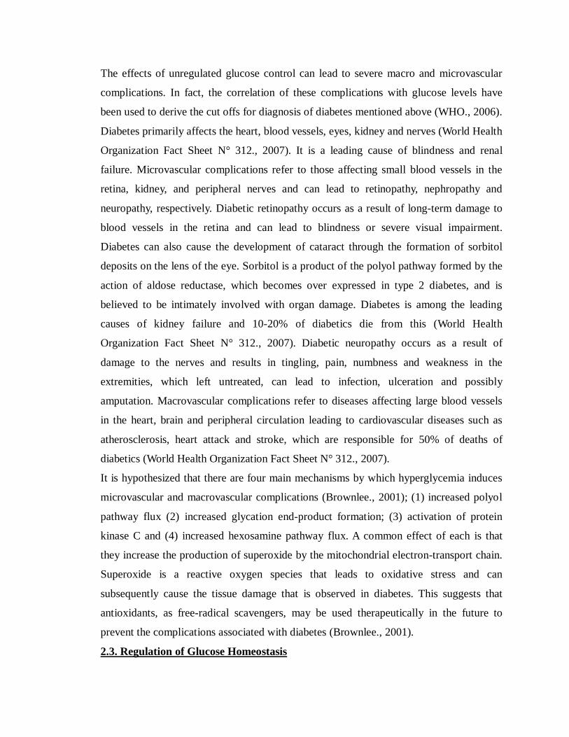
inducing weight loss. The structure of two thiazolidinediones, pioglitazone and
rosiglitazone, are shown in Figure 3.
Figure 3- Structure of thiazolidinediones, rosiglitazone and pioglitazone
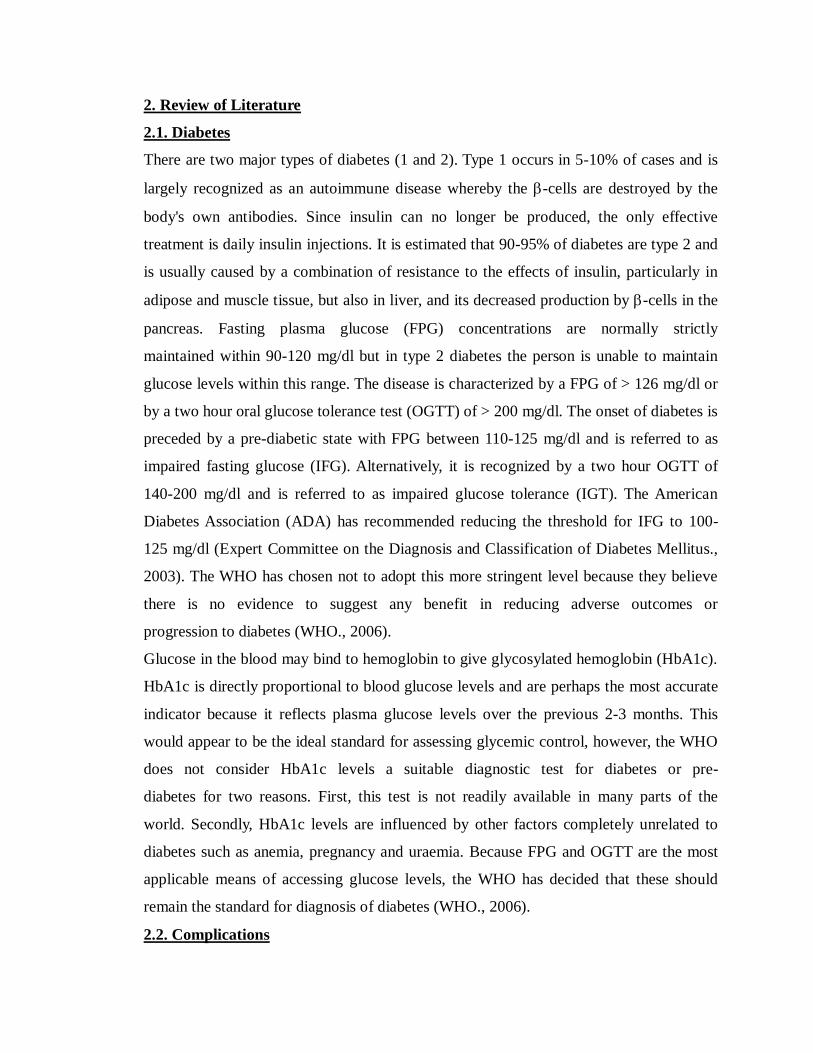
-Glucosidase inhibitors competitively inhibit the enzymes responsible for conversion of
disaccharides to monosaccharides. They prevent the digestion and absorption of
carbohydrates because only monosacchaides can be readily absorbed through the gut. -
Glucosidase inhibitors currently available include acarbose, voglibose and miglitol
(Figure 4). These drugs are capable of limiting postprandial glucose levels without the
risk of hypoglycemia and should be taken with food for optimal effect. They have only
modest antidiabetic activity by themselves and are usually used in combination therapy.
Side effects include GI disturbances such as flatulence, diarrhea and abdominal pain.
Figure 4- Structures of a-glucosidase inhibitors
Combination therapy is an option when one drug is no longer particularly
effective. After 3 years monotherapy with either a sulfonylurea or metformin,
approximately 50% of patients have HbA1c above 7% and after 9 years this number
increases to approximately 75% (Turner et al., 1999). In this case, a second agent of a
different class is usually added to the regimen to restore glycemic control through an
additive or synergistic effect. The most common combination is metformin with a
sulfonylurea. Other useful combinations include metformin and a TZD, metformin with a
meglitinide, or an -glucosidase inhibitor with either metformin or a sulfonylurea. In
the case when two agents are no longer effective a third agent of another class might also
be added (i.e. TZD to a combination of metformin and a sulfonylurea). Finally, when oral
hypoglycemic therapy has failed to achieve therapeutic goals in type 2 diabetes,
subcutaneous insulin injections are required to prevent hyperglycemia.
These hypoglycemic agents are useful in limiting hyperglycemia, but they do not address

the associated dyslipidemia and atherosclerotic cardiovascular disease, nor do they alter
the natural progression of the disease. Therapies which can increase or even preserve -
cell mass would represent a major advance. While a cure is not currently
available, research has led to a greater understanding of the etiology of the disease and
has resulted in the emergence of novel targets that are being exploited for possible use.
GLP-1 based therapy represents such a target and has already been successful.
2.6. Incretin and incretin effect
Eating provokes the secretion of multiple gastrointestinal hormones involved in the
regulation of gut motility, secretion of gastric acid and pancreatic enzymes, gall bladder
contraction, and nutrient absorption. Gut hormones also facilitate the disposal of absorbed
glucose through the stimulation of insulin secretion from the endocrine pancreas. The
observation that enteral nutrition provided a more potent insulinotropic stimulus compared
with isoglycaemic intravenous challenge led to the development of the incretin concept
(Elrick et al., 1964). The first incretin to be identified, glucose-dependent insulinotropic
polypeptide (GIP), was purified from porcine intestinal extracts and had weak effects on
gastric acid secretion. But more potent insulinotropic actions in human beings (Dupré et
al., 1973). GIP is a 42-amino acid hormone synthesised in duodenal and jejunal
enteroendocrine K cells in the proximal small bowel.
A second incretin hormone, glucagon-like peptide-1 (GLP-1) was identified after the
cloning of the cDNAs and genes encoding proglucagon (figure 5). GLP-1 exists in two
circulating equipotent molecular forms, GLP-1(7-37) and GLP-1(7-36) amide, although
GLP-1(7- 36) amide is more abundant in the circulation after eating. Most GLP-1 is made
in enteroendocrine L cells in the distal ileum and colon, but plasma levels of GLP-1, like
GIP, also increase within minutes of eating. Hence a combination of endocrine and neural
signals probably promote the rapid stimulation of GLP-1 secretion well before digested
food transits through the gut to directly engage the L cell in the small bowel and colon.
Plasma levels of GLP-1 are low in the fasted state, in the range of 5–10 pmol/L and
increase rapidly after eating, reaching 15–50 pmol/L. The circulating levels of intact GLP-
1 and GIP decrease rapidly because of enzymatic inactivation, mainly dipeptidyl
peptidase-4 (DPP-4), and renal clearance (Ørskov et al., 1993). Whether additional
proteases, such as human neutral endopeptidase, are also essential determinants of GLP-1

inactivation is being investigated. Both GIP and GLP-1 contain alanine at position 2, and
hence are excellent substrates for DPP-4. Indeed, DPP-4 is essential for incretin
inactivation, and mice with targeted inactivation of the DPP-4 gene have raised levels of
plasma GIP and GLP-1, increased insulin secretion, and reduced glucose excursion after
glycaemic challenge (Marguet et al., 2000). As a result of DPP-4 activity, intact,
biologically active GLP-1 represents only 10–20% of total plasma GLP-1 (Deacon et al.,
1995).
Figure 5- Physiology of GLP-1 secretion and action on GLP-1 receptors in different
organs and tissues
Both GIP and GLP-1 exert their actions by the engagement of structurally distinct G-
protein-coupled receptors (GPCRs). The GIP receptor is predominantly expressed on islet
cells, and to a lesser extent, in adipose tissue and in the central nervous system. By
contrast, the GLP-1 receptor (GLP-1R) is expressed in islet and ß cells and in peripheral
tissues, including the central and peripheral nervous systems, heart, kidney, lung,
gastrointestinal tract (figure 5). Activation of both incretin receptors on cells leads to
rapid increases in levels of cAMP and intracellular calcium, followed by insulin
exocytosis, in a glucose-dependent manner (Drucker et al., 1987). More sustained incretin
receptor signalling is associated with activation of protein kinase A, induction of gene
transcription, enhanced levels of insulin biosynthesis, and stimulation of -cell
proliferation (Drucker., 2006). Both GLP-1R and GIP receptor activation also promote
resistance to apoptosis and enhanced -cell survival, in both rodent (Li et al., 2003) and
human islets (Farilla et al., 2003). Consistent with the distribution of GLP-1R expression,
GLP-1 also inhibits glucagons secretion, gastric emptying, and food ingestion, and
promotes enhanced glucose disposal through neural mechanisms (Burcelin et al., 2001),
actions that also contribute to the control of glucoregulation. Notably, effects on glucagon
secretion like those on insulin secretory responses, are glucose- dependent, whereas
counter-regulatory release of glucagons in response to hypoglycaemia is fully preserved
even in the presence of pharmacological concentrations of GLP-1 (Nauck et al., 2002).
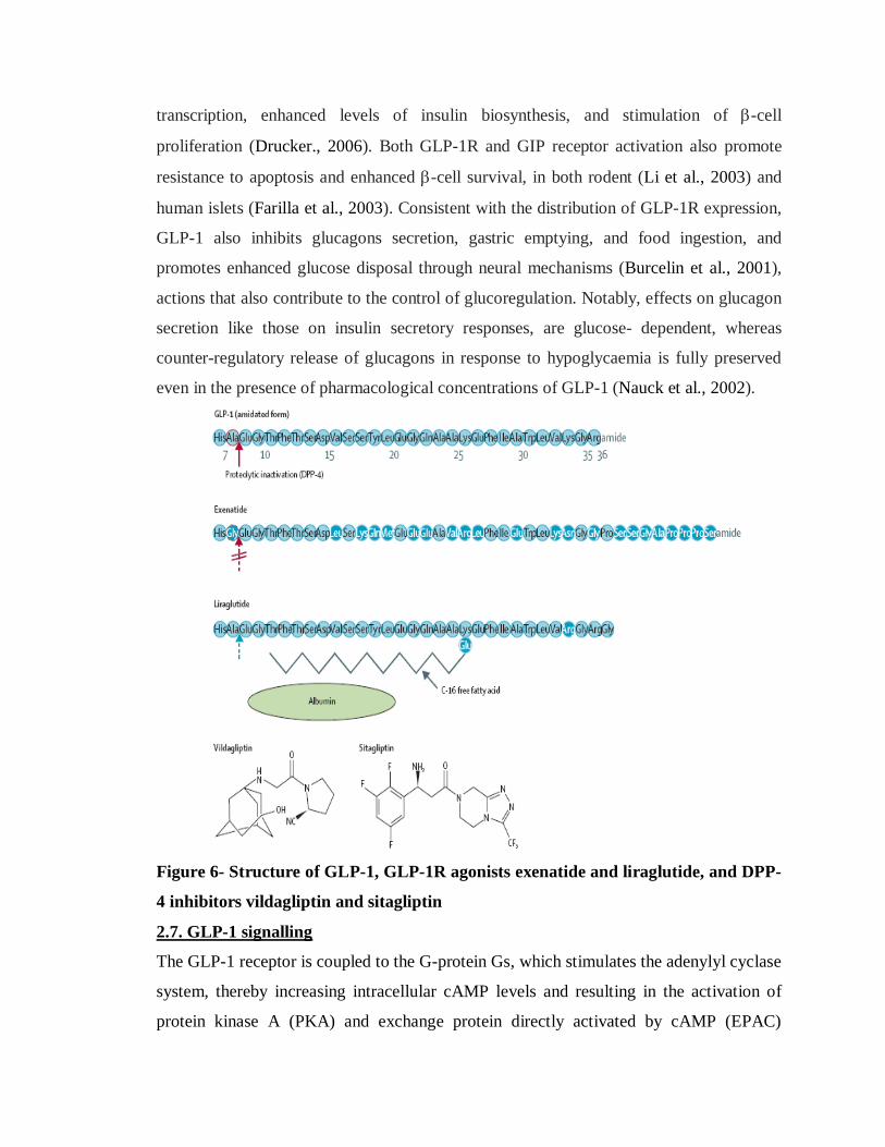
Figure 6- Structure of GLP-1, GLP-1R agonists exenatide and liraglutide, and DPP-
4 inhibitors vildagliptin and sitagliptin
2.7. GLP-1 signalling
The GLP-1 receptor is coupled to the G-protein Gs, which stimulates the adenylyl cyclase
system, thereby increasing intracellular cAMP levels and resulting in the activation of
protein kinase A (PKA) and exchange protein directly activated by cAMP (EPAC)

(Figure 7). In β-cells, GLP-1 induces KATP channel closure (through phosphorylation by
PKA), followed by membrane depolarisation and an increase in intracellular Ca2+ ,
resulting from the opening of Ca2+ channels (PKA pathway) and the mobilization of
intracellular Ca2+ stores (EPAC pathway), ultimately resulting in insulin exocytosis
(Ahren., 2006). Furthermore, GLP-1 not only regulates the expression of the insulin gene
but also other β -cell genes implicated in insulin secretion, such as those encoding
glucokinase and GLUT2. This effect is mediated by increased expression and activity of
pancreatic duodenal homeobox-1 (PDX1), which has been shown to be dependent on
cAMP/PKA (Wang et al., 2001).
Figure 7- Signal transduction pathways coupling GLP-1 receptor activation to insulin
secretion and β-cell mass protection. Binding of GLP-1 to its receptor in β -cells results
in an increase in intracellular cAMP, leading to the stimulation of insulin exocytosis by
two different pathways: PKA-dependent and PKA-independent (EPAC). GLP-1 also
increases the activity of PDX1, leading to the regulation of gene expression. The anti-
apoptotic effect of GLP-1 is mediated by the activation of CREB via the phosphorylation
by PKA and its interaction with a coactivator named TORC2. To migrate into the
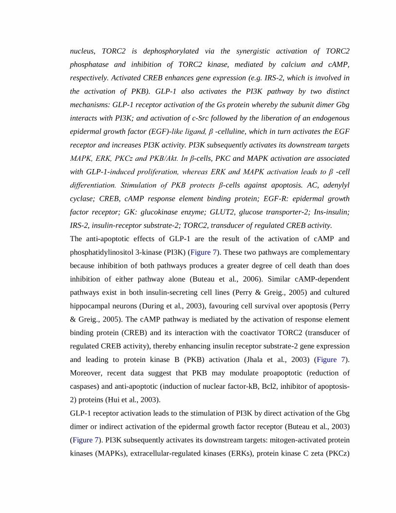
nucleus, TORC2 is dephosphorylated via the synergistic activation of TORC2
phosphatase and inhibition of TORC2 kinase, mediated by calcium and cAMP,
respectively. Activated CREB enhances gene expression (e.g. IRS-2, which is involved in
the activation of PKB). GLP-1 also activates the PI3K pathway by two distinct
mechanisms: GLP-1 receptor activation of the Gs protein whereby the subunit dimer Gbg
interacts with PI3K; and activation of c-Src followed by the liberation of an endogenous
epidermal growth factor (EGF)-like ligand, β -celluline, which in turn activates the EGF
receptor and increases PI3K activity. PI3K subsequently activates its downstream targets
MAPK, ERK, PKCz and PKB/Akt. In β-cells, PKC and MAPK activation are associated
with GLP-1-induced proliferation, whereas ERK and MAPK activation leads to β -cell
differentiation. Stimulation of PKB protects β-cells against apoptosis. AC, adenylyl
cyclase; CREB, cAMP response element binding protein; EGF-R: epidermal growth
factor receptor; GK: glucokinase enzyme; GLUT2, glucose transporter-2; Ins-insulin;
IRS-2, insulin-receptor substrate-2; TORC2, transducer of regulated CREB activity.
The anti-apoptotic effects of GLP-1 are the result of the activation of cAMP and
phosphatidylinositol 3-kinase (PI3K) (Figure 7). These two pathways are complementary
because inhibition of both pathways produces a greater degree of cell death than does
inhibition of either pathway alone (Buteau et al., 2006). Similar cAMP-dependent
pathways exist in both insulin-secreting cell lines (Perry & Greig., 2005) and cultured
hippocampal neurons (During et al., 2003), favouring cell survival over apoptosis (Perry
& Greig., 2005). The cAMP pathway is mediated by the activation of response element
binding protein (CREB) and its interaction with the coactivator TORC2 (transducer of
regulated CREB activity), thereby enhancing insulin receptor substrate-2 gene expression
and leading to protein kinase B (PKB) activation (Jhala et al., 2003) (Figure 7).
Moreover, recent data suggest that PKB may modulate proapoptotic (reduction of
caspases) and anti-apoptotic (induction of nuclear factor-kB, Bcl2, inhibitor of apoptosis-
2) proteins (Hui et al., 2003).
GLP-1 receptor activation leads to the stimulation of PI3K by direct activation of the Gbg
dimer or indirect activation of the epidermal growth factor receptor (Buteau et al., 2003)
(Figure 7). PI3K subsequently activates its downstream targets: mitogen-activated protein
kinases (MAPKs), extracellular-regulated kinases (ERKs), protein kinase C zeta (PKCz)
and PKB/Akt. PKC and MAPK pathways are associated with the GLP-1-induced
proliferative signal in β-cells and the trophic effect in cultured neuronal cells.
Furthermore, in the pancreas, activation of ERK and MAPK leads to β-cell
differentiation, whereas stimulation of PKB protects β-cells against apoptosis induced by
elevated glucose and/or fatty acids (Buteau et al., 2004). By inhibiting forkhead
transcription factor (FoxO1) through PI3K activation, GLP-1 increases Pdx1 and Foxa2
[proteins are the family of transcription factor that plays important role in regulating
genes involved in cell growth, proliferation, differentiation and longevity] expression
(Buteau et al., 2006), leading to proliferation (Brubaker et al., 2004) and antiapoptotic
effects in the pancreas (Buteau et al., 2006).
2.8. Dipeptidyl-peptidase IV (DPP-4) and DPP-4 inhibitors
Dipeptidyl-peptidase IV (DPP-4) is a ubiquitous enzyme that can be detected in the
endothelium of different organs and that is measurable as circulating enzymatic activity
in plasma. The incretins, namely glucagon-like peptide-1 (GLP-1) and glucose-dependent
insulinotropic peptide (GIP), are the only substrates of DPP-4 that have been well
validated in humans. DPP-4 has also been implicated in the regulation of several
additional peptides, such as pituitary adenylate cyclase-activating polypeptide (PACAP)
and gastrin-releasing peptide (GRP); however, in humans, these peptides have not been
definitively shown to be relevant in vivo substrates for this enzyme (Mest & Mentlein.,
2005) DPP-4 cleaves and inactivates GLP-1 within a few minutes (Mentlein., 1999). The
mechanism underlying the rapid degradation and elimination of the incretin hormones
GLP-1 and GIP has been described elsewhere. DPP-4 preferentially cleaves peptides with
the amino acid alanine or proline in position 2 of the N-terminus of the peptide chain.
Active GLP-1(7–36) amide is cleaved by DPP-4 to yield a dipeptide (His-Ala) and GLP-
1(9–36) amide (Mentlein., 1999, Gault et al., 2002, Knudsen & Pridal., 1996). DPP-4 is
also expressed on the cell membrane of activated T lymphocytes as CD26 (De Meester et
al., 1999); however, there is no compelling evidence that the catalytic activity of the
enzyme is important in immune function. Indeed, in clinical studies with DPP-4
inhibitors, no serious side effects or adverse events on immunological regulatory
mechanisms have been observed (Drucker & Nauck., 2006). Recently, 2-year safety data

on sitagliptin from pooled clinical trials have been published (Williams-Herman et al.,
2008).
Due to rapid cleavage and inactivation, a therapy with native GLP-1 administered
parenterally is not feasible for the continuous treatment of type 2 diabetes, and thus
incretin mimetics that are resistant to cleavage by DPP-4 are being pursued. DPP-4
inhibition is an alternate therapeutic option, in that inhibition of this enzyme results in an
increase in the circulating levels, of biologically active GLP-1. DPP-4 inhibitors are
orally active in contrast to incretin mimetics ((Drucker & Nauck., 2006). Furthermore,
they inhibit the degradation of GIP, and potentially other peptides involved in regulating
glucose homeostasis. They could therefore have additional beneficial effects in the
treatment of diabetes. DPP-4 is a member of a family of endopeptidases, and there is
evidence to suggest that selective inhibition of DPP-4 may be important to an optimal
safety profile for this new class of anti-hyperglycaemic agents (Lankas et al., 2005). The
DPP-4 inhibitors, namely sitagliptin and vildagliptin, are two compounds of the DPP-4
inhibitor class that have been approved in various countries. Alogliptin and saxagliptin
are currently under review for approval, and several other DPP-4 inhibitors are in
development (Gallwitz., 2008, Deacon., 2008, Huttner et al., 2008). The structures of the
DPP-4 inhibitors that have been approved and currently under review are shown in Fig. 8.
2.9. Pharmacology of DPP-4 inhibitors
Sitagliptin, vildagliptin, saxagliptin and alogliptin are competitive inhibitors with high
affinity for DPP-4. In humans, the pharmacokinetic and pharmacodynamic properties,
efficacy, safety and tolerability have been assessed in numerous clinical studies; the most
abundant database is available for sitagliptin and vildagliptin (Ahren ., 2008). After a
standard meal, active endogenous GLP-1 concentrations are increased two- to threefold
by these compounds. Both sitagliptin and vildagliptin have been shown to have clinically
meaningful efficacy for the treatment of type 2 diabetes in both monotherapy and in
combination with established oral anti-glycaemic agents, including metformin, in studies
of at least 6 months duration and up to 2 years for sitagliptin (Ahren., 2008).
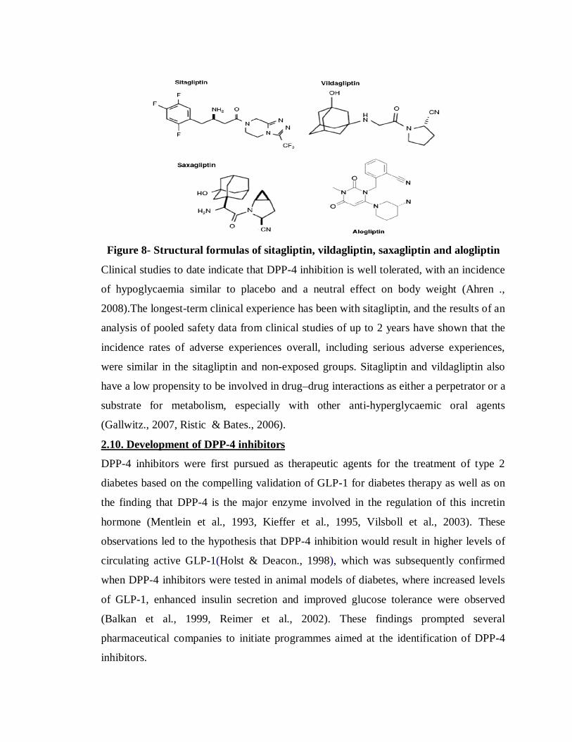
Figure 8- Structural formulas of sitagliptin, vildagliptin, saxagliptin and alogliptin
Clinical studies to date indicate that DPP-4 inhibition is well tolerated, with an incidence
of hypoglycaemia similar to placebo and a neutral effect on body weight (Ahren .,
2008).The longest-term clinical experience has been with sitagliptin, and the results of an
analysis of pooled safety data from clinical studies of up to 2 years have shown that the
incidence rates of adverse experiences overall, including serious adverse experiences,
were similar in the sitagliptin and non-exposed groups. Sitagliptin and vildagliptin also
have a low propensity to be involved in drug–drug interactions as either a perpetrator or a
substrate for metabolism, especially with other anti-hyperglycaemic oral agents
(Gallwitz., 2007, Ristic & Bates., 2006).
2.10. Development of DPP-4 inhibitors
DPP-4 inhibitors were first pursued as therapeutic agents for the treatment of type 2
diabetes based on the compelling validation of GLP-1 for diabetes therapy as well as on
the finding that DPP-4 is the major enzyme involved in the regulation of this incretin
hormone (Mentlein et al., 1993, Kieffer et al., 1995, Vilsboll et al., 2003). These
observations led to the hypothesis that DPP-4 inhibition would result in higher levels of
circulating active GLP-1(Holst & Deacon., 1998), which was subsequently confirmed
when DPP-4 inhibitors were tested in animal models of diabetes, where increased levels
of GLP-1, enhanced insulin secretion and improved glucose tolerance were observed
(Balkan et al., 1999, Reimer et al., 2002). These findings prompted several
pharmaceutical companies to initiate programmes aimed at the identification of DPP-4
inhibitors.
Interest in DPP-4 inhibition, as a new approach to the treatment of type 2 diabetes, was
driven not only by the proven glucose-lowering efficacy of GLP-1, but also by the
prospects of improvements in safety and tolerability over well-established oral agents.
Specifically, because GLP-1 stimulates insulin release and inhibits glucagon production
in a strictly glucose-dependent manner, this mechanism was anticipated to have a low
risk of hypoglycaemia. Secondly, no weight gain was anticipated with DPP-4 inhibitors.
Interest in this class was also driven by an emerging body of evidence that GLP-1
analogues have beneficial effects on -cell mass in rodents, suggesting the potential of
DPP-4 inhibitors for improvements in glycaemic durability.
Extensive structure–activity relationship studies in many different laboratories have
resulted in the identification of a number of potent, competitive DPP-4 inhibitors and
clinical candidates. Clinical proof of concept for DPP-4 inhibition in diabetic patients
was first reported in 2002 with an early inhibitor from Novartis (DPP-728), where
significant reductions in fasting plasma glucose and HbA1c were observed in a 4-week
study (Ahren et al., 2002).
Sitagliptin was approved in 2006 by the Food and Drug Administration (FDA) for use in
monotherapy, and in combination with metformin or a thiazolidinedione. Subsequently,
sitagliptin was approved for use with sulphonylurea and sulphonylurea plus metformin.
The European Commission has approved the use of: (1) sitagliptin in combination with
metformin, sulphonylureas, thiazolidinediones and with metformin plus sulphonylureas
and (2) vildagliptin in combination with metformin, sulphonylureas or thiazolidinediones.
2.11. Mechanisms of DPP-4 inhibitor action
DPP-4 has a well-established physiological role in the regulation of the incretin
hormones, GLP-1 and GIP. In animals that are genetically deficient in DPP-4, or with
pharmacological treatment with a DPP-4 inhibitor, increased active GLP-1, GIP and
improved glucose tolerance were observed (Balkan et al., 1999, Reimer et al., 2002,
Marguet et al., 2000, Conarello et al., 2003 ) Increased insulin and decreased glucagon
levels were also observed both in DPP-4-deficient mice and, upon pharmacological
treatment with inhibitors, in rodents and humans, consistent with the role of this enzyme
in incretin regulation and metabolic control. DPP-4 inhibitors do not improve glucose
tolerance in mice deficient in both GLP-1 and GIP receptors, indicating that these

incretins are exclusively responsible for the improved glucose tolerance that is observed
in these animals (Hansotia et al., 2004). Taken together, these data unequivocally
establish that these incretins are endogenous substrates for DPP-4. This enzyme has been
implicated in the regulation of peptides in addition to GLP-1 and GIP, including growth-
hormone-releasing hormone (GHRH), glucagon-like peptide 2 (GLP-2), pituitary
adenylate cyclase-activating polypeptide (PACAP) and gastrin-releasing peptide (GRP)
(Mest & Mentlein., 2005, De Meester et al., 2000). Several neuropeptides and
chemokines are also in vitro substrates for this enzyme. Although many of these peptides
are cleaved efficiently in vitro, it is difficult to determine if these peptides are regulated in
vivo by DPP-4, largely because suitable assays for measurement of the endogenous levels
of the putative substrates and products are not available. Further work will be required to
obtain a comprehensive understanding of the biology of this enzyme. DPP-4-deficient
mice are healthy and fertile, and thus if other proteins and/or peptides are regulated by
this enzyme, there are no obvious important consequences related to growth and
development, reproductive capacity or health. Moreover, the results of clinical studies
indicate that selective DPP-4 inhibitors are well tolerated and do not suggest any
functions for this enzyme beyond its role in metabolic control (Ahren., 2008).
2.12. DPP-4 inhibitor selectivity
DPP-4 is a member of a family of proteases that includes dipeptidyl-peptidase 8 (DPP8),
dipeptidylpeptidase 9 (DPP9) and fibroblast activation protein (FAP) (Sedo & Malik.,
2001). DPP8 and DPP9 are ubiquitously expressed and highly conserved across species
(Abbott et al., 2000, Ajami et al., 2004). Unlike DPP-4, which is expressed on the cell
surface, DPP8 and DPP9 are cytosolic enzymes. No endogenous substrates have been
identified, and thus the specific functions of these enzymes are unknown. There is
evidence to suggest that selective inhibition may be important to an optimal safety profile
with long-term administration of the DPP-4 inhibitor. Early DPP-4 inhibitors with off-
target activity against DPP-8 and DPP-9 were shown to cause profound toxicity in
preclinical species (Lankas et al., 2005). The results of subsequent toxicity studies with
highly selective DPP-4 inhibitors, and an inhibitor of DPP8 and DPP9, demonstrated that
the DPP-8/9 inhibitor produced toxicity in preclinical species, but that no toxicities were
observed with the selective DPP-4 inhibitor (Lankas et al., 2005)). These results provided

evidence that inhibition of DPP8/9 or a highly related enzyme, but not selective DPP-4
inhibition, is associated with multi-organ toxicities in preclinical species. The relevance
of the finding that a DPP8/9 inhibitor produces toxicity in preclinical species to the
development of DPP-4 inhibitors for clinical use is unknown.
2.13. Effects of DPP-4 inhibitors on glucagon secretion
After a meal, active endogenous GLP-1 and GIP concentrations are increased two- to
threefold by DPP-4 inhibitors. This action leads not only to an increase in insulin
secretion as long as hyperglycaemia is present, but also to a suppression of glucagon
secretion. Under hypoglycaemic conditions, counter-regulation of glucagon secretion by
the alpha cells is not impaired; on the contrary, treatment with a DPP-4 inhibitor may
even improve alpha cell sensitivity to glucose (Ahren & Foley., 2003).
2.14. Beta-cell function and beta-cell mass in animal studies with DPP-4 inhibitors
Based on beneficial effects on -cell mass that have been observed with GLP-1
analogues in rodents, there is interest in knowing if DPP-4 inhibitors are able to influence
the disease progression of type 2 diabetes favourably by slowing or even inhibiting loss
of beta-cell mass and function. To further understand this potential, animal studies were
designed to study the effect of sitagliptin and vildagliptin on islet function and beta cell
mass (Pospisilik et al., 2003, Mu et al., 2006). Diabetic mice treated with DPP-4
inhibitors showed a significant, dose-dependent reduction of glycaemic parameters. In
these studies, postprandial and fasting hyperglycaemia, HbA1c and lipid parameters
(plasma triglycerides and free fatty acids) were improved. Furthermore, DPP-4 inhibitors
increased the number of insulin-positive beta cells in islets and the beta-to-alpha cell ratio
in different diabetic animals was normalised. Likewise, the islet insulin content was
found to be increased and glucose-stimulated insulin secretion in isolated islets was found
to be improved in comparison to suklphonylurea-treated mice. In addition, DPP-4-
deficient mice are resistant to streptozotocin-induced beta-cell destruction (Conarello et
al., 2003). These results have led to the hypothesis that DPP-4 inhibitors may have the
potential to delay or prevent disease progression in type 2 diabetes and to improve beta-
cell mass and function (Gallwitz., 2007, Ristic & Bates., 2006).
2.15. Drug Profile
2.15.1. Vildagliptin

After 4 weeks of treatment with 100 mg/day, vildagliptin inhibits selectively DPP-4 more
than 90%. It is excreted by both digestive and urinary route. Its half-life is shorter than
sitagliptin (1.7 vs 12 h). Its action is yet extended in spite of this fast elimination. In
monotherapy, it decreases HbA1c in a range of 0.4 to 1.2% vs placebo (De Jager et al.,
2007, Ristic et al., 2005). In combination therapy, the decrease of HbA1c was of 1.1%
when vildagliptin was added to metformin (Bosi et al., 2007), and of 1.0% when added to
pioglitazone. In the meta-analysis from Amori et al. (Amori et al., 2007), HbA1c levels
in the different vildagliptin studies varied from +0.40% to -1.20%. In the nine studies
comparing vildagliptin to placebo, which gathered 1786 patients, the mean change of
HbA1c was -0.73%. Vildagliptin has also been added to insulin (Fonseca et al., 2006): in
the combination group compared to the group given insulin alone, mean HbA1c level was
reduced by 0.5% vs 0.2%, and hypoglycaemias were less frequent (113 events in 33
patients versus 185 events in 45 patients).
Table 1- Comparison of the Currently Available DPP-4 Inhibitors
2.15.2. PKF-275-055
The vildagliptin analogue PKF-275-055 was synthesized as a selective, long-acting
inhibitor of dipeptidyl peptidase 4 for the treatment of diabetes. The ability of the
compound to prevent and improve established peripheral diabetic neuropathy was
evaluated in rats with streptozotocin induced diabetes. According to oral glucose
tolerance test data, it caused body and muscle weight gain and improved glucose
tolerance when used as a preventative treatment or in established peripheral diabetic
neuropathy. While nerve conduction velocity was reduced by 36% at 10 weeks in
streptozotocin- treated rats compared with non-diabetic animals, it was only reduced by
17% in animals treated with PKF-275-055. The agent also reduced the decrease in
Na+/K+- ATPase activity in diabetic nerves by 50%, partially protected diabetic rats
against mechanical hypoalgesia and progressively prevented the increase in hind paw
thermal response latencies seen in diabetic animals (Bianchi et al., 2009).