review clinical review: update on neurally adjusted
TRANSCRIPT
Introduction
Pressure-support ventilation (PSV) is a widely used mode
of assisted mechanical ventilation (MV), notably during
the weaning phase [1,2]. Although PSV has been proven
valuable in several acute clinical conditions [3,4], pre-
defi ned ventilator settings – for example, airway pressure
(Paw) – that remain unchanged from breath to breath are
unlikely to provide optimal assistance all of the time.
To improve the match between the patient’s needs and
the assistance delivered by the ventilator, manufacturers
have developed several new modes of MV [5,6]. Among
these new modes we identifi ed proportional-assist
ventilation (PAV) and neurally adjusted ventilatory assist
(NAVA).
PAV is a mode of support in which the ventilator
pressure is proportional to instantaneous fl ow and volume,
and hence to pressure generated by the respiratory
muscles [7]. Previous studies have demonstrated that
PAV improves the synchrony between patient and
ventilator, during several clinical conditions [8-12]. Based
on the principles of the equation of motion, software
(PAV+; Covidien, Boulder, Colorado, USA) has been
developed that auto matically adjusts the fl ow assist and
the volume assist so that they always represent constant
unloading fractions of the measured values of resistance
and elastance loadings of the respiratory system [13-15].
Recent studies demonstrated that PAV+ is a safe and
effi cient ventilator mode in critically ill intubated patients
[16,17]. During PAV the ventilator provides support only
during the remaining duration of inspiratory eff ort,
Abstract
Conventional mechanical ventilators rely on pneumatic pressure and fl ow sensors and controllers to detect breaths.
New modes of mechanical ventilation have been developed to better match the assistance delivered by the
ventilator to the patient’s needs. Among these modes, neurally adjusted ventilatory assist (NAVA) delivers a pressure
that is directly proportional to the integral of the electrical activity of the diaphragm recorded continuously through
an esophageal probe. In clinical settings, NAVA has been chiefl y compared with pressure-support ventilation, one
of the most popular modes used during the weaning phase, which delivers a constant pressure from breath to
breath. Comparisons with proportional-assist ventilation, which has numerous similarities, are lacking. Because of
the constant level of assistance, pressure-support ventilation reduces the natural variability of the breathing pattern
and can be associated with asynchrony and/or overinfl ation. The ability of NAVA to circumvent these limitations has
been addressed in clinical studies and is discussed in this report. Although the underlying concept is fascinating,
several important questions regarding the clinical applications of NAVA remain unanswered. Among these questions,
determining the optimal NAVA settings according to the patient’s ventilatory needs and/or acceptable level of work
of breathing is a key issue. In this report, based on an investigator-initiated round table, we review the most recent
literature on this topic and discuss the theoretical advantages and disadvantages of NAVA compared with other
modes, as well as the risks and limitations of NAVA.
© 2010 BioMed Central Ltd
Clinical review: Update on neurally adjusted ventilatory assist – report of a round-table conferenceNicolas Terzi*1,2,3, Lise Piquilloud4, Hadrien Rozé5, Alain Mercat6,7, Frédéric Lofaso8,9, Stéphane Delisle10, Philippe Jolliet4,
Thierry Sottiaux11, Didier Tassaux4, Jean Roesler12, Alexandre Demoule13, Samir Jaber14, Jordi Mancebo15, Laurent
Brochard9,16 and Jean-Christophe Marie Richard16,17,18
Report from the Geneva Round Table
R E V I E W
*Correspondence: [email protected] Caen, Service de Réanimation Médicale, Caen F-14000, France
Full list of author information is available at the end of the article
Terzi et al. Critical Care 2012, 16:225 http://ccforum.com/content/16/3/225
© 2012 BioMed Central Ltd

which can cause limitation when dynamic hyperinfl ation
is present and when the inspiratory trigger is delayed due
to intrinsic end-expiratory pressure.
Th e other support mode is NAVA, which will be
discussed in this article. Th ere are several similarities
between PAV and NAVA, but this fi rst round-table
meeting focused on NAVA. A vast literature also exists
concerning PAV, but this topic would require a whole
chapter and will not be discussed in this current paper;
hopefully PAV will be the topic of a diff erent round table.
Th e present article is based on an investigator-initiated
round-table meeting. Th e article aims to review the
available knowledge on the physiological rationale and
feasibility of the recently introduced NAVA MV modality.
Th roughout the article, we place emphasis on the most
recent fi ndings concerning adjustment of the NAVA
settings; on the one hand considering specifi c issues
associated with assisted modes of MV, and on the other
considering the expecta tions placed upon NAVA.
NAVA is an assist mode of MV that delivers a pressure
proportional to the integral of the electrical activity of the
diaphragm (EAdi) [18], and therefore proportional to the
neural output of the patient’s central respiratory command.
Th e level of pressure delivered is thus determined by the
patient’s respiratory-center neural output. With NAVA,
the ventilator is triggered and cycled-off based on the
EAdi value, which directly refl ects the activity of the
neural respiratory command. Th e inspiratory airway
pressure applied by the ventilator is determined by the
following equation:
Paw = NAVA level × EAdi,
where Paw is the instantaneous airway pressure (cmH2O),
EAdi is the instantaneous integral of the diaphragmatic
electrical activity signal (μV), and the NAVA level
(cmH2O/μV or per arbitrary unit) is a proportionality
constant set by the clinician.
In February 2011, several European and Canadian
investigators with clinical results about NAVA available
in publication or in abstract format organized a round-
table discussion at the Geneva University Hospital to
describe and discuss recent advances regarding NAVA. A
representative of the company that commercializes the
NAVA machine (Maquet Critical Care SA, Sölna,
Sweden) was invited to attend the meeting in order to
answer only technical questions. Maquet Critical Care
SA agreed to sign a disclosure form before the meeting
specifying that neither the minutes of the meeting nor
the content of the report could be modifi ed and/or used
for commercial purposes. Th e main purpose of this
meeting was for all of the investigators and participants
to expose their standpoint and questions about NAVA,
and to share the main results of their studies. Maquet
Critical Care SA agreed to provide fi nancial support for
organiz ing the meeting, as detailed at the end of the
manuscript, but was not responsible for choosing partici-
pants and did not take any part in the writing of this
report. We here describe the content of the round-table
discussion, focusing on a selection of the most recent
studies [19-33] (Table 1).
Main problems with conventional ventilation
modalities in the ICU
Assisted modes generally aim at synchronizing the
ventilator insuffl ation to the patient’s eff ort, both to
optimize comfort and to minimize the work of breathing.
Th e price to pay for this strategy is a risk of patient–
ventilator asynchrony, which can be defi ned as a mis-
match between the patient’s neural output and the venti-
lator’s inspiratory and expiratory times [34-37]. Th ille
and colleagues reported that one-quarter of patients had
high rates of asynchrony during assisted ventilation [34].
Frequent asynchrony is associated with a longer duration
of MV [34,38].
Compelling evidence accumulated over the last decade
also supports the use of tidal volume (VT) values that are
lower than those traditionally used. Lower VT values than
traditionally used have several main advantages: they
diminish the risk of ventilator-induced lung injury
[39-41]; they preserve spontaneous breathing by avoiding
respiratory alkalosis, thus preventing diaphragmatic disuse
atrophy associated with MV [42-48]; they diminish several
types of patient–ventilator asynchrony [49]; and they may
improve the effi ciency of gas exchange [50]. Assisted
modes of ventilation that maintain at least part of the
patient’s spontaneous breathing activity contribute to
preventing these pulmonary and muscular complications.
Th e new challenge in developing ventilation strategies
thus consists of minimizing the risk of lung injury,
avoiding disuse atrophy of the diaphragm, and improving
the match between the patient’s needs and the assistance
delivered by the ventilator [6]. New ventilation modes
have been designed to meet this challenge [5], and NAVA
is a pressure-assisted mode in which the pressure
delivered by the ventilator is proportional to the electrical
activity of the diaphragm recorded continuously through
an esophageal probe [18]. NAVA theoretically delivers
pressure proportional to the neural output of the patient’s
central respiratory command. During NAVA, however,
reliable positioning of the catheter is mandatory in order
to obtain a representative EAdi signal from the dia-
phragm. Barwing and colleagues have evaluated whether
a formula based on the measurement from nose to ear
lobe to xiphoid process of the sternum (the NEX
distance) modifi ed for the EAdi catheter (NEXmod) is
adequate for predicting the accurate position of the
esophageal probe [51]. Th ey observed in 18 of 25 patients
Terzi et al. Critical Care 2012, 16:225 http://ccforum.com/content/16/3/225
Page 2 of 13

Ta
ble
1.
Ma
in a
va
ila
ble
cli
nic
al
stu
die
s
D
iag
no
sis
(n
um
be
r
Stu
dy
o
f p
ati
en
ts)
De
sig
n
Du
rati
on
M
ajo
r fi
nd
ing
s
Co
lom
bo
an
d
colle
agu
es,
20
08
[2
1]
Acu
te r
esp
irat
ory
failu
re
(un
sele
cte
d)
(n =
14
)
Cro
sso
ver
– P
SV s
et
to o
bta
in V
T 6 t
o 8
ml/
kg p
red
icte
d b
od
y w
eig
ht;
NA
VA v
s. P
SV
incr
eas
ed
or
de
cre
ase
d b
y 5
0%
. Eff
ect
s o
f m
od
ifi ca
tio
n o
f as
sist
leve
l
20
min
ute
s ×
3N
AVA
ave
rte
d t
he
ris
k o
f o
vera
ssis
tan
ce a
nd
imp
rove
d s
ynch
ron
y
Wu
an
d
colle
agu
es,
20
09
[1
9]
AR
DS
(n =
18
)P
SV v
s. N
AVA
– r
and
om
ize
d s
tud
y. In
cre
me
nta
l PSV
an
d N
AVA
ru
n r
and
om
ly in
fou
r
ste
ps.
Th
e P
SV le
vel w
as g
rad
ual
ly in
cre
ase
d b
y 5
cm
H2O
eve
ry 5
min
ute
s fr
om
5 t
o 2
0
cmH
2O
. In
cre
me
nta
l NA
VA w
as in
div
idu
ally
se
t in
ste
ps
of
0.2
to
1.0
cm
H2O
/μV
eve
ry
5 m
inu
tes
to d
ete
rmin
e t
he
NA
VA le
vel p
rovi
din
g a
n a
irw
ay p
ress
ure
in e
ach
ste
p
eq
uiv
ale
nt
to t
hat
wit
h P
SV. E
valu
atio
n o
f p
atie
nt–
ven
tila
tor
syn
chro
ny
(tri
gg
er
de
lay,
ine
ff e
ctiv
e e
ff o
rt);
eff
ect
of
assi
st le
vel
5 m
inu
tes
×4
Imp
rove
d s
ynch
ron
y w
ith
NA
VA
Bra
nd
er
and
colle
agu
es,
20
09
[2
0]
Acu
te r
esp
irat
ory
failu
re (u
nse
lect
ed
)
(n =
15
)
NA
VA le
vel i
ncr
eas
ed
pro
gre
ssiv
ely
. Me
tho
d fo
r ti
trat
ing
th
e N
AVA
leve
l3
0 m
inu
tes
+
3 h
ou
rs
Pro
gre
ssiv
e im
ple
me
nta
tio
n o
f N
AVA
may
be
a m
eth
od
for
de
term
inin
g t
he
ad
eq
uat
e le
vel –
do
wn
reg
ula
tio
n o
f EA
di
con
fi rm
ed
Sch
mid
t an
d
colle
agu
es,
20
10
[2
3]
Mai
nly
acu
te lu
ng
inju
ry (
n =
12
)
Lon
git
ud
inal
ob
serv
atio
nal
stu
dy
– P
SV s
et
to o
bta
in V
T 6 t
o 8
ml/
kg p
red
icte
d b
od
y
we
igh
t; N
AVA
vs.
PSV
wit
h in
cre
asin
g a
ssis
t. B
reat
h-b
y-b
reat
h v
aria
bili
ty o
f fl
ow
an
d
EAd
i-re
late
d v
aria
ble
s q
uan
tifi
ed
by
the
co
effi
cie
nt
of
vari
atio
n a
nd
au
toco
rre
lati
on
anal
ysis
10
min
ute
s ×
4 =
40
min
ute
s
NA
VA in
cre
ase
s b
reat
hin
g p
atte
rn v
aria
bili
ty
Co
ise
l an
d
colle
agu
es,
20
10
[2
7]
Po
sto
pe
rati
ve
pat
ien
ts (
n =
15
)
Cro
sso
ver
ran
do
miz
ed
– P
SV s
et
to o
bta
in V
T 6 t
o 8
ml/
kg p
red
icte
d b
od
y w
eig
ht;
NA
VA v
s. P
SV. E
ff e
cts
on
bre
ath
ing
pat
tern
, gas
exc
han
ge
, an
d v
aria
bili
ty o
f re
spir
ato
ry
cycl
es
(eva
luat
ed
by
coe
ffi c
ien
t o
f va
riat
ion
)
24
ho
urs
Var
iab
ility
of,
tid
al v
olu
me
an
d m
inu
te v
en
tila
tio
n w
ere
sig
nifi
can
tly
hig
he
r w
ith
NA
VA t
han
wit
h P
SV. V
aria
bili
ty o
f
ele
ctri
cal d
iap
hra
gm
atic
act
ivit
y w
as s
ign
ifi ca
ntl
y lo
we
r w
ith
NA
VA t
han
wit
h P
SV. O
xyg
en
atio
n in
cre
ase
d d
uri
ng
NA
VA
Terz
i an
d
colle
agu
es,
20
10
[2
5]
AR
DS
(n =
11
)C
ross
ove
r ra
nd
om
ize
d –
PSV
se
t to
ob
tain
VT 6
to
8 m
l/kg
pre
dic
ted
bo
dy
we
igh
t;
NA
VA v
s. P
SV. E
ff e
ct o
f n
eu
ral t
rig
ge
r vs
. fl o
w t
rig
ge
r. A
ssis
t le
vel w
as r
and
om
ize
d t
o
be
20
%, 4
0%
, or
60
% o
ver
the
bas
al le
vel.
Ass
ess
me
nt
of
the
ph
ysio
log
ical
re
spo
nse
to v
aryi
ng
PSV
an
d N
AVA
leve
ls in
se
lect
ed
AR
DS
pat
ien
ts a
nd
th
e e
ff e
ct o
f n
eu
ral
trig
ge
rin
g
5 m
inu
tes
×4
× 3
=
60
min
ute
s
Co
mp
are
d w
ith
PSV
, NA
VA li
mit
ed
th
e r
isk
of
ove
rass
ista
nce
,
pre
ven
ted
pat
ien
t–ve
nti
lato
r as
ynch
ron
y, a
nd
imp
rove
d o
vera
ll
pat
ien
t–ve
nti
lato
r in
tera
ctio
ns.
Co
mp
are
d w
ith
th
e p
ne
um
atic
trig
ge
r, th
e n
eu
ral t
rig
ge
r (f
rom
NA
VA)
con
sid
era
bly
de
cre
ase
d
pat
ien
t–ve
nti
lato
r as
ynch
ron
y
Spah
ija a
nd
colle
agu
es,
20
10
[2
2]
CO
PD
(1
4)
Pro
spe
ctiv
e, c
om
par
ativ
e c
ross
ove
r –
PSV
se
t to
ob
tain
VT 6
to
8 m
l/kg
pre
dic
ted
bo
dy
we
igh
t; N
AVA
vs.
PSV
. Pat
ien
ts w
ere
ve
nti
late
d fo
r 1
0-m
inu
te p
eri
od
s, u
sin
g t
wo
PSV
leve
ls (
low
est
to
lera
ble
an
d 7
cm
H2O
hig
he
r) a
nd
tw
o N
AVA
leve
ls (
sam
e p
eak
pre
ssu
res
and
ext
ern
al P
EEP
as
wit
h P
SV),
de
live
red
in r
and
om
ord
er
10
min
ute
s ×
2N
AVA
imp
rove
d p
atie
nt–
ven
tila
tor
syn
chro
ny
by
red
uci
ng
th
e
trig
ge
rin
g a
nd
cyc
ling
de
lays
, esp
eci
ally
at
hig
he
r le
vels
of
assi
st,
wh
ile p
rese
rvin
g b
reat
hin
g a
nd
mai
nta
inin
g b
loo
d g
as e
xch
ang
e
Pass
ath
an
d
colle
agu
es,
20
10
[2
8]
Un
sele
cte
d
pat
ien
ts (
n =
20
)
Lon
git
ud
inal
ob
serv
atio
nal
stu
dy.
Eva
luat
ion
of
eff
ect
s o
f P
EP o
n b
reat
hin
g p
atte
rn
and
ne
uro
ven
tila
tory
effi
cie
ncy
du
rin
g N
AVA
. Ad
eq
uat
e N
AVA
leve
l was
de
term
ine
d
as t
he
NA
VA le
vel e
arly
aft
er
the
tra
nsi
tio
n f
rom
an
init
ial s
tee
p in
cre
ase
in P
aw a
nd
VT t
o a
less
ste
ep
incr
eas
e o
r e
ven
pla
teau
of
Paw
an
d V
T, as
de
scri
be
d b
y B
ran
de
r
and
co
lleag
ue
s. P
EEP
was
se
t at
20
cm
H2O
th
en
de
cre
ase
d t
o 1
cm
H2O
. VT/E
Ad
i was
eva
luat
ed
as
an in
dic
ato
r o
f n
eu
rove
nti
lato
ry e
ffi c
ien
cy
20
min
ute
s ×
3D
uri
ng
ad
eq
uat
e-a
ssis
t N
AVA
, in
cre
asin
g P
EEP
re
du
ces
resp
irat
ory
dri
ve. P
atie
nts
ad
apt
the
ir n
eu
rove
nti
lato
ry e
ffi c
ien
cy s
uch
th
at
the
ind
ivid
ual
ve
nti
lato
ry p
atte
rn is
pre
serv
ed
ove
r a
wid
e r
ang
e
of
PEE
P le
vels
. Mo
nit
ori
ng
VT/E
Ad
i du
rin
g P
EEP
ch
ang
es
allo
ws
ide
nti
fi ca
tio
n o
f a
PEE
P le
vel a
t w
hic
h t
idal
bre
ath
ing
occ
urs
at
min
imal
EA
di c
ost
Co
nti
nu
ed o
verl
eaf
Terzi et al. Critical Care 2012, 16:225 http://ccforum.com/content/16/3/225
Page 3 of 13

(72%) that at NEXmod the EAdi signal was suitable for
running NAVA. Th e NAVA mode was possible at the
optimal position in four patients – the optimal position
being defi ned by checking three criteria: stable EAdi
signals, electrical activity highlighted in central leads of
the catheter positioning tool, and an absence of the p-
wave in the distal lead. Th e authors thus concluded that
positioning the EAdi catheter using NEXmod gives a
good approximation in most of the patients.
Moreover, the body position, positive end-expiratory
pressure (PEEP) and intra-abdominal pressure are factors
known to infl uence the position of the diaphragm.
Barwing and colleagues therefore enrolled 20 patients in
order to evaluate the eff ects of these factors on catheter
position [52]. Th ey evaluated six diff erent situations
regarding the PEEP, body position and intra-abdominal
pressure. Th eir results demonstrated that these factors
may modify the EAdi catheter optimal position, although
not compromising a stable signal due to the wide
electrode array. One can therefore conclude that the
optimal catheter position should be adjusted after major
changes in ventilator settings, clinical condition or
patient positioning.
Management of patient–ventilator synchrony
Th e time lag between the neural inspiratory input and
the occurrence of a ventilator breath aff ects all steps of
the respiratory cycle (initiation, insuffl ation, and cycling-
off for expiration) [53]. Among the diff erent forms of
asynchrony, ineff ective triggering (also known as wasted
eff ort) is the most common during invasive MV. During
noninvasive ventilation (NIV), leaks at the patient–
ventilator interface impair the function of the pneumatic
trigger and cycling system [54], thus promoting specifi c
asynchronies (autotriggering and prolonged insuffl ation)
[55].
Ineff ective eff orts are explained both by patients’
characteristics and by ventilator settings. Th e presence of
intrinsic PEEP increases the patient eff ort required to
trigger the ventilator, thereby increasing the likelihood
that the patient’s inspiratory eff ort will fail to trigger a
ventilator breath [36,53,56]. A weak inspiratory eff ort,
which may occur during situations of low respiratory
drive such as excessive ventilation, is also a risk factor
and is common in patients receiving high assist levels
[22] or sedation [38]. An excessive level of pressure
support is also associated with prolonged insuffl ation,
thus promoting hyperinfl ation and intrinsic PEEP. Reduc-
tion of ineff ective eff orts is often possible through a
careful optimization of ventilator settings, at least in
short-term studies. Reducing VT during PSV can improve
most factors contributing to ineff ective eff orts [49]. Th ille
and colleagues showed that wasted eff orts could be
decreased without increasing the patient’s work of Ta
ble
1.
Co
nti
nu
ed
D
iag
no
sis
(n
um
be
r
Stu
dy
o
f p
ati
en
ts)
De
sig
n
Du
rati
on
M
ajo
r fi
nd
ing
s
Piq
uill
ou
d a
nd
colle
agu
es,
20
11
[2
6]
Un
sele
cte
d
pat
ien
ts (
n =
22
;
CO
PD
n =
8/2
2)
Pro
spe
ctiv
e in
terv
en
tio
nal
stu
dy
– t
hre
e c
on
secu
tive
pe
rio
ds
of
ven
tila
tio
n:
PSV
–N
AVA
–P
SV. A
irw
ay p
ress
ure
, fl o
w, a
nd
tra
nse
sop
hag
eal
dia
ph
rag
mat
ic
ele
ctro
myo
gra
ph
y w
ere
re
cord
ed
co
nti
nu
ou
sly.
To
de
term
ine
wh
eth
er,
com
par
ed
wit
h P
SV, N
AVA
re
du
ced
tri
gg
er
de
lay,
insp
irat
ory
tim
e e
xce
ss, a
nd
th
e n
um
be
r o
f
pat
ien
t–ve
nti
lato
r as
ynch
ron
y e
ven
ts
20
min
ute
s ×
3N
AVA
re
du
ces
trig
ge
r d
ela
y, im
pro
ves
exp
irat
ory
syn
chro
ny
(in
spir
ato
ry t
ime
exc
ess
was
re
du
ced
) an
d r
ed
uce
s to
tal
asyn
chro
ny
eve
nts
Ro
zé a
nd
colle
agu
es,
20
11
[2
4]
Un
sele
cte
d
pat
ien
ts (
n =
15
)
To d
ete
rmin
e t
he
feas
ibili
ty o
f d
aily
tit
rati
on
of
the
NA
VA le
vel i
n r
ela
tio
n t
o t
he
max
imal
dia
ph
rag
mat
ic e
lect
rica
l act
ivit
y (E
Ad
i max
SBT
) m
eas
ure
d d
uri
ng
a S
BT
du
rin
g P
SV. E
Ad
i max
SBT
was
de
term
ine
d d
aily
du
rin
g a
SB
T u
sin
g P
SV w
ith
7 c
mH
2O
insp
irat
ory
pre
ssu
re a
nd
no
PEE
P. If
th
e S
BT
was
un
succ
ess
ful,
NA
VA w
as u
sed
an
d t
he
leve
l was
th
en
ad
just
ed
to
ob
tain
an
EA
di o
f 6
0%
of
the
EA
di m
axSB
T. A
rte
rial
blo
od
gas
anal
yse
s w
ere
pe
rfo
rme
d 2
0 m
inu
tes
afte
r e
ach
ch
ang
e in
NA
VA le
vel
Un
til e
xtu
bat
ion
Dai
ly t
itra
tio
n o
f N
AVA
leve
l wit
h a
n e
lect
rica
l go
al o
f 6
0%
EAd
i max
SBT
is fe
asib
le a
nd
we
ll to
lera
ted
ARD
S, a
cute
resp
irato
ry d
istr
ess
synd
rom
e; C
OPD
, chr
onic
obs
truc
tive
pulm
onar
y di
seas
e; E
Adi,
elec
tric
al a
ctiv
ity o
f the
dia
phra
gm; N
AVA
, neu
rally
adj
uste
d ve
ntila
tory
ass
ist;
Paw
, airw
ay p
ress
ure;
PEE
P, po
sitiv
e en
d-ex
pira
tory
pre
ssur
e; P
EP, e
nd-e
xpira
tory
pre
ssur
e; P
SV, p
ress
ure-
supp
ort v
entil
atio
n; S
BT, s
pont
aneo
us b
reat
hing
tria
l; VT, t
idal
vol
ume.
Terzi et al. Critical Care 2012, 16:225 http://ccforum.com/content/16/3/225
Page 4 of 13

breath ing, with the main goal of decreasing the pressure-
support level to obtain VT values of about 6 ml/kg
predicted body weight [49]. Because high pressure-
support levels are associated with prolonged insuffl ation
beyond the end of the patient’s neural inspiratory time,
another useful means of decreasing wasted eff orts
consists of adjusting the inspiratory time by increasing
the fl ow threshold of the cycling criterion [49,57].
Neurally adjusted ventilatory assist and asynchrony
NAVA involves the transesophageal recording of dia-
phrag matic electrical activity using specifi cally designed
technology to minimize measurement errors. Th e EAdi
signal reliably monitors and controls the ventilatory assist
[58]. During NAVA, the EAdi triggers the assist when the
patient initiates an inspiratory eff ort – even during
expiration with intrinsic PEEP – and a decrease in EAdi
terminates the assist. NAVA does not therefore depend
on measurements of airway pressure or fl ow and keeps
the assist synchronous with the inspiratory eff orts (inde-
pendent of the presence of leaks or intrinsic PEEP)
[19,21,22,25,29,59]. NAVA thus has two important
features: the delivered pressure is, in theory, synchronous
with the diaphragmatic activity, and the VT is completely
controlled by the output of the patient’s respiratory
control center [18].
A frequent form of minor patient–ventilator asyn-
chrony is a long inspiratory trigger delay (time lag
between the onset of neural inspiration, then the
detection of a breath initiated by the patient and, fi nally,
the onset of ventilator pressurization). Several factors
may increase the inspiratory trigger delay during PSV,
including the presence of intrinsic PEEP and suboptimal
ventilator performance [60]. Th e cycling-off delay is the
time diff erence between the end of the neural inspiratory
ramp and the end of ventilator pressurization. Piquilloud
and colleagues compared these delays and their conse-
quences between NAVA and PSV in a group of 22
patients intubated for acute respiratory failure. Th e
inspiratory trigger delay, the excess inspiratory time, and
the frequency of patient–ventilator asynchrony were
compared between the two modes [26]. Compared with
PSV, NAVA substantially improved patient–ventilator
synchrony by reducing the inspiratory trigger delay and
the total number of asynchrony events, and by improving
expiratory cycling-off .
Increasing the level of ventilatory assist with standard
modes may expose the patient to potentially dangerous
levels of volume and pressure, and to uncoupling between
the patient’s neural output and ventilator assistance. In
contrast to PSV, there is good evidence that NAVA off ers
protection against excessive Paw and VT values because
there is a downregulation of EAdi in response to increas-
ing assistance levels: the net result is a decrease in the
amount of assistance provided [20,21,61-63]. Th e absence
of a VT increase with increasing NAVA levels suggests
that the Hering–Breuer refl ex is operative [64], stopping
the output from the respiratory control center at the
same VT level, irrespective of the NAVA level. Unloading
of the respiratory muscles is always partial, as some level
of spontaneous activity is maintained, and patient–
ventilator synchrony is improved.
Several studies have evaluated the impact of increasing
PSV levels versus NAVA levels using similar methods of
setting the ventilator [20-22,25]. Inspiratory pressure
support was titrated in order to obtain 6 to 8 ml/kg
predicted body weight during active inspiration. During
PSV, the ventilator function ‘NAVA Preview’ estimates
the NAVA level that would achieve the same peak
inspiratory pressure. All studies performed in the ICU
consistently showed that NAVA, in contrast to PSV,
averted the risk of overassistance when the assist level
was increased gradually. NAVA also improved patient–
ventilator synchrony, in contrast to PSV, regardless of the
underlying diagnosis. Very high levels of NAVA, however,
might result in unstable periodic breathing patterns with
delivery of high tidal volume followed by periods of
apnea and signs of discomfort [65]. To separate the
eff ects of neural triggering and those of proportional
assis tance, Terzi and colleagues studied a selected popu-
la tion of patients recovering from acute respiratory distress
syndrome, using NAVA with two inspiratory triggers: the
EAdi signal and the inspiratory fl ow threshold used
previously for PSV [25] (Figure 1a,b). Not only propor-
tional assistance but also neural triggering improved
patient–ventilator synchrony in these patients during the
weaning process.
All of the available studies of NAVA in ICU patients
have limitations regarding the clinical applicability of the
results. Except for two studies [19,25], the patient popu-
lation was heterogeneous in terms of the cause of respira-
tory failure. Th e evaluation time was relatively short in
eight studies, but not for two studies [24,27].
Matching alveolar ventilation to metabolic
demand: role for the neural controller – variability
Interestingly, and for reasons that are not yet fully
understood, NAVA compared with PSV seemed to im-
prove the partial pressure of oxygen in arterial blood in
some studies independent of changes in the partial
pressure of carbon dioxide in arterial blood (PaCO2)
[25,27]. One hypo thesis is that the continuous spon ta-
neous inspira tory activity during NAVA improves the
matching between ventilation and perfusion. Earlier
studies had established that partial ventilatory support
allowing some degree of spontaneous breathing activity
using modes of ventilation other than NAVA improved
the ventilation/perfusion relationship compared with
Terzi et al. Critical Care 2012, 16:225 http://ccforum.com/content/16/3/225
Page 5 of 13

fully controlled MV [66]. In addition, NAVA allows a
more natural breathing pattern characterized by greater
variability, which may also contribute to improve gas
exchange [67] (see below).
According to the principle of homeostasis, the closed
loop that regulates PaCO2 comprises: sensors (or detec-
tors), which are chemoreceptors; a controller (or com-
para tor), which is the central respiratory command; and
Figure 1. Example of recording during neurally adjusted ventilatory assist and pressure-support ventilation. (a) Neurally adjusted
ventilatory assist using the neural trigger: no asynchrony was observed. (b) Pressure-support ventilation: wasted eff orts are underscored. Each
wasted eff ort is identifi ed by a blue rectangle.
40.040.040.0
0.0
10.0
20.0
30.0
cmH2O
Paw
1250.00.0
10.0
20.0
30.0
cmH2O
Paw
1250.00.0
10.0
20.0
30.0
cmH2O
Paw
1250.0
-1550.0
-850.0
-150.0
550.0
mL/min
Déb
it
350 0
500.0-1550.0
-850.0
-150.0
550.0
mL/min
Déb
it
350 0
500.0-1550.0
-850.0
-150.0
550.0
mL/min
Déb
it
350 0
500.0
-100.0
50.0
200.0
350.0
mL
Volume
5.9
-100.0
50.0
200.0
350.0
mL
Volume
5.9
-100.0
50.0
200.0
350.0
mL
Volume
5.9
0.0
2.0
3.9
μVolts
EAdi
0.0
2.0
3.9
μVolts
EAdi
0.0
2.0
3.9
μVolts
EAdi
4 sec4 sec4 sec
14.8
19.5
14.8
19.5
5.2
10.0
cmH2O
Paw
5.0
493.6
982.2
mL/min
Déb
it
5.2
10.0
cmH2O
Paw
5.0
493.6
982.2
mL/min
Déb
it
-972.2
-483.6
mD
86.3
250.0
413.7
577.5
mL
Volume
-972.2
-483.6
mD
86.3
250.0
413.7
577.5
mL
Volume
-77.5
86 3
0 0
1.5
3.0
4.5
6.0
μVolts
EAdi
-77.5
86 3
0 0
1.5
3.0
4.5
6.0
μVolts
EAdi
0.0
4 sec
0.0
4 sec
(a)
(b)
Terzi et al. Critical Care 2012, 16:225 http://ccforum.com/content/16/3/225
Page 6 of 13

eff ectors, which are the respiratory muscles. Each com-
po nent controls the next component in the loop, and the
eff ectors change their activity (that is, adapt) to keep the
PaCO2 value relatively constant. In other words, EAdi
and therefore the breathing pattern must adapt to a
variety of conditions to maintain PaCO2 within the
normal range. Another regulatory mechanism is optimi-
za tion of the work of breathing. For example, the rate
and/or the depth of breathing can be adjusted to mini-
mize the energy expenditure at a given respiratory eff ort
and/or to minimize the stretch on the lungs.
Any strategy based on automated feedback control of
ventilatory support should ideally require neural infor-
mation on the lung volume, rate of lung volume change,
and transpulmonary pressure, which are provided by
mechano receptors in the lungs and chest wall. Finally, the
varia bility and complexity of the breathing pattern are
infl u enced by several factors, including the load–capacity
relationship of the respiratory system [68-70], vagal aff er-
ent traffi c to the brain [71], and the activity of the central
pattern generators [72].
Ventilatory activity is nonlinear in nature and exhibits
chaos-like mathematical complexity [72,73]. Variability is
a mathematically complex notion, often expressed using
the coeffi cient of variation, which is the ratio of the
standard deviation over the mean. However, the com-
plexity of fl ow and EAdi variability can also be described
using noise titration, the largest Lyapunov exponent,
Kolmogorov–Sinai entropy, and three-dimensional phase
portraits [74,75]. Schmidt and colleagues used these
methods to compare respiratory variability and com-
plexity during PSV and NAVA [23]. Compared with PSV,
NAVA increased breathing pattern variability and fl ow
complexity without changing EAdi complexity. Accord-
ingly, when the NAVA level was increased from zero to a
high level in healthy individuals, they adapted their
inspiratory activity to the NAVA level in order to control
VT and to regulate PaCO
2 over a broad range of NAVA
settings [63]. In contrast, during high-level PSV, VT
became almost entirely determined by the ventilator and
hypocapnia developed as previously shown in healthy
subjects [76,77]. Th ese diff erences between NAVA and
PSV establish that with NAVA, even at a high level of
assis tance, VT is not imposed by the ventilator but remains
under the control of the patient’s central respiratory
command. NAVA therefore decreases the risk of over-
assistance. Th e extent to which the preserved variability
associated with NAVA is benefi cial remains to be estab-
lished. Whether variability restoration could be used to
adapt NAVA settings also warrants further studies, as
well as the development of specifi c tools for assessing
variability at the bedside.
Patients with respiratory failure probably adjust their
breathing activity to achieve the best compromise
between the muscular eff ort needed to breathe and the
sensory cost of tolerating elevated PaCO2 levels. NAVA
acts as an additional external cost-free muscle controlled
by the central respiratory command. NAVA therefore
does not seem to alter the closed loop that controls the
PaCO2 and respiratory pattern optimization. Accordingly,
when introducing NAVA in patients with respiratory
failure, progressively increasing the NAVA level allows
the PaCO2 (that is, V
T) to improve to the optimal value.
Further NAVA level increases then lead to respiratory
eff ort adjustments aimed at maintaining this optimal
PaCO2 value, but do not change V
T [20].
Moreover, Karagiannidis and colleagues intended
recently to evaluate the physiological eff ect of extra cor-
poreal membrane oxygenation on the pattern of
breathing in patients with severe lung failure treated with
NAVA [78]. Th ey demonstrated that a downregulation of
extra corporeal exchange gas transfer caused an imme-
diate upregulation of ventilation. Eucapnia under NAVA
was preserved because the patients adjusted their minute
ventilation to their needs. Th ese interesting data high-
lighted once again that the ventilatory adaptation to
maintain normocapnia remains under NAVA.
How can the optimal NAVA level be determined?
Determining the optimal NAVA level remains challeng-
ing, and several methods have been suggested. Contrary
to PSV and as already described, NAVA generates VT
levels that can remain constant independent of the assist
level once the patient’s ventilation needs appear to be
satisfi ed [20]. Consequently, NAVA settings cannot be
adjusted based solely on VT (and/or the corresponding
PaCO2 target).
Brander and colleagues tried to fi nd the best NAVA
level using breathing pattern analysis during a titration
procedure [20]. Titration consisted of starting at a
minimal assist level of around 3 cmH2O and then
increasing the NAVA level every 3 minutes in steps of
1 cmH2O per arbitrary unit (the amount of microvolts
recorded from the EAdi signal). Th e response in terms of
VT and Paw was biphasic. During the fi rst phase, V
T and
Paw increased while the esophageal pressure–time
product (that is, inspiratory muscle eff ort) and EAdi
decreased. Further increases in the NAVA level (second
phase) did not signifi cantly change Paw or VT but
continued to decrease the esophageal pressure–time
product and EAdi. Th e fi rst phase may thus indicate an
insuffi cient NAVA level to supplement the patient’s weak
breathing eff ort, while the beginning of the second phase
may correspond to the minimal assist level that satisfi es
the patient’s respiratory demand. Th e optimal (or
adequate) NAVA level may thus be indicated by the
infl ection point of the airway pressure trend graph during
a stepwise increase in the NAVA level (Figure 2). In this
Terzi et al. Critical Care 2012, 16:225 http://ccforum.com/content/16/3/225
Page 7 of 13
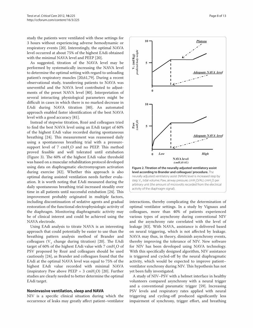
study the patients were ventilated with these settings for
3 hours without experiencing adverse hemodynamic or
respira tory events [20]. Interestingly, the optimal NAVA
level occurred at about 75% of the highest EAdi obtained
with the minimal NAVA level and PEEP [20].
As suggested, titration of the NAVA level may be
performed by systematically increasing the NAVA level
to determine the optimal setting with regard to unloading
patient’s respiratory muscles [20,61,79]. During a recent
observational study, transferring patients to NAVA was
uneventful and the NAVA level contributed to adjust-
ments of the preset NAVA level [80]. Interpretation of
several interacting physiological parameters might be
diffi cult in cases in which there is no marked decrease in
EAdi during NAVA titration [80]. An automated
approach enabled faster identifi cation of the best NAVA
level with a good accuracy [81].
Instead of stepwise titration, Rozé and colleagues tried
to fi nd the best NAVA level using an EAdi target of 60%
of the highest EAdi value recorded during spontaneous
breathing [24]. Th is measurement was reassessed daily
using a spontaneous breathing trial with a pressure-
support level of 7 cmH2O and no PEEP. Th is method
proved feasible and well tolerated until extubation
(Figure 3). Th e 60% of the highest EAdi value threshold
was based on a muscular rehabilitation protocol developed
using data on diaphragmatic electromyogram activation
during exercise [82]. Whether this approach is also
optimal during assisted ventilation needs further evalu-
ation. It is worth noting that EAdi measured during the
daily spontaneous breathing trial increased steadily over
time in all patients until successful extubation [24]. Th is
improvement probably originated in multiple factors,
including discontinuation of sedative agents and gradual
restoration of the functional electrophysiologic activity of
the diaphragm. Monitoring diaphragmatic activity may
be of clinical interest and could be achieved using the
NAVA electrode.
Using EAdi analysis to titrate NAVA is an interesting
approach that could potentially be easier to use than the
breathing pattern analysis method of Brander and
colleagues (VT change during titration) [20]. Th e EAdi
target of 60% of the highest EAdi value with 7 cmH2O of
PSV proposed by Rozé and colleagues should be used
cautiously [24], as Brander and colleagues found that the
EAdi at the optimal NAVA level was equal to 75% of the
highest EAdi value recorded with minimal NAVA
(inspiratory Paw above PEEP = 3 cmH2O) [20]. Further
studies are clearly needed to better determine the optimal
EAdi target.
Noninvasive ventilation, sleep and NAVA
NIV is a specifi c clinical situation during which the
occurrence of leaks may greatly aff ect patient–ventilator
interactions, thereby complicating the determination of
optimal ventilator settings. In a study by Vignaux and
colleagues, more than 40% of patients experienced
various types of asynchrony during conventional NIV
and the asynchrony rate correlated with the level of
leakage [83]. With NAVA, assistance is delivered based
on neural triggering, which is not aff ected by leakage.
NAVA may thus, in theory, diminish asynchrony events,
thereby improving the tolerance of NIV. New software
for NIV has been developed using NAVA technology.
With this specifi cally designed algorithm, NIV assistance
is triggered and cycled-off by the neural diaphragmatic
activity, which would be expected to improve patient–
ventilator synchrony during NIV. Th is hypothesis has not
yet been fully investigated.
A study of NIV–PSV with a helmet interface in healthy
volunteers compared asynchrony with a neural trigger
and a conventional pneumatic trigger [59]. Increasing
PSV levels and respiratory rates applied with neural
trigger ing and cycling-off produced signifi cantly less
impair ment of synchrony, trigger eff ort, and breathing
Figure 2. Titration of the neurally adjusted ventilatory assist
level according to Brander and colleagues’ procedure. The
neurally adjusted ventilatory assist (NAVA) level is increased step by
step. VT, tidal volume; Paw, airway pressure; cmH
2O/AU, cmH
2O per
arbitrary unit (the amount of microvolts recorded from the electrical
activity of the diaphragm signal).
Plateau
dy w
eigh
t
Plateau
VT (m
l/Kg)
pred
icte
d bo
d
5
Adequate NAVA level
0
10
Paw
(cm
H2O
)
5
Adequate NAVA level
NAVA level (cmH2O/AU)
0 Low High
10
Terzi et al. Critical Care 2012, 16:225 http://ccforum.com/content/16/3/225
Page 8 of 13

comfort, compared with conventional pneumatic trigger-
ing and cycling-off .
Cammarotta and colleagues recently compared NAVA
and NIV–PSV delivered through a helmet interface in
postextubation hypoxemic patients [32]. Ten patients
underwent three 20-minute trials of helmet NIV in PSV,
NAVA, and PSV again. Th e authors demonstrated that
there was less asynchrony during NAVA than during PSV
and no diff erence in gas exchange, although there were
more leaks during NAVA. Moreover it is important to
underline that the PSV mode chosen was specifi cally
dedicated to NIV, whereas the NAVA mode dedicated to
NIV that is now currently available did not exist at the
time of this study.
Recent data obtained in low-birth-weight infants
indicate that NAVA can maintain synchrony – both in
terms of timing and proportionality – even after extu-
bation in patients with an excessively leaky interface
under NIV (all infants in this study were ventilated using
a single nasal prong) [29].
Another consideration for NIV that deserves attention
in the near future is the impact on swallowing, phonation,
and sleep quality, most notably when NIV is used for
several days. Improvements in swallowing performance
have been reported in neuromuscular patients receiving
MV compared with spontaneous breathing [84,85]. Th e
close relationship between the muscles involved in
swallowing and those contributing to inspiration was
evidenced by Orlikowski and colleagues using an original
method of tongue-strength measurement. Th e signifi cant
tongue weakness observed in 16 weak patients with
Guillain–Barré syndrome correlated with the alterations
in respiratory parameters [86]. Additional physiological
studies are required to document the potential benefi ts of
NAVA on swallowing–breathing interactions during NIV.
Sleep quality during NIV has been shown to be a
predictor of success or failure [87]. Sleep quality can also
be improved compared with standard NIV settings by
careful physiological titration of the ventilator settings
[88]. Patient–ventilator asynchrony can cause sleep dis-
rup tion. Bosma and colleagues demonstrated that PAV, a
mode of partial ventilatory support in which the venti-
lator applies pressure in proportion to the inspiratory
load, was more eff ective than PSV in matching the
ventilatory requirements to the level of ventilator assis-
tance, thereby resulting in fewer patient–ventilator
asynchronies and better quality of sleep [11]. Delisle and
colleagues recently obtained sleep recordings during a
Figure 3. Change in neurally adjusted ventilatory assist according to maximum diaphragmatic electrical activity during spontaneous
breathing. Electrical activity of the diaphragm (EAdi) values during 1 hour, each point representing the mean value over 1 minute. EAdi variations
occurred before, during, and after a spontaneous breathing trial (SBT). Maximum EAdi was 21 μV after a SBT of 3 minutes and allowed a reduction
in the neurally adjusted ventilatory assist (NAVA) level from 2.4 to 2.2 cmH2O/μV in order to obtain EAdi values after the SBT of about 13 μV (60% of
maximum EAdi). Arterial blood gases were not changed by the NAVA level modifi cation.
Terzi et al. Critical Care 2012, 16:225 http://ccforum.com/content/16/3/225
Page 9 of 13

crossover study comparing NAVA and PSV in 14 mecha-
nically ventilated patients [89]. Each condition was
studied for 4 hours, and recordings were obtained over
19 consecutive hours in all. Patient–ventilator asyn-
chrony varied signifi cantly across sleep stages, and no
asynchrony occurred with NAVA. Overassistance occur-
red only with PSV, which probably explained the
improvements in physiological indices of sleep quality
observed with NAVA.
Neurally adjusted ventilatory assist in children and
infants
MV in children and in low-birth-weight infants is more
diffi cult to apply than in adults and has several speci-
icities. First infants take a very small tidal volume, have a
rapid respiratory rate, have a limited chest wall muscu-
lature, and have variable and fl uctuating lung compliance.
Second, most neonatal units use uncuff ed tracheal tubes
for fears of pressure necrosis and air leak is always
present, making reliable measurements and triggering
problematic. Th ird, ventilators that are effi cient in adults
are not systematically effi cient in children and infants,
mainly because the inspiratory triggers are not suffi ci-
ently sensitive for early detection of infants’/children’s
inspiratory eff ort [90].
Whether or not the respiratory drive of the preterm
infant is suitable to control MV is unknown. Beck and
colleagues fi rst evaluated patient–ventilator interaction
with NAVA in seven very-low-birth-weight infants [29].
As suggested by previous animal studies [91], they
demonstrated that NAVA could be implemented for a
short-term period, both invasively and noninvasively, in
infants with body weight as low as 640 g up to 3 years old.
During invasive ventilation with NAVA, EAdi and venti-
lator pressure were correlated and patient–ventilator
synchrony was improved compared with the other mode.
Moreover, this synchrony persisted after extubation while
ventilating the patient with an excessively leaky interface.
After this fi rst physiological demonstration, Bengtsson
and Edberg demonstrated the clinical feasibility and
safety with use of NAVA in pediatric patients [30].
Similarly, Breatnach and colleagues compared NAVA
(with a neural trigger) and PSV (with a pneumatic trigger)
in 16 ventilated infants [31]. Th is prospective crossover
comparison demonstrated that ventilation with NAVA
improved patient–ventilator synchrony.
Furthermore, Alander and colleagues recently compared
NAVA with pressure-controlled ventilation for newborns
and with pressure-regulated controlled ventilation for
children older than 3 months (with conventional trigger
modes: pressure and fl ow trigger) [92]. In this prospective
cross-over study, 18 patients requiring MV were random-
ized for 10 minutes with the diff erent modes. During
NAVA, the peak airway pressure was lower, the
respira tory rate was 10 breaths/minute higher than in the
pressure group, and patient–ventilator synchronization
was improved. However, there were no diff erences in
tidal volume and in oxygen saturation.
To evaluate the eff ects of the neural trigger on trigger
delay, ventilator response time, or work of breathing,
Clement and colleagues conducted a study in 23 pediatric
patients aged 0 to 24 months with a diagnosis of bron-
chio litis presenting respiratory failure requiring MV [33].
Th e authors compared the neural trigger and the
pneumatic trigger using similar NAVA assistance, and
observed that the trigger delay, the ventilator response
time, and the work of breathing were reduced by the
neural trigger.
Finally, all of these studies seem to demonstrate the
feasibility of and a potential advantage for NAVA in
children compared with the other assisted ventilatory
modes. Because patient–ventilator synchrony is improved
with NAVA, the children may require lower doses of
sedation with this mode of MV [93], which could reduce
the time of MV.
Future research
Clinical studies obtained in critically ill patients confi rm
many of the expected short-term physiological benefi ts
associated with NAVA, as discussed above.
Particularly, NAVA seems to markedly improve the
problems of nonsynchronization between the patient and
the ventilator and the problems of risk of overventilation –
including the risk of ineff ective or missed inspiratory
eff orts due to intrinsic PEEP observed in chronic
obstructive pulmonary disease patients, or to a rapid
breathing frequency with a very small tidal volume
observed in pediatric patients.
In addition, NAVA minimizes the risk of overinfl ation
because the duration and level of pressurization remain
under respiratory-center control, and minimizes the risk
of diaphragmatic inactivity because the presence of
pressure assistance requires the presence of this inspira-
tory activity.
A preserved respiratory muscle function is pivotal for
weaning from MV [44]. By using NAVA, which out-
performs the previous modes of MV for adequately
assisting the patient’s inspiratory eff ort without inducing
patient/ventilator dyssynchrony, a reduction in the dura-
tion of MV could be expected. Studies are needed to
evaluate the best time to begin the weaning process with
NAVA.
Th e NAVA setting is an important question not yet
fully resolved. If clinicians are accustomed to set a PSV
level, this is not the case for NAVA. Furthermore, because
the breathing pattern is less modifi ed by the NAVA
setting than during PSV, it is much less informative for
NAVA adjustment. As described above, the literature
Terzi et al. Critical Care 2012, 16:225 http://ccforum.com/content/16/3/225
Page 10 of 13

suggests that the adjustment should consider the electro-
myographic activity of the diaphragm, but this method is
not simple. As recently proposed, a direct evalu ation of
patient comfort and sense of dyspnea for the NAVA
setting should be evaluated [23].
Finally, the next research step will be to evaluate NAVA
over longer periods, in order to know whether this mode
can replace the modes usually used during MV and the
weaning period, like PSV. Appreciating the safety, the
feasibility and the constraints of this technology will be
useful. It is therefore necessary to test, during the total
weaning period, the eff ectiveness of the esophageal probe
and to know whether it is regularly necessary to adjust
the probe position. One of the most diffi cult questions to
address, however, is in which situations it is not desirable
to let the respiratory centers drive the ventilation.
Situations of severe metabolic acidosis, of high
respiratory drive and of high catecholamine levels may
induce situations of extreme hyperventilation, which may
be dangerous for the lungs. When sedation and/or paralysis
become necessary is therefore an important question to
address before widespread use of this mode [94].
Conclusion
NAVA, which is based on an original physiological
concept, adds new knowledge on patient–ventilator
interactions during spontaneous breathing, thus helping
to unravel the complex mechanisms involved in breathing
control during MV. Th ere is compelling evidence that
NAVA, as well as the PAV+ software, improves patient–
ventilator interactions and increases respiratory varia-
bility in comparison with PSV. Th is advantage holds
potential for many applications. Th e short-term and
long-term experi ence with NAVA, however, remains
scant. Further clinical studies are needed to assess the
feasibility and safety of NAVA. A key challenge is how to
determine the best NAVA settings according to the
patient’s ventilatory needs and the acceptable level of
work of breathing.
Abbreviations
EAdi, electrical activity of the diaphragm; MV, mechanical ventilation;
NAVA, neurally adjusted ventilatory assist; NEXmod, nose to ear lobe to
xiphoid process of the sternum distance modifi ed for the EAdi catheter; NIV,
noninvasive ventilation; PaCO2, partial pressure of carbon dioxide in arterial
blood; PAV, proportional-assist ventilation; Paw, airway pressure; PEEP, positive
end-expiratory pressure; PSV, pressure-support ventilation; VT, tidal volume.
Competing interests
SJ received a grant from Maquet Critical Care SA in 2009 and 2010 for clinical
research. PJ’s research laboratory received grants from Maquet Critical Care
SA Drager, Res-Med, Hamilton, for clinical research over the last 5 years. LB’s
research laboratory received a grant from Maquet Critical Care SA in 2009 and
2010 for clinical research. LB’s research laboratory has also received research
grants from Drager, Covidien, General Electric and Fisher Paykel over the last
5 years. J-CMR and AM received grants from Drager, Covidien, and General
Electric over the last 5 years for their research group. AD’s research laboratory
received a grant from Maquet Critical Care SA in 2008 and 2009 for clinical
research. AD coordinates a multicenter clinical study on NAVA. The remaining
authors declare that they have no competing interests.
Author contributions
NT, J-CMR and LB initiated and wrote the manuscript; all authors contributed
to the revision of this. All authors read and approved the fi nal manuscript.
Acknowledgements
The round-table meeting was initiated and organized solely by the
authors. Maquet Critical Care SA provided fi nancial support for travel and
accommodation costs, thus allowing the authors to meet for a half-day
conference, but had no infl uence on the content of the manuscript.
Author details1INSERM U1075, Caen F-14000, France. 2Université de Caen, Caen F-14000,
France. 3CHRU Caen, Service de Réanimation Médicale, Caen F-14000, France. 4Adult Intensive Care and Burn Unit, Lausanne University Hospital (CHUV), Rue
du Bugnon 46, 1011 Lausanne, Switzerland. 5Department of Anaesthesiology
and Intensive Care 2, Bordeaux University Hospital, F-33000 Bordeaux, France. 6LUNAM Université, Angers, France Université Angers, CHU Angers, Medical
ICU, 4 rue Larrey, 49933 Angers, Cedex 09 France. 7University Angers, CHU
Angers, Réanimation Médicale, 49933 Angers, France. 8Centre d’Investigation
Clinique – Innovations Technologiques, Services de Physiologie – Explorations
Fonctionnelles, Hôpital Raymond Poincaré, AP-HP, E.A. 4497, Université de
Versailles – Saint Quentin en Yvelines, 92380 Garches, France. 9INSERM U955,
94000 Créteil, France. 10Hôpital du Sacré-Coeur de Montréal, 5400 boul.
Gouin Ouest Montréal, Québec, H4J 1C5, Canada. 11Intensive Care Unit,
Clinique Notre Dame de Grâce, Gosselies Hospital, 212 Chaussée de Nivelles,
B-6041-Gosselies, Belgium. 12Intensive Care Unit, University Hospital St-Luc,
10 avenue Hippocrate, 1200 Brussels, Belgium. 13Medical Intensive Care Unit
and Respiratory Division, Groupe Hospitalier Pitié-Salpêtrière, Université
Pierrer et Marie Curie and INSERM974, Paris, France. 14Department of Critical
Care Medicine and Anesthesiology (DAR B), Saint Eloi University Hospital and
Universite of Montpellier, INSERM U1046, 80 Avenue Augustin Fliche, 34295
Montpellier, France. 15Intensive Care Unit, Hospital de Sant Pau C. St Quinti
89, 08041 Barcelona, Spain. 16Intensive Care Unit, Geneva University Hospital,
School of Medicine, University of Geneva, Rue Gabrielle-Perret-Gentil 4, 1205
Geneva, Switzerland. 17Medical Intensive Care Unit, University Hospital, Charles
Nicolle, 76000 Rouen, France. 18UPRES EA3830, Rouen, France.
Published: 20 June 2012
References
1. Esteban A, Anzueto A, Alía I, Gordo F, Apezteguía C, Pálizas F, Cide D,
Goldwaser R, Soto L, Bugedo G, Rodrigo C, Pimentel J, Raimondi G, Tobin MJ:
How is mechanical ventilation employed in the intensive care unit? An international utilization review. Am J Respir Crit Care Med 2000,
161:1450-1458.
2. Esteban A, Ferguson ND, Meade MO, Frutos-Vivar F, Apezteguia C, Brochard L,
Raymondos K, Nin N, Hurtado J, Tomicic V, González M, Elizalde J, Nightingale
P, Abroug F, Pelosi P, Arabi Y, Moreno R, Jibaja M, D’Empaire G, Sandi F,
Matamis D, Montañez AM, Anzueto A; VENTILA Group: Evolution of mechanical ventilation in response to clinical research. Am J Respir Crit Care
Med 2008, 177:170-177.
3. Cereda M, Foti G, Marcora B, Gili M, Giacomini M, Sparacino ME, Pesenti A:
Pressure support ventilation in patients with acute lung injury. Crit Care
Med 2000, 28:1269-1275.
4. Nava S, Bruschi C, Fracchia C, Braschi A, Rubini F: Patient–ventilator interaction and inspiratory eff ort during pressure support ventilation in patients with diff erent pathologies. Eur Respir J 1997, 10:177-183.
5. Navalesi P, Costa R: New modes of mechanical ventilation: proportional assist ventilation, neurally adjusted ventilatory assist, and fractal ventilation. Curr Opin Crit Care 2003, 9:51-58.
6. Moerer O: Eff ort-adapted modes of assisted breathing. Curr Opin Crit Care
2012, 18:61-69.
7. Younes M, Puddy A, Roberts D, Light RB, Quesada A, Taylor K, Oppenheimer L,
Cramp H: Proportional assist ventilation. Results of an initial clinical trial. Am Rev Respir Dis 1992, 145:121-129.
8. Giannouli E, Webster K, Roberts D, Younes M: Response of ventilator-dependent patients to diff erent levels of pressure support and proportional assist. Am J Respir Crit Care Med 1999, 159:1716-1725.
9. Alexopoulou C, Kondili E, Vakouti E, Klimathianaki M, Prinianakis G,
Georgopoulos D: Sleep during proportional-assist ventilation with load-adjustable gain factors in critically ill patients. Intensive Care Med 2007,
33:1139-1147.
Terzi et al. Critical Care 2012, 16:225 http://ccforum.com/content/16/3/225
Page 11 of 13

10. Ranieri VM, Giuliani R, Mascia L, Grasso S, Petruzzelli V, Puntillo N, Perchiazzi G,
Fiore T, Brienza A: Patient–ventilator interaction during acute hypercapnia: pressure-support vs. proportional-assist ventilation. J Appl Physiol 1996,
81:426-436.
11. Bosma K, Ferreyra G, Ambrogio C, Pasero D, Mirabella L, Braghiroli A,
Appendini L, Mascia L, Ranieri VM: Patient–ventilator interaction and sleep in mechanically ventilated patients: pressure support versus proportional assist ventilation. Crit Care Med 2007, 35:1048-1054.
12. Grasso S, Puntillo F, Mascia L, Ancona G, Fiore T, Bruno F, Slutsky AS, Ranieri
VM: Compensation for increase in respiratory workload during mechanical ventilation. Pressure-support versus proportional-assist ventilation. Am J
Respir Crit Care Med 2000, 161(3 Pt 1):819-826.
13. Younes M, Webster K, Kun J, Roberts D, Masiowski B: A method for measuring passive elastance during proportional assist ventilation. Am J
Respir Crit Care Med 2001, 164:50-60.
14. Younes M, Kun J, Masiowski B, Webster K, Roberts D: A method for noninvasive determination of inspiratory resistance during proportional assist ventilation. Am J Respir Crit Care Med 2001, 163:829-839.
15. Kondili E, Prinianakis G, Alexopoulou C, Vakouti E, Klimathianaki M,
Georgopoulos D: Respiratory load compensation during mechanical ventilation – proportional assist ventilation with load-adjustable gain factors versus pressure support. Intensive Care Med 2006, 32:692-699.
16. Xirouchaki N, Kondili E, Vaporidi K, Xirouchakis G, Klimathianaki M, Gavriilidis G,
Alexandopoulou E, Plataki M, Alexopoulou C, Georgopoulos D: Proportional assist ventilation with load-adjustable gain factors in critically ill patients: comparison with pressure support. Intensive Care Med 2008, 34:2026-2034.
17. Costa R, Spinazzola G, Cipriani F, Ferrone G, Festa O, Arcangeli A, Antonelli M,
Proietti R, Conti G: A physiologic comparison of proportional assist ventilation with load-adjustable gain factors (PAV+) versus pressure support ventilation (PSV). Intensive Care Med 2011, 37:1494-1500.
18. Sinderby C, Navalesi P, Beck J, Skrobik Y, Comtois N, Friberg S, Gottfried SB,
Lindstrom L: Neural control of mechanical ventilation in respiratory failure. Nat Med 1999, 5:1433-1436.
19. Wu XY, Huang YZ, Yang Y, Liu SQ, Liu HG, Qiu HB: [Eff ects of neurally adjusted ventilatory assist on patient-ventilator synchrony in patients with acute respiratory distress syndrome]. Zhonghua Jie He He Hu Xi Za Zhi 2009,
32:508-512.
20. Brander L, Leong-Poi H, Beck J, Brunet F, Hutchison SJ, Slutsky AS, Sinderby C:
Titration and implementation of neurally adjusted ventilatory assist in critically ill patients. Chest 2009, 135:695-703.
21. Colombo D, Cammarota G, Bergamaschi V, De Lucia M, Corte FD, Navalesi P:
Physiologic response to varying levels of pressure support and neurally adjusted ventilatory assist in patients with acute respiratory failure. Intensive Care Med 2008, 34:2010-2018.
22. Spahija J, de Marchie M, Albert M, Bellemare P, Delisle S, Beck J, Sinderby C:
Patient–ventilator interaction during pressure support ventilation and neurally adjusted ventilatory assist. Crit Care Med 2010, 38:518-526.
23. Schmidt M, Demoule A, Cracco C, Gharbi A, Fiamma MN, Straus C, Duguet A,
Gottfried SB, Similowski T: Neurally adjusted ventilatory assist increases respiratory variability and complexity in acute respiratory failure. Anesthesiology 2010, 112:670-681.
24. Rozé H, Lafrikh A, Perrier V, Germain A, Dewitte A, Gomez F, Janvier G,
Ouattara A: Daily titration of neurally adjusted ventilatory assist using the diaphragm electrical activity. Intensive Care Med 2011, 37:1087-1094.
25 . Terzi N, Pelieu I, Guittet L, Ramakers M, Seguin A, Daubin C, Charbonneau P,
du Cheyron D, Lofaso F: Neurally adjusted ventilatory assist in patients recovering spontaneous breathing after acute respiratory distress syndrome: physiological evaluation. Crit Care Med 2010, 38:1830-1837.
26 . Piquilloud L, Vignaux L, Bialais E, Roeseler J, Sottiaux T, Laterre PF, Jolliet P,
Tassaux D: Neurally adjusted ventilatory assist improves patient–ventilator interaction. Intensive Care Med 2011, 37:263-271.
27 . Coisel Y, Chanques G, Jung B, Constantin JM, Capdevila X, Matecki S, Grasso S,
Jaber S: Neurally adjusted ventilatory assist in critically ill postoperative patients: a crossover randomized study. Anesthesiology 2010, 113:925-935.
28 . Passath C, Takala J, Tuchscherer D, Jakob SM, Sinderby C, Brander L:
Physiologic response to changing positive end-expiratory pressure during neurally adjusted ventilatory assist in sedated, critically ill adults. Chest
2010, 138:578-587.
29 . Beck J, Reilly M, Grasselli G, Mirabella L, Slutsky AS, Dunn MS, Sinderby C:
Patient–ventilator interaction during neurally adjusted ventilatory assist in low birth weight infants. Pediatr Res 2009, 65:663-668.
30 . Bengtsson JA, Edberg KE: Neurally adjusted ventilatory assist in children: an observational study. Pediatr Crit Care Med 2010, 11:253-257.
31 . Breatnach C, Conlon NP, Stack M, Healy M, O’Hare BP: A prospective crossover comparison of neurally adjusted ventilatory assist and pressure-support ventilation in a pediatric and neonatal intensive care unit population. Pediatr Crit Care Med 2010, 11:7-11.
32 . Cammarota G, Olivieri C, Costa R, Vaschetto R, Colombo D, Turucz E, Longhini
F, Della Corte F, Conti G, Navalesi P: Noninvasive ventilation through a helmet in postextubation hypoxemic patients: physiologic comparison between neurally adjusted ventilatory assist and pressure support ventilation. Intensive Care Med 2011, 37:1943-1950.
33 . Clement KC, Thurman TL, Holt SJ, Heulitt MJ: Neurally triggered breaths reduce trigger delay and improve ventilator response times in ventilated infants with bronchiolitis. Intensive Care Med 2011, 37:1826-1832.
34 . Thille AW, Rodriguez P, Cabello B, Lellouche F, Brochard L: Patient–ventilator asynchrony during assisted mechanical ventilation. Intensive Care Med
2006, 32:1515-1522.
35 . Tobin MJ, Jubran A, Laghi F: Patient–ventilator interaction. Am J Respir Crit
Care Med 2001, 163:1059-1063.
36 . Parthasarathy S, Jubran A, Tobin MJ: Cycling of inspiratory and expiratory muscle groups with the ventilator in airfl ow limitation. Am J Respir Crit Care
Med 1998, 158(5 Pt 1):1471-1478.
37 . Beck J, Tucci M, Emeriaud G, Lacroix J, Sinderby C: Prolonged neural expiratory time induced by mechanical ventilation in infants. Pediatr Res
2004, 55:747-754.
38 . de Wit M, Pedram S, Best AM, Epstein SK: Observational study of patient–ventilator asynchrony and relationship to sedation level. J Crit Care 2009,
24:74-80.
39 . Dreyfuss D, Saumon G: Ventilator-induced lung injury: lessons from experimental studies. Am J Respir Crit Care Med 1998, 157:294-323.
40 . Ricard JD, Dreyfuss D, Saumon G: Ventilator-induced lung injury. Eur Respir J
Suppl 2003, 42:2s-9s.
41 . The Acute Respiratory Distress Syndrome Network: Ventilation with lower tidal volumes as compared with traditional tidal volumes for acute lung injury and the acute respiratory distress syndrome. N Engl J Med 2000,
342:1301-1308.
42 . Levine S, Nguyen T, Taylor N, Friscia ME, Budak MT, Rothenberg P, Zhu J,
Sachdeva R, Sonnad S, Kaiser LR, Rubinstein NA, Powers SK, Shrager JB: Rapid disuse atrophy of diaphragm fi bers in mechanically ventilated humans. N Engl J Med 2008, 358:1327-1335.
43 . Sieck GC, Mantilla CB: Eff ect of mechanical ventilation on the diaphragm. N Engl J Med 2008, 358:1392-1394.
44 . Jaber S, Petrof BJ, Jung B, Chanques G, Berthet JP, Rabuel C, Bouyabrine H,
Courouble P, Koechlin-Ramonatxo C, Sebbane M, Similowski T, Scheuermann V,
Mebazaa A, Capdevila X, Mornet D, Mercier J, Lacampagne A, Philips A, Matecki S:
Rapidly progressive diaphragmatic weakness and injury during mechanical ventilation in humans. Am J Respir Crit Care Med 2011, 183:364-371.
45 . Anzueto A, Peters JI, Tobin MJ, de los Santos R, Seidenfeld JJ, Moore G, Cox
WJ, Coalson JJ: Eff ects of prolonged controlled mechanical ventilation on diaphragmatic function in healthy adult baboons. Crit Care Med 1997,
25:1187-1190.
46 . Sassoon CS: Ventilator-associated diaphragmatic dysfunction. Am J Respir
Crit Care Med 2002, 166:1017-1018.
47 . Sassoon CS, Caiozzo VJ, Manka A, Sieck GC: Altered diaphragm contractile properties with controlled mechanical ventilation. J Appl Physiol 2002,
92:2585-2595.
48 . Sassoon CS, Zhu E, Caiozzo VJ: Assist-control mechanical ventilation attenuates ventilator-induced diaphragmatic dysfunction. Am J Respir Crit
Care Med 2004, 170:626-632.
49 . Thille AW, Cabello B, Galia F, Lyazidi A, Brochard L: Reduction of patient–ventilator asynchrony by reducing tidal volume during pressure-support ventilation. Intensive Care Med 2008, 34:1477-1486.
50 . Pinto Da Costa N, Di Marco F, Lyazidi A, Carteaux G, Sarni M, Brochard L: Eff ect of pressure support on end-expiratory lung volume and lung diff usion for carbon monoxide. Crit Care Med 2011, 39:2283-2289.
51 . Barwing J, Ambold M, Linden N, Quintel M, Moerer O: Evaluation of the catheter positioning for neurally adjusted ventilatory assist. Intensive Care
Med 2009, 35:1809-1814.
52 . Barwing J, Pedroni C, Quintel M, Moerer O: Infl uence of body position, PEEP and intra-abdominal pressure on the catheter positioning for neurally adjusted ventilatory assist. Intensive Care Med 2011, 37:2041-2045.
Terzi et al. Critical Care 2012, 16:225 http://ccforum.com/content/16/3/225
Page 12 of 13

53 . Parthasarathy S, Jubran A, Tobin MJ: Assessment of neural inspiratory time in ventilator-supported patients. Am J Respir Crit Care Med 2000,
162(2 Pt 1):546-552.
54 . Calderini E, Confalonieri M, Puccio PG, Francavilla N, Stella L, Gregoretti C:
Patient–ventilator asynchrony during noninvasive ventilation: the role of expiratory trigger. Intensive Care Med 1999, 25:662-667.
55 . Vignaux L, Tassaux D, Carteaux G, Roeseler J, Piquilloud L, Brochard L, Jolliet P:
Performance of noninvasive ventilation algorithms on ICU ventilators during pressure support: a clinical study. Intensive Care Med 2010,
36:2053-2059.
56 . Leung P, Jubran A, Tobin MJ: Comparison of assisted ventilator modes on triggering, patient eff ort, and dyspnea. Am J Respir Crit Care Med 1997,
155:1940-1948.
57 . Tassaux D, Gainnier M, Battisti A, Jolliet P: Impact of expiratory trigger setting on delayed cycling and inspiratory muscle workload. Am J Respir
Crit Care Med 2005, 172:1283-1289.
58 . American Thoracic Society/European Respiratory Society: ATS/ERS Statement on respiratory muscle testing. Am J Respir Crit Care Med 2002,
166:528-547.
59 . Moerer O, Beck J, Brander L, Costa R, Quintel M, Slutsky AS, Brunet F, Sinderby
C: Subject–ventilator synchrony during neural versus pneumatically triggered non-invasive helmet ventilation. Intensive Care Med 2008,
34:1615-1623.
60 . Thille AW, Lyazidi A, Richard JC, Galia F, Brochard L: A bench study of intensive-care-unit ventilators: new versus old and turbine-based versus compressed gas-based ventilators. Intensive Care Med 2009, 35:1368-1376.
61 . Allo JC, Beck JC, Brander L, Brunet F, Slutsky AS, Sinderby CA: Infl uence of neurally adjusted ventilatory assist and positive end-expiratory pressure on breathing pattern in rabbits with acute lung injury. Crit Care Med 2006,
34:2997-3004.
62 . Brander L, Sinderby C, Lecomte F, Leong-Poi H, Bell D, Beck J, Tsoporis JN,
Vaschetto R, Schultz MJ, Parker TG, Villar J, Zhang H, Slutsky AS: Neurally adjusted ventilatory assist decreases ventilator-induced lung injury and non-pulmonary organ dysfunction in rabbits with acute lung injury. Intensive Care Med 2009, 35:1979-1989.
63 . Sinderby C, Beck J, Spahija J, de Marchie M, Lacroix J, Navalesi P, Slutsky AS:
Inspiratory muscle unloading by neurally adjusted ventilatory assist during maximal inspiratory eff orts in healthy subjects. Chest 2007,
131:711-717.
64 . Guz A: Hering and Breuer revisited in humans: an invasive study before the days of ethics committees. Am J Respir Crit Care Med 2001,
164:1110-1111.
65 . Patroniti N, Bellani G, Saccavino E, Zanella A, Grasselli G, Isgro S, Milan M, Foti
G, Pesenti A: Respiratory pattern during neurally adjusted ventilatory assist in acute respiratory failure patients. Intensive Care Med 2012, 38:230-239.
66 . Putensen C, Rasanen J, Lopez FA, Downs JB: Eff ect of interfacing between spontaneous breathing and mechanical cycles on the ventilation–perfusion distribution in canine lung injury. Anesthesiology 1994,
81:921-930.
67 . Gama de Abreu M, Spieth PM, Pelosi P, Carvalho AR, Walter C, Schreiber-Ferstl
A, Aikele P, Neykova B, Hubler M, Koch T: Noisy pressure support ventilation: a pilot study on a new assisted ventilation mode in experimental lung injury. Crit Care Med 2008, 36:818-827.
68 . Brack T, Jubran A, Tobin MJ: Eff ect of elastic loading on variational activity of breathing. Am J Respir Crit Care Med 1997, 155:1341-1348.
69 . Brack T, Jubran A, Tobin MJ: Eff ect of resistive loading on variational activity of breathing. Am J Respir Crit Care Med 1998, 157(6 Pt 1):1756-1763.
70 . Brack T, Jubran A, Tobin MJ: Dyspnea and decreased variability of breathing in patients with restrictive lung disease. Am J Respir Crit Care Med 2002,
165:1260-1264.
71 . Sammon MP, Bruce EN: Vagal aff erent activity increases dynamical dimension of respiration in rats. J Appl Physiol 1991, 70:1748-1762.
72 . Fiamma MN, Straus C, Thibault S, Wysocki M, Baconnier P, Similowski T: Eff ects of hypercapnia and hypocapnia on ventilatory variability and the chaotic dynamics of ventilatory fl ow in humans. Am J Physiol Regul Integr Comp
Physiol 2007, 292:R1985-R1993.
73 . Wysocki M, Fiamma MN, Straus C, Poon CS, Similowski T: Chaotic dynamics of resting ventilatory fl ow in humans assessed through noise titration. Respir Physiol Neurobiol 2006, 153:54-65.
74 . Schurmann T, Grassberger P: Entropy estimation of symbol sequences.
Chaos 1996, 6:414-427.
75 . Briggs K: An improved method for estimating Liapunov exponents of chaotic time series. Phys Lett A 1990, 151:27-32.
76 . Lofaso F, Isabey D, Lorino H, Harf A, Scheid P: Respiratory response to positive and negative inspiratory pressure in humans. Respir Physiol 1992,
89:75-88.
77 . Scheid P, Lofaso F, Isabey D, Harf A: Respiratory response to inhaled CO2 during positive inspiratory pressure in humans. J Appl Physiol 1994,
77:876-882.
78 . Karagiannidis C, Lubnow M, Philipp A, Riegger GA, Schmid C, Pfeifer M,
Mueller T: Autoregulation of ventilation with neurally adjusted ventilatory assist on extracorporeal lung support. Intensive Care Med 2010,
36:2038-2044.
79 . Lecomte F, Brander L, Jalde F, Beck J, Qui H, Elie C, Slutsky AS, Brunet F,
Sinderby C: Physiological response to increasing levels of neurally adjusted ventilatory assist (NAVA). Respir Physiol Neurobiol 2009, 166:117-124.
80 . Barwing J, Linden N, Ambold M, Quintel M, Moerer O: Neurally adjusted ventilatory assist vs. pressure support ventilation in critically ill patients: an observational study. Acta Anaesthesiol Scand 2011, 55:1261-1271.
81 . Ververidis D, Van Gils M, Passath C, Takala J, Brander L: Identifi cation of adequate neurally adjusted ventilatory assist (NAVA) during systematic increases in the NAVA level. IEEE Trans Biomed Eng 2011, 58:2598-2606.
82 . Sinderby C, Spahija J, Beck J, Kaminski D, Yan S, Comtois N, Sliwinski P:
Diaphragm activation during exercise in chronic obstructive pulmonary disease. Am J Respir Crit Care Med 2001, 163:1637-1641.
83 . Vignaux L, Vargas F, Roeseler J, Tassaux D, Thille AW, Kossowsky MP, Brochard
L, Jolliet P: Patient–ventilator asynchrony during non-invasive ventilation for acute respiratory failure: a multicenter study. Intensive Care Med 2009,
35:840-846.
84 . Terzi N, Orlikowski D, Aegerter P, Lejaille M, Ruquet M, Zalcman G, Fermanian
C, Raphael JC, Lofaso F: Breathing–swallowing interaction in neuromuscular patients: a physiological evaluation. Am J Respir Crit Care
Med 2007, 175:269-276.
85 . Terzi N, Prigent H, Lejaille M, Falaize L, Annane D, Orlikowski D, Lofaso F:
Impact of tracheostomy on swallowing performance in Duchenne muscular dystrophy. Neuromuscul Disord 2010, 20:493-498.
86 . Orlikowski D, Terzi N, Blumen M, Sharshar T, Raphael JC, Annane D, Lofaso F:
Tongue weakness is associated with respiratory failure in patients with severe Guillain–Barre syndrome. Acta Neurol Scand 2009, 119:364-370.
87 . Roche Campo F, Drouot X, Thille AW, Galia F, Cabello B, d’Ortho MP, Brochard
L: Poor sleep quality is associated with late noninvasive ventilation failure in patients with acute hypercapnic respiratory failure. Crit Care Med 2010,
38:477-485.
88 . Fanfulla F, Delmastro M, Berardinelli A, Lupo ND, Nava S: Eff ects of diff erent ventilator settings on sleep and inspiratory eff ort in patients with neuromuscular disease. Am J Respir Crit Care Med 2005, 172:619-624.
89 . Delisle S, Ouellet P, Bellemare P, Tetrault JP, Arsenault P: Sleep quality in mechanically ventilated patients: comparison between NAVA and PSV modes. Ann Intensive Care 2011, 1:42.
90 . Fauroux B, Leroux K, Desmarais G, Isabey D, Clement A, Lofaso F, Louis B:
Performance of ventilators for noninvasive positive-pressure ventilation in children. Eur Respir J 2008, 31:1300-1307.
91 . Beck J, Campoccia F, Allo JC, Brander L, Brunet F, Slutsky AS, Sinderby C:
Improved synchrony and respiratory unloading by neurally adjusted ventilatory assist (NAVA) in lung-injured rabbits. Pediatr Res 2007,
61:289-294.
92 . Alander M, Peltoniemi O, Pokka T, Kontiokari T: Comparison of pressure-, fl ow-, and NAVA-triggering in pediatric and neonatal ventilatory care. Pediatr Pulmonol 2012, 47:76-83.
93 . Rhoney DH, Murry KR: National survey of the use of sedating drugs, neuromuscular blocking agents, and reversal agents in the intensive care unit. J Intensive Care Med 2003, 18:139-145.
94. Papazian L, Forel JM, Gacouin A, Penot-Ragon C, Perrin G, Loundou A, Jaber S,
Arnal JM, Perez D, Seghboyan JM, Constantin JM, Courant P, Lefrant JY, Guérin
C, Prat G, Morange S, Roch A; ACURASYS Study Investigators: Neuromuscular blockers in early acute respiratory distress syndrome. N Engl J Med 2010,
363:1107-1116.
doi:10.1186/cc11297Cite this article as: Terzi N, et al.: Clinical review: Update on neurally adjusted ventilatory assist – report of a round-table conference. Critical Care
2012, 16:225.
Terzi et al. Critical Care 2012, 16:225 http://ccforum.com/content/16/3/225
Page 13 of 13