reversible inhibition of calcineurin by the polyphenolic ... · can inhibition by these drugs is...
TRANSCRIPT

- 1 -
Reversible Inhibition of Calcineurin
by the Polyphenolic Aldehyde Gossypol
Ria Baumgrass*, Matthias Weiwad*, Frank Erdmann*, Jun O. Liu§ , Dirk Wunderlich+,
Susanne Grabley+, and Gunter Fischer*#
* Max Planck Research Unit for
Enzymology of Protein Folding
Weinbergweg 22
D-06120 Halle/Saale
Germany
§ Department of Pharmacology and Molecular Sciences and Department of Neuroscience,
Johns Hopkins University School of Medicine, Baltimore, MD 21205, USA
+Hans-Knöll-Institute for Natural Products Research
Beutenbergstrasse 11a
D-07745 Jena
#Corresponding author:
Gunter Fischer
Max-Planck-Unit for Enzymology of Protein Folding
Weinbergweg 22
D-06120 Halle/Saale, Germany
Tel.: +49 345 55 22800
Fax: +49 345 55 11972
e-mail: [email protected]
Copyright 2001 by The American Society for Biochemistry and Molecular Biology, Inc.
JBC Papers in Press. Published on October 11, 2001 as Manuscript M103273200 by guest on A
ugust 25, 2018http://w
ww
.jbc.org/D
ownloaded from

- 2 -
Running title: Gossypol is a new type of calcineurin inhibitor
SUMMARY
The reversible inhibition of calcineurin (CaN), which is the only Ca2+/calmodulin dependent
protein Ser/Thr phosphatase, is thought to be a key functional event for most cyclosporin A-
(CsA) and FK506-mediated biological effects. Beside CaN inhibition, however, CsA and
FK506 have multiple biochemical effects due to their action in a gain of function model that
requires prior binding to immunophilic proteins. We screened a small molecule library for
direct inhibitors of calcineurin using calcineurin mediated dephosphorylation of [33P]-labelled
19-residue phosphopeptide substrate (RII phosphopeptide)1 as an assay and found the
polyphenolic aldehyde gossypol as a novel calcineurin inhibitor. Unlike CsA and FK506,
gossypol does not require a matchmaker protein for reversible calcineurin inhibition with an
IC50 value of 15 µM. Gossypolone, a gossypol analog, showed improved inhibition of both RII
phosphopeptide and p-nitrophenylphosphate dephosphorylation with an IC50 of 9 µM and 6
µM, respectively. In contrast, apogossypol hexaacetate was inactive. Gossypol acts
noncompetitively, interfering with the binding site for the Cyp18/CsA-complex in calcineurin.
In contrast to FK506 and cyclosporin A, gossypol does not inactivate the peptidyl prolyl
cis/trans isomerase (PPIase) activity of immunophilins. Similar to CsA and FK506, NFATC1
translocation from the cytosol into the nucleus in response to stimulation by PMA/ionomycin
is inhibited by gossypol in a dose-dependent manner, and T cell signaling is suppressed
dose-dependently in a NFAT-luciferase reporter gene assay.
by guest on August 25, 2018
http://ww
w.jbc.org/
Dow
nloaded from

- 3 -
Introduction
In vivo inhibition by membrane-penetrable low-molecular mass compounds plays an
important role in evaluating the biological function of enzymes involved in protein
phosphorylation/dephosphorylation. The functional discrimination with specific inhibitors of
the four major protein Ser/Thr phosphatases, protein phosphatase 1 (PP1), protein
phosphatase 2A (PP2A), protein phosphatase 2B (PP2B, calcineurin, CaN) and protein
phosphatase 2C (PP2C) has proven to be successful (1). Okadaic acid and microcystin are
strong inhibitors of PP1 and PP2A, but exhibit poor inhibition of the calcium/calmodulin
regulated CaN and PP2C. It was difficult to identify CaN function in cell signaling until the
membrane-penetrable cyclopeptide cyclosporin A (CsA) and the peptidomacrolide FK506
were found to inhibit CaN specifically under certain conditions (2). Currently, most reports
about CaN involvement in cellular processes are based on CsA and FK506 susceptibility of
the appropriate bioassays. CaN inhibition by these drugs is characterized by their prior
binding to the 18-kDa cyclophilin (Cyp18) and 12-kDa FK506-binding protein (FKBP12),
respectively, indicating a gain-of-function mechanism (3). These mammalian prototypes of
two different families of the enzyme class of peptidyl prolyl cis/trans isomerases (EC 5.2.1.8;
PPIases) function as molecular matchmakers because the drugs cannot bind to CaN on their
own. The prototypic PPIases are themselves tightly inhibited in the course of formation of the
CaN-inhibitory PPIase/drug complex (4).
It is essential to recognize the uncertainties in the interpretation of the biological effects found
for the application of CsA and FK506 in cellular assays. They include the above-discussed
multifunctional biochemical properties released through the PPIase/drug interaction and the
often-unknown cellular content and isoenzyme composition of the matchmaker proteins.
Recent analyses of the human genome revealed the existence of 18 cyclophilins and 16
FKBPs, at least, most of which having the potential to be enzymatically active and to form
PPIase/drug complexes with mostly unknown affinity for CaN (5, 6, 7).
In fact, the CaN-inhibitory complexes of different affinity for CaN are formed from the
matchmaker proteins in competition for the limiting amounts of the drug rendering inhibition
by guest on August 25, 2018
http://ww
w.jbc.org/
Dow
nloaded from

- 4 -
less predictable. Other drug effects may result from the facilitated dissociation of
receptor/PPIase complexes (8, 9) and the slow rate of formation of the PPIase/drug
complexes (10, 11). In addition, the PPIase-mediated intracellular accumulation of the drugs
prevents the reliable analysis of dose-response curves. Although the molarity of the
administered dose is known the variable intracellular PPIase concentration does not permit
calculations of the biologically effective dose.
It has now become apparent that the demonstration of an influence of CsA and FK506 on the
signal of any biological assay does not by itself constitute final proof as to the involvement of
CaN in cell signaling.
While the use of drug derivatives lacking the CaN effector domain has permitted the
application of CaN inactive PPIase inhibitors (12, 13, 14, 15), corresponding experiments
with specific small-molecule CaN inhibitors, which are inactive toward PPIases, are still
missing. The results of biological studies using pyrethroid insecticides as CaN inhibitors (16)
were at variance with recent investigation which could not find any CaN inhibition with this
class of compounds (17, 18). The tyrphostin class of tyrosine kinase inhibitors possesses
CaN inhibitory potency in the micromolar range but lacks CaN specificity. The CsA-like anti-
human immunodeficiency virus-type 1 (HIV-1) replication inhibition of ring-substituted
benzothiophen-2-carboxamide was attributed to its CaN inactivating properties (19).
However, CaN inactivation has not been characterized with purified enzyme, and CsA does
not act on HIV-1 replication via CaN inhibition (13).
Irreversible CaN inactivation by 4-(fluoromethyl) phenyl phosphate is active in the millimolar
range but does not exhibit CaN specificity (20). A similar disadvantage must be considered
for cantharidin and endothall as CaN inhibitors, because they cannot differentiate between
CaN and PP1 or PP2A (21, 22). The inhibition of CaN-mediated dephosphorylation RII
phosphopeptide by the dihydroisobenzofuran dibefurin exhibits an IC50 of 46 µM but the
specificity of inhibition remained unknown. Interestingly, this compounds was active in the
mixed lymphocyte reaction assay (23).
by guest on August 25, 2018
http://ww
w.jbc.org/
Dow
nloaded from

- 5 -
Alternatively, CaN-inhibitory polypeptides as a 25 residue oligopeptide excised from the
autoinhibitory CaN domain (IC50= 10 µM) (24), the 97 amino acid residue autoinhibitory
oligopeptide of CaN (IC50= 5 µM) (25), and the AKAP79 protein (IC50= 4.2 µM) (26) may
serve as possible functional probes but have high molecular masses and transport
limitations.
To provide a more reliable tool for investigating the role of CaN in cellular processes, we
screened a compound library for low-molecular mass inhibitors of CaN-mediated
dephosphorylation of RII phosphopeptide. The experimental approach is based on
scintillation proximity concept with streptavidin-coated scintillation wells. The polyphenolic
aldehyde gossypol was identified to be a reversible inhibitor that is specific for CaN among
the Ser/Thr phosphatases, and does not require prior binding to a molecular matchmaker.
Furthermore, we have characterized the ability of gossypol to inhibit CaN-mediated cellular
dephosphorylation of the cytoplasmic component of the transcription factor NFAT by using
western blotting and the NFAT-luciferase reporter gene assay.
by guest on August 25, 2018
http://ww
w.jbc.org/
Dow
nloaded from

- 6 -
Experimental Procedures
Materials
Streptavidin coated scintillation wells were purchased from Wallac (Turku, Finland). The
biotinylated and nonbiotinylated 19-residue peptides of a partial sequence of the subunit of
the bovine cAMP-dependent protein kinase (DLDVPIPGRFDRRVSVAAE-OH) were
synthesized. The purity of the peptides was assessed by analytical reversed-phase HPLC.
Peptides were characterized by ESI-MS. Recombinant human Cyp18, human FKBP12 and
human Pin1 have been prepared as described elsewhere (27, 28) The catalytic subunit of
bovine heart cAMP dependent protein kinase (PKA) was obtained from Roche (Mannheim,
Germany). Protein Phosphatase 1 (recombinant rabbit muscle α−isoform) was purchased
from Calbiochem (Bad Soden, Germany) and Protein Phosphatase 2C (recombinant human
α-isoform) from Upstate biotechnology (Biomol, Hamburg, Germany). Trimeric Protein
Phosphatase 2A with a subunit composition of Cα/ß Aα Bα was kindly provided by A. Werner
(University Halle, Germany). Calmodulin, (+/-)-gossypol, (+/-)-gossypolone, (+/-)-
apogossypol hexaacetate, buffers and salts were purchased from Sigma (Taufkirchen,
Germany). Expression and purification of the recombinant human CaN α (rhCaN) from the
Escherichia coli strain BL21-(pLysS)/pETCNα/pBB131 was performed as published
previously (29).
The compound library „Natural Products Pool“ (Hans-Knöll-Institute for Natural Products
Research, Jena, Germany) at the time comprising approximately 5.000 pure compounds has
been used (30).
Enzyme activity assays
RII phosphopeptide
The biotinylated and nonbiotinylated 19-residue peptides of a partial sequence of the subunit
of the bovine cAMP-dependent protein kinase (PKA) were phosphorylated according to a
procedure previously described (31). In brief, the reaction mixture contained the following
concentrations 700 µM peptide, 100 µCi [γ-33P]ATP with a specific activity of approximately
by guest on August 25, 2018
http://ww
w.jbc.org/
Dow
nloaded from

- 7 -
3000 Ci/mmol from a stock solution of 10 µCi/µl (ICN, Eschwege, Germany), and 125 µM
ATP in a final volume of 100 µl buffer (20 mM MES, pH 6.5, 0.4 mM EDTA, 0.2 mM EGTA,
50 µM CaCl2 and 5 mM MgCl2). The phosphorylation of the peptides in the reaction mixture
were performed with 10 mU PKA at 30°C for 1h. Then, the peptides and ATP were separated
by a 1ml RP-C2 clean-up extraction column (Amchro, Sulzbach, Germany). The peptides
was eluted with 70% acetonitril/water, then freeze-dried, and dissolved in water prior to use.
The level of peptide phosphorylation was 24 %. The calculation takes into account the total
amount of radioactivity in the peptide fraction, the specific activity of incorporated [33P] and
the ratio of radioactive and nonradioactive ATP in the reaction mixture.
RII-phosphopeptide based calcineurin activity assay
The scintillation proximity concept (32, 33) has been applied to measure CaN activity using
scintillation wells coated with streptavidin.
Preincubation of calmodulin (50 nM), CaN (1.32 nM), and inhibitor at the required
concentrations in assay buffer (40 mM Tris/HCl, pH 7.5; 100 mM NaCl, 6 mM MgCl2, 0.5 mM
DTT, 1 mM CaCl2, 0.1 mg/ml BSA) was carried out at 22°C for 30 min in a 96-well microtiter
plate (Costar, Bodenheim, Germany). 10 pmol biotinylated [33P] RII phosphopeptide were
added to each well in a total assay volume of 100 µl. After dephosphorylation of the modified
RII phosphopeptide by CaN at 30°C for 20 min, a 90-µl sample of the reaction mixture was
transferred to a scintillation well coated with streptavidin. Biotinylated RII phosphopeptide
was allowed to bind to streptavidin for 20 min at 22°C. The well was washed once with water,
and the RII phosphopeptide associated [33P] radioactivity was measured in a MicroBeta
top-counter (Wallac, Turku, Finland).
Assay of protein phosphatases using [32P]-labeled protein substrates
Protein phosphatase 1 and 2A were assayed by using [32P]-labeled phosphorylase a as
described in (34). For CaN and protein phosphatases 2C [32P] casein was used as substrate
by guest on August 25, 2018
http://ww
w.jbc.org/
Dow
nloaded from

- 8 -
(35). Procedures for [32P]-labeling are detailed in ref. 36 and 37. Phosphorylase b ,
phosphorylase kinase and casein were purchased from Sigma (Taufkirchen, Germany).
Inhibition of calcineurin
Stock solutions of substances from the product library (10 mg/ml DMSO) tested for CaN
inhibition were stored at -70°C. Each substance was assayed at a final concentration of 100
µg/ml in the reaction mixture.
The CaN inhibition was measured in a concentration range of 0.5 to 200 µM of gossypol and
gossypol-derivatives at optimal Ca2+ and calmodulin concentrations. The obtained data were
fitted and computed with the SigmaPlot program (SPSS Inc., San Rafael, U.S.A.).
For competition experiments, the CaN/gossypol mixture was equilibrated in the assay buffer
at 22°C for 30 min. Subsequent use of the incubation mixture in the RII phosphopeptide-
based CaN assay yielded a final concentration of 1.3 nM CaN/12 µM gossypol. Residual
CaN activity was determined in the simultaneous presence of gossypol and 10 µM CsA at
varying concentrations of rhCyp18, and under similar conditions for CsA and gossypol alone.
CaN activity was referenced to the assay lacking additional compounds.
For kinetic analyses, biotinylated [33P] RII phosphopeptide and nonbiotinylated RII
phosphopeptide were mixed at a concentration ratio of 1:200 in the assay buffer. A total RII
phosphopeptide concentration of 5 to 10 µM was used for each concentration of gossypol
(5-20 µM).
pNPP-based calcineurin activity assay
Phosphatase activity was measured at room temperature using p-nitro phenylphosphate
(pNPP) as substrate in phosphatase assay buffer (see above). After preincubation of CaN
and calmodulin with the inhibitor in the assay buffer at room temperature for 20 min, the
reaction was initiated by the addition of pNPP to final concentrations up to 25 mM. The
release of p-nitrophenol was continuously measured on a Dynatec MR7000 micotiterplate
reader at 410 nm for 30 min.
by guest on August 25, 2018
http://ww
w.jbc.org/
Dow
nloaded from

- 9 -
PPIase assay
PPIases activity was determined with oligopeptide substrates using protease-coupled assays
as described elsewhere (38, 39). Typically, experiments were performed with the PPIase
concentrations in the low nM range and gossypol (120 µM) within the assay. The effect of
gossypol on the PPIase activity was calculated from the remaining activity after preincubation
of the enzyme and the inhibitor for 20 min.
Reversibility of calcineurin inhibition
The equilibrated gossypol/CaN mixture (120 µM/13.2 nM stock solution) in assay buffer
containing calmodulin (50 nM) was used to examine the reversibility of inhibition by dialysis
experiments. The mixture was placed on a Pierce system microdialyser equipped with a
Mr-3000 cut-off dialysis membrane and dialyzed against the assay buffer at 4°C. Reference
activity was determined with a CaN sample treated similarly but lacking gossypol.
Aliquots of 10 µl dialyzed enzyme or gossypol/enzyme complex were assayed using RII
phosphopeptide substrate.
Ferrous ammonium sulfate at a final concentration of 50 to 500 µM was used to study the
ability to recover the activity of gossypol-inhibited CaN according to the procedure previously
described (40). CaN activity was assayed using the pNPP substrate.
Elution of calcineurin from a Cyp18/CsA affinity column by gossypol
For preparation of the column 500 µg rhCyp18 were immobilized on 250 µg Affi-Gel 10
(Biorad). Remaining reactive groups of the gel were blocked by addition of 1 M Tris/HCl pH
7.5. Subsequently the column was preincubated with 300 nmol CsA for 1 h. Before and after
binding of 30 µg calcineurin on the column the beads were washed several times with PBS.
Elution of calcineurin was performed by incubation for 10 minutes with 100 µM apogossypol
hexaacetate, following 100 µM gossypol and 15 µM preformed Cyp18/CsA-complex. Eluates
by guest on August 25, 2018
http://ww
w.jbc.org/
Dow
nloaded from

- 10 -
were analysed by western blotting using in-house rabbit anti human polyclonal antibodies
specific for CaN.
Inhibition of other protein Ser/Thr phosphatases
The activities of the three protein phosphatases PP1, PP2A, and PP2C were measured with
RII phosphopeptide, as described for CaN. For assaying PP2C a final concentration of 30
mM MgCl2 was also included. The protein phosphatase concentrations were adjusted to an
activity level of approximately 80% dephosphorylation of 10 pmol RII phosphopeptide within
20 min. Protein phosphatase inhibition was evaluated for gossypol at a concentration range
of 1 to 100 µM.
T cell purification and cell culture
Human PBMC were obtained from healthy volunteers using Ficoll-Hypaque gradient
centrifugation. CD4 and CD8 T cells (>97% pure) were purified by positive selection using
magnetic Multisort-MicroBeads (Miltenyi Biotech, Bergisch Gladbach, Germany) according to
the manufacturer’s instructions.
The selected cells were cultured at 3 x 10⁷ /ml in RPMI 1640 (Biochrom, Berlin, Germany)
supplemented with 10 % FBS, 2 mM L-glutamine, 100 units/ml penicillin, and 100 µg/ml
streptomycin overnight. Then, lymphocytes were split and preincubated with one of the
following compounds at various concentrations as indicated: CsA (AWD, Dresden,
Germany), apogossypol hexaacetate or gossypol at 37°C for 10 min. Stock solutions of the
three compounds in DMSO (Sigma, Taufkirchen, Germany) were added to a final
concentration of 0.5 % DMSO in each cell sample. T cells were then stimulated with PMA (40
nM) and ionomycin (2 µM) at 37°C for 20 min. Cells were lysed immediately in hypotonic
lysis buffer (10 mM HEPES, pH 7.5, 0.1 mM EDTA, 10 mM KCl and 0.625 % NP-40) freshly
supplemented with Complete Protease Inhibitor Mixture (Boehringer Mannheim, Germany)
and 1 mM DTT on ice for 20 min and cleared by centrifugation.
by guest on August 25, 2018
http://ww
w.jbc.org/
Dow
nloaded from

- 11 -
Western Blotting
The proteins were separated in 10% SDS-PAGE gels and transferred onto polyvinyliden
difluoride membranes (Schleicher & Schuell, Germany). Membranes were probed with
monoclonal antibodies against NFATC1 and Actin (Santa Cruz Biotechnology, CA) or
polyclonal antibodies against CaN followed by horseradish peroxidase-labeled secondary
antibodies and visualized with the enhanced chemiluminescence reaction (Amersham
Pharmacia Biotech).
Luciferase reporter gene assay
Sorted T cells transfected with the NFAT-luciferase reporter plasmid (Stratagene,
Netherlands) by electroporation were cultured in RPMI 1640 with 10% FCS for 16 h at 37°C
in 5% CO2. The cells were incubated with CsA, gossypol, or apogossypol hexaacetate for 30
min and then stimulated with 40 nM PMA and 2 µM ionomycin for 5 h. The level of the
extracted luciferase from these cells was determined by bioluminescence measurement
using the luciferase assay system (Promega, Mannheim, Germany ).
RESULTS
Calcineurin activity assay
Scintillation wells coated with streptavidin-bound biotinylated [33P] RII phosphopeptide were
used to determine human recombinant CaN activity. About 15 pmol/well biotinylated RII
phosphopeptide saturated the binding capacity of the streptavidin wells. All experiments were
performed at 10 pmol/well RII phosphopeptide. Figure 1A shows the amount of
dephosphorylated RII phosphopeptide for a constant incubation time at different CaN
concentrations. The arrow depicts the CaN concentration of 1.32 nM, which was used in all
other experiments. {insert Fig. 1A and 1B} As could be inferred from Figure 1B the linear
range of the dephosphorylation rate for RII phosphopeptide at 1.32 µM CaN covers the 15
min period of incubation. Although the time course of RII dephosphorylation deviates by 8 %
from linearity for the 20 min dephosphorylation time point the IC50 values determined agreed
by guest on August 25, 2018
http://ww
w.jbc.org/
Dow
nloaded from

- 12 -
within experimental error with the IC50 values at lower incubation times. However, the larger
amount of product produced after 20 min ensures a high signal-to-noise ratio throughout the
experiments. A calmodulin concentration of 50 nM ensures sufficient CaN activation (data
not shown). A standard deviation less than 7% was obtained for the data points of Figure 1
when using the above assay protocol. The low CaN concentration of the scintillation
proximity CaN assay is an essential prerequisite for the kinetic evaluation of effective
inhibition as found for Cyp23/CsA (IC50=50 nM) or FKBP12/FK506 (IC50=50 nM) (5). The
advantages of this described screening assay lies at its high sensitivity by using [33P]-labeled
RII phosphopeptide and thus small amounts of CaN are needed. In contrast to pNPP-assay
colored natural compounds of the pool do not interfere in this test.
Characterization of CaN inhibition by gossypol
In the library screened for CaN inhibition the cottonseed oil product gossypol turned out to be
the only compound active in the lower micromolar range. Gossypol inhibits CaN with an
IC50-value of 17±1 µM with the RII phosphopeptide as a substrate and 14±1 µM with pNPP
as a substrate. Gossypolone displays a higher inhibitory potency (IC50=9±1 µM and 6±1 µM
respectively), whereas apogossypol hexaacetate is less active in inhibiting CaN (Fig. 2).
{insert Fig. 2A and 2B} CaN recovery via dialysis from inhibition in the presence of 120 µM
gossypol was obtained with a final yield of 72 % of enzyme activity of a gossypol-free control
treated similarly. This finding indicates that inhibition was reversible. After a prolonged
preincubation (> 2h) of gossypolone and CaN, an addition irreversible term of inhibition of
about 15 % was observed. Therefore, all kinetic experiments have been performed with a
preincubation time of 30 min. On the other hand, 500 µM ferrous ammonium sulfate did not
reverse the degree of CaN inactivation in the presence of 20 µM gossypol indicating that the
inhibitor does not act via complexation of divalent metal ions.
The alternative CaN substrate pNPP revealed a similar inhibitory potency for gossypol and,
when assayed at different pNPP concentrations, gives the first indication of noncompetitive
inhibition (Fig. 3). {insert Fig. 3}
by guest on August 25, 2018
http://ww
w.jbc.org/
Dow
nloaded from

- 13 -
To examine the type of CaN inhibition for peptide dephosphorylation, a series of kinetic
experiments with a mixture of [33P]-labeled biotinylated and unlabeled RII phosphopeptides
(1: 200 concentration ratio) were performed. This mixture exhibits a KM value of 20±3 µM
when determined by a Lineweaver-Burk plot (data not shown) that corresponds to the KM of
23 µM (41) and 25 µM (21) reported for nonbiotinylated RII phosphopeptide under similar
reaction conditions. As in the pNPP-based assay, noncompetitive inhibition of CaN by
gossypol was seen based on the Dixon plot (Fig. 4). Owing to the complexity of the reaction
mixture deviations from the linear behaviours of the plot becomes visible which are more
prominent in the absence of gossypol. However, the Ki value of 17 µM estimated from the
Dixon plot is similar to the aforementioned IC50 values of 17 µM and 14 µM (Fig. 2) . {insert
Fig. 4}
To determine whether or not gossypol and Cyp18/CsA complex share common CaN binding
sites, we assayed the residual RII phosphopeptide dephosphorylating CaN activity at 12 µM
gossypol in the presence of different concentrations of the Cyp18/CsA complex (Fig. 5).
{insert Fig. 5} The concentration of the Cyp18/CsA complex was assumed to be identical to
the Cyp18 concentration in the assay buffer at 10 µM CsA because of the high affinity
binding of CsA to Cyp18 with a Ki value of 2.6 nM (38). The IC50 value of 210 nM for the
Cyp18/CsA complex calculated from the data of Figure 5 agrees with the reported Ki value of
270 nM (42). The simultaneous presence of 12 µM gossypol and 10 µM CsA at varying
Cyp18 concentrations allows the inhibitors to be bound at either identical or independent
binding sites (Fig. 5). Obviously, inhibition increases in the presence of both compounds.
However, the magnitude of residual CaN activity is different for both inhibition models where
a lower degree of inhibition can be expected for competing binding sites. The feature of the
curve shapes based on either the experimental or the calculated data points (Figure 5)
indicates the inhibition model with common drug interaction sites. A close fit of the
experimental and the calculated curves (Fig. 5) can be achieved by minor changes of our
experimental IC50 values used for calculating the theoretical curve with this model only.
by guest on August 25, 2018
http://ww
w.jbc.org/
Dow
nloaded from

- 14 -
To directly test this possibility, the Cyp18/CsA was formed by adding CsA to affigel-bound
Cyp18 (2), followed by elution with various inhibitors. CaN was eluted from the column only
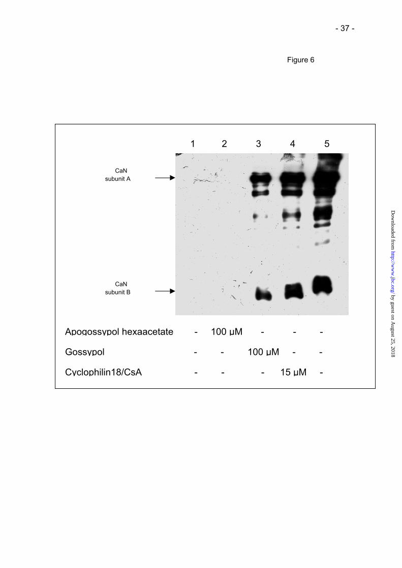
by gossypol or Cyp18/CsA, but not by apogossypol hexaacetate (Fig. 6). {insert Fig. 6} The
influence of calmodulin on CaN inhibition by gossypol is shown in Figure 7. The average
IC50-value was (18±2 µM) for four different calmodulin concentrations (25–150 nM) indicating
that gossypol does not target calmodulin for CaN inhibition. {insert Fig. 7}
Specificity of gossypol
Gossypol was tested as an inhibitor of other protein phosphatases utilizing the biotinylated
RII phosphopeptide as a general substrate of protein phosphatases. Despite the lower
efficiency relative to CaN, PP2A, and PP2C readily dephosphorylate the RII phosphopeptide
(43). In the case of PP1 phosphorylase a was chosen as a substrate, because
phosphorylated proteins are superior to phosphopeptide substrates regarding both specific
activity and higher signal to noise ratio. Except for CaN, gossypol at up to 100 µM
concentrations has no inhibitory effect on the other protein phosphatases (Table 1). {insert
Tab. 1} These results were confirmed by using the protein substrates [32P]-labeled
phosphorylase a for PP2A, and [32P]-labeled casein for CaN and PP2C (data not shown). To
exclude the possibility that in cells gossypol inactivates PPIases, human prototypic members
of the three PPIase families were tested at a concentration of 120 µM gossypol in PPIase
assays. Residual activities of 86% (Cyp18), 92 % (FKBP12) and 87 % (Pin1) were found.
Inhibition of NFAT translocation in activated T cells
To investigate the effect of gossypol on CaN in cells, we examined the NFATC1
disappearance from the cytosol after PMA/ionomycin activation of T cells. The transcription
factor NFAT is present in the cytoplasm of resting T cells and translocates into the nucleus
only after dephosphorylation by CaN, which is activated immediately after T cell stimulation
with a specific antigen, anti CD3/CD28 antibodies or PMA/ionomycin (44). {insert Fig. 8}
by guest on August 25, 2018
http://ww
w.jbc.org/
Dow
nloaded from

- 15 -
Human T cells were purified to 98% from human PBMC by magnetic cell sorting of CD4 or
CD8 positive cells. The isolated cells were preincubated with cyclosporin A, apogossypol
hexaacetate or gossypol and then stimulated with PMA/Ionomycin in the presence of these
compounds. The cytosolic portion of NFATC1 is detected in the cytosolic extracts by
Western blot analysis. Both CsA and gossypol inhibited NFATC1 disappearance from the
cytosol in a concentration-dependent manner (Fig. 8). In contrast, apogossypol hexaacetate
neither inhibited CaN in vitro (Fig. 2) nor prevented NFATC1 disappearance from cytosol
after T cell stimulation (Fig. 8).
The NFAT-luciferase reporter gene assay was used to show, that the cytoplasmic
disappearance of NFAT after stimulation indeed corresponds to NFAT activation by NFAT
dephosphorylation, and therefore its nuclear appearance with concomitant, DNA binding. It
was confirmed by luciferase activity measurement, that gossypol and CsA inhibit this NFAT
activation but apogossypol hexaacetate did not (Fig. 9). {insert Fig. 9}
by guest on August 25, 2018
http://ww
w.jbc.org/
Dow
nloaded from

- 16 -
DISCUSSION
In search for direct small molecule inhibitors of calcineurin, the naturally occurring
polyphenolic aldehyde gossypol (C30H30O8) was found to inhibit CaN reversibly with a
potency in the micromolar range (Fig. 10). {insert Fig. 10} There was no previous evidence
for the interaction of gossypol with phosphatases. Furthermore, an about two-fold increase in
inhibitory potency was observed for the chinoid metabolic derivative gossypolone in
comparison with gossypol, with an IC50 value of 7 µM. Both CsA and FK506 noncompetitively
inhibit CaN with IC50-values of 270 nM (42) and 57 nM (45), respectively, but each requires
the prior binding of a matchmaker protein. Despite the significant homology between the
catalytic subunits of the Ser/Thr protein phosphatases the CaN specificity of the
PPIase/CsA(FK506) complexes among the four protein phosphatase families is high
because the inhibitory complex does not involve the active site (46, 47). Similarly, gossypol
is highly specific for CaN among the Ser/Thr protein phosphatase families including PP1,
PP2A, and PP2C as judged by its specific inhibition of CaN at concentrations up to 100 µM
(Table 1). Unlike CsA and FK506, however, gossypol directly binds to CaN independent of
any matchmaker proteins. The ability of gossypol to inhibit CaN without affecting the enzyme
activity of members of the three PPIase families makes it potentially more specific probes for
calcineurin function than CsA and FK506 for which inhibition of PPIases is a prerequisite for
inhibition of CaN.
CaN is a binuclear Fe-Zn metallophosphatase, which is activated by Ca2+, calmodulin and
bivalent metal ions such as Mn2+ and Ni2+. The catalytically active form of Fe in the active
center of CaN is still under debate. It is assumed to be either Fe2+ (40) or Fe3+ (48). CaN has
been reported to be inactivated by oxidative processes and can be reactivated by ferrous
ammonium sulfate (40), DTT or the dithiol oxidoreductase thioredoxin (49). Current
hypotheses assume that either the binuclear metal center (40) or modification of cysteine
residues (49) can be the target for the oxidative inactivation of CaN. From our experiments,
we conclude that gossypol does not inactivate CaN by a redox mechanism, since (i) high
concentrations of DTT (5 mM) did not prevent inactivation, (ii) ferrous ammonium sulfate
by guest on August 25, 2018
http://ww
w.jbc.org/
Dow
nloaded from

- 17 -
failed to reverse the inactivation, and (iii) dialysis recovered a major part of CaN activity. In
contrast, gossypol in the micromolar concentration range inactivates sperm adenylate
cyclase by chelating the essential metal ion of the enzyme. High manganese concentration
protects this enzyme from inactivation.
Gossypol inhibits CaN independent of the nature of the three substrates used here. This
finding is in contrast with the effect of the Cyp18/CsA (FKBP12/FK506) complexes, which
inhibits the dephosphorylation of the RII phosphopeptide but activated the phosphatase
activity of CaN towards a small substrate pNPP by 2-3 fold (2, 5).
Like Cyp18/CsA (31), gossypol inhibits CaN noncompetitively, raising the possibility that it
may bind to a site overlapping that for Cyp18/CsA. In fact, the competition experiment
(Figure 5) implies that both complexes might exhibit similarity in the way these inhibitors
block CaN-catalyzed dephosphorylation. The specific elution by gossypol of CaN from a
matrix-bound ternary complex Cyp18/CsA/CaN (Fig. 6) supports this inhibition model. The
structure of the CaN/Cyp18/CsA complex has not been published so far but may relate to the
ternary inhibitory complex of FK506 (47). Here, the CaN-interacting parts of the binary
FKBP12/FK506 complex are remote to the active site of the phosphatase. Because the
active sites of CaN, PP1, PP2A and PP2C display considerable similarities inhibitory
specificity requires inhibitor binding at remote sites. Accordingly, the observed specificity of
gossypol among the protein phosphatases is in accordance with the noncompetitive type of
inhibition. Active-site directed CaN inhibitors like microcystin or the mixed type inhibitor
okadaic acid do not show CaN specificity among the protein phosphatases (31).
Because the use of pNPP substrate in the CaN assay revealed gossypol to inhibit CaN with
IC50 = 14±1 µM, which is similar to the IC50 value with the RII phosphopeptide as a substrate,
a binding regime closer to the active site is implied, but specificity is still retained.
In addition to pNPP and the RII phosphopeptide, we also examined the effect of gossypol
and analogs on the dephosphorylation of a protein substrate in vivo. Thus, the CaN
dependent activation of the transcription factor NFAT was investigated in the presence of
by guest on August 25, 2018
http://ww
w.jbc.org/
Dow
nloaded from

- 18 -
gossypol and apogossypol hexaacetate in human PBMCs. Gossypol is advantageous over
peptidic inhibitors (25, 26) for cellular experiments as it is cell permeable (50). In our assay,
the phosphoprotein NFATC1 migrating with an apparent molecular mass of 90-115 kDa in
SDS-polyacrylamide gels serves as the CaN substrate. It was previously shown that the
Cyp18/CsA (or FKBP12/FK506) complex-mediated inhibition of dephosphorylation of NFAT
is responsible for the inhibition of IL-2 transcription, and thus T-cell proliferation (46, 51).
Gossypol, but not apogossypol hexaacetate, prevented the dephosphorylation and nuclear
translocation of NFATC1 by CaN in a concentration-dependent manner. This effect is similar
to that observed for the Cyp18/CsA complex observed previously (51) (Fig. 8) The NFAT-
luciferase reporter gene assay was used to show, that the cytoplasmic disappearance of
NFAT after stimulation indeed corresponds to NFAT activation by NFAT dephosphorylation,
and therefore its nuclear appearance with concomitant DNA binding and gene transcription.
It was confirmed by luciferase activity measurement that gossypol and CsA inhibit the NFAT
activation but apogossypol hexaacetate did not (Fig. 9).
Inhibition of CaN by gossypol was found to be independent of CaN activation by calmodulin
suggesting that neither cofactor requirement nor Calmodulin binding site competition was
characteristic of gossypol-mediated inhibition even thought gossypol was also reported to
bind to calmodulin at an independent site (52).
The discovery of gossypol as a specific CaN inhibitor is also expected to shed light on how
this compound plays the well-known role in male antifertilization, inhibition of proliferation of
tumor cells, and how it mediates side effects of the contraceptive therapy (53, 54, 55, 56).
Gossypol has been already reported to inactivate intracellular dehydrogenases, protein
kinases, steroidogenic adrenal enzymes, cathepsin L, and topoisomerase II. Noncovalent
enzyme/gossypol complexes are formed by PKC (IC50=100 µM) (57), cyclic AMP-dependent
protein kinase (IC50=10 µM) (58), and at the NADH cofactor binding site of LDH (IC50=1 µM)
(59). Rapid binding of gossypol followed by slow covalent protein modification by Schiff base
formation at the N-terminal amino group results in the inactivation of phospholipase A2 (58).
by guest on August 25, 2018
http://ww
w.jbc.org/
Dow
nloaded from

- 19 -
Typically, gossypol concentrations ranging from 10 to 20 µM were antiproliferative in cancer
cell lines (60) irreversibly blocking cells in S phase. Spermicidial activity and alteration in
morphology of carcinoma cells can also be found with gossypolone, although it is less
efficient than gossypol (60, 61) and therefore suggest a CaN independent pathway. Yet there
exist few studies that examine the impact of gossypol on protein phosphorylation. Treatment
of human cancer cell lines with gossypol decreased the ratio of phosphorylated to
unphosphorylated cell cycle regulatory retinoblastoma protein at inhibitor concentrations
compatible with CaN inhibition but suggests a protein kinase as a putative gossypol target
(62). On the other hand, it is interesting to note that the increase in the activity of ornithine
decarboxylase, and subsequent proliferation following prolactin-stimulation of Nb 2 rat
lymphoma cells can be blocked with both CsA and gossypol. (63).
In conclusion, the novel protein Ser/Thr phosphatase inhibitor gossypol, which is specific for
CaN among different members of the phosphatase superfamily, allows CsA(FK506)-
mediated CaN and PPIase effects to be differentiated. Previous attempts to distinguish
among the enzymes were limited to monofunctional PPIase inhibitors that have no effect on
CaN. Now the possibility has been extended to a monofunctional CaN inhibitor that does not
require the mediation of any PPIases.
Our experiments with gossypol derivatives showed that gossypol may serve as a lead for the
development of specific and potent CaN inhibitors active in the nanomolar range, and with
improved specificity for CaN while discriminating against other gossypol-sensitive enzymes.
by guest on August 25, 2018
http://ww
w.jbc.org/
Dow
nloaded from
- 20 -
REFERENCES
1. Mackintosh, C. R. W. (1994) Trends Biochem. Sci. 19, 444-448
2. Liu, J., Farmer, J. D. Jr., Lane, W. S., Friedman, J., Weissman, I., and Schreiber, S. L.
(1991) Cell 66, 807-15
3. Schreiber, S. L., Albers, M. W., and Brown, E. J. (1993) Accounts Chem. Res. 26, 412-
420
4. Fischer, G. (1994) Angew. Chem. Int. Edit. 33, 1415-1436
5. Swanson, S. K., Born, T., Zydowsky, L. D., Cho, H., Chang, H. Y., Walsh, C. T., and
Rusnak, F. (1992) P. Natl. Acad. Sci. USA 89, 3741-3745
6. Sewell, T. J., Lam, E., Martin, M. M., Leszyk, J., Weidner, J., Calaycay, J., Griffin, P.,
Williams, H., Hung, S., Cryan, J., Sigal, N.H., and Wiederrecht, G.J. (1994) J. Biol. Chem.
269, 21094-21102
7. Baughman, G., Wiederrecht, G. J., Campbell, N. F, Martin, M. M., and Bourgeois, S.
(1995) Mol. Cell. Biol. 15, 4395-4402
8. Wang, T. W., Li, B. Y., Danielson, P. D., Shah, P. C., Rockwell, S., Lechleider, R. J.,
Martin, J., Manganaro, T., and Donahoe, P. K. (1996) Cell 86, 435-444
9. Lopez-Ilasaca, M., Schiene, C., Kullertz, G., Tradler, T., Fischer, G., and Wetzker, R.
(1998) J. Biol. Chem. 273, 9430-9434
by guest on August 25, 2018
http://ww
w.jbc.org/
Dow
nloaded from

- 21 -
10. Zarnt, T., Lang, K., Burtscher, H., and Fischer, G. (1995) Biochem. J. 305, 159-164
11. Janowski, B., and Fischer, G. (1997) Bioorg. Med. Chem. 5, 179-186
12. Nelson, P. A. , Akselband, Y., Kawamura, A., Su, M., Tung, R. D., Rich, D. H., Kishore,
V., Rosborough, S. L, Decenzo, M. T., Livingston, D. J., and Harding, M.W. (1993) J.
Immunol. 150, 2139-2147
13. Bartz, S. R., Hohenwalter, E., Hu, M. K., Rich, D.H., and Malkovsky, M. (1995) P. Natl.
Acad. Sci. USA 92, 5381-5385
14. Abraham, R. T., and Wiederrecht, G. J. (1996) Annu. Rev. Immunol. 14, 483-510
15. Gold, B. G. (2000) Expert Opin. Inv. Drug. 9, 2331-2342
16. Richter, A., Davies, D. E., and Alexander, P. (1995) Biochem. Pharmacol. 49, 367-373
17. Enz, A., and Pombovillar, E. (1997) Biochem. Pharmacol. 54, 321-323
18. Fakata, K. L., Swanson, S. A., Vorce, R. L., and Stemmer, P. M. (1998) Biochem.
Pharmacol. 55, 2017-2022
19. Gualberto, A., Marquez, G., Carballo, M., Youngblood, G. L., Hunt, S. W., Baldwin, A. S.,
and Sobrino, F. (1998) J. Biol. Chem. 273, 7088-7093
20. Born, T. L, Myers, J. K., Widlanski, T. S, and Rusnak, F. (1995) J. Biol. Chem. 270,
25651-25655
by guest on August 25, 2018
http://ww
w.jbc.org/
Dow
nloaded from

- 22 -
21. Enz, A., Zenke, G., and Pombovillar, E. (1997) Bioorg. Med. Chem. Lett. 7, 2513-2518
22. Tatlock, J. H., Linton, M. A., Hou, X. J., Kissinger, C. R., Pelletier, L. A., Showalter, R. E.,
Tempczyk, A., and Villafranca, J. E. (1997) Bioorg. Med. Chem. Lett. 7, 1007-1012
23. Brill, G., M., Premachandran, U., Karwowski, J., P., Henry, R., Cwik, D., K., Traphagen,
L., M., Humphrey, P., E., Jackson, M., Clement, J., J., Burres, N., S., Kadam, S., Chen,
R., H., McAlpine, J., B. (1996) J. Antibiot. 49(2), 124-128
24. Perrino, B. A. (1999) Arch. Biochem. Biophys. 372, 159-165
25. Sagoo, J. K., Fruman, D. A., Wesselborg, S., Walsh, C. T., and Bierer, B. E. (1996)
Biochem. J. 320, 879-884
26. Coghlan, V. M., Perrino, B. A., Howard, M., Langeberg, L. K., Hicks, J. B., Gallatin, W.
M., and Scott, J. D. (1995) Science 267, 108-111
27. Tradler, T., Stoller, G., Rucknagel, K. P., Schierhorn, A., Rahfeld, J. U., and Fischer, G.
(1997) FEBS Lett. 407, 184-190
28. Schutkowski, M., Bernhardt, A., Zhou, X. Z., Shen, M. H., Reimer, U., Rahfeld, J. U., Lu,
K. P., and Fischer, G. (1998) Biochemistry 37, 5566-5575
29. Mondragon, A., Griffith, E. C., Sun, L., Xiong, F., Armstrong, C., and Liu, J. O. (1997)
Biochemistry 36, 4934-4942
30. Koch, C., Neumann, T.,Grabley, S., Thiericke, R., (1999) Drug Discovery from Nature,
Springer-Verlag Heidelberg, Heidelberg, Germany
by guest on August 25, 2018
http://ww
w.jbc.org/
Dow
nloaded from

- 23 -
31. Enz, A., Shapiro, G., Chappuis, A., and Dattler, A. (1994) Anal. Biochem. 216(1), 147-
153
32. Sullivan, E., Hemsley, P., and Pickard, A. (1997) J. Biomol. Screen. 2(1), 19-23
33. Nakayama, G. R., Nova, M. P., and Parandoosh, Z. (1998) J. Biomol. Screen. 3(1), 43-48
34. Pelech, S., and Cohen, P. (1985) Eur. J. Biochem. 148(2) , 245-51
35. Waelkens, E., Goris, J., Di Salvo, J., and Merlevede, W. (1984) Biochem.Biophys.
Res.Comm. 120(2) , 397-404
36. Krebs, E., G., Kent, A., B., and Fischer, E.,H. (1958) J. Biol. Chem. 231, 73-83
37. Khandelwal, R. L., Vandenheede, J. R., and Krebs, E. G. (1976) J. Biol. Chem. 251(16),
4850-8
38. Fischer, G., Liebold, B. W., Lang, K., Kiefhaber, T., and Schmid, F. X. (1989) Nature 337,
476-478
39. Zhou, X. Z., Kops, O., Werner, A., Lu, P. J., Shen, M. H., Stoller, G., Kullertz, G., Stark,
M., Fischer, G., and Lu, K. P. (2000) Mol. Cell 6, 873-883
40. Wang, X. T., Culotta, V. C., and Klee, C. B. (1996) Nature 383, 434-437
41. Chan, C. P., Gallis, B., Blumenthal, D. K., Pallen, C. J., Wang, J. H., and Krebs, E. G.
(1986) J. Biol. Chem. 261(21), 9890-5
by guest on August 25, 2018
http://ww
w.jbc.org/
Dow
nloaded from

- 24 -
42. Etzkorn, F. A., Chang, Z. Y., Stolz, L. A., and Walsh, C. T. (1994) Biochemistry 33, 2380-
2388
43. Donella-Deana, A., Meyer, H. E., and Pinna, L. A. (1991) Biochim. Biophys. Acta
1094(1), 130-133
44. Loh, C., Shaw, K. T. Y., Carew, J., Viola, J. P. B., Luo, C., Perrino, B. A., and Rao, A.
(1996) J. Biol. Chem. 271, 10884-10891
45. Dumont, F. J., Staruch, M. J., Koprak, S. L., Siekierka, J. J., Lin, C. S., Harrison, R.,
Sewell, T., Kindt, V. M., Beattie, T. R., Wyvratt, M., and a, l. (1992) J. Exp. Med. 176,
751-760
46. Liu, J., Albers, M. W., Wandless, T. J., Luan, S., Alberg, D. G., Belshaw, P. J., Cohen, P.,
Mackintosh, C., Klee, C. B., and Schreiber, S. L. (1992) Biochemistry 31, 3896-3901
47. Griffith, J. P., Kim, J. L., E. E, Sintchak, M. D., Thomson, J. A., Fitzgibbon, M. J., Fleming,
M. A., Caron, P.R, Hsiao, K, and Navia M. A. (1995) Cell 82, 507-522
48. Yu, L., Haddy, A., and Rusnak, F. (1995) J. Am. Chem. Soc. 117, 10147-10148
49. Bogumil, R., Namgaladze, D., Schaarschmidt, D., Schmachtel, T., Hellstern, S., Mutzel,
R., and Ullrich, V. (2000) Eur. J. Biochem. 267(5), 1407-1415
50. Vander Jagt, D. L., Deck, L. M., and Royer, R. E. (2000) Curr. Med. Chem. 7(4), 479-98
by guest on August 25, 2018
http://ww
w.jbc.org/
Dow
nloaded from

- 25 -
51. Shaw, K.T.Y, Ho, A.M, Raghavan, A, Kim, J, Jain, J.N, Park, J.C, Sharma, S, Rao,
Hogan, and P.G. (1995) P. Natl. Acad. Sci. USA 92, 11205-11209
52. Jinsart, W., Ternai, B., and Polya, G. M. (1991) Biol. Chem. Hoppe Seyler 372, 819-827
53. Poso, H., Wichmann, K., Janne, J., Luukkainen, T. (1980) Lancet 1(8173), 885-886
54. Coyle, T., Levante, S., Shetler, M., and Winfield, J. (1994) J. Neuro-oncol. 19, 25-35
55. Flack, M. R., Pyle, R. G., Mullen, N. M., Lorenzo, B., Wu, Y. W., Knazek, R. A., Nisula, B.
C., and Reidenberg, M. M. (1993) J. Clin. Endocr. Metab. 76(4), 1019-1024
56. Frick, J., Aulitzky, W., and Kalla, N. R. (1988) Contraception 37(2), 153-162
57. Gomez, M. S., Piper, R. C., Hunsaker, L. A., Royer, R. E., Deck, L. M., Makler, M. T., and
Jagt, D. L. V. (1997) Mol. Biochem. Parasit. 90, 235-246
58. Yu, B. Z., Rogers, J., Ranadive, G., Baker, S., Wilton, D. C., Apitz-Castro, R., and Jain,
M. K. (1997) Biochemistry 36(41), 12400-12411
59. Russell, D. H., Buckley, A. R., Montgomery, D. W., Larson, N. A., Gout, P. W., Beer, C.
T., Putnam, C. W., Zukoski, C. F., and Kibler, R. (1987) J. Immunol. 138, 276-284
60. Gilbert, N. E., Reilly, J. E. O., Chang, C. J., Lin, Y. C., and Brueggemeier, R. W. (1995)
Life Sci. 57, 61-67
61. Kim, I. C., Waller, D. P., Marcelle, G. B., Cordell, G. A., Fong, H. H., Pirkle, W. H., Pilla,
L., and Matlin, S. A. (1984) Contraception 30, 253-259
by guest on August 25, 2018
http://ww
w.jbc.org/
Dow
nloaded from

- 26 -
62. Ligueros, M., Jeoung, D., Tang, B., Hochhauser, D., Reidenberg, M. M., and Sonenberg,
M. (1997) Brit. J. Cancer 76, 21-28
63. Shidaifat, F., Canatan, H., Kulp, S. K., Sugimoto, Y., Zhang, Y., Brueggemeier, R. W.,
Somers, W. J., Chang, W. Y., Wang, H. C., and Lin, Y. C. (1997) Anticancer Res. 17,
1003-1009
ACKNOWLEDGMENTS
The authors wish to thank Cordelia Schiene-Fischer for critical reading of the manuscript. We
are also grateful to Martina Heidler for excellent technical assistance and Mario Drewello for
peptide synthesis and Jens Rahfeld for providing rhCyp18.
This work was supported by the Fonds der Chemischen Industrie.
1The abbreviations used are: CsA, cyclosporin A; FK506, tacrolimus; rhCyp18,
recombinant human cyclophilin 18; FKBP12, FK506-binding protein 12; PPIase, peptidyl
prolyl cis/trans isomerase; PP1, Ser/Thr protein phosphatase type 1; PP2A, protein
phosphatase type 2A; PP2B, protein phosphatase type 2B or calcineurin; PP2C, protein
phosphatase 2C; pNPP, p-nitrophenyl phosphate; RII phosphopeptide, 19-residue
phosphopeptide of the regulatory subunit of type II cAMP-dependent protein kinase; PKC,
protein kinase C; LDH, lactate dehydrogenase; PMA, phorbol 12-myristate 13-acetate;
PBMC, peripheral blood mononuclear cells; NFAT, nuclear factor of activated T cell
by guest on August 25, 2018
http://ww
w.jbc.org/
Dow
nloaded from

- 27 -
FIGURE LEGENDS
Fig. 1:
Assay characterization.
The Ca2+/calmodulin stimulated recombinant human CaN activity was determined using 10
pmol/well biotinylated RII phosphopeptide substrate at pH 7.5, 30°C.
(A) Initial rate of dephosphorylation of the RII phosphopeptide at different CaN
concentrations. The arrow depicts the CaN concentration of 1.32 nM, which was used in all
experiments. Initial rates were determined by measuring the amount of dephosphorylated
substrate after a 20 min incubation.
(B) Time course of CaN-catalyzed (1.32 nM) dephosphorylation of biotinylated RII
phosphopeptide. Dephosphorylation was terminated by addition of EGTA to a final
concentration of 10 mM.
The arrow depicts the incubation time of 20 min, which was constantly used in the
experiments. The data presented are means ± SD of triplicates from two independent
experiments.
Fig. 2:
Inhibition of CaN by gossypol and its derivatives.
Gossypol (square), gossypolone (triangle), and apogossypol hexaacetate (circle) were
preincubated with CaN (1.32 nM) and calmodulin (50 nM) for 30 min at 22°C.
(A) The CaN phosphatase activity was measured using RII phosphopeptide substrate as
described in Materials and Methods.
(B) 10 mM p-nitro phenylphosphate was used to determine protein phosphatase activity
Data are expressed as the activity, relative to reference values without inhibitor. IC50 values
were calculated by four-parameter curve fitting using the program Sigma Plot. The data
presented are mean ± SD of three independent experiments.
by guest on August 25, 2018
http://ww
w.jbc.org/
Dow
nloaded from

- 28 -
Fig. 3:
Inhibition of CaN by gossypol using the pNPP substrate.
CaN and calmodulin were preincubated with gossypol (see Fig. 2) and then CaN
phosphatase activity was measured with pNPP substrate as described (see "Materials and
Methods"). The IC50 values at pNPP concentrations of 1 mM (circle), 5 mM (square), and 25
mM (triangle) were calculated. The data presented are mean ± SD of three independent
experiments.
Fig. 4:
Dixon plot of CaN inhibition by gossypol.
Inhibition of CaN by gossypol was measured at a substrate concentration of 5 µM (circle), 6
µM (triangle), 8 µM (square), and 10 µM (diamond) RII phosphopeptide (mixture of
biotinylated and nonbiotinylated RII phosphopeptide by 1 : 200) and a CaN concentration of
0.66 nM. Each graph represents the average of 4 independent experiments. The data
presented are mean ± SD of three independent experiments.
Fig. 5:
Simultaneous inhibition of CaN by Cyp18/ CsA and gossypol.
Inhibition mediated by gossypol (12µM; squares), CsA (10µM; circles) and a mixture of CsA
and gossypol (10µM and 12µM, respectively; triangels) in dependence of the Cyp18
concentration was plotted. Residual CaN activity (1.32 nM CaN, 50 nM calmodulin) was
measured using the RII phosphopeptide substrate, and calculated relative to a reference
experiment lacking the effectors. Preincubation of CaN with effectors was performed at 22°C
for 30 min. The data presented are the mean ± SD of three independent experiments.
Graphs were calculated in terms of residual activity assuming either identical (dash-dot-dot)
or different (short dash) binding sites of gossypol and Cyp18/CsA.
by guest on August 25, 2018
http://ww
w.jbc.org/
Dow
nloaded from

- 29 -
The inhibitory Cyp18/CsA complex is formed in relation to the amount of Cyp18 applied
according to the binding constant of Cyp18 and CsA.
Fig. 6:
Elution of CaN by gossypol from a Cyp18/CsA affinity column.
A Cyp18/CsA affinity column was incubated with 30 µg calcineurin for 1 hour. Next the
column was washed with PBS (lane 1, last washing step). Subsequently calcineurin was
eluted with the following reagents: 100 µM apogossypol hexaacetate (lane 2), 100 µM
gossypol (lane 3) and 15 µM preformed Cyp18/CsA-complex (lane 4) after incubation for 10
minutes. Lane 5, western blot of a reference sample of CaN.
Fig. 7:
Effect of calmodulin on CaN inhibition by gossypol.
CaN was preincubated with gossypol and incubated with the RII phosphopeptide in presence
of 25 nM (square), 37.5 nM (triangle up), 50 nM (circle), and 150 nM (triangle down)
calmodulin. Residual CaN activity was plotted against gossypol concentration. The IC50 value
for each calmodulin concentration was calculated. The data presented are means ± SD of
three independent experiments.
Fig. 8:
Translocation of NFATC1 from the cytosol in T cells.
Purified human T cells were preincubated with 1 µM CsA (lane 3), 30 µM apogossypol
hexaacetate (lane 4) or gossypol at a concentration of 1 µM, 15 µM and 30 µM (lane 5, 6, 7)
at 37°C for 10 min. Then cells were stimulated with 40 nM PMA /2 µM ionomycin (lane 2 to 7)
in the presence of either the compounds or DMSO as control (lane 2) at 37°C for 20 min.
Depletion of NFATC1 from the cytosol was detected by immunoblotting with anti-NFATC1,
and actin monoclonal antibodies.
by guest on August 25, 2018
http://ww
w.jbc.org/
Dow
nloaded from

- 30 -
Fig. 9:
Luciferase reporter gene assay.
Sorted T cells transfected with a NFAT-luciferase reporter plasmid were stimulated with PMA
plus ionomycin for 5 h after incubation with CsA, gossypol, or apogossypol hexaacetate for
30 min. Data are expressed as luminescence activity of the extracted luciferase. The data
presented are mean ± SD of three independent experiments.
Fig. 10:
Structures of gossypol and gossypol analogues.
by guest on August 25, 2018
http://ww
w.jbc.org/
Dow
nloaded from

- 31 -
Table 1: Inhibitory effect of gossypol on enzyme activity of the major classes of protein
Ser/Thr phosphatases measured against RII phosphopeptide and in the case of
PP1 against phosphorylase a . The results are expressed as % of the respective
phosphatase activity without inhibitor (SD < 7%).
% of control activity
inhibitor concentration
20µM 100µM
PP1* 97 84
PP2A 104 107
PP2B (CaN) 38 2
PP2C 105 85
*phosphorylase a as substrate
by guest on August 25, 2018
http://ww
w.jbc.org/
Dow
nloaded from

- 32 -
Calcineurin [nM]0 2 4 6 8 10 12 14
Cal
cine
urin
act
ivity
(pm
ol/ m
in)
0,0
0,1
0,2
0,3
Time (min)
0 20 40 60 80
Dep
hosp
hory
latio
n (p
mol
/ wel
l)
0
2
4
6
8
A
B
Figure 1
by guest on August 25, 2018
http://ww
w.jbc.org/
Dow
nloaded from

- 33 -
Inhibitor [µM]1 10 100
0
20
40
60
80
100
120
Cal
cine
urin
act
ivity
(% o
f con
trol
)
Figure 2
Inhibitor [µM]
1 10 1000
20
40
60
80
100
120
Cal
cine
urin
act
ivity
(% o
f con
trol
)A
B by guest on August 25, 2018
http://ww
w.jbc.org/
Dow
nloaded from

- 34 -
Gossypol [µM]
0.1 1 10 100 1000
Cal
cine
urin
act
ivity
(% o
f con
trol
)
0
20
40
60
80
100
Figure 3
by guest on August 25, 2018
http://ww
w.jbc.org/
Dow
nloaded from

- 35 -
Figure 4
-30 -20 -10 0 10 20 300
2
4
6
8
10
12
14
Gossypol [µM]
1/v
(pm
ol-1
min
)
by guest on August 25, 2018
http://ww
w.jbc.org/
Dow
nloaded from

- 36 -
rh Cyp18 [nM]
Figure 5
10 100 1000
Cal
cine
urin
act
ivity
(% o
f con
trol
)
0
20
40
60
80
100
by guest on August 25, 2018
http://ww
w.jbc.org/
Dow
nloaded from
- 37 -
Apogossypol hexaacetate - 100 µM - - -
Gossypol - - 100 µM - -
Cyclophilin18/CsA - - - 15 µM -
1 2 3 4 5
subunit A
subunit B
CaN
CaN
Figure 6
by guest on August 25, 2018
http://ww
w.jbc.org/
Dow
nloaded from

- 38 -
Gossypol [µM]1 10 100
0.0
0.1
0.2
0.3
Cal
cine
urin
act
ivity
[pm
ol/ m
in]
Figure 7
by guest on August 25, 2018
http://ww
w.jbc.org/
Dow
nloaded from

- 39 -
NFATC1cyt
Actincyt
1 2 3 4 5 6 7
– + + + + + +PMA/Ionomycin
Cyclosporin A – – 1 µM – – – –Apogossypol hexaacetate – – – 30 µM – – –Gossypol – – – – 1 µM 15 µM 30 µM
Figure 8
by guest on August 25, 2018
http://ww
w.jbc.org/
Dow
nloaded from

- 40 -
Figure 9
PMA/Ionomycin - + + + + + +
Cyclosporin A - - 1 µM - - - -
Apogossypol hexaacetate - - - 30 µM - - -
Gossypol - - - - 1 µM 15 µM 30 µM
RLU
0
50
100
150
200
250
300
350
by guest on August 25, 2018
http://ww
w.jbc.org/
Dow
nloaded from

- 41 -
Figure 10
CH3
CH3
CH3 CH3
OH
OH
OHO
OHO
OH
OH
CH3CH3
H
H
CH3
CH3
CH3 CH3
OH
OH
O
OO
O
OO
OH
OH
CH3CH3
H
H
CH3
CH3
CH3 CH3
O
O
O
O
O
O
CH3CH3
CH3
O
CH3
OCH3
CH3
O
CH3 OCH3
O
O
Gossypol
Gossypolone Apogossypol hexaacetate
by guest on August 25, 2018
http://ww
w.jbc.org/
Dow
nloaded from

Susanne Grabley and Gunter FischerRia Baumgrass, Matthias Weiwad, Frank Erdmann, Jun O. Liu, Dirk Wunderlich,
Reversible inhibition of calcineurinby the polyphenolic aldehyde gossypol
published online October 11, 2001J. Biol. Chem.
10.1074/jbc.M103273200Access the most updated version of this article at doi:
Alerts:
When a correction for this article is posted•
When this article is cited•
to choose from all of JBC's e-mail alertsClick here
by guest on August 25, 2018
http://ww
w.jbc.org/
Dow
nloaded from