retinoblastoma and hirschsprung disease in a patient with interstitial deletion of chromosome 13
TRANSCRIPT

Brief Clinical Report
Retinoblastoma and Hirschsprung Disease in aPatient With Interstitial Deletion ofChromosome 13
Brenda J. Weigel,1 Mary Ella M. Pierpont,2 Terri L. Young,3 Scott B. Mutchler,4 andJoseph P. Neglia1*1Department of Pediatrics, Division of Hematology/Oncology, University of Minnesota, Minneapolis, Minnesota2Department of Pediatrics, Division of Genetics, University of Minnesota, Minneapolis, Minnesota3Department of Ophthalmology, University of Minnesota, Minneapolis, Minnesota4Department of Pediatrics, Fargo, North Dakota
Retinoblastoma is a rare pediatric malig-nancy (1/20,000) while Hirschsprung diseaseis a relatively common pediatric disorder (1/5,000). We describe a boy with bilateral ret-inoblastoma, Hirschsprung disease, mul-tiple minor anomalies, and an interstitialdeletion 13q (q13 → q22). This child and asimilar previously reported girl with retino-blastoma and Hirschsprung disease mayrepresent a previously unrecognized con-tiguous gene syndrome. Am. J. Med. Genet.77:285–288, 1998 © 1998 Wiley-Liss, Inc.
KEY WORDS: r e t i n o b l a s t o m a ; H i r -schsprung disease; chromo-some 13
INTRODUCTION
Previously there was only one report of Hir-schsprung disease and retinoblastoma in a child with adeletion of chromosome 13 [Sparkes et al., 1984]. Wenow describe a boy with Hirschsprung disease and bi-lateral retinoblastoma. These two patients may repre-sent a previously unrecognized contiguous gene syn-drome and may help to more clearly delineate the ge-netics of Hirschsprung disease and neuronaldevelopment.
CLINICAL REPORTThe patient is a boy born at 38 weeks of gestation
following an uncomplicated pregnancy and delivery.
He is the second child born to a 26-year-old mother.There is a healthy 2-year-old sister. The parents arenot consanguineous. There is no family history of reti-noblastoma, Hirschsprung disease, or congenitalanomalies. Another pregnancy of the mother ended in afirst trimester spontaneous abortion.
At birth, the Apgar scores were 6 and 7 at 1 and 5min, respectively. He was small for gestational agewith a birth weight of 2,580 g (5th centile), a length of47 cm (5th centile), and a head circumference of 35.2cm (50th centile). He required oxygen after delivery,and meconium aspiration was suspected. Over the next48 hr, abdominal distention and poor feeding were ob-served. Rectal biopsy demonstrated aganglionic mega-colon with a barium enema study confirming large seg-ment Hirschsprung disease. A diverting colostomy wasperformed.
On examination, the head was large for the body andthere was occipital prominence. The fontanel was largeand the forehead sloping. There was hypertelorism.The ears were large, posteriorly angulated, and appar-ently low-set with underdeveloped antihelices. The na-sal bridge was flattened and the nares anteverted. Thephiltrum was long with a thin upper lip and down-turned mouth. The palate was high arched, and micro-gnathia was present. The chest and lungs were normal.There was a grade 2/6 systolic ejection murmur. Pe-ripheral pulses were normal. The abdomen was protu-berant. The thumbs and great toes were broad, andthere were mild contractures at elbows, knees, andankles. The genitalia were normal. He had a weakhoarse cry and hypotonia. Reflexes were normal.
An ophthalmologic examination under anesthesia(EUA) at age 3 weeks showed a large white retinalmass occupying one half of the volume of the posteriorchamber and involving the macula of the right eye,consistent with retinoblastoma. There did not appearto be any vitreous seeding or anterior chamber involve-
*Correspondence to: Joseph P. Neglia, M.D., University of Min-nesota, Box 484 UMHC, 420 Delaware Street S.E., Minneapolis,MN 55455.
Received 13 October 1997; Accepted 11 February 1998
American Journal of Medical Genetics 77:285–288 (1998)
© 1998 Wiley-Liss, Inc.
ment at the time of examination. The left eye appearednormal. A computerized tomography scan of the headand orbits without contrast demonstrated the right in-traocular lesion with no evidence of optic nerve or in-tracranial involvement. The cerebrospinal fluid did notcontain malignant cells, and a bone scan was normal.Enucleation of the right eye was performed at 3½weeks of age with no pathological evidence of tumorextension into the optic nerve, sclera, or episcleral ves-sels. The ciliary nerves of the enucleated right eye werealso carefully examined and were found to be normal.
At age 3 months, the patient underwent an EUAduring which it was discovered that he had developedfour retinoblastoma tumors of the left eye measuringone-half disc diameter to a full disc diameter in size.Chemoreduction with vincristine, carboplatin, and eto-poside was initiated, and laser therapy of the lesionswas planned after chemoreduction. Unfortunately, hetolerated the chemotherapy very poorly, and lasertherapy and cryotherapy were done after the firstcourse of chemotherapy. No further chemotherapy isplanned at this time unless there is extension into theoptic nerve of the left eye. The patient’s parents and2-year-old sister underwent ophthalmologic evalua-tions with no abnormal findings.
After birth, he had very slow feeding and inadequatecaloric intake. A gastrostomy was placed at age 6months. At age 10 months he receives all feedings viathe gastrostomy, has poor head control, and is unableto sit unassisted. He will be evaluated regularly byEUA for evidence of retinoblastoma progression in theleft eye. Surgical reversal of his colostomy is plannedfor age 12–14 months.
CYTOGENETIC STUDIES
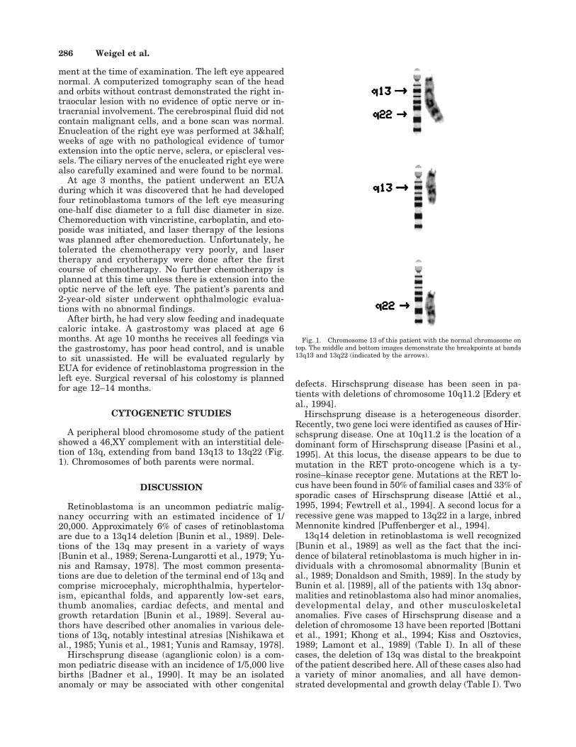
A peripheral blood chromosome study of the patientshowed a 46,XY complement with an interstitial dele-tion of 13q, extending from band 13q13 to 13q22 (Fig.1). Chromosomes of both parents were normal.
DISCUSSION
Retinoblastoma is an uncommon pediatric malig-nancy occurring with an estimated incidence of 1/20,000. Approximately 6% of cases of retinoblastomaare due to a 13q14 deletion [Bunin et al., 1989]. Dele-tions of the 13q may present in a variety of ways[Bunin et al., 1989; Serena-Lungarotti et al., 1979; Yu-nis and Ramsay, 1978]. The most common presenta-tions are due to deletion of the terminal end of 13q andcomprise microcephaly, microphthalmia, hypertelor-ism, epicanthal folds, and apparently low-set ears,thumb anomalies, cardiac defects, and mental andgrowth retardation [Bunin et al., 1989]. Several au-thors have described other anomalies in various dele-tions of 13q, notably intestinal atresias [Nishikawa etal., 1985; Yunis et al., 1981; Yunis and Ramsay, 1978].
Hirschsprung disease (aganglionic colon) is a com-mon pediatric disease with an incidence of 1/5,000 livebirths [Badner et al., 1990]. It may be an isolatedanomaly or may be associated with other congenital
defects. Hirschsprung disease has been seen in pa-tients with deletions of chromosome 10q11.2 [Edery etal., 1994].
Hirschsprung disease is a heterogeneous disorder.Recently, two gene loci were identified as causes of Hir-schsprung disease. One at 10q11.2 is the location of adominant form of Hirschsprung disease [Pasini et al.,1995]. At this locus, the disease appears to be due tomutation in the RET proto-oncogene which is a ty-rosine–kinase receptor gene. Mutations at the RET lo-cus have been found in 50% of familial cases and 33% ofsporadic cases of Hirschsprung disease [Attie et al.,1995, 1994; Fewtrell et al., 1994]. A second locus for arecessive gene was mapped to 13q22 in a large, inbredMennonite kindred [Puffenberger et al., 1994].
13q14 deletion in retinoblastoma is well recognized[Bunin et al., 1989] as well as the fact that the inci-dence of bilateral retinoblastoma is much higher in in-dividuals with a chromosomal abnormality [Bunin etal., 1989; Donaldson and Smith, 1989]. In the study byBunin et al. [1989], all of the patients with 13q abnor-malities and retinoblastoma also had minor anomalies,developmental delay, and other musculoskeletalanomalies. Five cases of Hirschsprung disease and adeletion of chromosome 13 have been reported [Bottaniet al., 1991; Khong et al., 1994; Kiss and Osztovics,1989; Lamont et al., 1989] (Table I). In all of thesecases, the deletion of 13q was distal to the breakpointof the patient described here. All of these cases also hada variety of minor anomalies, and all have demon-strated developmental and growth delay (Table I). Two
Fig. 1. Chromosome 13 of this patient with the normal chromosome ontop. The middle and bottom images demonstrate the breakpoints at bands13q13 and 13q22 (indicated by the arrows).
286 Weigel et al.

of these patients had eye abnormalities: one with reti-noblastoma and deletion 13q14.1 → 13q22.3 [Sparkeset al., 1984], and the second with bilateral optic nervehypoplasia and deletion 13q21.2 → 13q22 [Khong et al.,1994].
Intestinal atresia has been reported with retinoblas-toma. Yunis and Ramsay [1978] reported on a boy withdeletion of chromosome 13 at bands q14 → q21 withminor anomalies, developmental and growth delay, je-junal atresia, and bilateral retinoblastoma. A secondcase of intestinal atresia, minor anomalies, and unilat-eral retinoblastoma was reported by Yunis et al.[1981]. This boy had a large interstitial deletion from13q14 → 13q31.
The only patient with retinoblastoma and Hir-schsprung disease published previously was one re-ported by Sparkes et al. [1984]. This was a girl withbilateral retinoblastoma, developmental delay, andHirschsprung disease associated with a deletion at13q14.1 → 13q22.3. Our patient and that of Sparkes etal. [1984] share the common region on chromosome 13from q14.1 to q22. The deletion of part of chromosome13 in this region may unmask a recessive mutant allelefor Hirschsprung disease on the normal chromosome.
These two patients (ours and that of Sparkes et al.[1984]) may represent a previously unrecognized con-tiguous gene syndrome. These findings suggest a rela-tionship between chromosome 13 and neuronal devel-opment of the gastrointestinal tract and the eye. If anindividual has retinoblastoma, the possibilities of Hir-
schsprung disease and deletion of chromosome 13 needto be considered.
ACKNOWLEDGMENTS
The authors would like to thank Dr. Betsy Hirschand Rosalee Phillips of the Cytogenetics Laboratory atthe University of Minnesota for the cytogenetic analy-sis and chromosome images of this patient.
REFERENCES
Attie T, Pelet A, Sarda P, Eng C, Edery P, Mulligan LM, Pomder BAJ,Munnich A, Lyonnet S (1994): A 7 bp deletion of the RET proto-oncogene in familial Hirschsprung’s disease. Hum Mol Genet 3:1439–1440.
Attie T, Pelet A, Edery P, Eng C, Mulligan LM, Amiel J, Boutrand L,Beldjord C, Nihoul-Fekete C, Munnich A, Ponder BAJ, Lyonnet S(1995): Diversity of RET proto-oncogene mutations in familial and spo-radic Hirschsprung disease. Hum Mol Genet 4:1381–1386.
Badner JA, Sieber WK, Garver KL, Chakravarti A (1990): A genetic studyof Hirschsprung disease. Am J Hum Genet 46:568–580.
Bottani A, Xie Y, Binkert F, Schinzel A (1991): A case of Hirschsprungdisease with a chromosome 13 microdeletion, del(13)(q32.2q33.2): po-tential mapping of one disease locus. Hum Genet 87:748–750.
Bunin GR, Emanuel BS, Meadows AT, Buckley JD, Woods WG, HammondGD (1989): Frequency of 13q abnormalities among 203 patients withretinoblastoma. J Natl Cancer Inst 81:370–374.
Donaldson SS, Smith LM (1989): Retinoblastoma: biology, presentation,and current management. Oncology 3:45–52.
Edery P, Pelet A, Mulligan LM, Abel L, Attié T, Dow E, Bonneau D,David A, Flintoff W, Jan D, Journel H, Lacombe D, Le Merrer M,Meijers C, Parent P, Philip N, Plauchu H, Sarda P, Verloes A, Nihoul-Fekete C, Williamson R, Ponder BAJ, Munnich A, Lyonnet S (1994):
TABLE I. A Summary of Studies Involving Deletions in Chromosome 13q and Hirschsprung Disease or Intestinal Anomalies
Deletion Eye anomalyIntestinalanomaly
Otheranomaly Reference
13q13 → 13q22 Bilateralretinoblastoma
Hirschsprung disease Syndromal,a SGA,b developmentaldelay
Current patient
13q14.1 → 13q22.3 Bilateralretinoblastoma
Hirschsprung disease Developmental delay, growthretardation, hypotonia
Sparkes et al.,1984
13q14 → 13q21 Bilateralretinoblastoma
Jejunal atresia Syndromal, developmental delay,hypotonia
Yunis andRamsay, 1978
13q14 → 13q31 Unilateralretinoblastoma
Agenesis of colonicmesenteries,redundant sigmoid,hypoplastic gallbladder
Small stature, vertebralanomalies, syndromal,cardiomegaly, hypoplasticthymus
Yunis et al., 1981
13q21.2 → 13q22 Bilateral optic nervehypoplasia
Duodenal and jejunalatresia, Hirschsprungdisease
SGA, umbilical artery ulceration,polysplenia, syndromal
Khong et al., 1994
13q22.1 → 13q32.1 Normal Hirschsprung disease Growth retardation, syndromal,renal anomalies, vertebralanomalies, dislocation of hips,undescended testes
Lamont et al.,1989
13q21.2 → 13q32.3 Normal Hirschsprung diseaseand malrotation
Global delay, syndromal,cryptorchidism
13q22 → qter Normal Hirschsprung disease Syndromal, cryptorchidism,developmental delay
Kiss andOsztovics, 1989
13q32.3 → 13q33.2 Normal Hirschsprung disease Syndromal, developmental delay Bottani et al.,1991
13q14 Bilateralmicrophthalmos
Jejunal and ileal atresia Syndromal, SGA Nishikawa et al.,1985
aSyndromal includes a combination of the following: occipital prominence, large fontanel, hypertelorism, large, low-set, posteriorly angulated ears,underdeveloped external ears, flattened nasal bridge, anteverted nares, long philtrum, thin upper lip, downturned mouth, high arched palate, micro-gnathia, broad thumbs and great toes, epicanthal folds, nipple hypoplasia, inguinal hernias, prominent calcani, clinodactyly of fingers, simian crease,ptosis, umbilical hernia, syndactly of toes.bSGA 4 small for gestational age.
Retinoblastoma and Hirschsprung Disease 287
Long segment and short segment familial Hirschsprung’s disease: vari-able clinical expression at the RET locus. J Med Genet 31:602–606.
Fewtrell MS, Tam PKH, Thomson AH, Fitchett M, Currie J, Huson SM,Mulligan LM (1994): Hirschsprung’s disease associated with a deletionof chromosome 10 (q11.1q21.2): a further link with the neurocristopa-thies? J Med Genet 31:325–327.
Khong TY, Ford WDA, Haan EA (1994): Umbilical cord ulceration in as-sociation with intestinal atresia in a child with deletion 13q and Hir-schsprung’s disease. Arch Dis Child 71:F212–F213.
Kiss P, Osztovics M (1989): Association of 13q deletion and Hirschsprung’sdisease. J Med Genet 26:793–794.
Lamont MA, Fitchett M, Dennis NR (1989): Interstitial deletion of distal13q associated with Hirschsprung’s disease. J Med Genet 26:100–104.
Nishikawa A, Mitomori T, Matsuura A, Inoue A, Mori H, Takahashi M(1985): A 13q-syndrome with extensive intestinal atresia. Acta Paedi-atr 74:305–308.
Pasini B, Borrello MG, Greco A, Bongarzone I, Luo Y, Mondellini P, AlbertiL, Miranda C, Arighi E, Bocciardi R, Seri M, Barone V, Radice MT,
Romeo G, Pierotti MA (1995): Loss of function effect of RET mutationscausing Hirschsprung disease. Nat Genet 10:35–40.
Puffenberger ER, Kauffman ER, Bolk S, Matis TC, Washington SS, An-grist M, Weissenbach J, Garver KL, Mascari M, Ladda R, Slaugen-haupt SA, Chakravarti A (1994): Identity-by-descent and associationmapping of a recessive gene for Hirschsprung disease on human chro-mosome 13q22. Hum Mol Genet 3:1217–1225.
Serena-Lungarotti M, Calabro A, Mariotti G, Mastroiacovo PP, Proven-zano S, Dallapiccola B (1979): Interstitial deletion 13q syndromes: Areport on two unrelated patients. Hum Genet 52:269–274.
Sparkes RS, Sparkes MC, Kalina RE, Pagon RA, Salk DJ, Disteche CM(1984): Separation of retinoblastoma and esterase D loci in a patientwith sporadic retinoblastoma and del(13)(q14.1q22.3). Hum Genet 68:258–259.
Yunis E, Zuniga R, Ramirez E (1981): Retinoblastoma, gross internal mal-formations, and deletion 13q14→q31. Hum Genet 56:283–286.
Yunis JJ, Ramsay N (1978): Retinoblastoma and subband deletion of chro-mosome 13. Am J Dis Child 132:161–163.
288 Weigel et al.