release of bioactive peptides from milk proteins …vuir.vu.edu.au/21473/1/khaled_elfahri.pdf ·...
TRANSCRIPT

RELEASE OF BIOACTIVE PEPTIDES FROM MILK PROTEINS BY LACTOBACILLUS SPECIES
A thesis submitted in completion of requirements
of the degree of Master of Science
By
KHALED ELFAHRI
June 2012
SCHOOL OF BIOMEDICAL AND HEALTH SCIENCES,
FACULTY OF HEALTH, ENGINEERING AND SCIENCE
VICTORIA UNIVERSITY
MELBOURNE, VICTORIA

I. ABSTRACT
Proteolytic activity is very important characteristic of Lactic Acid Bacteria (LAB) They
produce therapeutic benefits and also increase physiological activity of cultured dairy
products by liberating a number of biologically active peptides. The main aim of this project
was to determine the release of bioactive peptides from milk proteins by selected
Lactobacillus species. Ten strains of Lactobacillus species (Lactobacillus helveticus 474,
Lactobacillus helveticus 1188, Lactobacillus helveticus 1315, Lactobacillus helveticus 953,
Lactobacillus delbrueckii ssp. bulgaricus 734, Lactobacillus delbrueckii ssp. bulgaricus
756, Lactobacillus delbrueckii ssp. bulgaricus 857, Lactobacillus delbrueckii ssp. lactis
1210, Lactobacillus delbrueckii ssp. lactis 1307, Lactobacillus delbrueckii ssp. lactis 1372
were assessed for growth characteristics, proteolytic activity and release of in vitro
angiotensin-converting enzyme inhibitory peptides in reconstituted skim milk (RSM). One
percent of each culture was initially propagated three times successfully in MRS broth at
37°C. After that, milk with14% total solid milk was inoculated with 1% of each activated
strain and cell growth, pH changes, proteolytic and ACE-inhibitory activities were assessed
during 0, 2, 4, 6, 8, 10 and 12 h of incubation at 37oC. The viability of selected Lactobacillus
species and their proteolytic activities were individually assessed in fermented milk at
different incubation times stated. All selected strains achieved the desired level of 6.00 log
cfu/g in the milk as growth medium. Lactobacillus strains showed good growth ability during
incubation period regardless of termination pH. The cell counts of L. helveticus strains
increased to 109 by the end of incubation. The presence of selected LAB enhanced
proteolysis significantly compared to the control. The proteolytic activity varied with changes
in pH, but was also appeared to be strain dependent. The increase of proteolysis improved
survival of all selected strains during incubation time resulting in further decrease in pH

II
compared to the control, whose pH was constant. All strains released bioactive peptides with
ACE-inhibitory activities between 1.26 and 48.69%. Inhibition of angiotensin I-converting
enzyme (ACE) activity results in an overall antihypertensive effect. Among ten strains used
in this study, three strains of L. helveticus (474, 1188 and 1315) that showed high proteolytic
and antihypertensive activity were selected for further studies. Their enzyme activity and
effect of crude proteinase extract (CPE) on production of antihypertensive, antioxidative and
immunostimulative peptides from milk proteins were evaluated. Aminopeptidase activity was
found in both extracellular (EE) and intracellular extract (IE) in various extents in all three
selected L. helveticus strains, while only oligopeptidase activity was observed in EE.
Antioxidative activity was evaluated using DPPH (1, 1-diphenyl-2-picrylhydrazyl) radical
model system to determine the free radical scavenging ability of bioactive peptides produced
by crude proteinases extracted from the selected strains. Antioxidative activity of all soluble
freeze dried samples at 12 h was significantly (P< 0.0001) higher than samples at 0 and 6 h
under the same condition, while the control had no activity was observed. The highest
development of DPPH scavenging and ACE-I activity were observed in the soluble freeze
dried peptides of CPE of L. helveticus 1188 compared to the other strains used at 12 h of
incubation time. These activities appeared to be time and strain specific. The effects of
soluble peptides produced by CPEs of individually selected L. helveticus strains on cytokine
production by human peripheral blood mononuclear cells (PBMCs) were determine by
ELISA (Enzyme-linked immunosorbent assay) method. Effects of soluble peptide samples
on the stimulation of all tested cytokine Th2 Interleukin-10 and Th1 Interferon-γ production
were detected in varied levels at 6 and 12 h. These bioactive peptides might have capability
to drive immune responses in opposite directions in vitro and thus may bring about imbalance
in the Th1/Th2 type cytokines.

III
II. Declaration
“I, , declare that the MSc thesis entitled “Release of Bioactive Peptides from Milk Proteins by
Lactobacillus Species ” is no more than 60,000 words in length including quotes and
exclusive of tables, figures, appendices, bibliography, references and footnotes. This thesis
contains no material that has been submitted previously, in whole or in part, for the award of
any other academic degree or diploma. Except where otherwise indicated, this thesis is my
own work”.
Signature: Date:

IV
III. Acknowledgements
I am extremely grateful to my supervisor, Assoc. Professor Todor Vasiljevic, School of
Biomedical and Health Sciences, Victoria University, for giving me the opportunity to study
under his continued assistance, guidance and encouragement throughout the entire period of
my study, without his support, this thesis would not exist. I sincerely thank him for all that he
has done for me.
I would also like to thank my co-supervisors, Dr. Ossana Donkor, School of Molecular
Sciences, Faculty of Health, Engineering and Science, Victoria University, Werribee
Campus, Werribee, for his unlimited support, contribution and valuable discussions during
the study.
I am thankful to our Lab- technicians, particularly Mr. Joseph Pelle, Mrs. Stacey Lloyd, for
their cooperation and technical help during my research.
I deeply appreciate the support and cooperation provided by all my laboratory mates,
particularly Mr. Mutamed Ayyash, Dr. Lata Ramchandran, Miss. Zeinab Ahmed and Dr.
Muditha Dissanayake.
Special thanks to my friends here in Australia for their kind support.
My thanks are due to my country (Libya) for providing with a scholarship to undertake my
study.
Finally, I am grateful and thankful to my mother, Sabria, and wife, Laila, who never stop
praying for me to be successful in my academic career and my life.
To my mother Sabria and wife, Laila
To my sons and daughters (Malak, Ahmed, Alaa and Farah)
To my brothers and sisters,
I dedicate this simple work
Khaled Elfahri
Werribee, Australia
Date:

V
Table of contents
Chapter Page
1.1. Background 2
1.2 Research objectives 5
2 Literature Review 6
2.1. Functional foods 7
2.2. Sources of bioactive peptides 11
2.3. Properties of lactic acid bacteria 19
2.4. Lactose metabolism 20
2.5. Genus Lactobacillus 22
2.6. Proteolytic activity of LAB 23
2.7. Lactic acid bacteria proteinases 26
2.8. Amino acid and peptide transport systems 29
2.9. Peptidases of LAB 30
2.10. Peptidase specificity 32
2.11. Viability of LAB 36
2.12. Factors affecting viability of LAB 36
2.13. Physiological functions of dairy derived bioactive peptides 37
2.14. Angiotensin converting enzyme inhibitory peptides 39
2.15. Antihypertensive effect of bioactive peptides 43

VI
2.16. Antioxidant activity of fermented milks generated using LAB 47
2.17. Immunoregulatory activity 48
3 Materials and Methods 57
3.1. The cultures and their propagation 57
3.2. Culture performance during cultivation in milk 57
3.3. Determination of proteolytic activity. 58
3.4. Preparation of released soluble peptides and chromatographic
determination.
59
3.5 ACE inhibitory activity. 60
3.6. Preparation of intracellular and cell wall extracts. 61
3.7. Aminopeptidase activity. 61
3.8. Assessment of X-prolyl-dipeptidyl aminopeptidase activity. 62
3.9. Extraction of crude proteinase. 63
3.10. Hydrolysis of milk proteins by L. helveticus crude proteinase extract for the release of oligopeptides.
64
3.11. Determination of radical scavenging activity of oligopeptides. 64
3.12. Isolation of human peripheral blood mononuclear cells from buffy
coat.
65
3.13. Immunomodulatory activity of peptides. 65
3.14. Statistical analysis. 66
4 Results and Discussion 67
4.1. The growth performance of the selected strains. 68
4.2. Proteolytic activity. 74
4.3. R P-HPLC profiling peptides. 78

VII
4.4. In vitro ACE inhibitory activities of fermented milk. 83
4.5 Strain selection for further investigation. 87
4.6. Aminopeptidase activity. 87
4.7 Protein hydrolysis by crude proteinase extract of selected Lb.
helveticus.
90
4.8. In vitro ACE inhibitory activities from soluble peptides produced by
CPE of selected Lb helveticus strains in milk.
92
4.9. Determination of antioxidative capacity. 94
4.10. Effects of fermented milk derived peptides on cytokine production. 97
5 Overall Conclusions and Future Directions 100
5.1. Overall Conclusions 101
5.2. Future Research Directions. 102
6 References. 104

VIII
List of Tables
Table page
2.1 Different types of functional foods. 9
2.2 Modern classification of bovine milk proteins. 12
2.3 Some examples of the identified bioactive peptides in fermented milk
and their corresponding physiological activity.
17
2.4 Commercial dairy products and ingredients with health or function
claims based on bioactive peptides.
18
2.5 Specificities of lactobacilli PrtP on αs1-CN f1-23. 28
2.6 Peptidases of Lactic acid bacteria. 31
2.7 Summary of ACE-inhibitory activities of milk protein fermented with
LAB.
41
2.8 Reported antihypertensive effects of milk protein derived bioactive
peptides.
46
4.1 Specific enzyme activity in extracellular (EE) and intracellular (IE)
extracts of selected L. helveticus strains.
89

IX
List of Figures
Figure Page
2.1 The structure of casein micelle in the sub-micelles model showing the
protruding C-terminal parts of κ-casein as proposed by Walstra.
14
2.2 Lactose metabolism by lactic acid bacteria: 1. Embden-Meyerhof- Parnas pathway (glycolysis); 2. tagatose pathway; 3. LeLoir pathway; 4. phosphoketolase pathway; *PEP-PTS – phosphoenolpyruvate dependent-phosphatotransferase system; **LPS – lactose permease.
21
2.3 Schematic representation of the proteolytic system identified in LAB. 25
2.4 Possible pathways for the release of milk derived bioactive peptides. 38
2.5 Regulation of blood pressure: role of Angiotensin-I-converting enzyme. 39
2.6 Active site of ACE showing the three subsites for interaction. 40
2.7 Overview of the human immune response system. 50
4.1 pH decline during growth of selected Lactobacillus species in sterile skim milk for 12 h at 37ºC. Legend A = L. helveticus strains; B = L. bulgaricus strains, C = L. lactis.
70
4.2 Change of cell concentration during growth of selected Lactobacillus species in sterile skim milk for 12 h at 37ºC. Legend A = L. helveticus strains; B = L. bulgaricus strains, C = L. lactis.
73
4.3 Extent of proteolysis measured using OPA method during growth of selected Lactobacillus species in sterile skim milk for 12 h at 37ºC. Legend A = L. helveticus strains; B = L. bulgaricus strains, C = L. lactis.
77
4.4 RP HPLC profile of the water-soluble peptides released in milk during growth of L. helveticus 118 (H1), L. helveticus 1315 (H2), L. helveticus 953 (H3) and L. helveticus 474 (H4) cultures at zero (A) h, 6 (B) and 12 (C) h at 37ºC by using a linear gradient from 100% to 0% solvent A (0.1% TFA in water) and solvent B (0.1% TFA in 90%, v/v acetonitrile in water) over 40 min at a flow rate of 0.75 mLmin_1. The eluted peptides were detected at 214 nm.
80
4.5 RP HPLC profile of the water-soluble peptides released in milk during growth of L. bulgaricus 734 (D1), L. bulgaricus 756 (D2) and L. bulgaricus 857 (D3) cultures at zero (A) h, 6 (B) and 12 (C) h at 37ºC by using a linear gradient from 100% to 0% solvent A (0.1% TFA in water)
81

X
and solvent B (0.1% TFA in 90%, v/v acetonitrile in water) over 40 min at a flow rate of 0.75 mLmin_1. The eluted peptides were detected at 214 nm.
4.6 RP HPLC profile of the water-soluble peptides released in milk during growth of L. lactis 1210 (L1), L. lactis 1307 (L2) and L. lactis 1372 (L3) cultures at zero (A) h, 6 (B) and 12 (C) h at 37ºC by using a linear gradient from 100% to 0% solvent A (0.1% TFA in water) and solvent B (0.1% TFA in 90%, v/v acetonitrile in water) over 40 min at a flow rate of 0.75 mLmin_1. The eluted peptides were detected at 214 nm.
82
4.7 In vitro ACE inhibitory activity during growth of selected Lactobacillus species in sterile skim milk for 12 h at 37ºC. Legend A = L. helveticus strains; B = L. bulgaricus strains, C = L. lactis.
86
4.8 RP-HPLC profile of the water-soluble peptides released during incubation of milk after 12 h and at 37°C with individual crude proteinase extracts obtained from L. helveticus 474 (B), L. helveticus 118 (C) or L. helveticus 1315 (D). Untreated milk (A) served as a control. The chromatographs were obtained eluting samples using a linear gradient from 100% to 0% solvent A (0.1% TFA in water) and solvent B (0.1% TFA in 90%, v/v acetonitrile in water) over 40 min at a flow rate of 0.75 mLmin_1. The eluted peptides were detected at 214 nm.
91
4.9 Angiotensin I-converting enzyme inhibitory (ACE-I) activity of liberated peptides samples obtained after 0, 6 or 12h incubation of milk at 37°C with individual crude proteinase extracts obtained from L. helveticus 474, L. helveticus 118 or L. helveticus 1315.
93
4.10 Antioxidative capacity of liberated peptides samples obtained after 0, 6 or 12 h incubation of milk at 37°C with individual crude proteinase extracts obtained from L. helveticus 474, L. helveticus 118 or L. helveticus 1315.
96
4.11 IL-10 (A) and IFN-γ (B) cytokine production induced by stimulation of human (PBMCs) with soluble milk peptides incubated for 72 h at 37°C in a humidified 5% CO2 incubator. The peptide samples were obtained after 0, 6 or 12 h incubation of milk at 37°C with individual crude proteinase extracts obtained from L. helveticus 474, L. helveticus 118 or L. helveticus 1315.
99

XI
List of abbreviations
ACE-I = angiotensin-converting enzyme inhibitory ATP = Adenosine triphosphate ABC = A member of the Binding Cassette BSA = blood serum albumin BP = blood pressure β- CN = beta casein α- CN = alpha casein Cfu = colony forming units CPE = crude proteinase extract Ca = calcium CE = capillary electrophoresis 0C = degree Celsius CaCl = calcium chloride CN = casein CVD = cardiovascular diseases DPPH = 1, 1-diphenyl-2-picrylhydrazyl EE = extracellular extract ELISA = enzyme-linked immunosorbent assay EDTA = ethylene diamine tetra-acetic acid FDA = food and drug administration f = fragment GIT = gastrointestinal tract g = gram HIV = human immune deficiency virus HCl = hydrochloric acid h = hour HEPES = (4-(2-hydroxyethyl)-1-piperazine ethanesulfonic acid) IL-10 = interleukin 10 IPP = Ile-Pro-Pro IE = intracellular extract IFN-γ = interferon gama IMDM = Iscoves Modified Dulbeccos Medium kDa = kilo Dalton κ- CN = kappa- casein LAB = Acid Bacteria L = Lactobacillus L. del = Lactobacillus delbrueckii Lc = lactococcus LPS = lactose permease MRS = de Mann Rogosa and Sharpe µ = micro mM = millimolar M = molar mM = millimolar mL = millilitre min = minute NH3 = amino groups NaCl = sodium chloride

XII
NK = natural killer OPA = o-phthaldialdihyde Opp = oligopeptides PBMCs = human peripheral blood mononuclear cells PEP-PTS = phosphoenolpyruvate dependent-phosphatotransferase system PAGE = polyacrylamide gel electrophoresis PrtP = proteinase PepA = aminopeptidase A PepC = aminopeptidase C PepL = aminopeptidase L PepN = aminopeptidase N PepP = aminopeptidase P PepX = aminopeptidase X PepV = dipeptidase V PepD = dipeptidase D PepT = tripeptidase T PepI = proiminopeptidase PepQ = prolidase PepR = prolinase PepF = endopeptidase F PepO = endopeptidase O PepE = endopeptidase E PepG = endopeptidase G PNA = para-nitroanilide rpm = revolution per minute RSM = reconstituted skim milk RP-HPLC = reverse phase- high performance liquid chromatography sp. = species ssp. = subspecies St. = streptococcus thermophilus S= Saccharomyces cerevisiae Th = T helper cells TCA = trichloroacetic acid TFA = trifluoroacetic acid UV = ultra violet v/v = volume per volume VPP = Val-Pro-Pro VIC = Victoria w/w = weight per weight WHO = World Health Organization x g = times gravitational force

13
Chapter 1
Introduction to Thesis

14
1.1. Background
Increased public consciousness of diet related health issues has resulted in a
consumers’ orientation towards healthy foods. Numerous scientific studies have confirmed
that many chronic diseases including osteoporosis, cancer, coronary heart diseases and
hypertension are linked to unbalanced diet. Furthermore, some reported that milk and other
dairy products have long been recognized as a significant component of a balanced diet. Milk
is a natural source which contains various essential nutrients and biologically active
compounds with potential health benefits (Rogelj, 2000, Shah, 2000, Lourens-Hattingh and
Viljoen, 2001b). Epidemiological studies have reported that individuals who constantly
consumed milk were much less likely to suffer from heart attack than those who did not
(Rogelj, 2000). Similarly some other studies have shown that people, who consumed dairy
products had a lower incidence of diabetes type II (Korhonen, 2009a). Fairly recently, milk
proteins have been recognized as one of the most significant sources of bioactive peptides.
Upon consumption, peptides with potent physiological activities may be liberated from milk
proteins by the action of proteolytic enzymes in the gut and thus influence the major body’s
systems including endocrine, nervous, digestive, cardiovascular and immune systems (Clare
and Swaisgood, 2000, Pihlanto and Korhonen, 2003, Meisel, 2005, Silva and Malcata, 2005,
Pihlanto, 2006b).
Milk is an excellent source of highly valuable proteins which are in general divided
into caseins and whey proteins. Caseins and whey proteins comprise approximately 80% and
20%, respectively, of total milk proteins (Haque and Chand, 2006). Numerous health
advantages of milk protein derived bioactive peptides have been claimed for commercial
interests in the environment of health sustaining-functional foods (Pihlanto, 2006b). Möller et
al (2008) defined bioactive peptides as substances that can affect the biological processes of

15
the body functions with beneficial effects. Dziuba and Darewicz (2007) reported that
bioactive peptides are protein sequences that remain inactive in the native protein primary
structure, but when released, for example by proteolytic enzymes, may regulate the most
body’s physiological functions. Bioactive peptides have been isolated from many protein
sources such as soy proteins, gelatine, fish proteins; and maize, but milk proteins appear to be
the most important sources of bioactive peptides identified thus far (Farnworth, 2003).
Milk proteins have been recognised as potential sources of biological active peptides
that are latent and encrypted in their native form. These biologically active peptides can be
generated and activated by different mechanisms including: (a) protein hydrolysis by
digestive enzymes (b) food processing and (c) proteolytic activity by enzymes derived from
microorganisms, especially lactic acid bacteria. Potent biologically active peptides have been
isolated from a number of fermented dairy products such as cheese, fermented milk and
yoghurt (Korhonen and Pihlanto, 2006). Due to growth requirements, dairy starter cultures
have developed highly sophisticated proteolytic system capable of breaking down milk
proteins, mainly αs1- and β-caseins. The lactic acid bacteria (LAB) proteolytic structure and
their activities in dairy products including yoghurt and cheese have been studied extensively
(Christensen et al., 1999).
Lactobacillus (L.) strains are the most important starter cultures used in traditional
fermented milk manufacturing. Their application mainly stems from two important
properties: rapid utilization of lactose (milk sugar) leading to fast acidification of milk as
growth medium, and highly developed proteolytic system capable of supplying essential
amino acids required by a fast growing organism (Kunji et al., 1996). A number of small and
oligo-peptides with different physiological functions has been released from milk proteins
through microbial proteolysis and has been well recognized and assessed (Hannu, 2009). A
number of scientific studies was conducted over the past several years and they have

16
confirmed that L. helveticus strains, in particular, were able to form antihypertensive peptides
from milk proteins, including Val-Pro-Pro (VPP) and Ile-Pro-Pro (IPP) with demonstrated in
vivo antihypertensive activity in a rat model and human studies (Masuda et al., 1996, Seppo
et al., 2003, Hirota et al., 2007). Also yoghurt starter cultures and commercial probiotic
bacteria have been verified to form different bioactive peptides in milk during fermentation
(Donkor et al., 2007b). Virtanen et al (2007) also showed that a single industrial dairy culture
generated antioxidant activity in the whey protein fractions during milk fermentation. The
activity was positively correlated with the degree of proteolysis suggesting that peptides were
responsible for the antioxidative property. Similarly Chen et al (2007) observed that a
commercial starter culture mixture consisting of five LAB strains released peptides that
increased Angiotensine Converting Enzyme inhibitory (ACE-I) activity of the final
hydrolyzate. A body of literature on this topic is extensive and covered into a greater detail in
Chapter 2. For example, Donkor et al (2007b) studied growth, proteolytic and in vitro ACE
inhibitory activities in milk fermented by several dairy LAB cultures and probiotic strains (L.
acidophilus, Bifidobacterium. lactis, L. casei). Again they found that Lactobacillus strains
showed the greatest ACE-inhibitory activity. Pihlanto-Leppala et al (1998) studied the
potential of in vitro ACE-inhibitory peptides released from cheese whey and caseins during
fermentation by the action of various commercial dairy starters used in the manufacture of
yoghurt, ropy milk and sour milk. While there was no ACE-inhibitory activity detected
initially, after adding pepsin and trypsin, as digestive enzymes, to the hydrolysates, several
strong ACE-inhibitory peptides were produced and identified.
Much of the work has been conducted on detection and identification of bioactive
peptides with various physiological properties. Furthermore sources of these peptides have
also been suggested. Pihlanto-Leppälä et al (1998) found that ACE inhibitory peptides were
primarily released from αs1-casein and β-casein. However the role of cell wall bound

17
proteases and intracellular peptidases in liberation and further hydrolysis has not been
assessed in a great detail. For instance, Kilpi et al. (2007) studied the influence of general
aminopeptidase (PepN) and X-prolyl dipeptidyl aminopeptidase (PepX) activities of L.
helveticus CNRZ32 strain on the ACE-inhibitory peptides produced in fermented milk by
taking advantage of peptidase-negative derivatives of the same strain. They found that milk
fermented by the peptidase deficient mutants, may increase ACE-inhibitory activity. These
results suggest that PepN and PepX were involved in the release or degradation of ACE-
inhibitory peptides during the fermentation process. Similarly Donkor et al (2007b) observed
a decline in ACE inhibitory activity of yoghurt stored over a period of time, which suggested
that bacterial peptidases were responsible for continuing hydrolysis and thus inactivation of
previously released bioactive peptides. Therefore understanding the properties and function
of different proteases and peptidases in relation to kinetics of bioactive peptide released,
corresponding physiological activity and stability would be imperative for appropriate strain
selection with a defined physiological benefit.
1.2 Research objectives
The main objective of the project was to assess the potential of highly proteolytic
strains of Lactobacillus species (sp.) to liberate novel peptides encrypted in milk proteins,
with potent physiological activities.
The specific objectives were:
(1) To assess proteolytic and peptidase activities of Lactobacillus sp. cultivated in milk.
(2) To establish the kinetics and character of liberated oligopeptides that served as bioactive
peptides precursors.
(3) To investigate the released bioactive peptides with different physiological benefits from milk
based system by selected proteolytic Lactobacillus strains during cultivation.

18
CHAPTER 2
Literature Review

19
2.1. Functional foods
Modern era migrations and industrialization have introduced new eating habits
followed by innovative production and processing of foods, consumption of which have had
substantial social and health impacts. Metabolic syndrome has been related to high energy
dense foods, and thus an unbalanced diet has become a major health related challenge in most
developed countries around the world. New eating habits in the European Union, for
example, has caused a rise in modern era diet underlined diseases and conditions such as
obesity, osteoporosis, cancer, diabetes, allergies and dental problems. For instance, more than
11 million American people in the United States have type-2 diabetes (Cordain et al., 2005).
Moreover, as per a recent WHO report (2010), more than 1 billion adults have been deemed
overweight globally, which reached epidemic levels. About 1/3 of them are clinically obese.
Obesity is a medical condition diagnosed by genesis or increasing of body fat to the critical
level that it may have an adverse effect on health, and subsequently lead to reduced life
expectancy and/or increased health problems (Haslam and James, 2005). Almost 300,000
deaths each year and $117 billion in the United States alone are related to health ailments
with obesity as underlining cause (Yanovski, 1996). Furthermore, Soedamah-Muthu et al
(2011) and Erdmann et al (2008) reported that cardiovascular diseases are the primary cause
of death in the Western countries.
Recently, a great deal of attention has been paid by food scientists, nutritionists and
health professionals to functional foods and biological active compounds that can potentially
reduce the risk of chronic diseases beyond their basic nutritional functions (Parvez et al.,
2006). A wide variety of foods has been recognized as functional food with a range of
components affecting a vast number of human physiological functions relevant to either a
state of well-being and health and/or to the reduction of the risk of a disease. As a
consequence, the term “functional foods” has been defined in a number of different ways.

20
Based on some commonly used definitions, functional foods are broadly recognized as ‘food
and drink products derived from naturally occurring substances or those similar in appearance
to conventional food or that which encompasses potentially helpful products including any
modified food or food ingredient, that can and should be consumed as part of the daily diet
and has been demonstrated to possess particular physiological benefits when ingested and/or
reduce the risk of chronic disease beyond nutritional functions’ (Roberfroid, 1999). Food
Standards Australia and New Zealand, as the primary food regulatory agency in Australia,
defines functional foods as ‘...similar in appearance to conventional foods and intended to be
consumed as part of a normal diet, but modified to serve physiological roles beyond the
provision of simple nutrient requirements’ (http://www.afgc.org.au/food-issues/functional-
foods.html).
A food can be said to be functional if it meets one of the following criteria:
a. It includes a food component (being nutrient or not) which have positive effects on
one or a limited number of function(s) in the body.
b. It has physiological or psychological implication as a result of the traditional
nutritional effect.
Collectively, functional foods should have a positive impact on well-being and health
or lead to a reduction of risk of many diseases. The biological active substances in functional
foods can be either an essential macronutrient if it has specific physiological effects or an
essential micronutrient if its intake is over and above the daily recommendations.
Additionally, it seems that food component can be functional even though some of its
nutritive value is not listed as essential, such as some oligosaccharides, or it is of non-
nutritive value, such as live microorganisms or plant chemicals (Roberfroid, 1999). The
major types of functional foods are indicated in Table 2.1.

21
Table 2.1 Different types of functional foods (Source from Spence, 2006)
Type Description Some examples
Fortified products
Increasing the content of existing nutrients
Grain products fortified with folic acid, fruit juices fortified with additional vitamin C
Enriched products
Adding new nutrients or components not normally found in a particular food
Fruit juices enriched with calcium, foods with probiotics and prebiotics
Altered products
Replace existing components with beneficial components
Low-fat foods with fat replacers
Enhanced commodities
Changes in the raw commodities that have altered nutrient composition
High lysine corn, carotenoid containing potatoes, lycopene enhanced tomatoes
The demand for functional foods and bioactive components in the food industry has drown
great attention from consumers, food scientists and nutritionist. Steady growth rates of market
sales for functional foods have been reported (Marketresearch.com, 2008). In 2008, the
global functional foods market occupied very important section in the food industry. Rapid
growth is expected to continue in the following years. In 2010, functional foods are expected
to represent 5% of the total global food market. Currently, the predicted sales of global
functional foods market are between US $ 7 – 63 billion, depending on sources and
definitions of functional foods (Marketresearch.com, 2008). It was expected that this market
would reach US$ 167 billion by 2010 (Park, 2009) due to consumer demand for solutions that
address long and short term health ailments.
Functional foods are also well-known as designer foods, medicinal or therapeutic
foods (Shah, 2001). Nutritionally important foods, i.e. dairy products, may become functional
if modified in a particular way such as by addition of LAB (Shah, 2007). Dairy products
fermented by LAB are probably the most important among functional foods since
Metchnikoff postulated underlining reasons for the relationship between long life of

22
Bulgarians peasants and their consumption of fermented milk containing LAB (Kailasapathy
and Chin, 2000). Milk fermented by LAB has been known since thousands of years to
preserve milk for prolonged storage. In addition to the preservation role from spoilage,
fermented milk has been recognized to have other functionalities for human health.
Degradation of milk proteins during fermentation is a potential means to improve their
nutritional value for both humans and animals (Kilpi et al., 2007). Recently, a great attention
has been paid to milk protein hydrolysis as potential ingredients to health-promoting
functional foods targeting diet-related chronic diseases, such as cardiovascular disease,
diabetes mellitus type 2 (Mensink, 2006, Korhonen, 2009b) and obesity (Korhonen, 2009b,
Tudor et al., 2009). Rachid (2006) reported that a diet rich in cultured dairy products may
inhibit the proliferation of many cancerogenous cells. The same author also stated that the
epidemiological studies had suggested that the oral intake of LAB dairy products may
minimize the incidence of colon cancer. Similarly, Mensink (2006) reported that the
consumption of skimmed fermented dairy products such as yoghurt was associated with
reducing the risk of development of type 2 diabetes. It has also been reported that there was a
relationship between low fat dairy products consumption and the possibility of reducing the
overweight syndrome (Korhonen, 2009b). Furthermore, oral administration of milk and milk
products has been linked with the reduction of hypertension. All these health beneficial
effects may be due to the biological compounds derived from milk proteins hydrolysis and
other effectors, such as the weight control effects of milk calcium. These protein-derived
compounds known as bioactive peptides may exert a number of activities affecting the
digestive, endocrine, cardiovascular, immune and nervous systems under in vitro and in vivo
conditions.
Bioactive peptides were first reported in 1950 when casein-derived phosphorylated
peptides enhanced vitamin D-independent calcification in rachitic infants upon ingestion
23
(Hayes et al., 2007). Fitzgerald and Murray(2006) defined bioactive peptides as ‘peptides
with hormone- or drug-like activity that eventually regulate physiological function through
binding interactions to specific receptors on target cells leading to induction of physiological
responses’. In recent years, a number of in vitro studies has been provided evidence for the
existence of biological active peptides and proteins derived from foods that might have
beneficial effects on human health (Möller et al., 2008). These primary studies have opened a
new scientific field to examine the production of bioactive peptides from many types of
dietary proteins. Proteins in the diet have been increasingly acknowledged and confirmed by
new scientific findings as a great value of vital source of amino acids and biologically active
substances (Korhonen, 2009a). These biologically active peptides are hidden in their parent
protein sequence and can be released by gastrointestinal tract (GIT) enzymes, food
processing and fermentation. Various health benefits including anticarcinogenic, weight
management, antithrombotic, antioxidative, immunomodulatory and antihypertensive
properties, have been reported (Shah, 2000, Korhonen, 2009b).
2.2. Sources of bioactive peptides
In addition to milk proteins, as an important source of bioactive peptides, plants such
as wheat, maize, soy, rice, mushroom, pumpkin and sorghum, as well as meat, fish, eggs
from animals have been identified as other sources of bioactive peptides (Möller et al., 2008).
Milk as a complete diet for infants consists of critical nutritive elements including lactose, fat
and proteins, required for their growth and development. Milk proteins are the most
important constituents of milk due to their nutritional, physiological and functional
properties, which are extensively used in the food industry. These properties include:
• High heat stability - heat treatment allows dairy products to be sterilized without
major changes in the physical property of milk.

24
• Coagulability with Ca++ following limited rennet-induced proteolysis, which is
exploited in the manufacture of a wide range of cheeses and some functional proteins.
• Coagulability at their isoelectric point (pH 4.6), which is used in the making of many
types of fermented dairy products (Fox, 2001).
Based on chemical, physical properties and their biological function, milk proteins
can be classified in various ways. The old classification, which milk proteins grouping into
casein, albumin and globulin, has given way to a more adequate classification system. Table
2.2 shows an abbreviated list of milk proteins according to a modern and widely accepted
system.
Table 2.2 Modern classification of bovine milk proteins (Vasiljevic and Shah, 2009)
Type of protein g/Kg
Total protein 35.1 Total Caseins 28.6 Alpha S1 11.5 Alpha S2 3.0 Beta 9.5 kappa 3.4 β casein 1.2 Total Whey Proteins 6.1 alpha lactalbumin 1.2 beta lactoglobulin 3.1 Proteose peptone 1.0 Immunoglobulin 0.8 Serum albumin 0.4
Caseins
In all mammals, milk caseins are a family of phosphoproteins. They exist in milk as
complex micelles of the proteins and mineral calcium phosphate. About 80% of total milk
proteins are casein proteins in bovine, ovine, caprine, and buffalo milk. αs1- and αs2-caseins

25
(CN), ß-CN and κ-CN are the principal casein fractions (Swaisgood, 1992, Fox et al., 2000).
Moreover, bovine caseins contain minor proteins as a result of limited proteolysis by plasmin.
The action of plasmin on αs1-CN and β-CN produces λ-caseins and γ-caseins and proteose
peptones, respectively (Swaisgood, 1992, Fox and McSweeney, 1997). The isoelectric point
of casein is 4.6. Casein has a negative charge in milk at pH 4.6. The purified protein is not
soluble in water. Even though it is insoluble in neutral salt solutions as well, with dilute
alkalis and salt solutions such as sodium oxalate and sodium acetate, it is readily dispersible
(from, http://en.wikipedia.org/wiki/Casein, 2010). It is important to note that many
distinguishing properties of casein proteins are based on their charge distribution and as well
as their sensitivity to calcium precipitation within the group of caseins. Most of milk caseins
exist in a colloidal particle recognized as the casein micelle. The biological function of the
casein micelle is to convey amounts of highly insoluble colloidal calcium phosphate (CCaP)
to all mammalian young in liquid form and to form a clot in the stomach for required
nutrition. Moreover, the micelle also contains enzymes such as lipase and plasmin enzymes,
in addition to citrate, minor ions, and entrapped milk serum.
It is thought that there are two different kinds of casein sub micelle; with and without
κ-casein. Aggregation of the submicelles occurs via calcium phosphate bridges, hydrophobic
interaction, and hydrogen bonds. The hydrophobic core of the submicelles is composed of the
calcium-sensitive caseins and the N terminus of κ-CN. The hydrophilic C-terminus of κ-CN
protrudes from the micelle surface, forming a hairy layer that prevents further aggregation of
the submicelles (Figure 2.1).

26
Ca9 (PO4)6 cluster
Figure 2.1 The structure of casein micelle in the sub-micelles model showing the protruding
C-terminal parts of κ-casein as proposed by Walstra (adapted from Walstra, 1999).
In contrast to Walstra (1990), Holt (1992) suggested that calcium phosphate
nanoclusters are the centres from which casein micelles grow. Caseins bind to the calcium
phosphate via phosphoserine residues to form submicelles, which coalesce gradually due to
hydrophobic interaction. The κ-CN has a tendency to be on the outside, while the minerals
tend to be associated with the phosphoserine residues of the caseins. In this model, calcium

27
acts as a negative-charge neutralizer instead of a cross-linker. The resulting micelles have
discontinuous distribution of caseins and calcium phosphate.
Although many types of sub-micelle models have been described, recent studies using
improved microscopes have failed to confirm the presence of sub-micelles; in the
irregularities were considered to be microtubules. Three alternatives to the sub-micelle
models depict the micelle as being made up of casein molecules linked together by CCaP
microcrystals and hydrophobic bonds but differ in detail. Further refinement of these models
can be expected, especially as electron microscopes are improved (Fox and Brodkorb, 2008).
The following factors must be considered when assessing the stability of the casein
micelle: (http://www.foodsci.uoguelph.ca/dairyedu/chem.html#protein2, 2010).
- Calcium - high percentage of calcium in skim milk (more than 90%) is linked with the
casein micelle. Losing of Ca++ results in reversible dissociation of ß-CN without
micellar disintegration, while adding Ca++ leads to aggregation.
- Hydrogen bonding - this happens between the individual caseins in the micelle but does
not occur to a great extent due to lack of secondary structure in four milk caseins.
- Disulphide bonds - both αs1 and ß-CN do not have any cysteine residues. If there is any
creation of S-S bonds within the micelle, these would not be the driving force for
stabilization.
- Hydrophobic interactions of caseins - as one of the most hydrophobic proteins, these
interactions have a role in the stability of the micelle.
- Electrostatic interactions - some of the subunit interactions may be the result of ionic
bonding, but the overall micellar structure is very loose and open.
- Salt content effects - salt content may affect the calcium activity in the serum in addition
to calcium phosphate content of the micelles.

28
- Changing in pH - decreasing pH results in dissolution of calcium phosphate until at
isoelectric point (pH 4.6), all phosphate is dissolved and the caseins precipitate.
- Temperature - at 4 °C, β-casein starts to split up from the micelle. It is important to note
that there is no micellar aggregation at 0 °C; freezing produces a precipitate called cryo-
casein.
- Heat treatment – during heat treatment whey proteins become denatured and interact
with the micelle surface, altering the behaviour of the micelle.
- Dehydration - adding alcohol, ethanol for example, leads to aggregation of the micelles.
Whey proteins
In bovine milk, whey proteins comprise of four main types of proteins including β -
lactoglobulin (β - Lg, 50%), α-lactalbumin (α-La, 20%), blood serum albumin (BSA, 10%),
Lactoferrin (Lf) and immunoglobulins (Ig, 10%; mainly IgG1, with lesser amounts of IgG2,
IgA and IgM). In human milk, there is no β -Lg and the principal Ig is IgA. The principal
whey proteins are well characterized (Ha and Zemel, 2003). Whey proteins own secondary,
tertiary and in most cases, quaternary structures in high levels. It has been reported that whey
proteins are typical globular proteins and denature upon heating e.g. at 90°C for 10 min (Fox,
2001). Whey proteins are also not phosphorylated and insensible to Ca++ (Fox, 2001). They
have the most important biological role, such as carrying of calcium, zinc, copper, iron and
phosphate ions in the body. They also play a biological activity as an important source of a
number of different bioactive peptides (Korhonen et al., 1998). Dropping pH at 4.6 by
acidification or rennet coagulation allows keeping whey proteins in solution. However, other
methods such as ultra-centrifugation, gel filtration as well as membrane technologies are
ways that can be used to separate whole caseins from whey proteins (Léonil et al., 2000).

29
Milk and dairy products have been reported as an excellent source of biological active
peptides. Table 2.3 and 2.4 list the main types of bioactive peptides with their physiological
functions derived from different types of commercial fermented dairy products. Among these
diversities of bioactive substances released, only a few of them have been acknowledged,
such as hypotensive peptides, which have been assessed clinically in animal and human
studies (Jauhiainen and Korpela, 2007, Murray and FitzGerald, 2007, Korhonen, 2009b).
Therefore, these bioactive fractions serving antihypertensive effect may supply a healthy and
natural alternative for antihypertensive medicines. These properties of lactic acid bacteria
utilizing milk proteins to produce various chains of peptides during fermentation depend on
their proteolytic system which is different from one species to another and also from strain to
strain in the same species.

30
Table 2.3. Some examples of the identified bioactive peptides in fermented milk and their
corresponding physiological activity (Vasiljevic and Shah, 2008)
Sequence Microbial agent Precursor Bioactivity
Val-Pro-Pro Ile-Pro-Pro
L. helveticus CM4 & Saccharomices
cerevisae
β -& κ-casein Hypotensive
Val-Pro-Pro Ile-Pro-Pro
L. helveticus LBK16H β -& κ-casein Hypotensive
Phe-Pro-Glu-Val- Phe-Glu-Lys
Commercial products+ digestion
αs1-casein ACE inhibition
Lys-Val-Leu-Pro- Val-Pro-Glu
Commercial products+ digestion
β –casein Antioxidative
Lys-Thr-Thr-Met- Pro-Leu-trp
Commercial products+ digestion
αs1-casein Possible Immunomodulation
Asn-Leu-His-Leu-Pro-Leu-Pro-Leu-Leu
L. helveticus NCC2765 β –casein ACE inhibition
Tyr-Pro-Phe-Pro-Glu-Pro-Ile-Pro-Asn
L. helveticus NCC2765 β –casein Opioid
Tyr-Pro L. helveticus CPN4 Caseins ACE inhibition
Leu-Asn-Val-Pro-Gly-glu-Ile-Val-glu
L. delbrueckii ssp. bulgaricus SS1
β –casein ACE inhibition
Asn-Ile-Pro-Pro-Leu-Thr-Glu-Thr-Pro-Val
Lc. lactis ssp. cremoris FT4
β –casein ACE inhibition

31
Table 2.4 Commercial dairy products and ingredients with health or function claims based on
bioactive peptides (Korhonen, 2009a)a
ble
Brand name Type of product
Bioactive peptides sequence
Health function claims
Manufacturer
Calpis Sour milk Vall-Pro-Pro, Ill-ProPro
Reduction of blood pressure
Calpis Co, Japan
Evolus Ca enriched fermented milk drink
Vall-Pro-Pro, Ill-ProPro
Reduction of blood pressure
Valio, Finland
BioZate Hydrolysed WPI
β-Lactoglobulin fragments Reduction of blood pressure
Davisco, USA
BioPURE-GMP
WPI κ-Casein f(106–169) (Glycomacropeptide)
Prevention of dental caries, influence the clott-ing of blood, protection against viruses and bacteria
Davisco, USA
PRODIET F200/Lactium
Flavoured milk drink
αs1-casein f(91–100) (Tyr-Leu-Gly-Tyr-Leu- Glu-Gln-Leu-Leu-Arg)
Reduction of stress effects
Ingredia, France
Festivo Fermented low-fat
hard cheese
αs1-casein f(1–6), f(1–7), f(1–9)
No health claim MTT Agrifood Research Finland
Capolac Ingredient Caseinophosphopeptide Helps mineral absorption
Arla Foods Ingredients,
Sweden PeptoPro Ingredient/
hydrolysate Casein derived peptide Improves athletic
performance and muscle
recovery
DSM Food Specialties, the
Netherlands
Vivinal Alpha Ingredient/ hydrolysate
Whey derived peptide Aids relaxation and sleep
Borculo Domo Ingredients (BDI), the
Netherlands Recaldent Chewing
gum Calcium casein peptonecalcium
Phosphate
Anticariogenic Cadbury Adams, USA

32
2.3. Properties of lactic acid bacteria
The lactic acid bacteria are defined as Gram-positive cocci or rods with a low-GC.
These are acid-tolerant, generally non-spore forming bacteria and associated by their
common metabolic and physiological characteristics. Furthermore, LAB can be found in
spoiling plants and lactic products which produce lactic acid as the major metabolic end-
product as a result of carbohydrate fermentation. This characteristic has, throughout the
history, associated LAB with fermented food industry, as acidification impedes the growth of
other microbial spoilage organisms. Several strains of LAB produce proteinaceous
bacteriocins that create an additional hurdle for spoilage and pathogenic microorganisms.
Moreover, it seems that lactic acid and other metabolic activity products contribute to the
organoleptic and textural profile of a food item. Due to their ubiquitous presence in
fermented foods and their contribution to the intestinal microflora of human mucosal
surfaces, the industrial importance of the LAB is more manifested by their generally
recognized as safe (GRAS) status. The genera that comprise the LAB are at its core
Lactobacillus, Leuconostoc, Pediococcus, Lactococcus, and Streptococcus as well as the
more peripheral Aerococcus, Carnobacterium, Enterococcus, Oenococcus,
Sporolactobacillus, Tetragenococcus, Vagococcus, and Weisella; these belong to the order
Lactobacillales (Sonomoto, 2011).
Two main pathways for hexose fermentation have been used to classify LAB genera.
Under certain conditions (excess glucose and limited oxygen), homolactic LAB catabolize
one mole of glucose in the Embden-Meyerhof-Parnas path to produce two moles of pyruvate.
Intracellular redox balance can be maintained through the oxidation of NADH, associated
with pyruvate reduction to produce lactic acid. This process yields two moles of ATP per

33
mole of glucose consumed. Representative homolactic LAB genera include Lactococcus,
Enterococcus, Streptococcus, Pediococcus, and group I lactobacilli.
Heterofermentative LAB use the second pathway which is called pentose
phosphoketolase pathway. In this pathway, one mole of glucose-6-phosphate is initially
dehydrogenated to 6-phosphogluconate and afterward decarboxylated to yield one mole of
CO2. Pentose-5-phosphate formed is split into one mole glyceraldehyde phosphate (GAP) and
one mole acetyl phosphate. GAP also subjected to further metabolized to lactate as in
homofermentation, with the acetyl phosphate reduced to ethanol via acetyl-CoA and
acetaldehyde intermediates. In theory, end-products (including ATP) are produced in
equimolar quantities from the catabolism of one mole of glucose. Obligate heterofermentative
LAB include Leuconostoc, Oenococcus, Weissella, and group III lactobacilli (Sonomoto,
2011).
2.4. Lactose metabolism
The importance of LAB in the dairy industry for the production of fermented products
has led to extensive research on the lactose metabolism of LAB (Figure 2.2). Two systems
for lactose transport and metabolism have been established for dairy lactic acid bacteria: (i) a
phosphoenolpyruvate-lactose phosphotransferase system (PEP-PTS) with a phospho-β-
galactosidase enzyme, found in the lactococci and L casei, and (ii) a lactose permease system
with a β-galactosidase, found in the thermophilic L bulgaricus, L helveticus, and St
thermophilus and the mesophilic Leuconostoc lactis (Vaughan et al., 1996).
Lactose is taken inside the cell either via the PEP-PTS or by lactose permease
systems. During the transport in the cell membrane, lactose translocated via PEP-PTS system
is phosphorylated, and when inside the cell, cleaved by phospho-β-galactosidase. The glucose

formed
is conve
Bousqu
Figure 2
(glycoly
– phosp
(Vasilje
is metaboli
erted into ta
uet et al., 19
2.2 Lactose
ysis); 2. tag
phoenolpyru
evic and Sha
ized by enz
agatose and
96).
e metabolis
gatose pathw
uvate depen
ah, 2009).
zymes throu
d cleaved in
m by lactic
way; 3. LeL
ndent-phosp
ugh the glyc
nto trioses, e
acid bacter
oir pathway
phatotransfe
colytic path
entering the
ria: 1. Embd
y; 4. phosph
ferase system
way, while
glycolytic
den-Meyerh
hoketolase p
m; **LPS –
resulting g
pathway (C
hof-Parnas p
pathway; *P
– lactose p
34
galactose
Cocaign-
pathway
PEP-PTS
ermease

35
2.5. Genus Lactobacillus
Lactobacillus sp. have widely been used in animal feeds, milk and many dairy
products. Many species of this genus are commercially used as starter cultures in fermented
dairy production such as cheeses, sour milk and yoghurt. Although some Lactobacillus sp.
have been recognized as aerotolerant and may utilize oxygen through the enzyme
flavoprotein oxidase, others have been identified as strictly anaerobic. The optimum pH for
lactobacilli growth is between 5.5 and 5.8 and they have intricate nutritional needs for amino
acids, peptides, vitamins, minerals, fatty acids and carbohydrates (Axelsson, 2004).
Based on fermentation patterns, the genus of lactobacilli can be divided into three
groups including (1) homofermentative, this group produces more than 85% lactic acid from
glucose; (2) facultative heterofermentativewhich produces only 50% lactic acid and
considerable quantities of ethanol, acetic acid and carbon dioxide); and (3) obligate
heterofermentative species, in this group, species produce DL-lactic acid, acetic acid and
other chemicals such as formic acid, acetone, acetaldehyde, diacetyl, etc).
Lactobacilli are widely spread in nature and many species have been used in the food
industry. The beneficial health roles of some Lactobacillus sp. have been confirmed in
clinical studies. For example the ability of lactobacilli to convert lactose to lactic acid has
been used as a preventive measure in the alleviation of lactose intolerance. Lactobacillus sp.
also have ability to inhibit the growth of harmful pathogenic microorganisms through
reduction of pH of the intestinal tract, some strains are able to produce bacteriocins and other
metabolic products such as hydrogen peroxide (H2O2), carbon dioxide (CO2) and diacetyl
(Ouwehand and Vesterlund, 2004). The role of H2O2 as bactericidal effect has been attributed
to its strong oxidizing effect on the microbial cells. Some of the H2O2 creating anaerobic
condition that is unfavourable for certain organisms. It has also been suggested that H2O2

36
production by lactobacilli is important for colonization of the urinary and genital tract.
Colonization of lactobacilli may have a role to reduce the acquisition of human immune
deficiency virus (HIV) infection, gonorrhoea and urinary tract infections (Fontaine et al.,
1999). It has been reported that production of CO2 provides anaerobic atmosphere and in
addition that CO2 itself has an antimicrobial activity (Ouwehand and Vesterlund, 2004).
Some strains of Lactobacillus such as L. acidophilus, L. casei, L. helveticus, L. delbrueckii,
and L. lactis have been found to produce bacteriocins. This production of bacteriocins offers
a more defined antimicrobial spectrum, ranging from only related strains to a wide variety of
Gram positive and Gram negative bacteria (Ouwehand and Vesterlund, 2004). Lactobacilli sp
have been used role restore intestinal flora in the digestive tract after antibiotic treatment.
Some representatives of such bacteria include strains of L. acidophilus and L. rhamnosus and
they have been approved as dietary supplements by Food and Drug Administration (FDA).
Lactobacilli are normally found in the mouth, intestinal tract and vagina, where they play
major part in preventing from mouth sores and vaginal infections caused by bacteria and
yeast infection (Admin, 2010). L. bulgaricus is one of two bacterial cultures used for the
production of yoghurt; it is homofermentative bacterium and producing lactic acid that leads
to drop the pH of the medium to approximately 3.8 (Lim et al., 2000).
2.6. Proteolytic activity of LAB
Lactic acid bacteria isolated from fermented dairy products, need from 4 up to 14
amino acids for growth depending on the strain (Chopin, 1993). It has been confirmed that
the quantity of free amino acids and short peptides in milk is very low. Therefore, LAB use
developed proteolytic system allowing for degradation of milk proteins for their growth
(Juillard et al., 1995b). Caseins are composed of all amino acids required for the growth of

37
lactic acid bacteria in milk to high cell density. Nevertheless, only less than 1% of the total
casein constituents, is actually required (Kunji et al., 1996). It has been well established that a
number of Lactobacillus sp. grow well in skim milk (Gilbert et al., 1996b).
Amino acids and peptides produced by enzymatic hydrolysis of milk proteins by LAB
proteolytic system and utilization of these amino acids are a central and integral part of their
metabolic activity. During fermentation, milk, as stated above cannot supply all essential
amino acids required for LAB growth in free form; therefore, LAB have developed ability to
degrade milk proteins, mainly caseins, by their proteolytic system producing initially
peptides, and then amino acids needed for their growth (Savijoki et al., 2006). Milk proteins
during fermentation are subjected to slight proteolytic degradation resulting in a number of
potentially bioactive peptides which may vary between 2-20 amino acid residues. Many of
them are known to express multi-functional physiological properties, some of which are
presented in Table 2.5 (FitzGerald and Meisel, 2003). Proteolysis is a cascade process
involving a number of steps including (i) an extracellular proteinase initiating degradation of
casein into oligopeptides, (ii) transport systems that translocate peptides and amino acids
across the cell wall, (iii) various intracellular peptidases for further degradation of peptides
into amino acids, and (iv) different enzymes that convert liberated amino acids into various
components (Kunji et al., 1996).
Lactobacillus helveticus have been especially reported as a species with very high
proteolytic and peptidolytic activity in comparison to other LAB strains presumably due to
the structure of their proteolytic system consisting of proteases, peptidases and a transport
system. The proteolytic system of LAB is schematically presented in Figure 2.3. The protein
hydrolysis by LAB and subsequently released peptides has been studied extensively. The
proteolytic enzymes and their activity have been investigated using o-phthaldialdihyde (OPA)
method (Donkor et al., 2007b), polyacrylamide gel electrophoresis (PAGE) (Hayaloglu et al.,

38
2004), RP-HPLC, and capillary electrophoresis (CE), mainly to assess the extent of
hydrolysis and products of the degradation during growth and residence in various dairy
products including yoghurt and cheeses.
Figure 2.3 Schematic representation of the proteolytic system identified in LAB (Kunji et al., 1996)

39
2.7. Lactic acid bacteria proteinases
Numerous studies have shown that degradation of caseins starts with a single cell
envelop proteinases (CEP) (Smid et al., 1991, Tan et al., 1993, Pritchard and Coolbear, 1993,
Kok and Vos, 1994). In general, the proteinase is a monomeric serineproteinase with a
molecular mass between 180-190 kDa (Laan and Konings, 1989). The extracellular location
of PrtP has been well established. The proteinase can be liberated from the cell-wall with
Ca2+-free buffers (Tsakalidou et al., 1999) or lysozyme (Fernandez-Espla et al., 2000) .
Treatment with lysozyme yields a product of 180 kDa. The electron microscopy of immuno-
gold labelled proteinases has also confirmed a localization of the proteinase in the cell wall
(Hugenholtz et al., 1987).
Generally, LAB proteinases are classified into two groups based on their specificity of
the casein degradation (Pritchard and Coolbear, 1993). The PI type proteinase degrades
β-casein (β-CN) readily with slight tendency to act upon on αs1-CN and κ-CN. PIII type
proteinase also cleaves β-casein, but at different sites from the PI type and shows a greater
tendency towards κ-CN and αs1-CN (Law and Haandrikman, 1997, Stepaniak, 2004). Two
regions in the PrtP contribute to the differentiation in substrate specificity of PI type and PIII-
type PrtP (Kunji et al., 1996). The first region is around the centre of the active site and is
homologous with substrate-binding site of subtilising (Kunji et al., 1996). The second region
is the amino acid residues at 747-748 positions. The β-CN cleavage site of PI-type is
characterized by glutamine and serine residues and is usually located in the region having low
charge, high hydrophobicity, and high proline content (Tan et al., 1993). PIII-type PrtP
cleaves β-CN at GL-X-X or X-GL-X peptide bonds, where X is generally a hydrophobic
residue such as methionine, phenylanine, leucine or tyrocine.

40
Exterkate et al (1993) reported another scheme of lactococcal PrtP classification
based on specificity of hydrolysis of αs1-CN f1-23. The scheme classified PrtP into 7 groups,
i.e. group a to group g. Group a in this classification was formerly reported as PIII-type PrtP,
while group e was formerly PI-type PrtP (Table 2.5). Broadbent et al. (1998) added group h
PrtP to the Exterkate et al. (1993) classification. The latter PrtP was reported to produce αs1-
CN f1-9 that was responsible for bitterness in Cheddar cheese (Broadbent et al., 2002,
Broadbent et al., 1998). However, specificity of proteinase identified in Lactococcus sp. is
different from proteinases in lactobacilli (Bockelmann, 1995, Hébert et al., 2002, Oberg et al.,
2002). Tsakalidou et al. (1999) isolated a PrtP from L. delbrueckii ssp. lactis ACA-DC178,
which is similar to PI type lactococcal PrtP. Orbeg et al. (2002) reported the specificity of
PrtP from 14 strains of L. delbrueckii ssp. bulgaricus and 8 strains of L. helveticus on f1-23 of
αs1-CN. The results have shown 6 groups of lactobacilli PrtP specificities based on the
primary and secondary products from this fraction. Table 2.5 represents 6 groups of
lactobacilli PrtP specificities based on the primary and secondary products of αs1-CN f1-23
hydrolysis. Undetectable amounts of di- and tripeptides have been produced by Lactic acid
bacteria Proteinase activity, and only traces of phenylalanine were measured. More than 50%
of polypeptides originated from the C-terminal part of β-casein, while about half of them
remaining were derived from the 60-105 regions (Kunji et al., 1996). Degradation of β-casein
by bacterial proteinases has been analyzed in vitro by using purified enzymes from different
Lc. lactis and L. helveticus strains (Monnet et al., 1989, Yamamoto et al., 1993). According
to Kunji et al (1996), the same peptides might be visible by using HPLC profiles in each
hydrolysate, although in different amounts. The degradation of αs1-and αs2 –CN is
hydrolysed by PIII-type and an intermediate-type proteinase yielding various peptides, out of
which 25 major oligopeptides were identified with about 50% originating from the C terminal
(Kunji et al., 1996).

41
Table 2.5 Specificities of lactobacilli PrtP on αs1-CN f1-23 (Oberg et al., 2002)
Group Species Strains Primary products Secondary products
I
Ld
Lh
1,2,26
29
f 1-13 + f 14-23
f 1-16 + f 17-23
f 1-9 + f 10-13
f 1-6 + f 7-13
II Lh 10,12,36 f 1-9 + f 10-23
f 1-13 + f 14-23
f 1-6 + f 7-13
III Ld 4,5,6,8 f 1 f 1-13 + f 14-
23-9 + f 10-23
f 1-17
f 1-6 + f 7-13
f 1-9 + f 10-13
IV Ld 7 f 1-13 + f 14-23 f 1-9 + f 10-13
f 1-6 + f 7-13
f 1-7
V Ld
Lh
13
9,11
f 1-9 + f 10-23
f 1-16 + f 17-23
f 1-6 + f 7-13
VI Lh
Ld
3,37,41
38
f 1-8 + f 9-23
f 1-9 + f 10-23
f 1-13 + f 14-23
f 1-16 + f 17-23
f 1-6 + f 7-13

42
2.8. Amino acid and peptide transport systems
In order to utilize amino acids as cellular building blocks, bacterial cells must import
degradation products derived from the caseins into the cell. Early studies showed that
Lactococcus sp. possess at least 10 amino acid transport systems with a high specificity for
structurally similar amino acids, e.g. Glu/Gln, Leu/Ile/Val, Ser/Thr, Ala/Gly, Lys/Arg/Orn
(Kunji et al., 1996). It appears that the translocation is driven either by hydrolysis of ATP or
by the proton motive force, depending on the type of amino acids. For example, ATP driven
transport is used for Glu/Gln, Ash and Pro/Glycine-Betaine (Konings et al., 1989, Molenaar
et al., 1993, Poolman, 1993). On the other hand, the proton motive force is used for
translocation of Leu/Val/Ile, Ala/Gly, Ser/Thr and Met (Konings et al., 1989). A study on
peptide importers in Lc. lactis has shown that relatively hydrophilic di- and tripeptides are
transported by a proton motive force-driven transport system (Smid et al., 1989).
Furthermore, it has been shown that Lc. lactis also possesses a transporter that is
specific for oligopeptides (Opp) (Kunji et al., 1993). This Opp system is capable of
transporting peptides up to 8 residues (Tynkkynen et al., 1993) and appears to be ATP rather
than the proton motive force driven (Kunji et al., 1993). On the basis of gene sequence
comparisons the system has been classified as a member of the Binding Cassette (ABC)
superfamily (Higgins, 1992), containing five subunits including a peptide binding protein
(OppA), two integral membrane proteins (OppB and OppC), and two ATP-binding proteins
(OppD and OppF) (Kunji et al., 1996). OppA act as a receptor protein which captures and
brings peptides to the membrane-bound proteinase. The function of OppA is to bind peptides
with high attraction properties, which depend on size and amino acid composition (Tame et
al., 1994). OppB and OppC recognised as highly hydrophobic proteins and likely involved in
the pathway that enables the translocation of oligopeptides through the membrane. OppD and

43
OppF are homologous to the ATP binding protein(s) domains of the ABC-transporter
superfamily (Higgins, 1992). These proteins most likely couple the hydrolysis of ATP to
conformational changes in OppB/C that allow passage of the peptides across the membrane.
Generally, the OPP systems of other LAB are not widely investigated. However, oligopeptide
systems of streptococcus thermophilus (St. thermophiles) (Garault et al., 2002) and L.
bulgaricus (Peltoniemi et al., 2002) were reported that similar to that described for
Lactococcus sp.
2.9. Peptidases of LAB
After the casein degradation by cell envelop proteinases (CEP), peptides released are
taken up by the LAB cells and further degraded by action of peptidases with different
specificities (Kunji et al., 1996). Generally, peptidases of LAB are classified into
aminopeptidases, proline-specific peptidases, dipeptidases, tripeptidases, and endopeptidases
(Christensen et al., 1999). Table 2.6 represents the collated information about the intracellular
peptidases purified and characterized from LAB. Intracellular peptidases such as PepN,
PepC, PepO, PepO2, PepF, PepV, PepX, and PepQ were detected and isolated from
Lactococcus and Lactobacillus sp. (Christensen et al., 1999, Chen et al., 2003). It has been
however reported that PepA and PepP were found only in Lactococcus sp., whereas PepD,
PepR, PepL, PepE, PepO3, and PepG were identified only in Lactobacillus sp (Christensen et
al., 1999). Dako et al. (1995) and Sasaki et al. (1995) first noted that the peptidases activity of
lactobacilli was higher than that in lactococci.

44
Table 2.6 Peptidases of Lactic acid bacteria (Hutkins, 2001)
Peptidase Abbreviation Specificity
Aminopeptidase A
PepA
Glu/Asp↓(X)n
Aminopeptidase C PepC X↓(X)n
Aminopeptidase L PepL Leu↓X or Leu↓X−X
Aminopeptidase N PepN X↓(X)n
Aminopeptidase P PepP X↓Pro−(X)n
Aminopeptidase X PepX X−Pro↓(X)n
Dipeptidase V PepV X↓X
Dipeptidase D PepD X↓X
Tripeptidase T PepT X↓X−X
Proiminopeptidase PepI Pro↓X−(X)n
Prolidase PepQ X↓Pro
Prolinase PepR Pro↓X
Endopeptidase F PepF (X)n−X−X↓X−(X)n
Endopeptidase O PepO (X)n−X ↓ X−(X)n
Endopeptidase E PepE (X)n−X ↓ X−(X)n
Endopeptidase G PepG (X)n−X ↓ X−(X)n
The position of the hydrolysed peptide bonds is shown by arrows.

45
While the cellular location of the proteinases has been confirmed by a number of
studies, the location of the identified peptidases still remains a subject of controversies. In
their comprehensive review, Kunji et al. (1996) have shown that some LAB peptidases are
present in cell-wall fractions. They also reported that most immunological, biochemical, and
genetic data suggest an intracellular position for most peptidases studied to date. According
to previous assertions which emphasize that peptidases of LAB are required for the release of
essential amino acids, this theory has been changed since oligopeptides transport system
(Opp) able to convey several large casein derived peptides (Kunji et al., 1996).
2.10. Peptidase specificity
Aminopeptidase C
Aminopeptidase C (PepC) has been categorized and purified from number of strains
including L. helveticus, L. delbrueckii ssp. bulgaricus and St. thermophilus (Wohlrab and
Bockelmann, 1993). Significant activity of PepC has been found on residues that are acidic
(Glu and Asp), hydrophobic/uncharged (Ala and Leu), basic (Arg, His, and Lys), and
aromatic (Phe) by using AA-βNAP substrates. Similar activity was determined for the
corresponding AA-ρNA substrates, including Gly- and Met-ρNA. The activity of PepC on
Pro-ρNA, Pro-βNAP, Xaa-Pro-ρNA, or Xaa-Pro-βNAP substrates was not found. PepC
activity was also reported for a variety of di- and tripeptides with basic or uncharged residues
in the amino terminal position (Kunji et al., 1996; Christensen et al., 1999)

46
Aminopeptidase N
The purification and characterization of aminopeptidase N (PepN) have been carried
out from strains of L. casei, L. delbrueckii, L. helveticus, Lc. lactis and St. thermophilus
(Christensen et al., 1999). Generally, the highest specificity of PepN on рNA-AA substrates
is for the basic amino acids Lys and Arg, followed by the hydrophobic/uncharged residues
Leu and Ala. Significant activity is also observed for Met and рNA-Ph, while unclear activity
for Asp-, Glu-, and рNA-Gly was reported. General augment in activity with increase in the
hydrophobicity of the carboxyl terminal residue of an Arg-Xaa dipeptide was reported (Niven
et al., 1995). Additionally, a study using cell-free extracts prepared from peptidase mutants of
L. helveticus has shown that PepN is predominant in the liberation of amino-terminal Tyr
residue from β- casein f193-209 (Christensen et al., 1999). The proportion of total
aminopeptidase activity increases nearly six-fold during the same time frame. The fact that
PepN activity remains constant for 12 h into the stationary phase while PepN transcription
levels decrease suggests the enzyme is relatively stable and/or there is an elongated balance
of turnover and expression under pH controlled conditions. In contrast, a study of PepN
activity from MRS broth grown L. helveticus without pH control indicates a decrease in PepN
activity from end of exponential phase (pH ~4.8) and reduction by four-fold during transition
to stationary phase (Christensen et al., 1999). The activity of L. helveticus PepN accounts for
greater than 99% of the hydrolysis of the substrates (Lys-, Leu-, Met-, and Ala- рNA) used to
measure total aminopeptidase activity in the previously mentioned studies (Christensen et al.,
1999).

47
X-prolyl dipeptidyl aminopeptidase
X-prolyl dipeptidyl aminopeptidase (PepX) has been purified and characterized from
strains of L. acidophilus, L. delbrueckii, L. casei, L. helveticus, Lc. lactis and St. thermophilus
(Christensen et al., 1999). The capability of PepX is to cleave Xaa-Pro dipeptides from the N-
terminus of peptides (Gatti et al., 2004, Pan et al., 2005). It has been reported that the highest
activities for PepX on Xaa-Pro-ρNA substrates are when the N-terminal residues are
uncharged (Ala-, Gly-) or basic (Arg-) (Christensen et al., 1999). While liberation of amino
acids from dipeptides has not been detected to date, PepX releases Xaa-Pro dipeptides from
peptides ranging from three to seven amino acid residues. No kinetics data is reported for
comparison with respect to substrate size (Booth et al., 1990, Miyakawa et al., 1991, Pan et
al., 2005). Liberated Xaa-Pro dipeptides contain residues that are basic (Arg-, His-, Lys-),
aromatic (Phe-, Tyr-), and hydrophobic/uncharged (Ala-, Ile-, Val-, Gly-). PepX is also
capable of hydrolyzing Pro-Pro-(Xaa) n substrates, but little or no hydrolysis is observed for
Xaa-Pro-Pro (including when Xaa is Pro) (Miyakawa et al., 1994, Pan et al., 2005).
Amino peptidases (PepL, PepP) and Endopeptidase PepO
A peptidase, PepL, has been cloned and characterized from L. delbrueckii that
displays high specificity for Leu- рNA and Ala- рNA (Klein et al., 1995). Growth tests with a
Leu / Pro auxotrophic E. coli strain expressing recombinant PepL suggest hydrolysis occurs
with several di- and tripeptides with N-terminal Leu residues. Growth was also obtained with
Pro-Pro, Pro-Ser, and Gln-Pro as the sole source of proline (Christensen et al., 1999)
The aminopeptidase, PepP, has been purified and characterized from Lc. lactis (Mars
and Monnet, 1995). PepP liberates the N-terminal amino acid from peptides with general
specificity for Xaa-Pro-Pro-(Yaa) n sequences (Mars and Monnet 1995). The rate of
hydrolysis was highest for the pentapeptides Arg-Pro-Pro- Gly-Phe (bradykinin f1-5) and

48
Leu-Pro-Pro-Ser-Arg. Relatively high activity was observed with peptides ranging from three
to nine residues, but the dipeptides tested were not hydrolyzed. The specificity was observed
for the N terminal (Xaa-) residues Arg, Met, Lys, Leu and Tyr (Christensen et al., 1999).
Endopeptidase PepO has been purified and characterized from Lc. lactis (Lian et al.,
1996). PepO is able to hydrolyse oligopeptides ranging in length from five (Met- and Leu-
enkephalin) to thirty-five residues (αs1-casein f165-199) (Tan et al., 1991, Pritchard et al.,
1994, Stepaniak and Fox, 1995). Although PepO has capability to hydrolyze several large
casein derived fragments, no activity was detected on the native caseins (Tan et al., 1991,
Stepaniak and Fox, 1995).
Tripeptidase (PepT)
Tripeptidases have been isolated and characterized from L. delbrueckii, L. sake, and
Lc. Lactis (Sahlstrom et al., 1993, Bockelmann et al., 1997, Sanz et al., 1998). PepT has
ability to hydrolyse tripeptides from wide range amino acids including substrates consisting
of hydrophobic/uncharged, aromatic, basic, acidic, and sulfur-containing residues. This
activity is observed for Pro-Gly-Gly and Leu-Xaa-Pro (-Gly-, -Ala-), but no hydrolysis was
reported for any di-, tetra-, or larger oligopeptides (Christensen et al., 1999).
49
2.11. Viability of LAB
Viability and culture performance (growth rate, acid production, proteolytic acitivity)
are important detriments of the culture selection in industrial applications. It has been
suggested that the concentration of dairy lactic acid bacteria should be at least 107 colony
forming units (cfu) per gram of a medium to obtain desired health benefits (Rybka and Fleet,
1997, Gomes and Malcata, 1999a). A consumer should consume about 108 cfu/g of product
daily to recompense the possible losses during the transit in the gastro intestinal tract (GIT) as
well as regular washouts due to poor adhesion of these bacteria. Some of LAB such as L.
acidophilus and Bifidobacterium sp. have shown poor survival and viability in acidified
products including yoghurt (Shah et al., 1995, Dave and Shah, 1998).
2.12. Factors affecting viability of LAB
Many factors, such as species specificity, interactions between strains, acidity and
hydrogen peroxide production as a result of bacterial metabolism, have been reported to exert
their effect on the culture activity and viability (Donkor et al., 2007b). These factors can
affect the viability of fermented lactic acid bacteria even during fermentation (Dave and
Shah, 1998, Lankaputhra and Shah, 1996). Moreover, storage temperature, concentration of
produced organic acids in the medium, growth promoters and inhibitors, inoculation level,
fermentation time and post-acidification and oxygen content can also affect the viability and
growth of LAB during fermentation and storage in dairy products (Dave and Shah, 1998,
Shah and Ravula, 2000, Tamime et al., 2007). Similarly, Lourens-Hattingh and Viljoen
(2001a) reported that adding enhancers with appropriate LAB strains can improve the
viability and survival of LAB in fermented dairy products.
50
2.13. Physiological functions of dairy derived bioactive peptides
Fermented dairy foods, in addition to providing energy and nutrients, also are a source
of physiologically important peptides that have a positive impact on body’s functions. These
potential health benefits may be due to the production of microbial metabolites, such as cell
wall components (Nakajima et al., 1995), bacteriocins (Hernández et al., 2005) and the
hydrolysis of cell-free extracts containing proteinase and peptidase activities on milk proteins
substrates (Pan et al., 2005). Research carried out during the last 10-15 years has shown that
the caseins and whey proteins can be an important source of biologically active peptides or
bioactive peptides. Bioactive peptides are described as ‘food derived components that in
addition to their nutritional value exert a physiological effect in the body’ (Vermeirssen et al.,
2004). Bioactive peptides usually contain 3 to 20 amino acid residues per molecule. They
have been found to have specific activities, such as antihypertensive, antioxidative,
antimicrobial, immunomodulatory, opiod or mineral-binding activities. Many milk-derived
bioactive peptides reveal multifunctional properties, i.e., specific peptide sequences may
exert two or more different biological activities. Due to their physiological and
physicochemical versatility, milk-borne bioactive peptides are regarded as important
ingredients for health-promoting functional foods (Korhonen and Pihlanto-Leppa¨la¨, 2004).
The major means for producing biologically active peptides from milk peptides is shown in
Figure 2.4.
Production of bioactive peptides derived from fermented milk with starter cultures has
been studied extensively (Gobbetti et al., 2002, Fitzgerald and Murray, 2006). Such peptides
are inactive in their parent protein sequences, and these biologically active peptides can be
released by three ways including digestive enzymes during passage through the
gastrointestinal tract, during processing of foods and by fermentation. Upon consumption of
microbially fermented milk, bioactive peptides may affect many human physiological

51
systems including digestive, nervous, endocrine, immune and cardiovascular (Korhonen,
2009). Fitzgerald and Meisel (2003) reported that some of the well-recognized activities of
known bioactive peptides are antihypertensive and immunomodulatory activities. Moreover,
risks of acquiring some of chronic diseases and metabolic disorders that are associated with
unbalanced diet may be reduced by consumption of fermented dairy products. Some of the
explored risk lowering effects involved cancer, osteoporosis, coronary heart diseases,
hypertension and obesity.
Figure 2.4 Possible pathways for the release of milk derived bioactive peptides
(Ramchandran, 2009)

52
2.14. Angiotensin converting enzyme inhibitory peptides
Milk protein derived antihypertensive peptides are considered to be angiotensin
converting enzyme (ACE) inhibitors. This particular enzyme, which is a part of the renin-
angiotensin system, converts angiotensin-I into angiotensin-II, thus regulates the peripheral
blood pressure due to vasoconstricting action (Figure 2.5). Inhibition of ACE exerts an
antihypertensive effect through a decrease of angiotensin II and an increase of bradykinin.
Due to the multifunctional property of ACE, its inhibition may also have an effect on other
regulatory systems involved in immuno-defense and nervous system activities (Fitzgerald and
Meisel, 2003) and even affect the lipogenesis thus slowing down a fat deposition (Medenieks
and Vasiljevic, 2008).
Figure 2.5 Regulation of blood pressure: role of angiotensin-I-converting enzyme
(adapted from Li et al., 2004)
Angiotensin-I converting enzyme-inhibitors are presumably competitive substrates for
ACE. The primary structural feature governing this inhibitory response is the C-terminal

53
tripeptide sequence and thus these peptides may interact with subsites s1, s
1' and s
2 ' at the
active site of ACE (Hong et al., 2008). ACE preferentially interacts with substrates and
inhibitors containing hydrophobic amino acid residues in the three C-terminal positions
(Cheung et al., 1980). Generally, aliphatic, basic and aromatic residues are preferred in the
penultimate positions, while aromatic proline and aliphatic residues are preferred in the
ultimate positions. The positive charge of Arg or the ε-amino group of Lys at the C-terminus
has also been shown to contribute to the ACE-inhibitory potential of several peptides
(Cheung et al., 1980).
Lactic acid bacteria possess the proteolytic system that hydrolyses milk proteins
which present a potential for release of ACE-inhibitory peptides (Yamamoto et al., 1993).
ACE-inhibitory and antihypertensive peptides originating from milk usually contain up to 10
amino acids. The majority of milk protein derived ACE-inhibitors have moderate inhibitory
potencies, usually within an IC50 range from 100 – 500 µmol/L (Hayes et al., 2007) and it
appears to be highly strain specific (Donkor et al., 2007b). Additionally these peptides may
be released by action of extracellular proteolytic and human digestive enzymes. Bioactive
peptides exerting ACE inhibitory activity derived from fermented milk proteins by micro-
organisms are presented in Table 2.7.

54
Table 2.7 Summary of ACE-inhibitory activities of milk protein fermented with LAB (Hayes
et al, 2007)
Microorganism Substrate Activity
L. helveticus
Milk
ACE-I
L. helveticus Whey protein ACE-I
L. helveticus LBK 16H Milk ACE-I
L. helveticus CPN 4 Milk ACE-I
L. helveticus CH CC637 Milk ACE-I
L. helveticus R389 Milk ACE-I
L. helveticus CP 790 proteinase β-casein ACE-I
L. helveticus CM4 endopeptidase Val-Pro-Pro-Leu ACE-I
L. helveticus JCM 1004 cell-free extract Skim milk ACE-I
L. helveticus R211 and R389 Casein enriched milk ACE-I
L. delbrueckii ssp. bulgaricus SS1 Milk ACE-I
L. delbrueckii ssp. Bulgaricus Milk ACE-I
Commercial dairy starters from yoghurt and ropy milk treated with enzymes
Whey, casein ACE-I
Aspergillus oryzae protease Skim Milk ACE-I
Among these cultures that are commonly used in milk fermentations, L. helveticus has
shown the highest proteolytic activity during manufacturing of dairy fermented products.
Calpis, a fermented products produced by using Lactobacillus helveticus which is capable of
releasing antihypertensive peptides such as VPP and IPP (Nakamura et al., 1995, Seppo et al.,
2003). Similarly, L. delbrueckii sp. also showed the ability to produce ACE-I during 72 h
fermentation (Gobbetti et al., 2000). A study reported that fermented milk with L.
delbrueckii ssp bulgaricus was able to release oligo anti-hypertensive peptides such as Ser-
Lys-Val-Tyr-Pro-Phe-Pro-Gly-Pro-IIe derived from β-casein. Importantly, most of bioactive

55
peptides expressing ACE-I activity have a short chain with proline residue at the end of C-
terminal end (2004). Proline is known to escape digestive enzyme degradation and therefore,
these short sequences of bioactive peptides may cross to the blood circulation and exert an
antihypertensive activity (Yamamoto et al., 2003)
Proteolytic strains of the LAB species L. helveticus, L. casei, L. plantarum, L.
rhamnosus, L. acidophilus, L. lactis ssp. lactis and L. lactis ssp. cremoris, as well as the two
species used in traditional yogurt manufacturing L. delbrueckii ssp. bulgaricus and S.
thermophilus have been demonstrated to produce products containing a high number of
bioactive peptides including ACE-inhibitory and antihypertensive peptides (López-Fandiño et
al., 2006). Some ACE-inhibitory peptides are products of extracellular proteases alone such
as the large β-casein fragments produced by the extracellular proteases from L. helveticus
CP790 (Yamamoto et al., 1993) whereas others are most likely the result of the concerted
action of both proteases and peptidases such as a dipeptide (YP) isolated from a yogurt like
product fermented by L. helveticus CPN4 (Yamamoto and Takano, 1999). Algaron et al.
(2004) demonstrated that the specific peptidase activity of LAB would influence the bioactive
nature of the peptides produced. Yamamoto et al. (2004) also showed that pepO plays a role
in release of the antihypertensive peptides derived from L. helveticus fermentation of milk
proteins. Ueno et al. (2004) purified and characterized an endopeptidase from L. helveticus
CM4 and verified that this peptidase can generate the tripeptides IPP and VPP using
oligopeptides as substrate.
Fermented milk produced by a mixture containing several types of microorganisms
may contain a wider variety of functional substances than milk cultured with a single strain
(Kuwabara et al., 1995). Inclusion of probiotics to yogurt has been shown to enhance in vitro
ACE-inhibitory activity due to improved proteolytic activity (Donkor et al., 2007b). Nielsen
et al. (2009) found that the pH at the end of fermentation influences the ACE-inhibitory

56
activity of fermented milk which varies with the strain of LAB used. As a result of
intercellular peptidase activity, the residual levels of peptides with bioactivity, such as ACE-
inhibitory activity increase in fermented milks. The oligopeptides derived from milk proteins
as a result of fermentation can be an additional source for the liberation of bioactive peptides
when further degraded by intracellular peptidases after the lysis of bacterial cells. The
digestive enzymes in the gastrointestinal tract may also further degrade long oligopeptides
leading to possible release of bioactive peptides. Once liberated in the intestine, bioactive
peptides may act locally or pass through the intestinal wall into the blood circulation and end
up at a target organ.
2.15. Antihypertensive effect of bioactive peptides
Hypertension is defined as a sustained increase in blood pressure (BP) and is a
controllable risk factor in the development of a number of cardiovascular diseases (CVD)
such as stroke and coronary infarction. Uncontrolled high BP increases the risk for CVD,
stroke, heart failure and kidney disease. Even a small decrease in BP results in a significant
reduction in the risk of CVD and a 5 mm Hg reduction in diastolic BP reduces the risk of
heart disease by approximately 16% in hypertensive subjects(FitzGerald et al., 2004). Such a
reduction in diastolic BP corresponds to a decrease in systolic BP by 9-10 mm Hg. It has
been estimated that a decrease in systolic BP by 3 mm Hg would reduce the risk of stroke by
about 10-13% (Tuomilehto et al., 2004). The high cost of and potential adverse side effects
associated with pharmacological therapy for hypertension have encouraged individuals to
adopt lifestyle modifications such as weight reduction, low-fat dairy products and dietary
sodium reduction.(Miller et al., 2007) The blood pressure lowering effects of specific casein
and whey protein hydrolysates or fermented dairy products provide compelling evidence for a
57
beneficial role of dairy peptides to induce clinically significant reductions in systolic BP and
diastolic BP with no reported adverse effects (Huth et al., 2006).
The bioactive peptides such as ACE-inhibitory peptides must reach their target organ
intact to exert their effects in vivo. Degradation of peptides in the acidic environment of the
stomach, alkaline conditions of the small intestines as well as hydrolysis by the brush border
peptidases can either activate or deactivate ACE-inhibitory peptides before they reach the
portal circulation. Therefore, only those ACE-inhibitors that are not affected by the action of
angiotensin-II and gastrointestinal enzymes or those that are converted to stronger ACE-
inhibitors exert antihypertensive effects in vivo (Korhonen and Pihlanto, 2003a)
Due to the incomplete and often unknown bioavailability of the ACE-inhibitory
peptides following oral administration, it is difficult and unreliable to predict the in vivo
antihypertensive effect based on inhibitory activity in vitro (Erdmann et al., 2008). Although
valuable information can be obtained from in vitro model systems regarding the
proteolytic/peptideolytic stability and susceptibility to intracellular passage of these peptides,
the actual hypotensive effects can be reliably assessed only through in vivo studies
(FitzGerald and Meisel, 2003). The in vivo effects are tested in spontaneously hypertensive
rats (SHR), which constitute an accepted model for human essential hypertension (López-
Fandiño et al., 2006). Numerous rat studies have been performed to determine the
hypotensive effect of milk protein derived ACE-inhibitors. The maximal decrease in systolic
BP achieved in SHRs using various peptides from milk proteins is summarized in Table 2.8.
In general ACE-inhibitory peptides that have been found to have antihypertensive
activity in SHR have IC50
values lower than 150 μM. However, in some cases, the extent of
ACE-inhibitory activity of the peptide is not correlated with the antihypertensive activity
(Matar et al., 2003). Some peptides show strong antihypertensive activity at a low dose even
though they possess a low ACE-inhibitory activity (Maeno et al., 1996). Thus, there appears

58
to be no direct relationship between the extent of systolic BP decrease and the IC50
values for
the different peptides tested to date (FitzGerald et al., 2004). This implies that apart from
ACE-inhibition, milk peptides may exert antihypertensive effect through other mechanisms
also, such as inhibition of the release of endothelin-I by endothelial cells (Maes et al., 2004),
stimulation of bradykinin activity (Perpetuo et al., 2003), enhancement of endothelium-
derived nitric oxide production (Sipola et al., 2002), enhancement of the vasodilatory action
of binding to opiate receptors (Nurminen et al., 2000) and a vascular relaxing mechanism
(Miguel et al., 2007). Most LAB produce ACE-inhibitors during milk fermentation.
However, the activity, and thus the in vivo potential of the fermented milk, varies with the
strain (Fuglsang et al., 2003). Some studies have recorded lowering of blood pressure in
animals and human subjects fed on fermented milks. Seppo et al. (2002) found that daily
consumption of L. helveticus LBK-16H fermented milk reduced systolic and diastolic BP by
6.7 and 3.6 mm Hg in hypertensive patients while Tuomilehto et al. (2004) found a 11-16
mm Hg decrease in systolic BP but no difference in diastolic BP when subjects with mild
hypertension were fed sour milk fermented with L. helveticus. Chen et al. (2007) found that
the whey separated from low-fat milk fermented with five mixed LAB when fed orally to
SHR for 8 wks reduced the systolic and diastolic BP by 22 and 21.5 mm Hg. Tsai et al.
(2008) observed reduction in diastolic and systolic BP of SHR by 15.9 and 15.6 mm Hg
respectively, when fed with whey of milk fermented with St. thermophilus and L. delbrueckii
ssp. bulgaricus. Costa et al. (2007) reported a reduction in systolic BP of 25 mm Hg when
SHR were intraperitoneally administered whey protein hydrolysate.

59
Table 2.8 Reported antihypertensive effects of milk protein derived bioactive peptides
Milk protein Peptide fraction
Maximum decrease in systolic BP (mm Hg)
Reference
αs1
-Casein f(1-9) -9.3 Saito et al. (2000)
f(23-24) -34.0 Karaki et al. (1990)
f(90-94) -25.0 del Mar Contreras et al. (2009)
f(104-109) -13.0 Maeno et al (1996)
f(143-149) -20 del Mar Contreras et al. (2009)
f(146-147) -32.1 Yamamoto et al. (1999)
αs2
-Casein f(89-95) -15.0 del Mar Contreras et al. (2009)
f(198-202) -9.0 Maeno et al. (1996)
β-Casein f(59-61) -21.0 Abubakar et al. (1998)
f(59-64) -22.0 Abubakar et al. (1998)
f(60-68) -7.0 Saito et al. (2000)
f(74-76) -28.3 Nakamura et al. (1995)
f(80-90) -8.0 Abubakar et al. (1998)
f(84-86) -32.1 Nakamura et al. (1995)
f(169-174) -32.2 Maeno et al. (1996)
f(169-175) -31.5 Maeno et al. (1996)
f(177-183) -10.0 (Hideaki et al., 1990)
α-Lactalbumin f(50-53) -23.0 Mullally et al. (1997)
β-Lactoglobulin f(58-61) -20.0 Hernández-Ledesma et al. (2008)
f(78-80) -31.0 Abubakar et al. (1998)
f(103-105) -20.0 Hernández-Ledesma et al. (2008)
Bovine serum albumin
f(221-222) -27.0 Abubakar et al. (1998)
β2-Microglobulin f(18-20) -26.0 Abubakar et al. (1998)

60
2.16. Antioxidant activity of fermented milks generated using LAB
Oxidative metabolism is crucial for the survival of human cells (Pihlanto, 2006a).
However, the risk associated with this activity is that the production of free radicals may
cause oxidative changes. For example, in some of the age specific diseases, oxidative stress
has been reported to play an important role in the genesis of these diseases (Pihlanto, 2006a).
Free radicals have also been linked with many other pathological conditions such as
atherosclerosis, diabetes, rheumatoid arthritis (Abuja and Albertini, 2001, Gutteridge and
Halliwell, 2000, Halliwell and Whiteman, 2004). Inhibition of the free radicals formed in the
living body and foodstuff is an important way to protect body from these serious diseases.
Many artificial antioxidative agents are prohibited in some countries because of the risk
associated with their consumption (Pihlanto, 2006a). A number of natural antioxidative
agents have been produced from plants, and some dietary proteins have also been reported to
have antioxidant activity (Okada and Okada, 1998). Several food protein hydrolysates have
been found to exhibit antioxidant activity (Saiga et al., 2003, Dávalos et al., 2004). To date,
only a few details are available about antioxidant peptides derived from fermented dairy
products. Milk proteins have been suggested to have possible free radical scavenging by
amino acids such as tyrosine and cysteine (Pihlanto, 2006a). Peptides derived from casein
hydrolysis have been reported to have antioxidant activity (Cervato et al., 1999, Wong and
Kitts, 2003). Peptide produced from β-casein f (177–183), for example, which known as
ACE-I, it has been reported to have antioxidant activity . Furthermore, a potent antioxidant
activity was found in the peptide Tyr–Phe–Tyr–Glu-Pro–Leu. Casein hydrolysates were
reported to have higher concentration of histidine, lysine, proline and tyrosine, which are able
to react with free radical and serve as scavengers(Suetsuna et al., 2000). However, few
antioxidant peptides have been observed in microbial fermented milk. A κ-casein derived

61
peptide with 1, 1-diphenyl-2-picrylhydrazyl (DPPH) radical scavenging activity has been
found in milk fermented with L. delbrueckii ssp. bulgaricus (Kudoh et al., 2001).Liu et al
(2005) assessed the antioxidative activity of milk-kefir and soymilk- kefir and found that the
DPPH radical-scavenging activity of milk-kefir and soymilk-kefir was significantly higher
than that of milk and soymilk. These results suggest that this activity may be, in part,
attributed to the peptides derived from degradation of milk and soybean proteins. These
results of scavenging properties of free radicals by dairy cultures might be useful in food
manufacturing and can present additional sources of health enhancing antioxidants (Osuntoki
and Korie, 2010).
2.17. Immunoregulatory activity
The physiology of the immune system
The reactions of the immune system can be influenced by various factors. Many
reports demonstrate that bioactive peptides produced from milk proteins may interact with the
immune system at different levels. The immune system is a wide and complicated network of
cells, tissues, and organs that has evolved to defend the body against pathogens and foreign
material, generally called as “non-self”. Pathogens include infectious organisms as viruses,
bacteria and parasites and foreign material include such as toxins. All the non-self-substances
capable of triggering an immune response are known as antigens (from the National Cancer
Institute of USA, www.cancer.gov/cancertopics/understandingcancer/immunesystem/). The
immune system network is positioned throughout the body, and includes the bone marrow
that is involved in the production of the immune cells. Moreover, the thymus, where T
lymphocytes mature, the spleen and the lymph nodes contain specialized compartments
where immune cells gather and deal with antigens. Beside these organs, clusters of lymphoid
tissue are found in many parts of the body, especially in the linings of the digestive tract

62
which called Gut-Associated Lymphoid Tissue, the airways (Bronchus-Associated Lymphoid
Tissue) and the various mucosal compartments of the body (Mucosa-Associated Lymphoid
Tissue) (from the National Cancer Institute of USA). It has been considered that the cells of
the immune system are of various natures and each group has a particular function.
Neutrophils for example, are particularly active against bacteria. Circulation of monocytes in
the blood takes about one to three days, after which they typically move into tissues
throughout the body, where they differentiate into tissue resident macrophages or dendritic
cells. Circulating monocytes are responsible for phagocytosis of antigens. Basophils are
granulocytic cells and they release granules containing histamine and hence play a role in
both parasitic infections and allergies. Mast cells are similar to the basophil cells in
morphology and function but they resident cells of several types of tissues. Eosinophils are
granulocites with the main role of combating multicellular parasites and some infections.
Finally, Natural killer (NK) cells are a kind of cytotoxic lymphocyte; they play an important
role in the rejection of cancers and cells infected by viruses. The mechanism of this activity is
by liberating the proteins named perforin and granzyme, resulting in the target cell to die by
apoptosis (cell programmed death) (from http://en.wikipedia.org/wiki/Immune_system). In
particular, these cells populations constitute the first line defense against antigens. In fact
they are involved in the recruitment of the immune cells to the infection sites, through the
production of chemical factors. In addition they promote the clearance of dead cell and they
activate the process of inflammation that is one of the first reactions of the immune system
defenses to the infection sites or positions. The first response to an antigen is rapid and
important but it is not selective against the antigen and it is called innate immune response.
This means that the cells of the innate system recognize and respond to pathogens in a
generic way, not conferring long-lasting or protective immunity to the host (from
http://en.wikipedia.org/wiki/Immune_system). While adaptative immune system is the other

63
part of the immune system which composes of highly specialized, systemic cells and
processes that recognize and “remember” specific pathogens. In this way the response to the
pathogen is more selective and efficient each time the pathogen is encountered. The most
important cells intervening in this system are lymphocytes (Figure 2.6).
Figure 2.6 Overview of the human immune response system (adapted from
http://www.uta.edu/chagas/html/biolImS1.html.)
These cells attack the pathogens after antigen-presenting cells such as dendritic cells
(or macrophages) display the foreign substance in the form of antigen fragments.
Lymphocytes can be divided in different subgroups, T lymphocytes (T cells) and B
lymphocytes (B cells). When B cell turns into a plasma cell, thousands of specific antibodies

64
release into the bloodstream. Antibodies are large soluble proteins used to identify, recognize
and neutralize specific antigens. There are different categories of antibodies, differing in their
biological aspects; each one has developed to handle different types of antigens (from
http://en.wikipedia.org/wiki/Immune_system). The T cells organize the whole immune
reaction, they eliminate the viruses that hiding in diseased cells and contribute to the immune
defenses in a cell-mediated way. The T cells can be sub-grouped as follows - T helper cells
(TH cells) - these cells give help to the other white blood cells in immunologic developments,
which including maturating of B cells into plasma cells and triggering cytotoxic T cells and
macrophages, among other functions. Cytotoxic T cells (TC cells) destroy virus-related
infected cells and cancer cells, they are also involved in transplant rejection. After the
infection has resolved, another subset of antigen-specific T cells persist and they are called
Memory T cells. These cells increase rapidly in large amounts of effector T cells upon re-
contact to their cognate antigen, and hence providing the immune system with "memory"
against past infections. There are two subtypes of memory T cells: central memory T cells
(TCM cells) and effector memory T cells (TEM cells). Finally, regulatory T cells (Treg
cells), which are also known as suppressors. This type of cells is essential for the upkeep of
immunological tolerance. The main function of these cells is to shut down T cell-mediated
immunity in the direction of the end of an immune reaction.(from
http://en.wikipedia.org/wiki/T_cell). The efficient components of the immune system act
cooperatively to eliminate the infection. The messengers that responsible for the
communication between the different parts are specialized chemical mediators, called
cytokines. Many types of cytokines have been recognized as potent chemical messengers.
These cytokines are secreted by the immune cells. Cytokines include interleukins, interferons,
and growth factors. Cytokines secreted by lymphocytes, including both T cells and B cells,
are named lymphokines, while the cytokines secreted by monocytes and macrophages are

65
called monokines. Number of cytokines secreted is known as interleukins because they act as
a messenger between leukocytes. When receptors bind with specific substrates on the
targeted cells, cytokines employ number of different subsets of the immune system.
Cytokines have a role to cell growth, cell activation, direct cellular traffic, and terminate
diseased cells including cancer cells. Moreover, it is important to note that different kinds of
cells may secrete the same kinds of cytokines or single cytokine to act on several cell types.
Cytokines are redundant in their activity; this signifies that the same function might be
triggered by different types of cytokines. In particular, two important cytokines involved in
lymphocytes proliferation and activation are IL2 and INFγ. INFγ is an important
macrophage-activating lymphokine and it is involved in other cytokines induction,
particularly T helper 2 cytokines, such as IL4, IL5, and IL10. Because of its role in mediating
macrophages and NK cell activation, INFγ is essential in the immune system against
intracellular pathogens and viruses and against tumors responses thus influencing
downstream immunological responses (http://microvet.arizona.edu/courses/MIC419/
Tutorials/ cytokines.html).
Th1/Th2 balance
The equilibrium between Th1/Th2 is essential for the stability of immune system. The
polarization of immune reactions towards Th1 or Th2 underlies the development of many
immunoinflammatory syndromes. Allergies, as an example, are motivated by excesses of
activation of Th2 immune responses and inflammatory bowel disease (IBD) and autoimmune
disorders such as type 1 diabetes are mainly motivated by Th1 type immune responses.
According to suggestions from recent immune studies, malfunctioning of Treg cell activity
might be the crucial cause for the concurrent rise in Th1 and Th2-mediated diseases observed

66
within the past few decades. People who suffering type 1 diabetes, multiple sclerosis and
those who have a predisposition to allergy development are known to have malfunction of
Treg cell activity (Guarner et al., 2006). Th1 cells produce pro-inflammatory cytokines such
as interferon-γ (IFNγ), TNFα, interleukin- (IL) 2, and IL-12, while IL-4, IL-5, IL-6 and IL-13
cytokines are produced by Th2 cells. The functions of cytokines produced by Th1 cells are to
motivate the phagocytosis operation and destroy pathogenic microorganisms, while Th2
cytokines such as IL-4 are basically to stimulate the production of antibodies targeted to
extracellular parasites. IL-5 stimulates eosinophil reactions, and it’s also part of the immune
response against extracellular parasites (Donkor et al., 2010).
Immunomodulatory peptides derived from milk
Immunomodulatory milk peptides act on the immune system and cell proliferation
responses thus influencing downstream immunological responses and cellular functions.
Indeed, Jollés et al (1981) discovered that a tryptic hydrolysate of human milk possessed in
vitro immunostimulatory activity (more specifically, stimulation of phagocytosis of sheep red
blood cells and production of hemolytic antibodies against the same cells). In the following
years, a number of potentially immunoregulatory peptides were identified encrypted in
bovine caseins (Otani and Hata, 1995, Meisel, 1997) and whey proteins (Mattsby-Baltzer et
al., 1996, Shinoda et al., 1996), which can manifest different effects. Some casein-derived
peptides (residues 54-59 of human β-casein and residues 194-199 of αs1-casein) can stimulate
phagocytosis of sheep red blood cells by murine peritoneal macrophages (Parker et al., 1984),
exert a protective effect against Klebsiella pneumoniae (Migliore-Samour et al., 1989) or
modulate proliferative responses and immunoglobulin production in mouse spleen cell
cultures (fragment 1-28 of bovine β-casein, (Otani and Hata, 1995, Otani et al., 2001). More
recently, lactoferricin B, obtained by hydrolysis of lactoferricin by pepsin, was found to

67
promote phagocytic activity of human neutrophils (Miyauchi et al., 1998). Others fragments
(fragment 18-20 of β-casein, fragment 90-96 of αs1-casein) can either stimulate or inhibit
lymphocyte proliferation depending upon the concentration used (Kayser and Meisel, 1996),
while some whey-derived peptides can affect cytokine production from leucocytes (Mattsby-
Baltzer et al., 1996, Shinoda et al., 1996).
It seems, therefore, that the immunomodulatory potential of bovine milk and bovine
milk bioactive peptides is not restricted to the cells of bovine derivation. Although the precise
effects of these milk components may be different on target cells of different species,
however, the mechanisms by which these milk-derived peptides exert their
immunomodulatory effects or influence cell proliferation are not currently fully elucidated.
Some immunomodulatory peptides are multifunctional peptides and may modulate cell
proliferation by interacting with opioid receptors. This is the case of the opioid peptide β-
casomorphin derived from human β-casein that in vitro inhibits the proliferation of human
lamina propria lymphocytes via opiate receptor (Elitsur and Luk, 1991). Indeed, immune
system and opioid peptides are related and it has already been demonstrated that opioid
receptors are expressed on T lymphocytes (Carr et al., 1989, Wybran et al., 1979). Other
milk-derived peptides with immunomodulatory activity belong to the
caseinophosphopeptides class. For example, caseinophosphopeptide preparation CPP-III that
is commercially available and comprising mainly of the oligopeptides extracted from bovine
caseins αs2- CN (1-32) and β-CN (1–28), improves the proliferative response stimulated by
lipopolysaccharide, phytohaemagglutinin and concanavalin A (conA) stimulation, and
immunoglobulin production in cell cultures from mouse spleen (Hata et al., 1999, Hata et al.,
1998); this stimulation of immune activity was resulted from the o-phospho-L-serine residue,
and hence, there is a suggestion that this bioactivity is relatively resistant to proteinase action
in the intestinal tract (Otani et al., 2000). Furthermore, the study of Otani et al. (2003), which

68
aiming to determine the effects of CPP-III on serum and intestinal immunoglobulin G and
immunoglobulin A secretion in mice, showed that oral consumption of
caseinophosphopeptide is beneficial toward improvement of the mucosal immunity. In
addition, an alternative hypothesis involves a possible immunomodulatory action via ACE-
inhibitory mechanism. ACE-inhibitory peptides are well known as antihypertensive
properties through their role in inhibiting the conversion of angiotensin I to angiotensin II.
These peptides are also found to prevent hydrolysis of bradykinin that is mediated by ACE.
Bradykinin is able to stimulate macrophages, and thus enhance the migration of lymphocyte
cells and stimulate the secretion of cytokines from lymphocyte in culture as a mediator of the
acute inflammatory process. It should be noted that amino acid sequences of some ACE-
inhibitory and immunomodulatory peptides have arginine as the C-terminal residue.
Immunomodulatory milk-derived peptides may contribute to the overall immune response
and may ameliorate immune system function. Migliore-Samour (1989) suggested that
bioactive peptides derived from casein are involved in the stimulation of the immune system
of neonates. It cannot be excluded that these peptides may act as anti pathogenic bacteria and
viral infection in adults.
A recent study reported that peptides derived from casein led to Th1 response by
enhancing ConA-induced IL-2 production while no activity was observed on IL-10 (Phelan et
al., 2009). In vitro results also have shown that fractions obtained from casein degradation
have effect on the production of Th1 cytokines and, thus able to influence on cellular
immunity. It has been observed that human consumption of casein degraded by Lactobacillus
GG down-regulated IL-4 production but showed no effect on IFN-γ production (Sütas et al.,
1996). Dairy peptides have been demonstrated to modulate antibody production during
infancy (Juvonen et al., 1999). These results suggested that some peptides derived from
casein may have a function for the control of allergic responses.

69
Although the effects of LAB on stimulation of Th1/Th2 cells in cytokine production
are well characterized, studies of immunomodulatory effects of peptides derived from milk
proteins has not been widely investigated due to lack of characterization of such peptides
(Gauthier et al., 2006). Therefore this project further explored the in vitro immunomodulatory
effects of hydrolysed milk proteins by LAB on buffy coat PBMCs.

70
CHAPTER 3
MATERIALS AND METHODS

71
3.1. The cultures and their propagation
Lactobacillus helveticus ASCC474, L. helveticus ASCC118, L. helveticus
ASCC1315, L. helveticus ASCC953, L. delbrueckii subsp. bulgaricus ASCC734, L.
delbrueckii subsp. bulgaricus ASCC756, L. delbrueckii subsp. bulgaricus ASCC857, L.
delbrueckii subsp. lactis ASCC1210, L. delbrueckii subsp. lactis ASCC1307, and L.
delbrueckii subsp. lactis ASCC1372 were obtained from the Australian Starter Culture
Collection (Dairy Innovation Australia, Werribee, Australia). The organisms were stored at -
80 °C. The propagation for each strain was performed according to Donker et el (2007b).
Briefly, sterile 10 mL aliquots of de Mann Rogosa Sharpe (MRS) broth (Oxoid West
Heidelberg, Australia) were inoculated with 1% (100 µl) of each strain individually and
incubated at 37°C for 24h. After two successive transfers of 24 h incubations, the pre-inocula
cultures were prepared by transferring 1% (v/v) of activated culture to 10 mL aliquots of
reconstituted sterile skim milk (RSM) containing 14% (w/w) total milk solid. The skim milk
was autoclaved following the standard procedure (121°C for 15 min).
3.2. Culture performance during cultivation in milk
The fermentation trials were conducted as described by (Donkor et al., 2007a). Five
hundred millilitres of reconstituted skim milk (14% total solids) was prepared by dissolving
of skim milk powder (SMP) (Bonlac Foods Limited, Australia) in Milli Q water and heat
treated at 85°C for 30 min followed by cooling to approximately 40°C. This milk was
aseptically inoculated with 1% (v/v) of each single strains in this study and incubated at 37°C
for 12 h. Samples were taken for analysis at 2 h intervals: 0, 2, 4, 6, 8, 10 and 12 h during the
fermentation time. The pH change was estimated by a pH metre (model 8417; HANNA
Instruments, Singapore) at the times mentioned above.

72
Cell populations of each culture L. helveticus ASCC474, L. helveticus ASCC118, L.
helveticus ASCC1315, L. helveticus ASCC953, L. delbrueckii subsp. bulgaricus ASCC734,
L. delbrueckii subsp. bulgaricus ASCC756, L. delbrueckii subsp. bulgaricus ASCC857, L.
delbrueckii subsp. lactis ASCC1210, L. delbrueckii subsp. lactis ASCC1307, and L.
delbrueckii subsp. lactis ASCC1372 were assessed using pour plate technique (Donkor et al.,
2006). Briefly, one mL of each fermented sample taken was added to 9 mL of sterile 0.15%
(w/v) bacteriological peptone (Oxoid) and water diluent followed by vortexing using a MT
19 auto vortex mixer (Chiltern Scientific, Biolab Ltd., Auckland, New Zealand) for 30 s. The
resulting suspension was serially diluted in 0.15% of sterile peptone water (v/v) (Oxoid) and
one mL of the appropriate dilution was selected for enumeration by the pour plate technique.
All samples were enumerated on MRS-sorbitol agar under anaerobic incubation using
anaerobic kits at 37°C for 72 h. Plates containing 25–250 colonies were enumerated and the
colony forming units (cfu) per g of the product was calculated
3.3. Determination of proteolytic activity
The extent of proteolytic activity at time stated previously during fermentation of milk
was measured by assessing free amino groups using the o-phthaldialdehyde (OPA) method
(Donkor et al., 2007b). Briefly, three millilitre aliquots of the samples at each time point
selected were mixed with 3-mL of 1% (w/v) TCA (trichloroaceticacid) and vacuum-filtered
using an Advantec #231 filter paper (MFS Inc., Dublin, CA, USA). The permeate was
collected and approximately 150 µl was added to 3 mL of OPA reagent. The mixture was
held at room temperature (~ 20°C) for 2 minutes and the absorbance of each solution was
measured by using a spectrophotometer (Cary IE UV/visible spectrophotometer, Varian
Australia Pty. Ltd., Melbourne, Australia) at 340 nm. The proteolytic activity of the bacterial
cultures was expressed as the absorbance of OPA derivatives at 340 nm. A relative degree of

73
proteolysis was determined as the difference between proteolytic activities in fermented milk
to that of untreated milk.
3.4. Preparation of released soluble peptides and chromatographic determination
These samples were prepared according to the method of Donkor et al. (2007a).
Briefly, 50 mL of each fermented milk sample was taken at regular time intervals 0, 2, 4, 6, 8,
10 and 12 h during fermentation and subsequently pH was adjusted to 4.6 using either 1M
HCl or NaOH. All samples were centrifuged at 15,000 x g (JA20 rotor, Beckman Instruments
Inc., Palo Alto, CA, USA) for 30 min at 4°C and the resulting supernatant was subsequently
filtered separately through a 0.45 mm membrane filter (Schleicher & Schuell GmbH, Dassel,
Germany) and stored at -20°C for further analysis.
The filtered soluble peptides from all individually strains in RSM samples were profiled
using a reversed-phase HPLC (Varian Analytical Instruments, Walnut Creek, CA, USA)
equipped with C-18 monomeric column (5 µm, 300A, 250 mm x 4.6 mm; Grace Vydac,
Hesperia CA, USA) according to the method of Donkor et al. (2007a) with some
modifications. Filtered samples were applied using a 50 µl injection loop. The peptides were
eluted by a linear gradient from 100% to 0% of solvent A (0.1% trifluoroacetic acid (TFA) in
deionised water) in solvent B (0.1% TFA in 90%, v/v acetonitrile in deionised water) over 40
min. Separations were performed at room temperature (~20 °C) at a flow rate of 0.75 mL
min-1. The eluted peptides were detected using a Varian 9050 variable wavelength UV/Vis
detector at 214 nm. All solvents and samples were filtered throughout a 0.45 mm membrane
filter.

74
3.5 ACE inhibitory activity
The Angiotensin Converting Enzyme inhibitory (ACE-I) activity was determined
according to the method of Ramchandran et al. (2008). Briefly, 0.005 M Hip-His-Leu was
diluted in 0.1 M borate buffer containing 0.3 M NaCl at pH 8.3, as a substrate for the rabbit
lung ACE (0.1 units/mL). Approximately, 60 µl of the borate buffer was added to 200 μl of
the substrate solution, followed by the addition of 30 μl of the filtered sample and pre-
incubated at 37 °C for 5 min. The next step was addition of exactly 20 μl of the enzyme
solution (ACE) and incubation at 37 °C for 30 min. The reaction was stopped by adding 250
μl of 1 M HCl solution and then mixing with 1.7 mL ethyl acetate. After 10 min at room
temperature, 1.4 mL of the ethyl acetate layer was removed into a new test tubeand dried on a
boiling water bath and then in an oven at 80°C for 30 min. The remaining residue after the
removal of 1.4 mL ethyl acetate was discarded. The dried hippuric acid in the test tube was
dissolved in 1 mL of deionised water and the absorbance of the resulting solution was
measured at 228 nm using a UV/Vis spectrophotometer (Pharmacia, LKB-UltrospecIII). The
percent inhibition was calculated using the following formula:
100 )(1(%) ⋅−−−=− BA
DCinhibitionACE
where A is the absorbance in the presence of ACE and without sample, B is the absorbance
without ACE and sample, C is the absorbance with ACE and ACE- inhibitory component,
and D is the absorbance with sample but without ACE.

75
3.6. Preparation of intracellular and cell wall extracts
The cultures, which exhibited high proteolytic and ACE inhibition activities (L.
helveticus ASCC474, L. helveticus ASCC118 and L. helveticus ASCC1315), were selected
for further analysis. The preparation of intracellular (IE) and extracellular (EE) extracts was
done as described by Shihata and Shah (2000). Briefly, three selected strains were propagated
three successive times in RSM and finally in MRS broth. The latter was performed to avoid a
carryover of milk proteins and their interference with the assay. Thus after two subculturing
in MRS medium for 18 h at 37°C, cultures were cultivated separately in 100 mL batches of
MRS broth at 37°C. After that, cells were collected by centrifugation at 4000× g at 4°C for 30
min. The supernatant obtained was designated as the cell-free extracellular enzymatic extract
(EE). The cell pellet gained was washed two times using 0.9% (w/v) NaCl solution and
centrifuged each time to the NaCl solution. The washed pellet was dissolved in 1 mL 0.05 M
Tris-HCl buffer, pH 8.5. The resulting cell dispersion was sonicated for 5 min at 30 s
intervals at 4°C. The supernatant obtained after centrifugation at 4000× g for 30 min at 4°C
was designated as the intracellular enzymatic extract (IE).
3.7. Aminopeptidase activity
The aminopeptidase activity of the extracts of all the selected cultures separately was
determined by the methods of Donkor et al. (2007a) and Ramchandran et al. (2008) using
chromogenic substrates of p-nitroanilide (pNA) derivatives of glycine-pNA, arginin-pNA,
glycine-prolin- pNA and succinyl-alanin-alanin-prolin-phenylanin-pNA (Gly-pNA, Arg-pNA,
Gly-Pro-pNA and Suc-Ala-Ala-Pro-Phe-pNA) (Sigma-Aldrich Pty. Ltd. Australia). Each
assay mixture consisted of 100 μL of the IE, 400 μL of 0.05M Tris HCl buffer (pH 7.0), and
50 μL of 0.01M of each substrate. The mixture was incubated at 37 °C for 20 min and the
reaction was after that stopped by adding 1 mL (30%) of acetic acid. The concentration of p-

76
nitroanilide released was determined by measuring the absorbance at 410 nm using
NovaSpec®-II UV-Spectrophotometer. The enzyme activity was calculated using a molar
absorption coefficient of 8100 mol−1cm−1 and expressed as micromoles of substrate
hydrolyzed per milliliter of extract per minute. The specific activity was expressed per
milligram of protein. The protein content of the EE and IE extracts was estimated using the
method of Bradford (Bradford, 1976). A 0.1 mL aliquot of the enzyme solution and 3 mL
aliquot of the Bradford reagent (Sigma) were vortexed gently to mix thoroughly and the
samples were incubated at room temperature for 30 min after which the absorbance was
measured at 595 nm. Bovine serum albumin (Sigma-Aldrich Pty. Ltd, Australia) was used as
a standard. The protein concentration of the samples was determined by comparing the net
absorbance values obtained at 595 nm against the standard curve.
3.8. Assessment of X-prolyl-dipeptidyl aminopeptidase activity
X-prolyl-dipeptidyl aminopeptidase (PepX) activity of dairy cultures is a very
important feature required for production of fermented milk products due to high proline
content in milk protein. This activity was evaluated according to Pan et al. (2005) and
Donkor et al. (2007a) with glycyl-prolyl p-nitroanilide (Gly-Pro-pNA) as a substrate from
Sigma Aldrich (Australia). Briefly, 50 μL of 0.0064 M of chromogenic substrate was added
to 2.85 mL of 0.05 M Tris-HCl buffer pH 7.0, and 100 μL of cell-free intracellular extract
(IE) in 0.05M Tris-HCl buffer pH 7.0. The mixture was incubated at 37°C for 20 min and the
reaction after that was stopped by adding 500 μL of 30% acetic acid. The extent of hydrolysis
was measured with the Cary IE UV/visible spectrophotometer (Varian Australia Pty. Ltd.,
Melbourne, Australia) at 410 nm. The same experiment was also carried out with cell-free
extracellular (EE) enzymatic extracts. One unit of enzyme activity was defined as the amount
of enzyme required to release 1 μmol of p-nitroanilide per min under assay conditions. The

77
enzyme activity reported is expressed as specific Pep X activity defined as units of enzyme
activity per milligram of proteins. A unit of activity is derived from the enzyme content
required to liberate 1 μmol of p-nitroanilide per min under assay conditions.
3.9. Extraction of crude proteinase
The extraction of crude cell wall bound proteases was conducted according to the
method of Nas and Nissen-Meyer (1992). Briefly, cells of the three highly proteolytic L
helveticus strains were selected and individually harvested from 200 mL culture by
centrifugation at 10,000xg (JA20 rotor, Beckman Instruments Inc., Palo Alto, CA, USA) for
30 min. The cells obtained as a pellet were washed twice with 0.05-M of β-glycerophosphate
at pH 7.0 containing 0.02 M CaCl2 in order to minimize the release of the cell wall bound
proteinases. After washing, the extraction of the cell-wall proteinase was carried out by
resuspending the cells three successive times in 0.02M Bistris buffer adjusted to pH 6-0 with
using concentrated HCl, containing 0.01 M EDTA. The reason of adding EDTA with this
buffer was to optimize the release of proteinase (Nas and Nissen-Meyer, 1992). Each
extraction took an hour at room temperature (~20 °C). The supernatants obtained from the
extractions were filtered through 0.45 µ (Millipore) and the solution filtrated was designated
crude proteinase extract (CPE).

78
3.10. Hydrolysis of milk proteins by L. helveticus crude proteinase extract for the release
of oligopeptides
The crude proteinases extracts (CPE) obtained from the highly proteolytic L.
helveticus strains were kept frozen at -20 °C after extraction. Each single selected strain
proteinase was mixed with 200 mL (14% total solid) of reconstituted skim milk (RSM) and
incubated for 12 h at 37°C. During this incubation, samples were taken at 0, 6 and 12 h
followed by centrifugation at 15,000xg (Beckman Instruments Inc., Palo Alto, CA, USA) and
4°C for 30 min. Supernatants likely containing oligopeptides were poured out and freeze-
dried (Dynavac freeze drier; Dynavac Eng. Pty. Ltd., Melbourne, Australia). The freeze dried
samples were stored at -20 °C for further analysis.
3.11. Determination of radical scavenging activity of oligopeptides
The 1,1-diphenyl-2-picrylhydrazyl radical (DPPH) was used according to the method
of (Bhandari et al., 2010) to determine the free radical scavenging ability of oligopeptides
released by crude proteinase extracts in section 3.10. Briefly, 0.3 mL of a sample was added
to two millilitre of 0.1 mM DPPH dissolved in methanol in test tubes and dissolved solution
was shaken vigorously. The tubes were then incubated in the dark at room temperature for 30
min. A DPPH blank sample was prepared in free extract and methanol was used for the
baseline correction. Reduction of the absorbance at 517 nm was measured using a UV-Vis
spectrophotometer. All experiments were carried out in triplicate. The radical scavenging
activity was expressed as the inhibition percentage and was calculated using the following
formula:
100 (%)activity scavenging Radical ⋅−= ControlSampleControl
79
3.12. Isolation of human peripheral blood mononuclear cells from buffy coat
Human peripheral blood mononuclear cells (PBMC) were isolated from healthy donor
buffy coats (Australian Red Cross Blood Services (ARCBS), Melbourne, Australia) by Ficoll
gradient. PBMCs were isolated according to the methods described in Laffineur et al (1996)
with minor modifications. Briefly, buffy coat was diluted with an equal volume of PBS and
layered on Ficoll-Paque Plus (GE Healthcare, Bio-Sciences, Uppsala, Sweden) in a 50 mL
centrifuge tube. Cells at the interphase were collected after centrifugation (680xg) for 25 min
at 18°C (Sorvall® RT7 centrifuge, DuPont, Newtown, Ct. USA). Blood lymphocytes were
then washed once in cold PBS and after centrifugation at 18°C for 10 min at 680xg, the pellet
was resuspend in 2 mL ACK lysing buffer (8.024g/L NH4Cl, 1.001g/L KHCO3 and
0.003722g/L EDTA Na2.2H2O) to lyse any remaining erythrocytes and incubated for 2 min at
room temperature. The volume was then adjusted to 30 mL via the addition of sterile PBS
and centrifuged at 18 °C for 10 min at 680 x g. The above washing step with PBS was
repeated two times and the resulting pellet was resuspended in Iscoves Modified Dulbeccose
Medium (IMDM) (Sigma) supplemented with 10% fetal bovine serum (FBS, GIBCO,
Mulgrave, Australia), and antibiotic antimycotic solution (Sigma) at 10 mL/L ready for
further experimentation.
3.13. Immunomodulatory activity of peptides
Immunomodulatory activity of peptides was performed according to the method of
Laffineur et al (1996) to determine the in vitro immunostimulation of soluble peptides.
Briefly, PBMC count was obtained using 0.4% trypan blue stain and haemocytometer. The
average number of cells in one large square was determined by the dilution factor x 104 cells.
PBMCs (1 x l06 cells/ml) were co-cultured with 100 µL of each resuspended soluble peptide
sample in IMDM (20 µg protein) onto 24-well flat bottomed plate (Greiner bio-one, Cellstar)

80
and incubated for 72 h at 37°C in a humidified 5% CO2 incubator. PBMCs alone were
cultured for period as a control. Cytokine production by PBMCs were analysed from
supernatants collected after 72 h co-culture and centrifuged at 14,000 rpm for 10 min.
Interleukin (IL)-10 and interferon gama (IFN-γ) were measured by commercially available
ELISA kits (BD biosciences, Australia).
3.14. Statistical analysis
All the experiments were carried out in triplicate for each bacterial culture. Results
obtained were analysed as a split plot in time design with 2 main factors: strains and
replications as the main plot and time as a subplot. The statistical evaluations of the data were
performed using the General Linear Model (SAS, 1996). Significant differences between
treatments were tested by analysis of variance (ANOVA) followed by a comparison between
treatments performed by Fisher’s least significant difference (LSD) method, with a level of
significance of P < 0.05.

81
CHAPTER 4
RESULTS AND DISCUSSION

82
4.1. The growth performance of the selected strains
The strains selected for the study have been recognized as highly proteolytic and fast
growing cultures. The culture performance was assessed as the ability of the strains to
produce organic acids as the primary metabolites, which was measured by pH decline shown
in Figure 4.1. The growth was also assessed by determining viable cell counts. For
appropriate culture performance in milk, the cells require highly developed proteolytic and
glycolytic systems capable of providing essential compounds for the culture growth. As it
appears the performance was highly strain dependant. During 12 hours of incubation of
selected LAB strain in sterile milk, these cultures induced pH reduction in various degrees
ranging from 5.72 to 3.9 (Figure. 4.1A,B and C). All L. helveticus strains reached and even
passed ultimate pH of 4.6 as illustrated in Figure 4.1A. (Donkor et al., 2007b). This
performance was not followed by the strains belonging to L. delbrueckii ssp. bulgaricus or
lactis, which in majority failed to reach the ultimate pH (Figure 4.1.B, C). The only exception
among these strains was L. bulgaricus 1307, which decreased pH of milk down to 4.54 at the
end of the fermentation significantly (P< 0.05) lower than any other strain of this species.
The best performer among all the strain was L. helveticus 1315, which brought pH down to
3.92 at the end of the fermentation at 37°C. On the other hand, L. bulgaricus 1210 which
growth in milk resulted in pH decline only down to 5.72.
The rate of pH decline is indicative of the culture activity and performance when it
comes to the culture applications in the industry. Closer data analysis by curve fitting has
suggested that the rate of acid production and thus pH decline was the strain dependent
(Figure. 4.2 A, B and C). The greatest rate of the pH decrease was observed for L. helveticus
1315, which was -0.243 units h-1. All L. helveticus strains performed substantially better than
other Lactobacillus species assessed in this study, recording pH lowering rate on average of
-0.217 units h-1. Similarly, the same Lactobacillus strains showed very high specific growth

83
rate, with L. helveticus 1315 having the highest. Correlational analysis between specific
growth rate and pH decrease rate showed a weak relationship of -0.479. Surprisingly, L.
bulgaricus 1210 and 1372 showed appreciable specific growth rate of 0.235 and 0.221 cfu h-
1, respectively, although the rate by which they lowered pH was substantially lower than any
other strains. This observation could be only explained in the differences in the metabolic
ability and growth requirements. The pH decline depends on the amount of lactic acid and
other organic acids released, which is directly linked to the culture metabolic capacity.
Certain lactic acid bacteria strains can utilize lactose fully as opposed to some others than can
mainly convert a part of lactose, namely glucose, into lactic acid which could have been a
reason for a slow pH decline (Donkor et al., 2007b).
To efficiently modulate the balance of intestinal microflora and potentially improve
the health and wellbeing of the recipients, it is important that dairy starter cultures should be
alive in adequate numbers in fermented milk when consumed by consumers at the time of
consumption (Donkor et al., 2007a). To provide therapeutic effects, dairy starter cultures
should be presented in cultured milk to the minimum level of 106 log colony forming unit per
gram (cfu g-1) of fermented milk (Gomes and Malcata, 1999b).

84
Figure 4.1 pH decline during growth of selected Lactobacillus species in sterile skim milk for
12 h at 37ºC. Legend A = L. helveticus strains; B = L. bulgaricus strains, C = L. lactis.
Standard error of the mean for all samples was 1.537.

85
A population of all lactic acid bacteria (LAB) used (L. helveticus 474, L. helveticus
118, L. helveticus 1315, L. helveticus 953, L. bulgaricus 734, L. bulgaricus 756, L.
bulgaricus 857, L. lactis 1210, L. lactis 1307, L. lactis 1372) successfully achieved the
desired level, reaching at least 7.03 log cfu mL-1 at the end of incubation time in milk as the
growth medium, which is shown in Figure 4.2A, B and C. During the first half of the
incubation period, L. helveticus 474 and L. lactis 1372 reached the highest (P< 0.005) growth
level (8.55 and 8.08 cfu/g, respectively) compared with all other selected strains at the same
time of incubation. However, at the end of incubation time, the population of L. helveticus
474 and L. lactis 1210 were significantly higher (P< 0.005), which increased to 9.84 and 9.0
cfu/g, respectively, than other fermented cultures, which showed a consistent and gradual
increase in their cell concentration during incubation time stated. The decrease of the pH
during fermentation time did not affect the increase of growth level of these strains. This
desired observation could be probably due to their stability and ability to survive at various
pH during fermentation time. Donkor et al (2006) reported that pH is one of the most
important factors that can affect the growth of microorganisms. The growth of L. bulgaricus
strains were rather more inferior to other species examined during milk fermentation. These
results could be due to a weak ability of these strains to release or required amino acids from
milk proteins and probably attributed to the decline of pH changes during fermentation time.
On the other hand, nutrients such as free NH3 groups and peptides produced may also be
other factors affecting the viability of LAB growth (Donkor et al., 2007b). Chandan and
O’Rell (2006) found that L. bulgaricus has a poor ability to degrade peptides to free amino
acids in comparison to S. thermophilus. These results also suggest that L. delbrueckii ssp.
bulgaricus are more fastidious than other Lactobacillus species tested in this study and need
extra sources of amino acids needed for their growth. Hence, L. delbrueckii ssp. bulgaricus
growing in milk as the growth medium may take longer time to establish desired levels in

86
such a system that contains complex proteins as a source of nitrogen(Ramchandran and Shah,
2008). The growth of L. helveticus 474 in fermented milk reached the highest level among
those strains used in this study. More importantly, even though pH of some Lactobacillus
strains (L. lactis 1210, L. helveticus 315, L. helveticus 474, L. helveticus 118, L. lactis 1372
and L. helveticus 953) declined below 4.6, these strains in the milk as growth medium
appeared to maintain more appreciable growth than other strains tested under the same
conditions.

87
Figure 4.2 Change of cell concentration during growth of selected Lactobacillus species in sterile skim milk for 12 h at 37ºC. Legend A = L. helveticus strains; B = L. bulgaricus strains, C = L. lactis. Standard error of the mean for all samples was 0.02.
88
4.2. Proteolytic activity
Production of amino acids and peptides from the degradation of milk proteins by LAB
enzymes and utilization of these amino acids are a central metabolic activity of LAB
(Gobbetti et al 2002). Lactic acid bacteria, isolated from milk products, require from 4 up to
14 amino acids depending on the strain (Chopin, 1993). However, the amount of free amino
acids and peptides in milk is very low. Therefore, lactic acid bacteria depend on a proteolytic
system allowing degradation of milk proteins for the growth (Juillard et al., 1995a). Casein,
comprising the major part of milk proteins, contains all amino acids necessary for the growth
of lactic acid bacteria in milk to high cell density, yet only a minor fraction, less than 1% of
the total casein content, is actually needed (Kunji et al., 1996).
Amino acids and peptides produced by enzymatic hydrolysis of milk proteins by LAB
proteolytic system and utilization of these amino acids are a central and integral part of their
metabolic activity. During fermentation, milk, as stated in literature review, cannot supply all
essential amino acids needed for LAB growth in free form; therefore, these microorganisms
have developed their capability to degrade milk proteins, mainly caseins, by their proteolytic
system producing initially peptides and then amino acids needed for their growth(Donkor et
al., 2007a). It has been well established that a number of Lactobacillus sp. grow well in skim
milk (Gilbert et al., 1996a). Proteolysis is a cascade of processes involving a number of steps
including an extracellular proteinase initiating degradation of casein into oligopeptides,
transport systems that translocate peptides and amino acids across the cell wall, various
intracellular peptidases for further degradation of peptides into amino acids, and different
enzymes that convert liberated amino acids into various components (Kunji et al., 1996).
Proteolytic activity of all fermented strains used in this study was assessed during 12
h of incubation time at 37°C and is shown in figure 4.3.A, B and C. The free amino acid
content in all fermented milk samples was variable due to proteolytic system of these

89
microorganisms in the milk as the growth medium (Donkor et al., 2007b). Proteolysis as
assessed by the release of free NH3 groups by using OPA method, increased during the 12 h
of the incubation time. During fermentation, milk proteins were degraded by Lactobacillus
proteinases and peptidases resulting in producing a number of free amino groups and various
forms of peptides. As depicted in Figure 4.3 A, B and C, the amount of liberated amino
groups and peptides increased significantly during fermentation from 0 to 12 h for all strains
tested in this study. The extent of proteolysis varied among strains examined and showed to
be the time and strain dependant (Donkor et al., 2007a). The extent of proteolysis in the L.
helveticus strains fermented milk was significantly higher (P< 0.05) than that of the other
fermented strains in this study at the first half period of incubation time. The level of
proteolytic activity remained substantially higher (P< 0.05) than those of L. bulgaricus and L.
lactis strains, and that degree of proteolysis was depended significantly on incubation time
and strain. These results were similar as those reported by Leclerc et al. (2002), who
demonstrated a linear increase in the extent of proteolysis with fermentation time for L.
helveticus among the species of LAB studied. The primary enzymes in LAB responsible for
the hydrolysis of the proteins are proteinases and peptidases (Law and Haandrikman, 1997,
Shihata and Shah, 2000). L. helveticus 118 achieved the highest (P< 0.05) proteolytic activity
among strains studied followed by L. helveticus 474, L. helveticus 1315 and L. helveticus
953, of which absorbance were 2.226, 1.841 and 1.754 respectively. These results indicate
that the proteolytic activity of L. helveticus strains under the dropping of the pH, had a strong
effect on bacterial growth (P< 0.05) compared to other strains examined (Leclerc et al.,
2002). On the other hand, L. bulgaricus 1210 had the lowest (P< 0.05) proteolytic activity
comparing with L. helveticus strains in this study which only reached to 0.592 of absorbance.
As indicated previously L. bulgaricus 1210 showed appreciable growth in the medium,
however the levels of free amino acids and peptides in milk remained low. This strain may

90
have proteolytic enzymes affected by decreasing the pH (P< 0.05) during fermentation time
and thus might need longer time to adapt to the growth medium for proteolytic system
development. These differences in the amounts of amino groups released during
fermentation of milk observed could probably relate to the different proteinases and
peptidases of the strains and appeared to be strain dependent (Shihata and Shah, 2000).

91
Figure 4.3 Extent of proteolysis measured using OPA method during growth of selected
Lactobacillus species in sterile skim milk for 12 h at 37ºC. Legend A = L. helveticus strains;
B = L. bulgaricus strains, C = L. lactis. Standard error of the mean for all samples was 1.54.

92
4.3. R P-HPLC profiling peptides
The data obtained from RP-HPLC (reverse phase- high performance chromatography)
show the profile of the peptides released during the growth of selected cultures in milk over
12 h period at 37 °C. Initial increase in the released peptides was due to the developing
proteolytic activity of the selected Lactobacillus strains. At the beginning, only one peak was
observed (Fig. 4.4-4.6) at 10 min elution time for almost all L. helveticus strains. This peak
was not detected for L. helveticus 953. Similarly, there were two peaks detected for L.
bulgaricus strains indicating peptides at 14 and 30 min for L. bulgaricus 734 and one small
peak at 18 min for L. bulgaricus 756. Peptide profiles of L. lactis strains were similar to the
previous strains with the exception of L. lactis 1372 where two peaks were observed at 12
and 30 min. These peptides produced at zero time could probably be due to proteinases and
peptidase activities of these organisms to provide amino acids initially required for LAB
growth (Dnkor et al., 2007b), or might probably be due to small peptides existed as a result of
milk enzymes activity such as plasmin. The principal substrate for plasmin is β-CN. Alfa s2,-
Casein in solution is also hydrolysed very rapidly by plasmin at certain bonds(Crudden et al.,
2005). A number of peaks for all L. helveticus strains increased substantially after 6h
comparing with the beginning of the fermentation time. Seven peaks were detected at first
20min of elution for L. helveticus 474 followed by L. helveticus 118, which indicate that high
proteolytic activity by proteinases and peptidases of these strains. This is another evidence
that these activities are crucial for the performance of these strains in milk; for example, the
greatest pH decline and highest cell concentration (Donkor et al., 2007b). On the other hand,
the extent of protein hydrolysis showed as the breakdown of peptides by L. bulgaricus and L.
lactis strains did not change substantially in comparison to L. helveticus strains during the
same period. Some of the initially detected peaks released by L. bulgaricus and L. lactis
strains disappeared and were probably hydrolysed after 6 h of fermentation. It appears from

93
these findings that these strains may require longer time to adapt to develop their proteolytic
system for more amino acids and peptides. The fermentation temperature can be another
factor affecting proteolytic activity during incubation and in the current study was not
changed but maintained constant. At the end of the fermentation, the number of peaks
increased significantly for all the strains examined with the exception of L. bulgaricus 1372
with only a few sporadic peaks. These findings demonstrate again that the release of the
peptides is a very dynamic process and strictly strain specific (Ong et al., 2007). It can be
concluded from the chromatograms that milk fermented by L. helveticus strains showed
substantially higher levels of liberated peptides compared with those of L. delbrueckii sp
under the same conditions and L. helveticus strains were not affected by the acidity and
incubation time at 37 0 C. These peptides produced might have bioactive functions such as
ACE-I inhibitory, antioxidant and immunomodulatory activity. The peptide pattern of
Lactobacillus strains also substantially differed from one to another which indicated
differences in the proteolytic activity among these organisms.

94
Figure 4.4 RP HPLC profile of the water-soluble peptides released in milk during growth of
L. helveticus 118 (H1), L. helveticus 1315 (H2), L. helveticus 953 (H3) and L. helveticus 474
(H4) cultures at zero (A) h, 6 (B) and 12 (C) h at 37ºC by using a linear gradient from 100%
to 0% solvent A (0.1% TFA in water) and solvent B (0.1% TFA in 90%, v/v acetonitrile in
water) over 40 min at a flow rate of 0.75 mLmin_1. The eluted peptides were detected at 214
nm.

95
Figure 4.5 RP HPLC profile of the water-soluble peptides released in milk during growth of
L. bulgaricus 734 (D1), L. bulgaricus 756 (D2) and L. bulgaricus 857 (D3) cultures at zero
(A) h, 6 (B) and 12 (C) h at 37ºC by using a linear gradient from 100% to 0% solvent A
(0.1% TFA in water) and solvent B (0.1% TFA in 90%, v/v acetonitrile in water) over 40 min
at a flow rate of 0.75 mLmin_1. The eluted peptides were detected at 214 nm.

96
Figure 4.6 RP HPLC profile of the water-soluble peptides released in milk during growth of
L. lactis 1210 (L1), L. lactis 1307 (L2) and L. lactis 1372 (L3) cultures at zero (A) h, 6 (B)
and 12 (C) h at 37ºC by using a linear gradient from 100% to 0% solvent A (0.1% TFA in
water) and solvent B (0.1% TFA in 90%, v/v acetonitrile in water) over 40 min at a flow rate
of 0.75 mLmin_1. The eluted peptides were detected at 214 nm.

97
4.4. In vitro ACE inhibitory activities of fermented milk
The increase in blood pressure (BP) is a controllable risk factor in the development of
a number of cardiovascular diseases (CVD) such as stroke and coronary infarction.
Uncontrolled high BP results in more risk in the body such as CVD, stroke, heart failure and
kidney disease. The high cost of and potential adverse side effects associated with
pharmacological therapy for hypertension have encouraged individuals to adopt lifestyle
modifications such as weight reduction, low-fat dairy products, dietary sodium reduction and
regular physical activity to combat hypertension(FitzGerald et al., 2004). Uncontrolled high
BP can be also reduced by consumption of microbial fermented milk exhibiting angiotensin-
converting enzyme-inhibitory (ACE-I) (Lye et al., 2009).
Specific milk protein hydrolysates or fermented dairy products have been shown to
induce clinically significant reductions in systolic BP and diastolic BP with no reported
adverse effects (von Huth Smith et al., 2007). Bioactive peptides such as ACE-inhibitory
peptides must reach their target organ intact to exert their effects in vivo. Degradation of
peptides in the acidic environment of the stomach, alkaline conditions of the small intestines
as well as hydrolysis by peptidases can affect the activation or deactivation of ACE-
inhibitory peptides before they reach the portal circulation. Therefore, only those ACE-
inhibitors that are not affected by the action of angiotensin-II and gastrointestinal enzymes or
those that are converted to stronger ACE-inhibitors exert antihypertensive effects in vivo
(Korhonen and Pihlanto, 2003b, Vermeirssen et al., 2003). Proteolytic strains of the
Lactobacillus species have been demonstrated to produce products containing a high number
of bioactive peptides including ACE-inhibitory and antihypertensive peptides (López-
Fandiño et al., 2006). Some ACE-inhibitory peptides are products of extracellular proteases
alone such as the large β-casein fragments produced by the extracellular proteases from L.

98
helveticus CP790 (Yamamoto et al., 1993) whereas others are most likely the result of the
concerted action of both proteases and peptidases (Yamamoto et al., 1999).
In the current study, Figure 4.7 A, B and C depicts the in vitro ACE inhibitory activity
of filtered soluble peptides fractions from milk fermented by all bacterial strains at various
time intervals (0, 2, 4, 6, 8, 10 or 12 h) during incubation at 37°C. Ten strains of
Lactobacillus species were screened for their ACE inhibitory effect. The measured inhibitory
activities varied from 1.26 to 48.69% during the fermentation time. Among LAB tested in
this study, only L. helveticus strains have shown to possess strong proteolytic activity in milk-
based media and were also known to produce potent in vitro ACE-inhibitory peptides during
milk fermentation. This relationship has been reported earlier by (Pihlanto et al., 2010).The
higher ACE inhibitory activity (P< 0.05) in fermented milk was observed by L. helveticus
1315, which increased from 6.33% at the beginning to 48.69% at the end of the fermentation
period. This could be associated with the proteolytic activity of this strain and its ability to
produce ACE inhibitory peptides stronger than others (Fuglsang et al., 2003). However, ACE
inhibitory activity in filtered peptides from L. helveticus 474 increased remarkably (P< 0.05)
to 48.66% at 10 h and then followed by a significant reduction (P< 0.05) at the end of the
incubation time, declining to 24.34%. Similar fluctuation was observed in all strains tested at
different incubation times, with the exception of L. helveticus 1315. This instability in ACE-I
peptide production could be attributed to the activity of Lactobacillus proteinases and
peptidases which cause some ACE-I peptides to disappear and other new peptides to appear
(Donkor et al., 2007b). All milk fermented by L. bulgaricus and L. lactis strains showed a
weak and substantially lower (P<0.05) inhibition of ACE in comparison to L. helveticus
strains. These activities varied between 5.06 with L. lactis 1372 to 32% with L. lactis 1307.
The incubation time and pH reduction that affect the release of various peptides observed in
this study might have important consequences on the extent of in vitro ACE inhibitory

99
activity in fermented milk, which need more investigation (Donkor et al., 2007a). To sum up,
the ability of Lactobacillus strains to generate ACE inhibitory peptides during milk
fermentation was observed to be a strain specific characteristic (Pihlanto et al., 2010) which
might be connected to many factors such as bacterial growth, organic acid production and
proteolytic activity of these strains. Furthermore, the time-dependent release of various
peptides observed in our study might have important consequences on the extent of in vitro
ACE inhibitory activity in fermented milk, which deserves further elaboration.

100
Figure 4.7 In vitro ACE inhibitory activity during growth of selected Lactobacillus species in
sterile skim milk for 12 h at 37ºC. Legend A = L. helveticus strains; B = L. bulgaricus strains,
C = L. lactis. Standard error of the mean for all samples was 1.73.

101
4.5 Strain selection for further investigation
In the following part, three strains of L. helveticus (474, 118 and 1315), which have
shown the highest proteolytic activity and expressed greatest ACE-I activity than other strains
used in the previous phase, were selected. These selected strains were subjected to further
analysis to determine their enzyme activity using chromogenic substrates after successively
growing in sterile reconstituted skim milk (RSM) and finally in MRS to avoid the carryover
of proteins. More investigations were done to determine the role of crude cell proteinases of
these selected strains to produce bioactive oligopeptides serving as ACE-I, antioxidant and
immuno regulation activity.
4.6. Aminopeptidase activity
The specific activity of aminopeptidases in the extracellular and intracellular extracts
of all L. helveticus strain selected is shown in Table 4.1. All differences in the specific
activity of aminopeptidases were observed among the various strains in this species used and
also for the various chromogenic substrates. All strains of L. helveticus showed high
aminopeptidase activity (sum of IE and EE) to all chromogenic substrates used with
exception of Succinyl-Alanin-Alanin-Proline-Phenyline PNA. Among selected strains, EEs of
L. helveticus 118 and 1315 were able to hydrolyse p-nitroanilide derivatives of Glycine,
indicative of PepC activity. These EE activities were significantly higher (P < 0.05) than IE
of all strains with exception of L. helveticus 474, which peptidolytic activity was higher in IE
than in EE. L. helveticus 118, 1315 and 474 showed similar activity to para-nitroanilide
derivatives of Arginine, as substrate for PepN, in both IE and EE. Furthermore, specific
activity of aminopeptidase toward Arginin-PNA was significantly higher (P < 0.05) in the EE
of L. helveticus 474 and 118 than IE which might indicate the presence of aminopeptidase

102
activity includes PepN and PepC. These observations of extracellular enzyme activity could
be attributed to the cell lysis and hence release of intracellular enzymes into the medium
(Donkor et al., 2007a). The EE activity may further indicate that the extent of cell lysis may
be high with these cultures and likely lead to erroneous conclusions (Donkor et al., 2007a).
Amino peptidases activity at both IE and EE that was observed with all selected L.
helveticus strains maybe because that these strains secreted some general amino peptidases
such as PepC and PepN to release essential amino acids for L. helveticus strain
growth(Donkor et al., 2007a). Shihata and Shah (2000) found similar aminopeptidase
activity, which were detected for all bacterial strains used at both extracellular and
intracellular levels.
The activity of X-prolyl-dipeptidyl aminopeptidase (PepX) is very important for the
selection of dairy starter cultures due to high proline content in milk proteins. PepX cleaves
Xaa-Pro dipeptides from the N-terminus of peptides (Gatti et al., 2004, Pan et al., 2005). The
highest activity of this enzyme on Xaa-Pro-ρNA substrates particularly when N-terminal
residues are uncharged (Ala-Gly-) or basic (Arg-) (Christensen et al., 1999). All selected L.
helveticus strains exhibited PepX activity towards glycyl-prolyl p-nitroanilide (Gly-Pro-pNA)
as substrate in various extents of intracellular and extracellular extracts, which was
significantly higher (P< 0.05) at EE level of L. helveticus 474 and 118 than IE with exception
of L. helveticus 1315. Once again, these differences of the activity of this aminopeptidase
location could probably contribute to the production of free amino acids in fermented milk,
which are required for LAB growth as growth factors, and peptides which may have
biological function activity (Pan et al., 2005). These findings have shown clearly the
significance of aminopeptidases for the release of amino acids for growth of microorganisms
through the hydrolysis of peptides in the growth medium. Christensen and Steele (2003)
showed that the loss of aminopeptidases such as PepC, PepN, and PepX activities led to

103
significant impairment of growth rate in milk. Table 4.1 also shows the activity of
extracellular and intracellular peptidases towards Succinyl-Alanin-Alanin-Proline-Phenyline
PNA. All selected L. helveticus cultures appeared to have produced enzymes capable of
hydrolysing large peptides only at extracellular level. This activity might indicate that some
enzymes involved in milk peptides hydrolysis may include proteinases.
Table 4.1 Specific enzyme activity in extracellular (EE) and intracellular (IE) extracts of
selected L. helveticus strains.
Strain Enzyme location activity
Succinyl-Ala-Ala-Pro-Phe
PNA Gly PNA
Gly-Pro PNA
Arg PNA
Lh474 IE ND 0.042 0.140 0.140
EE 0.255 0.039 0.057 0.304
Lh118 IE ND 0.047 0.054 0.028
EE 0.162 0.114 0.072 0.044
Lh1315 IE ND 0.030 0.034 0.347
EE 0.163 0.101 0.073 0.304
SEM 0.005 0.005 0.007 0.097

104
4.7 Protein hydrolysis by crude proteinase extract of selected Lb. helveticus
The role of LAB proteinases is to degrade latent milk proteins at specific bonds to
produce more than one hundred different oligopeptides ranging from 4 to 18 amino acid
residues with most of them containing 4-8 amino acids residues (Kunji et al., 1996). Only
short peptides are able to cross the cell wall into the cytoplasm for further degradation by
peptidases to release amino acids required for nutrition. Longer peptides that remain in the
medium could serve as biological active peptides (Meisel and Bockelmann, 1999). In this
study, after cells were harvested, cell wall bound proteinases which were designed as crude
protease extract (CPE) were extracted and milk proteins served as substrates for CPE under
defined conditions (pH, temperature and incubation time). The profiles of released
oligopeptides were determined using RP-HPLC as shown in Figure 4.8. A number of
different peaks were detected for all milk batches inoculated with CPE of selected L.
helveticus strains while comparing with untreated milk. All peaks appeared between 15 to 40
min and were mostly of the similar size. Comparing the extent of protein hydrolysis for these
treated batches was difficult due to the similarities in the number of peaks and total area.
These results indicate that these cultures could probably have similar types of proteinases
which are the first in line for milk proteins degradation. Theses peptides detected could
probably exhibit bioactivity such as ACE-I, antioxidant and immuno regulatory activity.
Similar bio activities have been observed by Qian et al (2011) who evaluated the in vitro
antihypertensive, antioxidant and immunomodulatory peptides derived from skim milk
fermented with L. bulgaricus LB340. They found that peptides produced exhibited a good
antioxidant, angiotensin I-converting enzyme inhibition and good immunomodulatory
peptides. Further research is needed to evaluate the biofunctional activity of these peptides
produced in vivo.

105
Figure 4.8 RP-HPLC profile of the water-soluble peptides released during incubation of milk
after 12 h and at 37°C with individual crude proteinase extracts obtained from L. helveticus
474 (B), L. helveticus 118 (C) or L. helveticus 1315 (D). Untreated milk (A) served as a
control. The chromatographs were obtained eluting samples using a linear gradient from
100% to 0% solvent A (0.1% TFA in water) and solvent B (0.1% TFA in 90%, v/v
acetonitrile in water) over 40 min at a flow rate of 0.75 mLmin_1. The eluted peptides were
detected at 214 nm.

106
4.8. In vitro ACE inhibitory activities from soluble peptides produced by CPE of
selected Lb helveticus strains in milk
Most LAB produce ACE-inhibitors during milk fermentation. However, this activity
of the fermented milk varies with the strain (Fuglsang et al., 2003). In preliminary in vitro
experiments, inoculation of milk with CPEs of L. helveticus 474, 118 and 1315 strains as
illustrated in Figure 4.9 produced a weak ACE inhibitory activity comparing with their
activity in the first part in this study. The percentage of ACE inhibition activity of L.
helveticus 474 increased significantly (P< 0.05) at 6 h which reached to 10.3% followed by
significant reduction (P< 0.05) at 12 h declining to 7.1%. This reduction may indicate that
some ACE-I peptides appeared and others disappeared due to action by CPE which may also
contains some peptidases. However, there was no fluctuation in the activity of soluble
peptides of L. helveticus 118 and 1315 throughout the same period of incubation. Although
the ACE-I activity of the sample with CPE of L. helveticus 118 increased significantly (P<
0.05) at 6 and 12 h comparing with zero time and control (untreated milk). No significant
changes with L. helveticus 1315 were observed. These results could be attributed to the
production of ACE-I oligopeptides derived from milk proteins hydrolysis. The oligopeptides
produced might be unable to transport into the cell and can remain in the medium to exhibit
bioactivity (Meisel and Bockelmann, 1999). Interestingly, ACE-I activity of soluble peptides
of L. helveticus 118 CPE at 6 and 12 h of incubation was remarkably higher (P< 0.05) than L.
helveticus 474 and 1315 at the same time of incubation.
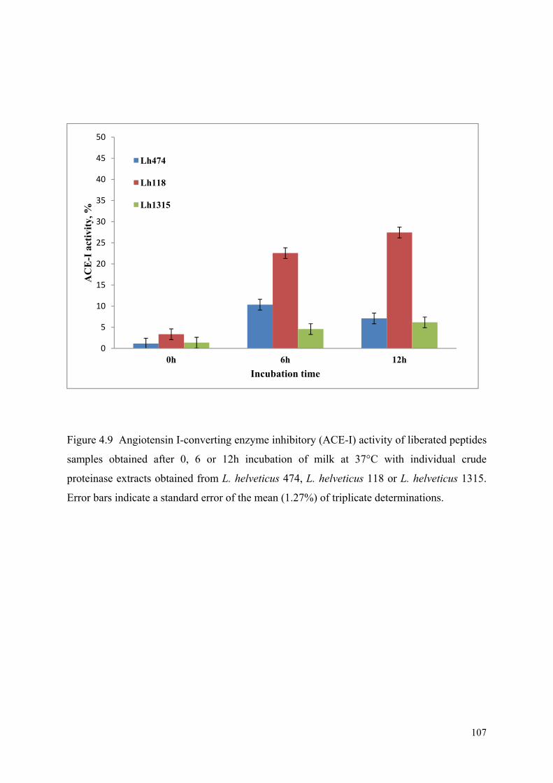
107
Figure 4.9 Angiotensin I-converting enzyme inhibitory (ACE-I) activity of liberated peptides
samples obtained after 0, 6 or 12h incubation of milk at 37°C with individual crude
proteinase extracts obtained from L. helveticus 474, L. helveticus 118 or L. helveticus 1315.
Error bars indicate a standard error of the mean (1.27%) of triplicate determinations.
0
5
10
15
20
25
30
35
40
45
50
0h 6h 12h
AC
E-I
act
ivity
, %
Incubation time
Lh474
Lh1188
Lh1315

108
4.9. Determination of antioxidative capacity
Oxidative metabolism is crucial for the survival of human body’s cells. The risk of
this activity is that the production of free radicals causes oxidative changes (Pihlanto, 2006a).
Free radicals have been linked with many pathological conditions such as atherosclerosis,
diabetes, rheumatoid arthritis (Abuja and Albertini, 2001, Gutteridge and Halliwell, 2000,
Halliwell and Whiteman, 2004). Inhibition of the free radicals formed in the living body and
foodstuff is the important way to protect body from above serious diseases.
1, 1-diphenyl-2-picrylhydrazyl (DPPH) is a popular indicator for showing free radical
scavenging activity of biological samples. In this study, peptides derived from milk proteins
by crude proteinases of selected highly proteolytic L. helveticus strains were assessed
separately for their antioxidant activity. The antioxidant activity of all peptides produced
from milk by using crude proteinase extract (CPE) from selected L. helveticus strains was
determined by DPPH•+ radical cation assay. This method is a type of inhibition assay in
which the extent of scavenging of a preformed free radical relative to that of a standard
antioxidant compound corresponds to the value of antioxidant activity. A more apparent
increase in the antioxidant activity was observed in the milk inoculated with individual CPE
from selected L. helveticus strains and incubated for 12 h at 37°C as shown in Figure 4.10.
The free radical scavenging activity in all samples changed significantly (P< 0.05) from zero
to 6 and 12 h comparing with untreated milk as a control, where no antioxidative activity was
detected (data not shown). Milk with crude proteinase from L. helveticus 118 had the highest
antioxidant capacity which increased from 18.8% at zero time to 23.5, 86.7 at 6 and 12 h
respectively, whereas similar trends were observed with L. helveticus 474 and 1315 during
the same incubation time which reached to 84.2% and 81.6%, respectively.
The variations of these activities may be attributed to the production of different
bioactive peptides, which may or may not have antioxidant properties and it is likely to be

109
strain dependent (Donkor et al., 2007b). It has been reported that DPPH radical-scavenging
activity of skim milk increased by fermentation with a L. casei strain and also suggested that
peptides produced in the fermented milk might be one of the factors enhancing radical-
scavenging activity (Nishino et al., 2000). A study published by Suetsuna et al (2000) also
showed that some peptides derived from the pepsinic hydrolysate of casein demonstrated
DPPH radical scavenging activities. Liu et al (2005) found that the DPPH radical-scavenging
activity of milk-kefir and soymilk-kefir was significantly higher than that of milk and
soymilk and they suggested that this activity may be, in part, attributed to the peptides
deriving from degradation of milk and soybean proteins. These results of scavenging
properties of free radicals by dairy cultures might be useful in food manufacturing and can
present additional sources of health enhancing antioxidants.

110
Figure 4.10 Antioxidative capacity of liberated peptides samples obtained after 0, 6 or 12 h
incubation of milk at 37°C with individual crude proteinase extracts obtained from L.
helveticus 474, L. helveticus 118 or L. helveticus 1315. Error bars indicate a standard error of
the mean (1.99%) of triplicate determinations.
0
10
20
30
40
50
60
70
80
90
100
0 h 6 h 12 h
Ant
ioxi
dativ
e ca
paci
ty, %
Incubation time
Lh474
Lh1188
Lh1315

111
4.10. Effects of fermented milk derived peptides on cytokine production
Inflammation has been reported as a host defence process that occurs at injured sites
and is strictly regulated. The organization of pro-inflammatory and anti-inflammatory
moderators is necessary in the regulation of the inflammatory response (Lee et al., 2002).
Regulation of host immunity is one of the most commonly proposed benefits of the
consumption of foods containing Lactobacillus strains. The main purpose of their use is to
prevent and treat the consumers from many diseases such as acute gastrointestinal infections,
allergy and inflammatory bowel diseases (Gill and Prasad, 2008). The beneficial effects of
these strains are based partly on their ability to regulate differentially the production of anti-
and pro-inflammatory cytokines and the balance of Th1/Th2 activity (Delcenserie et al.,
2008). Several milk proteins have been reported to modulate lymphocyte proliferation in
vitro (Phadke et al., 2005)such as whole casein, α-, β-, κ-casein (Phadke et al., 2005, Sütas et
al., 1996, Araujo et al., 2000). The actions of fermented LAB on immune system regulation
may also be strongly dependent on the lactic acid bacteria species, or on their metabolites
such as peptides released from proteins hydrolysis, which may or may not interact with
immune cells to induce distinct immunomodulatory response (LeBlanc et al., 2004).
In this study, the results of the effects of soluble peptides produced by CPEs of
individually selected L. helveticus strains on cytokine production by human peripheral blood
mononuclear cells (PBMCs) are shown in Figure 4.11A and B. The effects of soluble peptide
samples were detected in the stimulation of production of all tested cytokines. At 12 h of
incubation, all samples induced the production of cytokine IL-10 higher than the
nonstimulated PBMCs, comparing to untreated milk as a control and peptide samples at zero
hour, were no stimulation was detected (data not shown). The stimulation of PBMCs by
peptides at 37°C for 72 h from crude proteinase extract of L. helveticus 118 showed highest
production of IL-10 followed by L. helveticus 474 and 1315. However, peptides produced by

112
CPE from L. helveticus 474 induced the production of IFN-γ higher than other strains,
followed by L. helveticus 1315 and 118. In contrast, L. helveticus 118 was among the strains
inducing the production of the lowest levels of the Th1 cytokines IFN-γ, while inducing
higher IL-10 production. L. helveticus 474 induced a similar cytokine profile regarding IFN-γ
and higher production of IL-10. Therefore, the peptides produced of the same CPE species
are potentially able to drive immune responses into opposite directions in vitro.
These results clearly showed (Figure 4.11) that the soluble peptides derived from
degradation of milk proteins by selected L. helveticus strains stimulated the production of
both pro- and anti-inflammatory cytokines (IFN-γ and IL-10) in varying concentrations
compared with untreated milk or unstimulated PBMCs. The results also indicated that soluble
peptides may have induced Th1 and Th2 responses leading to the production of these
cytokines. It could be suggested from this study, that different peptides produced from the
same CPE may potentially be able to drive immune responses in opposite directions in vitro
and thus bring about an imbalance of Th1/Th2 type cytokines.
To sum up, the results showed that the lactobacillus strains used in this research may
release ACE-I, antioxidant and immunostimulative peptides during fermentation in bovine
milk. This bioactivity might rely on the growth level, proteolytic enzymes and pH changes in
addition to other factors that could affect the production of bioactive peptides such as
incubation time and the inoculation level of the fermented bacteria. Oligopeptides produced
by L helveticus proteinases and under certain conditions may serve as ACE-I, antioxidant and
immunostimulatory activity in different degrees.

113
Figure 4.11 IL-10 (A) and IFN- γ (B) cytokine production induced by stimulation of human
(PBMCs) with soluble milk peptides incubated for 72 h at 37°C in a humidified 5% CO2
incubator. The peptide samples were obtained after 0, 6 or 12 h incubation of milk at 37°C
with individual crude proteinase extracts obtained from L. helveticus 474, L. helveticus 118 or
L. helveticus 1315.
0200400600800
10001200140016001800
0 6 12 0 6 12 0 6 12
474 1188 1315
IL-1
0, p
g/m
l
A
0
50
100
150
200
250
300
350
400
450
0h 6hrs 12hrs 0h 6hrs 12hrs 0h 6hrs 12hrs
Lh474 Lh1188 Lh1315
IFN
-γ, p
g/m
L
B

114
CHAPTER 5
OVERALL CONCLUSIONS AND
FUTURE DIRECTIONS

115
5.1. Overall Conclusions
Ten selected strains of Lactobacillus helveticus 474, Lactobacillus helveticus 118,
Lactobacillus helveticus 1315, Lactobacillus helveticus 953, Lactobacillus delbrueckii ssp.
bulgaricus 734, Lactobacillus delbrueckii ssp. bulgaricus 756, Lactobacillus delbrueckii ssp.
bulgaricus 857, Lactobacillus delbrueckii ssp. lactis 1210, Lactobacillus delbrueckii ssp.
lactis 1307 and Lactobacillus delbrueckii ssp. lactis 1372 were assessed for their growth and
metabolic performance in reconstituted skim milk. All the cultures showed the appreciable
growth in RSM due to their proteolytic activities and some of them were not affected by the
rate of pH decrease or the final pH. The growth and the rate of acid production indirectly
showed as a pH decline was strictly strain dependant. L. helveticus 474 showed highest
growth among strains examined. The increase in growth was correlated with the time of
incubation and the decrease in pH during milk fermentation. The growth and metabolic
activity was also dependant on the extent of the proteolytic activity, which varied to a great
extent among the strains. Among all these organisms, L. helveticus strains exerted exemplary
proteolytic activity in comparison to other strains. On the other hand, L. bulgaricus and L.
lactis, as poor proteolytic activity strains, may need a longer time or different temperature or
even growth enhancers to establish similar cell concentrations to those of L. helveticus
strains. Due to their proteolytic activities, all organisms in this study were able to release a
number of bioactive peptides from milk proteins. These peptides were assessed for ACE
inhibitory activity in vitro and their potency varied with the strains. L. helveticus strains, as
highly proteolytic strains, released either more potent or more peptides that had higher ACE-I
activity than other strains. The time of fermentation, temperature, pH and as well as
proteolytic enzymes activity for each strain influenced the increase and decrease of ACE-I
activity for Lactobacillus strains during milk fermentation with exception of L. helveticus 118
which showed a minimal effect on ACE inhibition. Based on these findings, L. helveticus

116
474, 118 and 1315 were further examined. Extracellular and intracellular extracts of these
strains exhibited aminopeptidase activities towards all chromogenic substrates tested. On the
other hand, extracellular activity was characterised by the oligopeptidase activity. These
activities reflected the appreciable growth and proteolytic activity for these strains. The crude
proteinase extracts of these strains in milk produced a very low percentage ACE-I activity
while high levels of antioxidative activity. L. helvericus 118 produced the highest activity of
in vitro ACE-I and antioxidative activity at the end of fermentation. These findings showed
that bioactive peptides produced may rely on many factors such as temperature, inoculation
level of the crude proteinase extracts of selected L. helvericus strains. Furthermore, the
incubation time and strain used were the main possible factors influencing these bioactivities.
However in order to maintain high levels of these activities it would be necessary to prevent
further degradation. The oligopeptides produced were also assessed for their potential to
induce different cytokines. They induced release of interleukin-10 and interferon-γ, as main
representatives of anti- and pro-inflammatory cytokines, respectively. These bioactive
peptides might have capability to drive immune responses in opposite directions in vitro and
thus may bring about imbalance in the Th1/Th2 type cytokines.
5.2. Future Research Directions
Our results revealed that Lactobacillus species particularly L. helveticus strains used
in this study could release bioactive peptides from bovine milk proteins during fermentation.
Furthermore, the dairy industry which uses Lactobacillus species as starter cultures in dairy
products, should work closely with the medical professionals in order to substantiate the
health claims associated with these beneficial micro-organisms. A number of Lactobacillus
strains have been identified that are capable to release different peptides with varying ACE-
inhibitory, antioxidative and immunostimulatory activity from milk proteins. Although these

117
observed activities in this study were strictly strain and time dependant during fermentation,
their stability during storage of fermented dairy products by these strains and their resistance
to digestive enzymes need to be investigated. L. helveticus strains revealed higher proteolytic
and ACE-I activity than other strains studied. More research is required to develop and
enhance the proteolytic activity of Lactobacillus species to obtain products enriched with
bioactive peptides of specific or multi-functions that might be used to optimize the nutritional
and health effect of these compounds as therapeutic additives in dairy products.

118
CHAPTER 6
REFERENCES
ABUBAKAR, A., SAITO, T., KITAZAWA, H., KAWAI, Y. & ITOH, T. 1998. Structural
Analysis of New Antihypertensive Peptides Derived from Cheese Whey Protein by Proteinase K Digestion. Journal of Dairy Science, 81, 3131-3138.
ABUJA, P. M. & ALBERTINI, R. 2001. Methods for monitoring oxidative stress, lipid peroxidation and oxidation resistance of lipoproteins. Clinica Chimica Acta, 306, 1-17.
ADMIN. 2010. My Weight Online [Online]. Available: http://myweightonline.com/blog/culturelle-and-lactobacillus/5/ [Accessed 12.11.2010 2010].
ALGARON, F., MIRANDA, G., LE BARS, D. & MONNET, V. 2004. Milk fermentation by Lactococcus lactis with modified proteolytic systems to accumulate potentially bio-active peptides. Lait, 84, 115-123.
119
ARAUJO, M. C., MELO, R. L., CESARI, M. H., JULIANO, M. A., JULIANO, L. & CARMONA, A. K. 2000. Peptidase specificity characterization of C- and N-terminal catalytic sites of angiotensin I-converting enzyme. Biochemistry, 39, 8519-8525.
ASHAR, M. N. & CHAND, R. 2004. Fermented milk containing ACE-inhibitory peptides reduces blood pressure in middle aged hypertensive subjects. Milchwissenschaft, 59, 363-366.
AXELSSON, L. 2004. Lactic acid bacteria: classification and physiology. In: SALMINEN, S., VON WRIGHT, A. & OUWEHAND, A. (eds.) Lactic Acid Bacteria. Microbiological and Functional Aspects. New York: Marcel Dekker.
BHANDARI, P., KUMAR, N., SINGH, B. & AHUJA, P. S. 2010. Online HPLC-DPPH method for antioxidant activity of Picrorhiza kurroa Royle ex Benth. and characterization of kutkoside by ultra-performance LC-electrospray ionization quadrupole time-of-flight mass spectrometry. Indian Journal of Experimental Biology, 48, 323-328.
BOCKELMANN, W., GOLLAN, V. & HELLER, K. J. 1997. Purification of a second tripeptidase from Lactobacillus delbrueckii subsp. bulgaricus B14. Milchwissenschaft, 52, 500-503.
BOOTH, M., FHAOLÁIN, I. N., JENNINGS, P. V. & O'CUINN, G. 1990. Purification and characterization of a post-proline dipeptidyl aminopeptidase from Streptococcus cremoris AM2. Journal of Dairy Research, 57, 89-99.
BRADFORD, M. M. 1976. A rapid and sensitive method for the quantitation of microgram quantities of protein utilizing the principle of protein dye binding. Analytical Biochemistry, 72, 248-254.
CARR, D. J. J., DECOSTA, B. R., KIM, C. H., JACOBSON, A. E., GUARCELLO, V., RICE, K. C. & BLALOCK, J. E. 1989. Opioid receptors on cells of the immune system: Evidence for δ- and κ-classes. Journal of Endocrinology, 122, 161-168.
CERVATO, G., CAZZOLA, R. & CESTARO, B. 1999. Studies on the antioxidant activity of milk caseins. International Journal of Food Sciences and Nutrition, 50, 291-296.
CHEN, G.-W., TSAI, J.-S. & SUN PAN, B. 2007. Purification of angiotensin I-converting enzyme inhibitory peptides and antihypertensive effect of milk produced by protease-facilitated lactic fermentation. International Dairy Journal, 17, 641-647.
CHEN, Y. S., CHRISTENSEN, J. E., BROADBENT, J. R. & STEELE, J. L. 2003. Identification and characterization of Lactobacillus helveticus PepO2, an endopeptidase with post-proline specificity. Applied and Environmental Microbiology, 69, 1276-1282.
CHEUNG, H., WANG, F., ONDETTI, M., SABO, E. & DW., C. 1980. Binding of peptide substrates and inhibitors of angiotensinconverting enzyme. Importance of the COOH-terminal dipeptide sequence. J. Biol. Chem, 255, 401-407.
CHOPIN, A. 1993. Organization and regulation of genes for amino acid biosynthesis in lactic acid bacteria. FEMS Microbiology Reviews, 12, 21-37.
CHRISTENSEN, J. E., DUDLEY, E. G., PEDERSON, J. A. & STEELE, J. L. 1999. Peptidases and amino acid catabolism in lactic acid bacteria. Antonie van Leeuwenhoek, 76, 217-246.
CHRISTENSEN, J. E. & STEELE, J. L. 2003. Impaired growth rates in milk of Lactobacillus helveticus peptidase mutants can be overcome by use of amino acid supplements. Journal of Bacteriology, 185, 3297-3306.
CLARE, D. A. & SWAISGOOD, H. E. 2000. Bioactive milk peptides: A prospectus. Journal of Dairy Science, 83, 1187-1195.

120
COCAIGN-BOUSQUET, M., GARRIGUES, C., LOUBIERE, P. & LINDLEY, N. D. 1996. Physiology of pyruvate metabolism in Lactococcus lactis. Antonie van Leeuwenhoek, 70, 253-267.
CONTRERAS, M. D. M., CARRÓN, R., MONTERO, M. J., RAMOS, M. & RECIO, I. 2009. Novel casein-derived peptides with antihypertensive activity. International Dairy Journal, 19, 566-573.
CORDAIN, L., EATON, S. B., SEBASTIAN, A., MANN, N., LINDEBERG, S., WATKINS, B. A., O'KEEFE, J. H. & BRAND-MILLER, J. 2005. Origins and evolution of the Western diet: Health implications for the 21st century. American Journal of Clinical Nutrition, 81, 341-354.
CRUDDEN, A., AFOUFA-BASTIEN, D., FOX, P. F., BRISSON, G. & KELLY, A. L. 2005. Effect of hydrolysis of casein by plasmin on the heat stability of milk. International Dairy Journal, 15, 1017-1025.
DAKO, E., EL SODA, M., VUILLEMARD, J. C. & SIMARD, R. E. 1995. Autolytic properties and aminopeptidase activities of lactic acid bacteria. Food Research International, 28, 503-509.
DÁVALOS, A., MIGUEL, M., BARTOLOMÉ, B. & LÓPEZ-FANDIÑO, R. 2004. Antioxidant activity of peptides derived from egg white proteins by enzymatic hydrolysis. Journal of Food Protection, 67, 1939-1944.
DAVE, R. I. & SHAH, N. P. 1998. The influence of ingredient supplementation on the textural characteristics of yogurt. Aust. J. Dairy Technol, 53, 180-184.
DELCENSERIE, V., MARTEL, D., LAMOUREUX, M., AMIOT, J., BOUTIN, Y. & ROY, D. 2008. Immunomodulatory effects of probiotics in the intestinal tract. Current Issues in Molecular Biology, 10, 37-54.
DONKOR, O., N., HENRIKSSON, A., VASILJEVIC, T. & SHAH, N., P. 2007a. Proteolytic activity of dairy lactic acid bacteria and probiotics as determinant of growth and in vitro angiotensin-converting enzyme inhibitory activity in fermented milk. Lait, 87, 21-38.
DONKOR, O. N., HENRIKSSON, A., SINGH, T. K., VASILJEVIC, T. & SHAH, N. P. 2007b. ACE-inhibitory activity of probiotic yoghurt. International Dairy Journal, 17, 1321-1331.
DONKOR, O. N., HENRIKSSON, A., VASILJEVIC, T. & SHAH, N. P. 2006. Effect of acidification on the activity of probiotics in yoghurt during cold storage. International Dairy Journal, 16, 1181-1189.
DONKOR, O. N., VASILJEVIC, T. & GILL, H. S. 2010. Probiotics and Immunomodulation
Dietary Components and Immune Function. In: WATSON, R. R., ZIBADI, S. & PREEDY, V. R. (eds.). Humana Press.
DZIUBA, M. & DAREWICZ, M. 2007. Food Proteins as Precursors of Bioactive Peptides -- Classification Into Families. Food Science and Technology International, 13, 393-404.
ELITSUR, Y. & LUK, G. D. 1991. Beta-casomorphin (BCM) and human colonic lamina propria lymphocyte proliferation. Clinical and Experimental Immunology, 85, 493-497.
ERDMANN, K., CHEUNG, B. W. Y. & SCHRÖDER, H. 2008. The possible roles of food-derived bioactive peptides in reducing the risk of cardiovascular disease. The Journal of Nutritional Biochemistry, 19, 643-654.
FARNWORTH, E. R. 2003. Handbook of Fermented Functional Foods, FL, CRC Press, Boca Raton.

121
FERNANDEZ-ESPLA, M. D., GARAULT, P., MONNET, V. & RUL, F. 2000. Streptococcus thermophilus Cell Wall-Anchored Proteinase: Release, Purification, and Biochemical and Genetic Characterization. Applied and Environmental Microbiology, 66, 4772-4778.
FITZGERALD, R. J. & MEISEL, H. 2003. Milk protein hydrolysates and bioactive peptides. In: FOX, P. F. & MCSWEENEY, P. L. H. (eds.) Advanced dairy chemistry, Vol. 1: Proteins 3rd ed. New York, NY, USA: Kluwer Academic/Plenum Publishers.
FITZGERALD, R. J. & MURRAY, B. A. 2006. Bioactive peptides and lactic fermentations. International Journal of Dairy Technology, 59, 118-125.
FITZGERALD, R. J., MURRAY, B. A. & WALSH, D. J. 2004. Hypotensive Peptides from Milk Proteins. The Journal of nutrition, 134, 980S-988S.
FONTAINE, K. R., BAROFSKY, I., ANDERSEN, R. E., BARTLETT, S. J., WIERSEMA, L., CHESKIN, L. J. & FRANCKOWIAK, S. C. 1999. Impact of weight loss on Health-Related Quality of Life. Quality of Life Research, 8, 275-277.
FOX, P. F. 2001. Milk proteins as food ingredients. International Journal of Dairy Technology, 54, 41-55.
FOX, P. F. & BRODKORB, A. 2008. The casein micelle: Historical aspects, current concepts and significance. International Dairy Journal, 18, 677-684.
FOX, P. F., GUINEE, T. P., COGAN, T. M. & MCSWEENEY, P. L. H. 2000. Chemistry of milk constituents. In Fundamentals of Cheese Science. Gaithersburg, MD.: Aspen Publishers.
FOX, P. F. & MCSWEENEY, P. L. H. 1997. Rennets: their role in milk coagulation and cheese ripening. In: LAW, B. A. (ed.) Microbiology and Biochemistry of Cheese and Fermented Milk. London, UK.: Chapman and Hall.
FUGLSANG, A., RATTRAY, F., NILSSON, D. & NYBORG, N. 2003. Lactic acid bacteria: inhibition of angiotensin converting enzyme in vitro and in vivo. Antonie van Leeuwenhoek, 83, 27-34.
GARAULT, P., LE BARS, D., BESSET, C. & MONNET, V. 2002. Three Oligopeptide-binding Proteins Are Involved in the Oligopeptide Transport of Streptococcus thermophilus. Journal of Biological Chemistry, 277, 32-39.
GATTI, M., FORNASARI, M. E., LAZZI, C., MUCCHETTI, G. & NEVIANI, E. 2004. Peptidase activity in various species of dairy thermophilic lactobacilli. Journal of Applied Microbiology, 96, 223-229.
GAUTHIER, S. F., POULIOT, Y. & SAINT-SAUVEUR, D. 2006. Immunomodulatory peptides obtained by the enzymatic hydrolysis of whey proteins. International Dairy Journal, 16, 1315-1323.
GILBERT, C., ATLAN, D., BLANC, B., PORTAILER, R., GERMOND, J., LAPIERRE, L.
& MOLLET, B. 1996a. A new cell surface proteinase: sequencing and analysis of the prtB gene from Lactobacillus delbruekii subsp. bulgaricus. J. Bacteriol., 178, 3059-3065.
GILBERT, C., ATLAN, D., BLANC, B., PORTAILER, R., GERMOND, J. E., LAPIERRE, L. & MOLLET, B. 1996b. A new cell surface proteinase: sequencing and analysis of the prtB gene from Lactobacillus delbruekii subsp. bulgaricus. Journal of Bacteriology, 178, 3059-65.
GILL, H. & PRASAD, J. 2008. Probiotics, immunomodulation, and health benefits. GOBBETTI, M., LAVERMICOCCA, P., MINERVINI, F., DE ANGELIS, M. &
CORSETTI, A. 2000. Arabinose fermentation by Lactobacillus plantarum in

122
sourdough with added pentosans and αα-L-arabinofuranosidase: a tool to increase the production of acetic acid. Journal of Applied Microbiology, 88, 317-324.
GOBBETTI, M., STEPANIAK, L., DE ANGELIS, M., CORSETTI, A. & DI CAGNO, R. 2002. Latent bioactive peptides in milk proteins: Proteolytic activation and significance in dairy processing. Critical Reviews In Food Science And Nutrition, 42, 223-239.
GOMES, A. M. P. & MALCATA, F. X. 1999a. Bifidobacterium spp. and Lactobacillus acidophilus: biological, biochemical, technological and therapeutical properties relevant for use as probiotics. Trends in Food Science & Technology, 10, 139-157.
GOMES, A. M. P. & MALCATA, F. X. 1999b. Bifidobacterium spp. and Lactobacillus acidophilus: biological, biochemical, technological and therapeutical properties relevant for use as probiotics. Trends in Food Science & Technology, 10, 139-157.
GUARNER, F., BOURDET-SICARD, R., BRANDTZAEG, P., GILL, H. S., MCGUIRK, P., VAN EDEN, W., VERSALOVIC, J., WEINSTOCK, J. V. & ROOK, G. A. W. 2006. Mechanisms of Disease: the hygiene hypothesis revisited. Nat Clin Pract Gastroenterol Hepatol, 3, 275-284.
GUTTERIDGE, J. M. C. & HALLIWELL, B. 2000. Free Radicals and Antioxidants in the Year 2000: A Historical Look to the Future. Annals of the New York Academy of Sciences, 899, 136-147.
HA, E. & ZEMEL, M. B. 2003. Functional properties of whey, whey components, and essential amino acids: mechanisms underlying health benefits for active people (review). The Journal of Nutritional Biochemistry, 14, 251-258.
HALLIWELL, B. & WHITEMAN, M. 2004. Measuring reactive species and oxidative damage in vivo and in cell culture: how should you do it and what do the results mean? British Journal of Pharmacology, 142, 231-255.
HANNU, K. 2009. Milk-derived bioactive peptides: From science to applications. Journal of Functional Foods, 1, 177-187.
HAQUE, E. & CHAND, R. 2006. Milk protein derived bioactive peptides [Online]. Dairy Science. Available: http://www.dairyscience.info/exploitation-of-anti-microbial-proteins/111-milk-protein-derived-bioactive-peptides.html [Accessed Octoper 2010].
HASLAM, D. W. & JAMES, W. P. T. 2005. Obesity. Lancet, 366, 1197-1209. HATA, I., HIGASHIYAMA, S. & OTANI, H. 1998. Identification of a phosphopeptide in
bovine α(s1)-casein digest as a factor influencing proliferation and immunoglobulin production in lymphocyte cultures. Journal of Dairy Research, 65, 569-578.
HATA, I., UEDA, J. & OTANI, H. 1999. Immunostimulatory action of a commercially available casein phosphopeptide preparation, CPP-III, in cell cultures. Milchwissenschaft, 54, 3-6.
HAYALOGLU, A. A., GUVEN, M., FOX, P. F., HANNON, J. A. & MCSWEENEY, P. L. H. 2004. Proteolysis in Turkish White-brined cheese made with defined strains of Lactococcus. International Dairy Journal, 14, 599-610.
HAYES, M., ROSS, R. P., FITZGERALD, G. F. & STANTON, C. 2007. Putting microbes to work: Diary fermentation, cell factories and bioactive peptides. Part I: Overview. Biotechnology Journal, 2, 426-434.
HERNÁNDEZ-LEDESMA, B., RECIO, I. & AMIGO, L. 2008. β-Lactoglobulin as source of bioactive peptides. Amino Acids, 35, 257-265.
HERNÁNDEZ, D., CARDELL, E. & ZÁRATE, V. 2005. Antimicrobial activity of lactic acid bacteria isolated from Tenerife cheese: initial characterization of plantaricin TF711, a bacteriocin-like substance produced by Lactobacillus plantarum TF711. Journal of Applied Microbiology, 99, 77-84.
123
HIDEAKI, K., KUNIO, D., SHIGERU, S., HIDEYO, U., RYUJI, S., UMEJI, M. & SHIZUME, T. 1990. Antihypertensive effect of tryptic hydrolysate of milk casein in spontaneously hypertensive rats. Comparative Biochemistry and Physiology Part C: Comparative Pharmacology, 96, 367-371.
HIGGINS, C. F. 1992. ABC Transporters: From microorganisms to man. Annual Review of Cell Biology, 8, 67-113.
HIROTA, T., OHKI, K., KAWAGISHI, R., KAJIMOTO, Y., MIZUNO, S., NAKAMURA, Y. & KITAKAZE, M. 2007. Casein Hydrolysate Containing the Antihypertensive Tripeptides Val-Pro-Pro and Ile-Pro-Pro Improves Vascular Endothelial Function Independent of Blood Pressure-Lowering Effects: Contribution of the Inhibitory Action of Angiotensin-Converting Enzyme. Hypertens Res, 30, 489-496.
HOLT, C. 1992. Structure and Stability of Bovine Casein Micelles. In: C.B. ANFINSEN, F. M. R. J. T. E. & DAVID, S. E. (eds.) Advances in Protein Chemistry. Academic Press.
HONG, F., MING, L., YI, S., ZHANXIA, L., YONGQUAN, W. & CHI, L. 2008. The antihypertensive effect of peptides: A novel alternative to drugs? Peptides, 29, 1062-1071.
HUGENHOLTZ, J., VAN SINDEREN, D., KOK, J. & KONINGS, W. N. 1987. Cell Wall-Associated Proteases of Streptococcus cremoris Wg2. Applied and Environmental Microbiology, 53, 853-859.
HUTH, P. J., DIRIENZO, D. B. & MILLER, G. D. 2006. Major Scientific Advances with Dairy Foods in Nutrition and Health. Journal of Dairy Science, 89, 1207-1221.
HUTKINS, R. W. 2001. Metabolism of starter cultures. In: MARTH, E. H. & STEELE, J. L. (eds.) In Applied Dairy Microbiology. NY: Marcel Dekker,.
JAUHIAINEN, T. & KORPELA, R. 2007. Milk Peptides and Blood Pressure. The Journal of nutrition, 137, 825S-829S.
JOLLES, P., PARKER, F. & FLOC'H, F. 1981. Immunostimulating substances from human casein. Journal of Immunopharmacology, 3, 363-369.
JUILLARD, V., LAAN, H., KUNJI, E., JERONIMUS-STRATINGH, C., BRUINS, A. & KONINGS, W. 1995a. The extracellular PI-type proteinase of Lactococcus lactis hydrolyzes beta-casein into more than one hundred different oligopeptides. J. Bacteriol., 177, 3472-3478.
JUILLARD, V., LAAN, H., KUNJI, E. R., JERONIMUS-STRATINGH, C. M., BRUINS, A. P. & KONINGS, W. N. 1995b. The extracellular PI-type proteinase of Lactococcus lactis hydrolyzes beta-casein into more than one hundred different oligopeptides. Journal of Bacteriology, 177, 3472-8.
JUVONEN, P., MÅNSSON, M., KJELLMAN, N. I. M., BJÖRKSTÉN, B. & JAKOBSSON,
I. 1999. Development of immunoglobulin G and immunoglobulin E antibodies to cow’s milk proteins and ovalbumin after a temporary neonatal exposure to hydrolyzed and whole cow’s milk proteins. Pediatric Allergy and Immunology, 10, 191-198.
KAILASAPATHY, K. & CHIN, J. 2000. Survival and therapeutic potential of probiotic organisms with reference to Lactobacillus acidophilus and Bifidobacterium spp. Immunol Cell Biol, 78, 80-88.
KAYSER, H. & MEISEL, H. 1996. Stimulation of human peripheral blood lymphocytes by bioactive peptides derived from bovine milk proteins. FEBS Letters, 383, 18-20.
KILPI, E. E. R., KAHALA, M. M., STEELE, J. L., PIHLANTO, A. M. & JOUTSJOKI, V. V. 2007. Angiotensin I-converting enzyme inhibitory activity in milk fermented by wild-type and peptidase-deletion derivatives of Lactobacillus helveticus CNRZ32. International Dairy Journal, 17, 976-984.

124
KLEIN, J. R., DICK, A., SCHICK, J., MATERN, H. T., HENRICH, B. & PLAPP, R. 1995. Molecular cloning and DNA sequence analysis of pepL, a leucyl aminopeptidase gene from Lactobacillus delbrueckii subsp. lactis DSM7290. European Journal of Biochemistry, 228, 570-578.
KOK, J. & VOS, W. M. D. 1994. The proteolytic system of lactic acid bacteria. In: GASSON, M. J. & VOS, W. M. D. (eds.) Genetics and biotechnology of lactic acid bacteria. . Glasgow, UK Blackie Academic Professional.
KONINGS, W. N., POOLMAN, B. & DRIESSEN, A. J. 1989. Bioenergetics and solute transport in lactococci. Critical reviews in microbiology, 16, 419-476.
KORHONEN, H. 2009a. Milk-derived bioactive peptides: From science to applications. Journal of Functional Foods, 1, 177-187.
KORHONEN, H. & PIHLANTO-LEPPA¨LA¨ 2004. Milk-derived bioactive peptides: formation and prospects for health promotion. In: C. SHORTT & O’BRIEN, J. (eds.) Handbook of functional dairy products. Functional foods and nutraceuticals Boca Raton, Florida, FL, USA: CRC Press, Taylor Francis Group.
KORHONEN, H., PIHLANTO-LEPPÄLÄ, A., RANTAMÄKI, P. & TUPASELA, T. 1998. Impact of processing on bioactive proteins and peptides. Trends in Food Science and Technology, 9, 307-319.
KORHONEN, H. & PIHLANTO, A. 2003a. Bioactive peptides: New challenges and opportunities for the dairy industry. Australian Journal of Dairy Technology, 58, 129-134.
KORHONEN, H. & PIHLANTO, A. 2003b. Food-derived bioactive peptides - Opportunities for designing future foods. Current Pharmaceutical Design, 9, 1297-1308.
KORHONEN, H. & PIHLANTO, A. 2006. Bioactive peptides: Production and functionality. International Dairy Journal, 16, 945-960.
KORHONEN, H. J. 2009b. Bioactive milk proteins and peptides: From science to functional applications. Australian Journal of Dairy Technology, 64, 16-25.
KUDOH, Y., MATSUDA, S., IGOSHI, K. & OKI, T. 2001. Antioxidative peptide from milk fermented with Lactobacillus delbrueckii subsp. bulgaricus IFO13953. Nippon Shokuhin Kagaku Kogaku Kaishi, 48, 44-50.
KUNJI, E. R. S., MIERAU, I., HAGTING, A., POOLMAN, B. & KONINGS, W. N. 1996. The proteotytic systems of lactic acid bacteria. Antonie van Leeuwenhoek, 70, 187-221.
KUNJI, E. R. S., SMID, E. J., PLAPP, R., POOLMAN, B. & KONINGS, W. N. 1993. Di-tripeptides and oligopeptides are taken up via distinct transport mechanisms in Lactococcus lactis. Journal of Bacteriology, 175, 2052-2059.
KUWABARA, Y., NAGAI, S., YOSHIMITSU, N., NAKAGAWA, I., WATANABE, Y. & TAMAI, Y. 1995. Antihypertensive effect of the milk fermented by culturing with various lactic acid bacteria and a yeast. Journal of Fermentation and Bioengineering, 80, 294-295.
LAAN, H. & KONINGS, W. N. 1989. Mechanism of proteinase release from Lactococcus lactis subsp. cremoris Wg2. Applied and Environmental Microbiology, 55, 3101-3106.
LAFFINEUR, E., GENETET, N. & LEONIL, J. 1996. Immunomodulatory Activity of β-Casein Permeate Medium Fermented by Lactic Acid Bacteria. Journal of Dairy Science, 79, 2112-2120.
LANKAPUTHRA, W. E. V. & SHAH, N. P. 1996. Survival of Lactobacillus acidophilus and Bifidobacterium spp in the presence of acid and bile salts. Cult. Dairy Prod, 30, 2-7.

125
LAW, J. & HAANDRIKMAN, A. 1997. Proteolytic enzymes of lactic acid bacteria. International Dairy Journal, 7, 1-11.
LEBLANC, J., FLISS, I. & MATAR, C. 2004. Induction of a humoral immune response following an Escherichia coli O157:H7 infection with an immunomodulatory peptidic fraction derived from Lactobacillus helveticus-fermented milk. Clinical and Diagnostic Laboratory Immunology, 11, 1171-1181.
LECLERC, P.-L., GAUTHIER, S. F., BACHELARD, H., SANTURE, M. & ROY, D. 2002. Antihypertensive activity of casein-enriched milk fermented by Lactobacillus helveticus. International Dairy Journal, 12, 995-1004.
LEE, S. W., HONG, Y. S., CHUN, C. M., MOON, J. D., KIM, S. J., JUNG, I. C., YOON, Y. H., LEE, B. A., MOON, S. W., CHOI, S. H. & MOON, C. K. 2002. Anti-inflammatory effects of IL-4 and IL-10 on human polymorphonuclear leukocytes. Journal of Korean medical science, 17, 7-14.
LÉONIL, J., GAGNAIRE, V., MOLLÉ, D., PEZENNEC, S. & BOUHALLAB, S. D. 2000. Application of chromatography and mass spectrometry to the characterization of food proteins and derived peptides. Journal of Chromatography A, 881, 1-21.
LI, G.-H., LE, G.-W., SHI, Y.-H. & SHRESTHA, S. 2004. Angiotensin I–converting enzyme inhibitory peptides derived from food proteins and their physiological and pharmacological effects. Nutrition research (New York, N.Y.), 24, 469-486.
LIAN, W., WU, D., KONINGS, W. N., MIERAU, I. & HERSH, L. B. 1996. Heterologous expression and characterization of recombinant Lactococcus lactis neutral endopeptidase (neprilysin). Archives of Biochemistry and Biophysics, 333, 121-126.
LIM, E. M., EHRLICH, S. D. & MAGUIN, E. 2000. Identification of stress-inducible proteins in Lactobacillus delbrueckii subsp. bulgaricus. Electrophoresis, 21, 2557-2561.
LIU, J. R., CHEN, M. J. & LIN, C. W. 2005. Antimutagenic and antioxidant properties of milk-kefir and soymilk-kefir. Journal of Agricultural and Food Chemistry, 53, 2467-2474.
LÓPEZ-FANDIÑO, R., OTTE, J. & VAN CAMP, J. 2006. Physiological, chemical and technological aspects of milk-protein-derived peptides with antihypertensive and ACE-inhibitory activity. International Dairy Journal, 16, 1277-1293.
LOURENÇO DA COSTA, E., ANTONIO DA ROCHA GONTIJO, J. & NETTO, F. M. 2007. Effect of heat and enzymatic treatment on the antihypertensive activity of whey protein hydrolysates. International Dairy Journal, 17, 632-640.
LOURENS-HATTINGH, A. & VILJOEN, B. C. 2001a. Growth and survival of a probiotic yeast in dairy products. Food Research International, 34, 791-796.
LOURENS-HATTINGH, A. & VILJOEN, B. C. 2001b. Yogurt as probiotic carrier food. International Dairy Journal, 11, 1-17.
LYE, H. S., KUAN, C. Y., EWE, J. A., FUNG, W. Y. & LIONG, M. T. 2009. The improvement of hypertension by probiotics: Effects on cholesterol, diabetes, renin, and phytoestrogens. International Journal of Molecular Sciences, 10, 3755-3775.
MAENO, M., YAMAMOTO, N. & TAKANO, T. 1996. Identification of an Antihypertensive Peptide from Casein Hydrolysate Produced by a Proteinase from Lactobacillus helveticus CP790. Journal of Dairy Science, 79, 1316-1321.
MAES, W., VAN CAMP, J., VERMEIRSSEN, V., HEMERYCK, M., KETELSLEGERS, J. M., SCHREZENMEIR, J., VAN OOSTVELDT, P. & HUYGHEBAERT, A. 2004. Influence of the lactokinin Ala-Leu-Pro-Met-His-Ile-Arg (ALPMHIR) on the release of endothelin-1 by endothelial cells. Regulatory Peptides, 118, 105-109.
MARS, I. & MONNET, V. 1995. An aminopeptidase P from Lactococcus lactis with original specificity. Biochimica et Biophysica Acta (BBA) - General Subjects, 1243, 209-215.

126
MASUDA, O., NAKAMURA, Y. & TAKANO, T. 1996. Antihypertensive peptides are present in aorta after oral administration of sour milk containing these peptides to spontaneously hypertensive rats. The Journal of nutrition, 126, 3063-8.
MATAR, C., LEBLANC, J. G., MARTIN, L. & PERDIGON, G. 2003. Biologically active peptides released from fermented milk: role and functions. In: FARNWORTH, T. (ed.) released from fermented milk: role and functions. Boca Raton, Florida, FL, USA: CRC Press, Taylor and Francis Group.
MATTSBY-BALTZER, I., ROSEANU, A., MOTAS, C., ELVERFORS, J., ENGBERG, I. & HANSON, L. Å. 1996. Lactoferrin or a fragment thereof inhibits the endotoxin-induced interleukin-6 response in human monocytic cells. Pediatric Research, 40, 257-262.
MEDENIEKS, L. & VASILJEVIC, T. 2008. Underutilised fish as sources of bioactive peptides with potential health benefits. Food Australia, 60, 581-588.
MEISEL, H. 1997. Biochemical properties of regulatory peptides derived from milk proteins. Biopolymers - Peptide Science Section, 43, 119-128.
MEISEL, H. 2005. Biochemical properties of peptides encrypted in bovine milk proteins. Current Medicinal Chemistry, 12, 1905-1919.
MEISEL, H. & BOCKELMANN, W. 1999. Bioactive peptides encrypted in milk proteins: Proteolytic activation and thropho-functional properties. Antonie van Leeuwenhoek, International Journal of General and Molecular Microbiology, 76, 207-215.
MENSINK, R. P. 2006. Dairy products and the risk to develop type 2 diabetes or cardiovascular disease. International Dairy Journal, 16, 1001-1004.
MIGLIORE-SAMOUR, D., FLOC'H, F. & JOLLÈS, P. 1989. Biologically active casein peptides implicated in immunomodulation. Journal of Dairy Research, 56, 357-362.
MIGUEL, M., MANSO, M. A., LÓPEZ-FANDIÑO, R., ALONSO, M. J. & SALAICES, M. 2007. Vascular effects and antihypertensive properties of κ-casein macropeptide. International Dairy Journal, 17, 1473-1477.
MILLER, G. D., JARVIS, J. K. & MCBEAN, L. D. 2007. Dairy foods and hypertension. Handbook of Dairy Foods and Nutrition. 3rd. ed. CRC Press:Florida.
MIYAKAWA, H., KOBAYASHI, S., SHIMAMURA, S. & TOMITA, M. 1991. Purification and Characterization of an X-Prolyl Dipeptidyl Aminopeptidase from Lactobacillus delbrueckii ssp. bulgaricus LBU-147. Journal of Dairy Science, 74, 2375-2381.
MIYAUCHI, H., HASHIMOTO, S.-I., NAKAJIMA, M., SHINODA, I., FUKUWATARI, Y.
& HAYASAWA, H. 1998. Bovine Lactoferrin Stimulates the Phagocytic Activity of Human Neutrophils: Identification of Its Active Domain. Cellular Immunology, 187, 34-37.
MOLENAAR, D., HAGTING, A., ALKEMA, H., DRIESSEN, A. J. M. & KONINGS, W. N. 1993. Characteristics and osmoregulatory roles of uptake systems for proline and glycine betaine in Lactococcus lactis. Journal of Bacteriology, 175, 5438-5444.
MÖLLER, N., SCHOLZ-AHRENS, K., ROOS, N. & SCHREZENMEIR, J. 2008. Bioactive peptides and proteins from foods: indication for health effects. European Journal of Nutrition, 47, 171-182.
MULLALLY, M. M., MEISEL, H. & FITZGERALD, R. J. 1997. Angiotensin-I-converting enzyme inhibitory activities of gastric and pancreatic proteinase digests of whey proteins. International Dairy Journal, 7, 299-303.
MURRAY, B. A. & FITZGERALD, R. J. 2007. Angiotensin converting enzyme inhibitory peptides derived from food proteins: Biochemistry, bioactivity and production. Current Pharmaceutical Design, 13, 773-791.

127
NAKAJIMA, K., HATA, Y., OSONO, Y., HAMURA, M., KOBAYASHI, S. & WATANUKI, M. 1995. Antihypertensive effect of extracts of Lactobacillus casei in patients with hypertension. Journal of clinical biochemistry and nutrition 18, 181-187.
NAKAMURA, Y., YAMAMOTO, N., SAKAI, K., OKUBO, A., YAMAZAKI, S. & TAKANO, T. 1995. Purification and Characterization of Angiotensin I-Converting Enzyme Inhibitors from Sour Milk. Journal of Dairy Science, 78, 777-783.
NAS, H. & NISSEN-MEYER, J. 1992. Purification and N-terminal amino acid sequence determination of the cell-wall-bound proteinase from Lactobacillus paracasei subsp. paracasei. J Gen Microbiol, 138, 313-318.
NIELSEN, M. S., MARTINUSSEN, T., FLAMBARD, B., SØRENSEN, K. I. & OTTE, J. 2009. Peptide profiles and angiotensin-I-converting enzyme inhibitory activity of fermented milk products: Effect of bacterial strain, fermentation pH, and storage time. International Dairy Journal, 19, 155-165.
NISHINO, T., SHIBAHARA-SONE, H., KIKUCHI-HAYAKAWA, H. & ISHIKAWA, F. 2000. Transit of radical scavenging activity of milk products prepared by maillard reaction and Lactobacillus casei strain Shirota fermentation through the hamster intestine. Journal of Dairy Science, 83, 915-922.
NIVEN, G. W., HOLDER, S. A. & STRØMAN, P. 1995. A study of the substrate specificity of aminopeptidase N from Lactococcus lactis subsp. cremoris Wg2. Applied Microbiology and Biotechnology, 44, 100-105.
NURMINEN, M.-L., SIPOLA, M., KAARTO, H., PIHLANTO-LEPPÄLÄ, A., PIILOLA, K., KORPELA, R., TOSSAVAINEN, O., KORHONEN, H. & VAPAATALO, H. 2000. α-Lactorphin lowers blood pressure measured by radiotelemetry in normotensive and spontaneously hypertensive rats. Life Sciences, 66, 1535-1543.
OKADA, Y. & OKADA, M. 1998. Scavenging Effect of Water Soluble Proteins in Broad Beans on Free Radicals and Active Oxygen Species. Journal of Agricultural and Food Chemistry, 46, 401-406.
OSUNTOKI, A. & KORIE, I. 2010. Antioxidant activity of whey from milk fermented with Lactobacillus species isolated from Nigerian fermented foods. Food Technology and Biotechnology, 48, 505-511.
OTANI, H. & HATA, I. 1995. Inhibition of proliferative responses of mouse spleen lymphocytes and rabbit Peyer's patch cells by bovine milk caseins and their digests. Journal of Dairy Research, 62, 339-348.
OTANI, H., KIHARA, Y. & PARK, M. 2000. The immunoenhancing property of a dietary casein phosphopeptide preparation in mice. Food and Agricultural Immunology, 12, 165-173.
OTANI, H., NAKANO, K. & KAWAHARA, T. 2003. Stimulatory effect of a dietary casein phosphopeptide preparation on the mucosal IgA response of mice to orally ingested lipopolysaccharide from Salmonella typhimurium. Bioscience, Biotechnology and Biochemistry, 67, 729-735.
OTANI, H., WATANABE, T. & TASHIRO, Y. 2001. Effects of bovine beta-casein (1-28) and its chemically synthesized partial fragments on proliferative responses and immunoglobulin production in mouse spleen cell cultures. Biosci Biotechnol Biochem, 65, 2489-95.
OUWEHAND & VESTERLUND 2004. Antimicrobial components from lactic acid bacteria In: SALMINEN., S., WRIGHT, A. V. & OUWEHAND, A. (eds.) Lactic Acid Bacteria: Microbiological and Functional New York: Aspects, Marcel Dekker, Inc.

128
PAN, D., LUO, Y. & TANOKURA, M. 2005. Antihypertensive peptides from skimmed milk hydrolysate digested by cell-free extract of Lactobacillus helveticus JCM1004. Food Chemistry, 91, 123-129.
PARK, Y. W. 2009. Overview of Bioactive Components in Milk and Dairy Products. Bioactive Components in Milk and Dairy Products. Wiley-Blackwell.
PARKER, F., MIGLIORE-SAMOUR, D., FLOCH, F., ZERIAL, A., WERNER, G. H., JOLLÈS, J., CASARETTO, M., ZAHN, H. & JOLLÈS, P. 1984. Immunostimulating hexapeptide from human casein: amino acid sequence, synthesis and biological properties. European Journal of Biochemistry, 145, 677-682.
PARVEZ, S., MALIK, K. A., AH KANG, S. & KIM, H. Y. 2006. Probiotics and their fermented food products are beneficial for health. Journal of Applied Microbiology, 100, 1171-1185.
PELTONIEMI, K., VESANTO, E. & PALVA, A. 2002. Genetic characterization of an oligopeptide transport system from Lactobacillus delbrueckii subsp. bulgaricus. Archives of Microbiology, 177, 457-467.
PERPETUO, E. A., JULIANO, L. & LEBRUN, I. 2003. Biochemical and Pharmacological Aspects of Two Bradykinin-Potentiating Peptides Obtained from Tryptic Hydrolysis of Casein. Journal of Protein Chemistry, 22, 601-606.
PHADKE, S. M., DESLOUCHES, B., HILEMAN, S. E., MONTELARO, R. C., WIESENFELD, H. C. & MIETZNER, T. A. 2005. Antimicrobial peptides in mucosal secretions: The importance of local secretions in mitigating infection. Journal of Nutrition, 135, 1289-1293.
PHELAN, M., AHERNE, A., FITZGERALD, R. J. & O'BRIEN, N. M. 2009. Casein-derived bioactive peptides: Biological effects, industrial uses, safety aspects and regulatory status. International Dairy Journal, 19, 643-654.
PIHLANTO-LEPPÄLÄ, A., ROKKA, T. & KORHONEN, H. 1998. Angiotensin I Converting Enzyme Inhibitory Peptides Derived from Bovine Milk Proteins. International Dairy Journal, 8, 325-331.
PIHLANTO, A. 2006a. Antioxidative peptides derived from milk proteins. International Dairy Journal, 16, 1306-1314.
PIHLANTO, A. 2006b. Bioactive peptides: Functionality and production. Agro Food Industry Hi-Tech, 17, 24-26.
PIHLANTO, A. & KORHONEN, H. 2003. Bioactive peptides and proteins. PIHLANTO, A., VIRTANEN, T. & KORHONEN, H. 2010. Angiotensin I converting
enzyme (ACE) inhibitory activity and antihypertensive effect of fermented milk. International Dairy Journal, 20, 3-10.
POOLMAN, B. 1993. Energy transduction in lactic acid bacteria. FEMS Microbiology Reviews, 12, 125-147.
PRITCHARD, G. G. & COOLBEAR, T. 1993. The physiology and biochemistry of the proteolytic system in lactic acid bacteria. FEMS Microbiology Reviews, 12, 179-206.
PRITCHARD, G. G., FREEBAIRN, A. D. & COOLBEAR, T. 1994. Purification and characterization of an endopeptidase from Lactococcus lactis subsp. cremoris SK11. Microbiology, 140, 923-930.
QIAN, B., XING, M., CUI, L., DENG, Y., XU, Y., HUANG, M. & ZHANG, S. 2011. Antioxidant, antihypertensive, and immunomodulatory activities of peptide fractions from fermented skim milk with Lactobacillus delbrueckii ssp. bulgaricus LB340. Journal of Dairy Research, 78, 72-79.
RACHID, M., MATAR, C., DUARTE, J. & PERDIGON, G. 2006. Effect of milk fermented with a Lactobacillus helveticus R389(+) proteolytic strain on the immune system and

129
on the growth of 4T1 breast cancer cells in mice. FEMS Immunology & Medical Microbiology, 47, 242-253.
RAMCHANDRAN, L. 2009. PHYSICO-CHEMICAL AND THERAPEUTIC PROPERTIES OF LOW-FAT YOGURT AS INFLUENCED BY FAT REPLACERS, EXOPOLYSACCHARIDES AND PROBIOTICS. DOCTOR OF PHILOSOPHY, Victoria University.
RAMCHANDRAN, L. & SHAH, N. P. 2008. Growth, Proteolytic, and ACE-I Activities of Lactobacillus delbrueckii ssp. bulgaricus and Streptococcus thermophilus and Rheological Properties of Low-Fat Yogurt as Influenced by the Addition of Raftiline HP®. Journal of Food Science, 73, M368-M374.
ROBERFROID, M. B. 1999. Concepts in functional foods: The case of inulin and oligofructose. Journal of Nutrition, 129, 1398S-1401S.
ROGELJ, I. 2000. Milk, Dairy Products, Nutrition and Health. Food Technology and Biotechnology, 38, 143-147.
RYBKA, S. & FLEET, G. H. 1997. Populations of Lactobacillus delbrueckii ssp bulgaricus, Streptococcus thermophilus, Lactobacillus acidophilus and Bifidobacterium species in Australian yoghurts. Department of Food Science and Technology, 49, 471-475.
SAHLSTROM, S., CHRZANOWSKA, J. & SORHAUG, T. 1993. Purification and characterization of a cell wall peptidase from Lactococcus lactis subsp. cremoris IMN-C12. Applied and Environmental Microbiology, 59, 3076-3082.
SAIGA, A., TANABE, S. & NISHIMURA, T. 2003. Antioxidant Activity of Peptides Obtained from Porcine Myofibrillar Proteins by Protease Treatment. Journal of Agricultural and Food Chemistry, 51, 3661-3667.
SAITO, T., NAKAMURA, T., KITAZAWA, H., KAWAI, Y. & ITOH, T. 2000. Isolation and Structural Analysis of Antihypertensive Peptides That Exist Naturally in Gouda Cheese. Journal of Dairy Science, 83, 1434-1440.
SANZ, Y., MULHOLLAND, F. & TOLDRÁ, F. 1998. Purification and Characterization of a Tripeptidase from Lactobacillus sake. Journal of Agricultural and Food Chemistry, 46, 349-353.
SASAKI, M., BOSMAN, B. W. & TAN, P. S. T. 1995. Comparison of proteolytic activities in various lactobacilli. Journal of Dairy Research, 62, 601-610.
SAVIJOKI, K., INGMER, H. & VARMANEN, P. 2006. Proteolytic systems of lactic acid bacteria. Applied Microbiology and Biotechnology, 71, 394-406.
SEPPO, L., JAUHIAINEN, T., POUSSA, T. & KORPELA, R. 2003. A fermented milk high in bioactive peptides has a blood pressure–lowering effect in hypertensive subjects. The American Journal of Clinical Nutrition, 77, 326-330.
SEPPO, L., KEROJOKI, O., SUOMALAINEN, T. & KORPELA, R. 2002. The effect of a Lactobacillus helveticus LBK-16 H fermented milk on hypertension: a pilot study on humans. Milchwissenschaft, 57, 124-127.
SHAH, N. P. 2000. Effects of milk-derived bioactives: An overview. British Journal of Nutrition, 84, S3-S10.
SHAH, N. P. 2001. Functional foods from probiotics and prebiotics. Food Technology, 55, 46.
SHAH, N. P. 2007. Functional cultures and health benefits. International Dairy Journal, 17, 1262-1277.
SHAH, N. P., LANKAPUTHRA, W. E. V., BRITZ, M. L. & KYLE, W. S. A. 1995. Survival of Lactobacillus acidophilus and Bifidobacterium bifidum in commercial yoghurt during refrigerated storage. International Dairy Journal, 5, 515-521.
SHAH, N. P. & RAVULA, R. R. 2000. Microencapsulation of probiotic bacteria and their survival in frozen fermented dairy desserts. J. of Dairy Technol, 55, 139-144.

130
SHIHATA, A. & SHAH, N. P. 2000. Proteolytic profiles of yogurt and probiotic bacteria. International Dairy Journal, 10, 401-408.
SHINODA, I., TAKASE, M., FUKUWATARI, Y., SHIMAMURA, S., KÖLLER, M. & KÖNIG, W. 1996. Effects of Lactoferrin and Lactoferricin® on the Release of Interleukin 8 from Human Polymorphonuclear Leukocytes. Bioscience, Biotechnology and Biochemistry, 60, 521-523.
SILVA, S. V. & MALCATA, F. X. 2005. Caseins as source of bioactive peptides. International Dairy Journal, 15, 1-15.
SIPOLA, M., FINCKENBERG, P., KORPELA, R., VAPAATALO, H. & NURMINEN, M., A. 2002. Effect of long-term intake of milk products on blood pressure in hypertensive rats. Journal of Dairy Research, 69, 103-111.
SMID, E. J., DRIESSEN, A. J. & KONINGS, W. N. 1989. Mechanism and energetics of dipeptide transport in membrane vesicles of Lactococcus lactis. Journal of Bacteriology, 171, 292-298.
SMID, E. J., POOLMAN, B. & KONINGS, W. N. 1991. Casein utilization by lactococci. Applied and Environmental Microbiology, 57, 2447-2452.
SOEDAMAH-MUTHU, S. S., DING, E. L., AL-DELAIMY, W. K., HU, F. B., ENGBERINK, M. F., WILLETT, W. C. & GELEIJNSE, J. M. 2011. Milk and dairy consumption and incidence of cardiovascular diseases and all-cause mortality: dose-response meta-analysis of prospective cohort studies. The American Journal of Clinical Nutrition, 93, 158-171.
SONOMOTO, K. 2011. Lactic acid bacteria and bifidobacteria; current progress in advanced research, Book News, Inc.
SPENCE, J. T. 2006. Challenges related to the composition of functional foods. Journal of Food Composition and Analysis, 19, Supplement, S4-S6.
STEPANIAK, L. 2004. Dairy enzymology. International Journal of Dairy Technology, 57, 153-171.
STEPANIAK, L. & FOX, P. F. 1995. Characterization of the principal intracellular endopeptidase from Lactococcus lactis subsp. lactis MG1363. International Dairy Journal, 5, 699-713.
SUETSUNA, K., UKEDA, H. & OCHI, H. 2000. Isolation and characterization of free radical scavenging activities peptides derived from casein. The Journal of Nutritional Biochemistry, 11, 128-131.
SÜTAS, Y., HURME, M. & ISOLAURI, E. 1996. Down-regulation of anti-CD3 antibody-induced IL-4 production by bovine caseins hydrolysed with Lactobacillus GG-derived enzymes. Scandinavian Journal of Immunology, 43, 687-689.
SWAISGOOD, H. E. 1992. Chemistry of the caseins. In: F., F. P. (ed.) Advanced dairy chemistry, I. Protein. London: Elsevier.
TAME, J. R. H., MURSHUDOV, G. N., DODSON, E. J., NEIL, T. K., DODSON, G. G., HIGGINS, C. F. & WILKINSON, A. J. 1994. The structural basis of sequence-independent peptide binding by OppA protein. Science, 264, 1578-1581.
TAMIME, A. Y., SAARELA, M., SØNDERGAARD, A. K., MISTRY, V. V. & SHAH, N. P. 2007. Production and Maintenance of Viability of Probiotic Micro-Organisms in Dairy Products. Probiotic Dairy Products. Blackwell Science Ltd.
TAN, P. S., POOLMAN, B. & KONINGS, W. N. 1993. Proteolytic enzymes of Lactococcus lactis. Journal of Dairy Research, 60, 269-286.
TAN, P. S. T., POS, K. M. & KONINGS, W. N. 1991. Purification and characterization of an endopeptidase from Lactococcus lactis subsp. cremoris Wg2. Applied and Environmental Microbiology, 57, 3593-3599.

131
TSAI, J.-S., CHEN, T.-J., PAN, B. S., GONG, S.-D. & CHUNG, M.-Y. 2008. Antihypertensive effect of bioactive peptides produced by protease-facilitated lactic acid fermentation of milk. Food Chemistry, 106, 552-558.
TSAKALIDOU, E., ANASTASIOU, R., VANDENBERGHE, I., VAN BEEUMEN, J. & KALANTZOPOULOS, G. 1999. Cell-Wall-Bound Proteinase of Lactobacillus delbrueckii subsp. lactis ACA-DC 178: Characterization and Specificity for β-Casein. Applied and Environmental Microbiology, 65, 2035-2040.
TUDOR, M., HAVRANEK, J. & SERAFINI, M. 2009. Dairy foods and body weight management. Mljekarstvo, 59, 88-95.
TUOMILEHTO, J., LINDSTROM, J., HYYRYNEN, J., KORPELA, R., KARHUNEN, M. L., MIKKOLA, L., JAUHIAINEN, T., SEPPO, L. & NISSINEN, A. 2004. Effect of ingesting sour milk fermented using Lactobacillus helveticus bacteria producing tripeptides on blood pressure in subjects with mild hypertension. J Hum Hypertens, 18, 795-802.
TYNKKYNEN, S., BUIST, G., KUNJI, E., KOK, J., POOLMAN, B., VENEMA, G. & HAANDRIKMAN, A. 1993. Genetic and biochemical characterization of the oligopeptide transport system of Lactococcus lactis. Journal of Bacteriology, 175, 7523-7532.
UENO, K., MIZUNO, S. & YAMAMOTO, N. 2004. Purification and characterization of an endopeptidase that has an important role in the carboxyl terminal processing of antihypertensive peptides in Lactobacillus helveticus CM4. Letters in Applied Microbiology, 39, 313-318.
VASILJEVIC, T. & SHAH, N. P. 2008. Probiotics--From Metchnikoff to bioactives. International Dairy Journal, 18, 714-728.
VASILJEVIC, T. & SHAH, N. P. 2009. Cultured Milk and Yogurt. Dairy Processing & Quality Assurance. Wiley-Blackwell.
VAUGHAN, E. E., DAVID, S. & DE VOS, W. M. 1996. The lactose transporter in Leuconostoc lactis is a new member of the LacS subfamily of galactoside-pentose-hexuronide translocators. Applied and Environmental Microbiology, 62, 1574-1582.
VERMEIRSSEN, V., VAN CAMP, J., DEVOS, L. & VERSTRAETE, W. 2003. Release of Angiotensin I Converting Enzyme (ACE) Inhibitory Activity during in Vitro Gastrointestinal Digestion:� from Batch Experiment to Semicontinuous Model. Journal of Agricultural and Food Chemistry, 51, 5680-5687.
VERMEIRSSEN, V., VAN CAMP, J. & VERSTRAETE, W. 2004. Bioavailability of angiotensin I converting enzyme inhibitory peptides. British Journal of Nutrition, 92, 357-366.
VIRTANEN, T., PIHLANTO, A., AKKANEN, S. & KORHONEN, H. 2007. Development of antioxidant activity in milk whey during fermentation with lactic acid bacteria. Journal of Applied Microbiology, 102, 106-115.
VON HUTH SMITH, L., BORCH-JOHNSEN, K. & JØRGENSEN, T. 2007. Commuting physical activity is favourably associated with biological risk factors for cardiovascular disease. European Journal of Epidemiology, 22, 771-779.
WALSTRA, P. 1990. On the Stability of Casein Micelles. Journal of Dairy Science, 73, 1965-1979.
WALSTRA, P. 1999. Casein sub-micelles: do they exist? International Dairy Journal, 9, 189-192.
WOHLRAB, Y. & BOCKELMANN, W. 1993. Purification and characterization of a second aminopeptidase (pepC-like) from Lactobacillus delbrueckii subsp. bulgaricus B14. International Dairy Journal, 3, 685-701.

132
WONG, P. Y. Y. & KITTS, D. D. 2003. Chemistry of buttermilk solid antioxidant activity. Journal of Dairy Science, 86, 1541-1547.
WYBRAN, J., APPELBOOM, T., FAMAEY, J. P. & GOVAERTS, A. 1979. Suggestive evidence for receptors for morphine and methionine-enkephalin on normal human blood T lymphocytes. Journal of Immunology, 123, 1068-1070.
YAMAMOTO, N., AKINO, A. & TAKANO, T. 1993. Purification and specificity of a cell-wall-associated proteinase from Lactobacillus helveticus CP790. Journal of Biochemistry, 114, 740-745.
YAMAMOTO, N., EJIRI, M. & MIZUNO, S. 2003. Biogenic peptides and their potential use. Current Pharmaceutical Design, 9, 1345-1355.
YAMAMOTO, N., MAENO, M. & TAKANO, T. 1999. Purification and Characterization of an Antihypertensive Peptide from a Yogurt-Like Product Fermented by Lactobacillus helveticus CPN4. Journal of Dairy Science, 82, 1388-1393.
YAMAMOTO, N., SHINODA, T. & MIZUNO, S. 2004. Cloning and expression of an endopeptidase gene from Lactobacillus helveticus CM4 involved in processing antihypertensive peptides. Milchwissenschaft, 59, 593-597.
YAMAMOTO, N. & TAKANO, T. 1999. Antihypertensive peptides derived from milk proteins. Die Nahrung, 43, 159-164.
YANOVSKI, S. Z. 1996. Long-term pharmacotherapy in the management of obesity. Journal of the American Medical Association, 276, 1907-1915.