regulation of water and electrolytes - homepage |...
TRANSCRIPT

Regulation of Water and ElectrolytesBy J. RUSSELL ELKINTON, M.D.
TO INCLUDE a discussion of the regula-tion of total body water and electrolytes
in such a symposium OIn the cardiovascularsystem at first glanee may require some justi-fication. It should be quickly apparent, how-ever, that the total body fluids and the circula-tory system are so intimately interrelated thatconsideration of one without the other wouldbe quite incomplete. Part of the body fluids,the cellular fluids of erythrocytes and theplasma, are distributed within the vascularsystem and constitute the medium, so to speak,upon which the circulatory systenm operates.Being that which is circulated, these fluidphases are an integral part of the eirculatorysystem. Likewise the circulatory system is es-sential to the integrity of the fluids of thebody. The eirculation provides the mixingapparatus that maintains the homogeneity ofmultiple and widely separated portions of thebody fluids and the circulation is the link ofthe organs of exchange with the external enl-vironment. It would seem proper, therefore,to include water and electrolytes in this dis-cussion of regulation of the cardiovascularsystem.The concept of regulatioii as already defined
by our moderator can be applied to the bodyfluids as well as to the rest of the cardiovascu-lar system. In the consideration that follows.I shall attempt to show not only that physio-logic servomechanisms are operative but thata great deal remains to be learnied of theirmoduts operandi. The emphasis will be on theholes in, rather than the substance of, ourknowledge.
Evidence That the TotalBody Fluids are Regulated
Let us begin simply by considering the 2major dimensions of any fluid, including body
From the Chemical Section of the Department ofMedicine, University of Peniisylvania School of Medi-cine, Philadelphia, Pa.
fluids, namely, concentration and volume. Ithas been knowni for many years that the totalconcentration of solutes, or osmolality, ofplasma and extracellular fluid is maintainedwithin rather narrow and constant limits.This constancy of osmolality or tonicity wasone of the features of the milieu interieutr ofClaude Bernard. It holds in amphibians, birds,and mammals and has been the subject ofmuch investigation in the field of comparativephysiology. On the other hand, constancy ofvolume of the body fluids has received rela-tively little attention until recent years. In1916 that very shrewd observer of naturalphenomena, L. J. Henderson, presented beforethe National Academy of Science a short noteon the importaniee of volume in biology' inwhich he pointed out that at a given level ofdevelopment the maintenance of a constantvolume is one of the basic properties of livingorganisms. Evidenee for this phenomenon inour own bodies is to be found in the constancyof our body weights. Since body water con-stitutes such a large proportion of bodvweight, rapid fluctuations in the latter mustapproximate changes in the former. This istrue, of course, only if concomitant changes inbody fat are not being induced by neurogemiieor iatrogenic dietary manlipulations, or by themimore natural processes of growth or seneseeniee.On one occasion I measured my own basalbody weight daily for 56 days; the 2 standarddeviation variations about the mean did notexceed +1 per cent. This observation suggeststhat my total body water content was beingregulated. In figure 1 are shown, again inmyself, the oscillations in weight that occurredwithin one recent 24-hour period. On thisSaturday away from the hospital, intake offood and fluid at breakfast was followed byconsiderable loss of body water and salt dur-ing the heavy exercise of cutting grass. Atlunch a large thirst as well as hunger led toa copious intake; this was followed by a less
Circulation, Volume XXI, June 1960184
by guest on May 24, 2018
http://circ.ahajournals.org/D
ownloaded from

SYMPOSIUM-CARDIOVASCULAR REGULATION
breakfast lunch4. 4
mildexercise
dinner
heavy N
exercise Wt. flux:
Time - hour:
in * 2.9 kg.out 5 2.7totol . 5.6net =+0.2
8 o.m. 12 4 p.m. 8 12 4 a.m. 8
Figure 1Oscillations of body weight during a 24-hour period.
rapid loss of body water during an expeditionwith my younig son to the zoo. A mid-after-noon oscillation (dotted line) occurred due to
ingestion of peanuts and root beer but was
unmeasured. Intake at dinner was succeededby a slower rate of loss during the evening andsleep. At the end of the 24-hour period my
body weight had changed by only a plus0.2 Kg. while the total weight flux was 5.6 Kg.(2.9 in, 2.7 out). These oscillations approxi-mate the fluctuations of total body fluid anddelineate very nieely the operation of intakeand output servoimechanisms that regulatediurnally the volume of my total body water.
These servomuechanisms are the main sub-ject of nmy conitribution to this symposium. Itis a subject of concern to the physician as
well as to the physiologist and biologist. In
disease, servomechanisms go wrong; the resultis a sick patient requiring diagnosis and treat-ment. The primary pathogenic factor may bea lesion of the heart, the kidney, the adrenalcortex, the brain, or other organ; the resultmay be one or more disturbances in volume or
osmolal concentration that we label congestiveheart failure or edema, or dehydration, or
hypernatremia or hyponatremia, and so on.
But proper therapy needs an understandinigof the servomechanisms involved in the pa-
tient's disturbed homeostasis. Let us as phy-sicians consider these mechanisms of regula-tion.
Circulation, Volume XXI, June 1960
Regulation of Water Relative to SoluteOsmolal regulation through the neurohypo-
physeal antidiuretic hormone (ADH) systemnis perhaps the best delineated servomechanismin the physiology of body fluid. The classicexperiment in support of this mechanism was
that of Verney,2 who demonstrated that hy-pertonic solutions of sodium salts or sucrose,
but not urea, infused into the arterial bloodsupply of the hypothalamus of the dog wouldinhibit a pre-established water diuresis to thesame degree as a known amount of pitressin(fig. 2). From his observation Verney postu-lated that there must be receptor cells, sensi-tive to osmolal changes in the extracellularfluid bathing them, that control the rate ofrelease of antidiuretic hormone (ADH) fromthe posterior pituitary. These sensitive loci hedesignated "osmoreceptors," and he pre-
sented evidence that they responded to changesin osmolal concentration of the order of 1 to2 per cent.
It is now possible to characterize this servo-
mechanism that regulates body water contentrelative to solute content (fig. 3). The stimuluson the afferent side of the are is change in thetotal concentration in extracellular fluid or
plasma of solutes that do not readily penetratecells, (or, more precisely, change in the con-
centration of water). The effect of this stimu-lus on the receptor center is to alter the rateof release of an efferent humoral mediator,
Body
weight
I bs.
164-
163-
162-
161
R.E.
sleep
1185
by guest on May 24, 2018
http://circ.ahajournals.org/D
ownloaded from

ELKINTON
minuttes minutes
Figure 2One of Verney's experiments demonstrating theantidiuretic effect of hypertonic solutions injectedat different times into the carotid artery of a
water-loaded dog. In graph a are shown the effectsof 2 ml. of 1.28 M NaCl (A), 0.98 Mi Na2SO4 (B),2.39 M dextrose (C), anid 2.39 M urea (D). Ingraph b are shown the effects of 2.5 (A) and 3.5(B) mU of posterior pituitary extract. (FromVerney, E. B.2 Reproduced by permission of thepu-blisher and the author.)
antidiuretic hormone. This hormone, ADH,produces in the effector organ, the kidney, a
change in rate of tubular reabsorption ofwater, and hence the rate of water output. Theresult is a modification of the original osmolalstimulus-in cybernetic terms, correction ofan error by negative feedback.
It must here be pointed out that a variety-of other stimuli act on the neurohypophysealsystem to produce changes in the rate of re-
lease of ADH.3 These stimuli, listed in table1, strongly suggest that the hypothalamic cen-
ter is subject to signals from areas of the cen-
tral nervous system above the diencephalon.The experimental observation that changes involume somewhere in the intravascular por-
tion of the extracellular fluid also affect ADHrelease, is important and is discussed below.
So far we have discussed a servomechanismthat regulates water content relative to soluteonly by regulation of water output, i.e.,
through adjustment of overflow through thekidney. The kidney, however, can never makeup a deficit; equally important and essential to
Sti mukls + OscoL
Receptor
( hypotholamus
I\ \
- ADH + Mediator
renol
H20+ .4
Effector
(kidney)
Figure 3Regulation of osmolal concentration by water out-put.
the maintenance of a steady state is the regu-lation of water intake. Although thirst, theeffeetor system that leads to drinking, is a com-plex physiologic mechanism, there is no doubtthat one of the principal stimuli is a rise inosmolal concentration. The receptor centerfor this stimulus again is in the hypothalamusadjacent to and, indeed, overlapping the areacontaining the osmoreceptors controllingADH release. Figure 4 indicates that waterregulation involves dual integrated servo-mechanisms operating on both intake and out-put of water. As in the case of ADH release,stimuli other than extracellular osmolal con-centrationi stimulate thirst; these include habitpatterns, conditioned reflexes, oral sensations,and changes in volume of extracellular fluidand perhaps of intracellular fluid as well.4
This dual mechanism for the regulation ofbody water relative to solute, however, is onlypart of the story. Unless the latter also beregulated, the body might swell to the pointof bursting or shrink to the point of near-desiccation according to the fortuitous va-garies of solute content, the osmolal concen-trations being maintained withal within con-stant limits. Regulation of solute content,therefore, is primary to regulation of thewater or osmolal concentration if the volumeof body fluids is to remain constant.
Circulation, Volume XXI, June 1960
1186
A
by guest on May 24, 2018
http://circ.ahajournals.org/D
ownloaded from

SYMPOSIUM-CARDIOVASCULAR REGULATION
Table 1Osmolal Regulation. Factors Affecting the Rate ofRelease of Antidiuretic Hormone
Stimulation: - ADH release - Inhibition:H20 reabsorption t H20 reabsorption 4(H20 exeretion 4-) (H20 excretion 1)
1 Osmolal concentration in extracellular fluid 44- Volume: extracellular and intravascular fluid 1
Emotion AlcoholExercise Inhalation CO2SleepAnesthesiaNicotine
Regulation of Solute Content,and Hence of Volume
The servomechanisnms subserviiig this bodyfluid parameter are much less clearly under-stood than are those involved in osmolal con-centration. And yet the reason for suspectiugtheir existence is compelling, namely, the con-stancy of body weight to which I alluded atthe start of this presentation. What is knownof this servomechanism?John P. Peters in 19355 pointed out that the
excretion of sodium, the principal extracellu-lar electrolyte, must somehow be responsive tochanges in volume of the extracellular fluid,most probably to some intravascular portionof that fluid phase. Subsequently Borst6 in1948 suggested that the renal secretion of so-dium is related to cardiac output. Since thena host of investigators have provided evi-dence that changes in intravaseular fluid vol-ume and hemodynamics induced in the head,the thorax, the abdominal cavity, or the ex-tremities will lead to changes in rate of excre-tion of sodium and water; some of these fac-tors as experimentally demonstrated are listedin table 2 (modified from Robinson7). Al-though it is apparent that consideraffle uncer-tainty obtains in both the afferenit and efferentportions of this regulatory circuit, it is possi-ble to construct a tentative schema to outlinethe servomechanisms that must be involved,(fig. 5). The stimulus must be a change in vol-ume at one or more loci in the circulatory sys-tem. Changes in pressure in the left atriumhave been shown to lead to changes in the rate
Circulation, Volume XXI, June 1960
H20intake
thirst
//Osmol./conc.
ADH
renalreab.H20
Figure 4Regulation of osmolal concentration by water in-take and output.
of water excretion.8 This reflex appears to bedependent on continuous pulsatile stretch ofthe atrial wall9 and the afferent nerve fibersare carried in the vagus.10 In contrast, changesin excretion rate of sodium rather than ofwater are produced by alterations in pressurein, and stretch of, the right atrium and greatveins, apparently through the mediation ofaldosterone.1i In view of the variety of condi-tions that lead to changes in sodium excretionit is most probable that the degrees of fillingof the vascular tree in both the venous andthe arterial sides are stimuli in multiple re-ceptor sites. From these receptor sites afferentneural pathways must lead to an integratingcenter in the central nervous system, againlocation unknown.The efferent arc has been the subject of
considerable recent investigative activity; theeffector organ is the kidney but is the media-tor humoral or neural or both? Changes inrate of aldosterone secretion have been shownto be affected by experimental alterations inextracellular fluid, and especially in plasmavolume.12 Cross-circulation experiments tonormal dogs from dogs with constricted in-ferior venae cavae (an experimental prepara-tion known to lead to increased secretion of al-
I I87i
by guest on May 24, 2018
http://circ.ahajournals.org/D
ownloaded from

ELKINTON
Table 2Volume Regulation. Factors Affecting the Rate ofSodium Excretion
Stimulation: Inhibition:Na reabsorption Na reabsorption 4(Na excretion 1) (Na excretion t)
1 Volume: extracellular and intravascular fluid tExperimental
Hemorrhage, shock
Upright posture
Cuff on proximal portionof limbs
Abdominal compressionBalloon in inferior
vena eavaConstriction inferiorvena eava
Intravenous hyperoncoticalbumin solution
Intravenous hypotonicsaline in waterloaded subject
Intravenous isotonicsaline
Supine posture
Compression neck
Lowering central venouspressure in congestiveheart failure withdigitoxin
Compression of legs byelastic bandages
dosterone") have demonstrated that the trig-ger to increased aldosterone production in thesecond dog is a humoral agent.13 It is notACTH and, as far as I know, it has not beenidentified biochemically. Hence in figure 6 itis labeled X hormone, although some workershave called it glomerulotropin."1 These ex-
periments, of course, suggest that 2 humoralmediators (one from the adrenal cortex) are
involved in the efferen-t are of this regulatorycircuit. On the other hand, there are some
further observations that cast doubt on therole of an adrenoeortical steroid as the soleefferent mediator. Some of the experimentalmanipulations of fluid volumes, such as pro-
duction of intracranial congestion or rapidinfusion of hypotonic saline solutions, havebeen shown to be equally natriuretic in pa-
tients or animals with diseased or absentadrenal glands.14 Such observations have ledto the postulation by Dr. Homer Smith of an
antinatriuretic hormone ("ANH") that per-
haps is elaborated in the neurohypophysis andacts directly on the renal tubule.'5 These find-ings also have raised again the possibility ofa direet neural efferent pathway to the kid-ney. Only 2 experimental studies in supportof such neural pathway will be cited. One isthe finding that a completely denervated kid-
mediars/ /
\?*ANH I+ volume Stimulus
signaligue
Regulation+ oldosterone cne a etcl
\r enol\ reab /
Effectorkidney)
Figure 5Reguzlaltion of sodium content and extracellulctrfluid volume by sodium output.
ney, as transplanted from an identical twindonor, does not respond in a normal way tosome of the experimental volume manipula-tions listed in table 2.16 The other is the dem-onstration in the dog with experimental con-gestive heart failure that injection into onerenal artery of an epinephrine-norepinephrineantagonist will lead to unilateral changes insodium exeretioll by that kidney, therebysuggesting interferenee with the action of anabnormal sympathetic tone.'7 In view of thevariety, if not the contradictory nature, of aplethora of experimental evidenee, we mustconclude that much remains to be learned ofthe servomechanisms involved in the regula-tion of sodium content and volume of extra-cellular fluid.
Integration of Volume and Osmolal RegulationNevertheless, from what has been presented
we may feel sure of the existence of servo-mechanisms for regulation of these functionsor dimensions of the body fluids. That theprimary mechanism for regulation of sodiunmcontent and volume of extracellular fluidmust be closely coordinated with the second-ary mechanism of osmolal concentration, orwater -relative to solute, hardly needs to besaid. In figure 6 the relationship is presentedschematically of these double regulatory cir-cuits. On the left is shown that which con-cerns sodium content and fluid volume; apossible but unidemonstrated circuit involving
Circulation, Volume XXI, June 1960
1188
by guest on May 24, 2018
http://circ.ahajournals.org/D
ownloaded from

SYMPOSIUM-CARDIOVASCULAR REGULATION
Naintokee
\
appetite
\-;
thirst
cerebrolcortex
x horn
oldosl
Na
mone
terone
renalreab.Na
Regulation of
of soluteand hencefluid volume
amount( Na),of( E.C.F.)
FigureRelation of volume regulation to osmolal regult
appetite and sodium intake is indicated by a
broken line. Nothing has been said previouslyabout a servomechanism for regulating intakeof sodium because essentially little is knownabout such a mechanism; the kidney may
operate only on overflow of an entirely for-tuitous intake of this solute. In any case, theoutput regulatory circuit operates throughthe kidney as effector organ with an efferentmediator that is either neural or humoral (asshown) or both. The secondary regulation ofthe amount of water relative to the amountof solute, or osmolal concentration, is pre-
sented on the right in figure 6. The principalstimulus, change in osmolal concentration,acting on a single or on several closely inte-grated hypothalamic receptor centers, effectsa change in water intake through thirst andin water output by mediation of the antidiu-retic hormone acting on the kidney. In addi-
Circulation, Volume XXI, June 1960
H20
AD
renolrea b.H2O
H
2. Regulation of amountof water relativeto solute, i. e.,concentration
6ation.
tion, the receptors in these 2 circuits aredirectly stimulated by changes in volume ofsome portion of the extracellular fluid j3,4,18,19other stimuli from higher levels in the centralnervous system must play only an occasionaland intermittent role in this regulation. Thisschema as presented is deceptively oversim-plified but represents in broad outline thegeneral plan and relationships of these 2 dualintegrated servomechanisms that are respon-sible for the regulation of water and solutecontent of the extracellular fluid of the body.
Some of the Things That We Do Not KnowThese are many, and their appraisal is con-
ducive to humility. Such appraisal perhapsis best conducted within the frame of refer-ence of the concepts of cybernetics or infor-mation theory as outlined by our moderatorin his introduction to this symposium.
1189
by guest on May 24, 2018
http://circ.ahajournals.org/D
ownloaded from

ELKINTON
Figure 7Possible transfer functions of the osmoreceptor.
Let us first consider the mechanism ofosmolal regulation, since of the 2 servomecha-nisms that we have been discussing this is themore clearly delineated. Is the hypothalamicosmoreceptor one cell or a large number ofcells in an area ? Is the response to an osmolalstimulus a graded response per cell or is itan all-or-none response that produces a gradedtotal efferent signal according to the numberof cells responding? Does the stimulus act on
synapses or on neurones ? What are the trans-fer functions in the osmoreceptor ? A possibleschema for such is shown in figure 7. Here
it is suggested that a change in extracellularosmolal concentration produces (via an os-
motic shift of water) a change in receptorcell volume; the volume change at least tran-siently alters pressure, which in turn resultsin stretch of the cellular membrane. Thestretch is then converted into an informationsignal, presumably electrical in nature. Thissequence, of course, is almost entirely specu-
lative, for there is very little direct experi-mental evidence in its support; it is quitepossible that the change in osmolal concen-
tration produces a signal in some other way.
Some of my colleagues, however, have estab-lished that neurones growing in tissue cultureundergo changes in volume when the osmolalconcentration of the surrounding medium isaltered; currently they are investigating thepossibility that differential volume changesbetween dendrites and axon hillock may leadto alterations in electrical potential of thecell.20 Almost everything remains to be
learned of the conversion of a change in os-molal concentration into a signal of informa-tion.And given such an information signal, it
must then be compared, if a self-correctingservomechanism is operative, with an ideal or"normal set" value in order to quantitate theerror that needs to be corrected. In the caseof body fluid osmolal concentration the nor-mal set value is a phylogenetic inheritancethat is much the same throughout most ver-tebrate species. How it functions in the osmo-receptor is quite unknown. If change in vol-ume due to osmotic shift of water is thecritical transfer function, then the normalvalue must be set by the amount of osmotical-ly active solute present inside the receptorcell. Some years ago we hypothesized that analteration in this factor must be responsiblefor the abnormal set of the osmoreceptor-ADH system in certain patients with per-sistent hyponatremia or hypo-osmolality oftheir body fluids.21 But we are totally ignor-ant of the factors in health or disease thatcondition the quantity of osmotically activeintracellular solute in these particular recep-tor cells.
In any case, the error having been meas-ured, an efferent information symbol (i' infigure 7) must then be further integrated withthe signals from other efferent stimuli fromvolume receptors and from the cerebral cor-tex. The resultant modified information (i" isthen sent out on the efferent arcs of both theintake and output-regulating circuits. No at-tempt is made to list here the many questionsconcerning the efferent mechanism of thirstor concerning the mode of action of the anti-diuretic hormone in the renal tubule.Turning to the regulatory system for extra-
cellular solute content and hence for volumewe find an even greater number of unansweredquestions. The uncertainties about the precisestimuli and the sites of reception of thosestimuli have already been set forth. The trans-fer functions of such volume receptors arealso a matter of conjecture. There is more di-rect experimental evidence than in the case ofosmolal regulation to support a volume -
Circulation, Volume XXI, June 1960
1190
No-rolse?t
by guest on May 24, 2018
http://circ.ahajournals.org/D
ownloaded from
SYMPOSIUM-CARDIOVASCULAR REGULATION
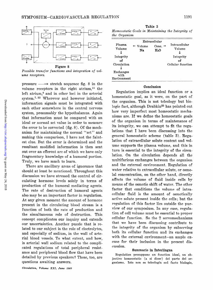
Figure 8Possible transfer functions and integration of vol-ume receptors.
pressure - stretch sequence fig. 8 in thevolume receptors in the right atrium," theleft atrium,8 and in other loci in the arterialsystem.7 22 Wherever and however initiated,information signals must be integrated witheach other somewhere in the central nervoussystem, presumably the hypothalamus. Againthat information must be compared with anideal or normal set value in order to measurethe error to be corrected (fig. 8). Of the mech-anism for maintaining the normal "set" andmaking this comparison, I have not the faint-est clue. But the error is determined and theresultant modified information is then sentout over an efferent arc of which we have onlyfragmentary knowledge of a humoral portion.Truly, we have much to learn.There are ancillary areas of ignorance that
should at least be mentioned. Throughout thisdiscussion we have stressed the control of cir-culating hormone levels solely in terms ofproduction of the humoral mediating agents.The rate of destruction of humoral agentsalso may be an important factor in regulation.At any given moment the amount of hormonepresent in the circulating blood stream is afunction of both the rate of production andthe simultaneous role of destruction. Thisconcept complicates our inquiry and extendsour uncertainties. Another puzzle that is re-lated to our subject is the role of electrolytes,and especially of sodium, in the wall of arte-rial blood vessels. To what extent, and how,is arterial wall sodium related to the compli-cated regulations of total peripheral resist-ance and peripheral blood flow that have beendetailed by previous speakers? These, too, arequestions awaiting answers.Circulation, Volume XXI, JIune 1960
Table 3Homeostatic Goals in Maintaining the Integrity ofthe Organism
ExtracellilarPlasma v Volume Cone. e IntracellularVolume Na 120 Volume
Integrity Integrityof of
Circulation Cellular function
Exchangeswith
Environment
ConclusionRegulation implies an ideal function or a
homeostatic goal, as it were, on the part ofthe organism. This is not teleology but bio-logic fact, although Drabkin23 has pointed outhow very imperfect most homeostatic mecha-nisms are. If we define the homeostatic goalsof the organism in terms of maintenance ofits integrity, we can attempt to fit the regu-lations that I have been discussing into thegeneral homeostatic scheme (table 3). Regu-lation of extracellular solute content and vol-ume supports the plasma volume, and this inturn is essential to the integrity of the circu-lation. On the circulation depends all themultifarious exchanges between the organismand the external environment. Regulation ofwater relative to extracellular solute, or osmo-lal concentration, on the other hand, directlyaffects the volume of fluid inside cells bymeans of the osmotic shift of water. The otherfactor that conditions the volume of intra-cellular fluid is the amount of osmoticallyactive solute present inside the cells; but theregulation of this factor lies outside the pur-view of our symposium. In any case, regula-tion of cell volume must be essential to propercellular function. So the 2 servomechanismsthat we have been discussing contribute tothe integrity of the organism by subservingboth its cellular funetion and its exchangeswith the external environment-an ample ex-cuse for their inclusion in the present dis-cussion.
Summario in InterlinguaRegulation presuppone un function ideal, un ob-
jectivo homeostatic (a si dicer) del parte del or-ganismo. Isto non es teleologia sed facto biologic,
7N.,,,,.lset
p-
-E.J-
z
1191
?
by guest on May 24, 2018
http://circ.ahajournals.org/D
ownloaded from

ELKINTON
ben que Drabkin ha signalate le extreme imperfectiondel majoritate del mechanismos homeostatic. Si nosdefini le objectivos homeostatic del organismo ab lepuncto de vista del mantenentia de su integritate,nos pote interprender le tentativa de visualisar lehic-discutite typos de regulation- illo del aqua e illodel electrolytos-in uni schema de homeostase general.Le regulation del contento extracellular de soluto edel volumine del liquido extracellular supporta levolumine del plasma, e isto-de su parte-es essentialpro le integritate del circulation. Oinne le multipleformas de excambio inter le organismo e le ambienteexterne depende del circulation. Del altere latere, leregulation de aqua in relation al solutos extracellular,o le concentration osmolal, affice directemente levolumine de liquido intra le cellulas per le agentiadel transition osmotic de aqua. Le altere factor queconditiona le volumine de liquido intracellular es lequantitate del osmoticamente active soluto que espresente intra le cellulas (sed le regulation de istefactor non es parte del thema formulate pro lepresente symposio). In omne caso, le regulation delvolumine cellular debe esser essential al normal func-tion cellular. Assi le duo servoinechanismos discutitein le presente analyse contribue al integritate delorganismo per le facto que illos es subserviente alfunction cellular e al excambio del organismo conle ambiente externe, e isto es plus que adequate comojustification de includer los in le presente symposio.
References1. HENDERSON, L. J.: On volume in biology. Proc.
Nat. Acad. Sc. 2: 654, 1916.2. VERNEY, E. B.: Croonian lecture. Antidiuretic
hormone and the factors which determine itsrelease. Proc. Roy. Soc., London, s.B. 135:25, 1947.
3. STRAUSS, M. B.: Body Water in Man: The Ac-quisition and Maintenance of the Body Fluids.Boston, Little, Browin & Company, 1957, Chap.7 and 8.
4. WOLF, A. V.: Thirst: Physiology of the Urge toDrink and Problems of Water Lack. Spring-field, Ill., Charles C Thomas, Publisher, 1958,Chap. 2.
5. PETERS, J. P.: Body Water: The Exchange ofFluids in Man. Springfield, Ill., Charles CThomas, Publisher, 1935, Chap. 11.
6. BORST, J. G. G.: Maintenance of adequate cardiacoutput by regulation of urinary excretion ofwater and sodium chloride; essential factor ingenesis of oedema. Acta med. Scandinav.Suppl. 207, 130: 1, 1948.
7. ROBINSON, J. R.: Reflections on Renal Function.Oxford, England, Blackwell Scientific Publi-cations, 1954, Chap. 7.
8. SIEKER, H. O., GAUER, 0. H., AND HENRY, J. P.:The effect of continuous negative pressurebreathing on water and electrolyte excretion
by the human kidney. J. Clin. Invest. 33: 572,1954.
9. LOVE, A. H. G., RODDIE, R. A., ROSENSWEIG, J.,AND SHANKS, R. G.: The effect of pressurechanges in the respired air on the renal excre-tion of water and electrolytes. Clin. Se. 16:281, 1957.
10. HENRY, J. P., AND PEARCE, J. W.: The possiblerole of cardiac atrial stretch receptors in theinduction of changes in urine flow. J. Physiol.131: 572, 1956.
11. FARRELL, G.: Regulation of aldosterone secretion.Physiol. Rev. 38: 709, 1958.
12. BARTTER, F. C., LIDDLE, G. W., DUNCAN, L. E.,JR., BARBER, J. K., AND DELEA, C.: The regula-tion of aldosterone secretion in man: The roleof fluid volume. J. Clin. Invest. 35: 1306, 1956.
13. YANKOPOULOS, N. A., DAVIS, J. O., KLIMAN, B.,AND PETERSON, R. E.: Evidence that a humoralagent stimulates the adrenal cortex to secretealdosterone in experimental secondary hyper-aldosteronism. J. Clin. Invest. 38: 1278, 1959.
14. ROSENBAUM, J. D., PAPPER, S., AND ASHLEY,M. M.: Variations in renal excretion of sodiumindependent of change in adrenocortical hor-mone dosage in patients with Addison's dis-ease. J. Clin.- Endocrinol. & Metab. 15: 1459,1955.
13. SMITH, H. W.: Salt and water volume recep-tors. Am. J. Med. 23: 623, 1957.
16. BRICKER, N. S., GUILD, W. R., REARDAN, J. B.,AND MERRILL, J. P.: Studies on the functionalcapacity of a denervated homotransplantedkidney in an identical twin with parallel ob-servations in the donor. J. Clin. Invest. 35:1364, 1956.
17. BARGER, A. C., LIEBOWITZ, R., AND MULDOWNEY,F. P.: The role of the kidney in the homeo-static adjustments of congestive heart failure.J. Chron. Dis. 9: 571, 1959.
18. MCCANCE, R. A.: Experimental sodium chloridedeficiency in man. Proc. Roy. Soc. s.B. 119:245, 1936.
19. LEAP, A., AND MAMBY, A. R.: An antidiureticmechanism not regulated by extracellular fluidtonicity. J. Clin. Invest. 31: 60, 1952.
20. SQUIRES, R. D., AND WILLIAMS, C.: Personalcommunication.
291. ELKINTON, J. R., AND SQUIRES, R. D.: The dis-tribution of body fluids in congestive heartfailure. I. Theoretic considerations. Circula-tion 4: 679, 1951.
22. EPSTEIN, F. H.: Renal excretion of sodium andthe concept of a volume receptor. Yale J.Biol. Med. 29: 282, 1956.
23. DRABKIN, D. L.: Imperfection: biochemical pho-bias and metabolic ambivalence. Perspect.Biol. Mled. 2: 473, 1959.
Circulation, Vol?cm e XXI, June 1960
1192
by guest on May 24, 2018
http://circ.ahajournals.org/D
ownloaded from

J. RUSSELL ELKINTONRegulation of Water and Electrolytes
Print ISSN: 0009-7322. Online ISSN: 1524-4539 Copyright © 1960 American Heart Association, Inc. All rights reserved.
is published by the American Heart Association, 7272 Greenville Avenue, Dallas, TX 75231Circulation doi: 10.1161/01.CIR.21.6.1184
1960;21:1184-1192Circulation.
http://circ.ahajournals.org/content/21/6/1184.citationlocated on the World Wide Web at:
The online version of this article, along with updated information and services, is
http://circ.ahajournals.org//subscriptions/
is online at: Circulation Information about subscribing to Subscriptions:
http://www.lww.com/reprints Information about reprints can be found online at: Reprints:
document. and Rights Question and Answer
Permissionsthe Web page under Services. Further information about this process is available in thewhich permission is being requested is located, click Request Permissions in the middle column ofClearance Center, not the Editorial Office. Once the online version of the published article for
can be obtained via RightsLink, a service of the CopyrightCirculationoriginally published in Requests for permissions to reproduce figures, tables, or portions of articlesPermissions:
by guest on May 24, 2018
http://circ.ahajournals.org/D
ownloaded from