recombinant parainfluenza virus 5 (piv5) expressing the influenza a virus hemagglutinin provides...
TRANSCRIPT

7) 139–150www.elsevier.com/locate/yviro
Virology 362 (200
Recombinant parainfluenza virus 5 (PIV5) expressing the influenza A virushemagglutinin provides immunity in mice to influenza A virus challenge
S. Mark Tompkins a, Yuan Lin b, George P. Leser c, Kari A. Kramer a, Debra L. Haas a,Elizabeth W. Howerth e, Jie Xu b, Mary J. Kennett b, Russell K. Durbin f, Joan E. Durbin f,
Ralph Tripp a, Robert A. Lamb c,d, Biao He b,g,⁎
a Department of Infectious Diseases, University of Georgia, Athens, GA, USAb Department of Veterinary and Biomedical Sciences, Pennsylvania State University, University Park, PA, USA
c Department of Biochemistry, Molecular Biology and Cellular Biology, Northwestern University, Evanston, IL, USAd Howard Hughes Medical Institute, Northwestern University, Evanston, IL, USA
e Department of Pathology, College of Veterinary Medicine, University of Georgia, Athens, GA, USAf Children’s Hospital, Ohio State University, Columbus, OH, USA
g Center for Molecular Immunology and Infectious Disease, Pennsylvania State University, University Park, PA, USA
Received 1 October 2006; returned to author for revision 23 October 2006; accepted 3 December 2006Available online 23 January 2007
Abstract
Parainfluenza virus type 5 (PIV5), formerly known as simian virus 5 (SV5), is a non-segmented negative strand RNA virus that offers severaladvantages as a vaccine vector. PIV5 infects many cell types causing little cytopathic effect, it replicates in the cytoplasm of infected cells, anddoes not have a DNA phase in its life cycle thus avoiding the possibility of introducing foreign genes into the host DNA genome. Importantly,PIV5 can infect humans but it is not associated with any known human illness. PIV5 grows well in tissue culture cells, including Vero cells, whichhave been approved for vaccine production, and the virus can be obtained easily from the media. To test the feasibility of using PIV5 as a livevaccine vector, the hemagglutinin (HA) gene from influenza A virus strain A/Udorn/72 (H3N2) was inserted into the PIV5 genome as an extragene between the hemagglutinin-neuraminidase (HN) gene and the large (L) polymerase gene. Recombinant PIV5 containing the HA gene ofUdorn (rPIV5-H3) was recovered and it replicated similarly to wild type PIV5, both in vitro and in vivo. The HA protein expressed by rPIV5-H3-infected cells was incorporated into the virions and addition of the HA gene did not increase virus virulence in mice. The efficacy of rPIV5-H3 as alive vaccine was examined in 6-week-old BALB/c mice. The results show that a single dose inoculation provides broad and considerableimmunity against influenza A virus infection.© 2006 Elsevier Inc. All rights reserved.
Keywords: Parainfluenza virus 5; Vaccine; Influenza virus; Viral vector
Introduction
PIV5, formerly known as simian virus 5 (SV5), is a non-segmented negative strand RNA virus in the paramyxovirusfamily. PIV5 contains an RNA genome of 15246 nucleotides thatis surrounded by a nucleocapsid protein and the genome encodes
⁎ Corresponding author. Department of Veterinary and Biomedical Sciences,Center for Molecular Immunology and Infectious Disease, The PennsylvaniaState University, 115 Henning Bldg., University Park, PA 16802, USA. Fax: +1814 863 6140.
E-mail address: [email protected] (B. He).
0042-6822/$ - see front matter © 2006 Elsevier Inc. All rights reserved.doi:10.1016/j.virol.2006.12.005
eight known viral proteins (Lamb and Kolakofsky, 2001).Nucleocapsid protein (NP), phosphoprotein (P), V and largeRNA polymerase (L) proteins are important for transcription andreplication of the RNA genome. Several studies suggest that theV protein has a role in evasion of host immune responses as wellas in regulating viral RNA synthesis (Didcock et al., 1999; He etal., 2002; Lin and Lamb, 2000; Lin et al., 2005; Sun et al., 2004).The fusion (F) glycoprotein mediates both virus to cell and cell tocell fusion in a pH independent manner. The hemagglutinin-neuraminidase (HN) glycoprotein, is the receptor binding proteinand its neuraminidase activity is important for virus release fromhost cells (Schmitt et al., 1999; Yuan et al., 2005). The matrix (M)

140 S.M. Tompkins et al. / Virology 362 (2007) 139–150
protein has an important role in the maturation of virus (Schmittet al., 2005). The SH integral membrane protein may have animportant role in inhibiting TNF-α-mediated apoptosis (Lin etal., 2003; Wilson et al., 2006).
Non-segmented negative strand RNA viruses (NNSVs) suchas PIV5 are potential viral vector candidates for vaccinedevelopment. As compared to DNA viruses, the NNSVs arepotentially safer because they do not have a DNA phase in theirlife cycles and most of them replicate in the cytoplasm, thusavoiding unintended consequences from genetic modificationsof host cell DNA that may be associated with recombination orinsertion. In addition, as compared to positive strand RNAviruses, the genome of NNSVs is stable. These characteristicsmake NNSVs useful as potential vaccine vectors.
Despite the advantages of using NNSV as vaccine vectors,only in recent years has it been possible to manipulate their RNAgenomes due to the development of methodologies for perform-ing reverse genetics (Neumann et al., 2002; Schnell et al., 1994).This has allowed for successful generation of recombinant NNSVvectors that include vesicular stomatitis virus (VSV), humanparainfluenza virus 3 (hPIV3) and Newcastle disease virus(NDV). VSV is a highly lytic NNSV which has been engineeredto express a hemagglutinin (HA) gene of influenza A virus. Therecombinant HA-VSV has been shown to provide a level ofimmunity in mice challenged with influenza A virus (Roberts etal., 1998). In addition, NDVhas been used to express the HAgeneof human (H1N1) influenza and the recombinant virus shown toprovide immunity against influenza virus challenge in mice(Nakaya et al., 2001). Very recently, NDVwas used to express theHA protein of avian (H5N1) influenza and this virus inducespotent protection against both influenza and NDV infection inpoultry (Park et al., 2006; Veits et al., 2006).
PIV5 infects a range of cell types including primary humancells (Arimilli et al., 2006). Indeed, there has been no report of acell line that is resistant to PIV5 infection. Importantly, PIV5causes very little cytopathic effect (CPE) in infected cells(Choppin, 1964; Zakstelskaya et al., 1976). PIV5 also infectsmost mammals including humans and is not associated with anyclinical disease with the exception of canine kennel cough (Cohnet al., 1996; Cornwell et al., 1976; McCandlish et al., 1978). Theability of PIV5 to infect a large spectrum of cells with littlecytopathic effect suggests that this virus could be used for geneexpression in a wide range of species to elicit a broad spectrumimmune response without associated inflammation and patho-genicity. Moreover, the lack of CPE allows PIV5 grow incommon cell lines, including MDBK cells, for more than 40 dayswhile the virions are continually released into media at titers up to8×108 PFU/ml (Choppin, 1964), providing an easy and costeffective means for mass production of the vaccine vector.
In previous studies, it was shown that PIV5 could be readilyrecovered from cloned DNA, and that a foreign gene encodinggreen fluorescent protein (GFP) could be inserted into the PIV5genome (rPIV5-GFP) with GFP expressed cytoplasmically (Heet al., 1997). Recombinant PIV5 containing GFP (rPIV5-GFP)replicates to similar titers as wild type (wt) PIV5 and insertion ofthe gene into the viral genome is stable. We tested the feasibilityof utilizing recombinant PIV5 as a vaccine vector by expressing
the HA gene of influenza A virus. We show that we can insertthe HA gene from influenza A/Udorn/72 (H3N2 subtype) virusinto the PIV5 genome at the gene junction between HN and Lgenes, recover infectious recombinant PIV5 containing the HAgene (rPIV5-H3), and that mice immunized with rPIV5-H3 areprotected from challenge with influenza virus.
Results
Generation of rPIV5-H3
The HA gene of influenza A virus (A/Udorn/72, H3N2subtype) was inserted between the HN and L genes of PIV5 in aplasmid (pBH311) containing the full length PIV5 genome aspreviously described for the GFP gene (Fig. 1A) (He et al.,1997). To ensure correct viral transcription initiation, termina-tion and polyadenlyation of transcripts, the HA gene wasflanked with gene start (GS), intergenic sequences (I) and geneend (GE) sequences from the junction region of the NP and V/Pgenes which gives high level transcription (He and Lamb,1999). Recovery of rPIV5-HA was performed as described byHe et al. (11) and recovery was as efficient as that of wild typePIV5. The authenticity of the rescued virus as rPIV5-H3 wasconfirmed by RT-PCR (Fig. 1A). Insertion of the HA geneincreases the expected size of RT-PCR fragment to 2.1 kb from400 base pairs (bp) when using the specific primers that annealto the HN and L genes (Fig. 1A). This 2.1 kb fragment wassequenced and found to match the input HA gene sequence(data not shown). It is known that PIV5 infects human cells(primary and established cell lines) (Arimilli et al., 2006). Thusto examine expression of HA in human primary cells, cells fromhuman peripheral blood mononuclear cells (PBMC) wereinfected with PIV5 and PIV5-H3. Virus gene expression wasdetected in the human primary cells for both viruses (Fig. 1B).
Expression of HA on the surface of cells infected withrPIV5-H3
Expression of the HA gene in cells infected with rPIV5-H3was examined by using both immunofluorescence and immu-noprecipitation techniques. rPIV5-H3-infected CV-1 cells werestained with anti-HA antibody and anti-F antibody and surfaceexpression of both HA and F could be detected (Fig. 1C). Cellsurface-expressed F protein was also detected using anF-specific mAb. Expression and cleavage of HA on the cellsurface were confirmed by treatment of cell surfaces withtrypsin. As shown in Fig. 1D, uncleaved HA0 could be cleavedby addition of exogenous trypsin to HA1 and HA2. Immuno-precipitation of the V/P proteins indicated that amounts of viralproteins expressed were similar between wild type PIV5 andrPIV5-H3 viruses.
Incorporation of HA into the rPIV5-H3 virion
To analyze the incorporation of HA into rPIV5-H3 virions,the virus was grown in MDBK cells and purified, andpolypeptides were analyzed by SDS-PAGE. HA was observed

Fig. 1. Generation of rPIV5-H3 and expression of HA in virus-infected cells. (A) Generation of rPIV5-H3. HA gene was inserted between HN and L genes of PIV5.The shaded area represents regions between two genes. It contains GE (gene end), I (intergenic) and GS (gene start) sequences which are important for RNAtranscription. A genomic nucleotide length divisible by six (the rule of six) was maintained. Oligomer BH195 that anneals to genome sense of HN gene was used forreverse-transcription; oligomer BH196 that anneals to mRNA sense of L gene and oligomer BH195 were used for PCR. The PCR product was resolved in 1% agarosegel. The DNA size markers are indicated. (B) Immunofluorescence of human primary cells infected with PIV5 and PIV5-H3. Cells obtained from primary humanPBMC cells (mostly macrophages) were infected with PIV5, PIV5-H3 or mock infection. At 2 days p.i., the cells on coverslips were fixed and treated with anti-NP(red) and anti-V/P (green) antibodies. (C) Expression of HA in infected cells. CV-1 cells were infected by the rPIV5 or rPIV5-H3 and the cells were processed forimmunofluorescence or immunoprecipitation at 20 h p.i. CV-1 cells on coverslips were fixed and treated with anti-HA and anti-F antibodies as indicated. (D)Expression of HA on cell surface. Infected CV-1 cells were labeled with 35S-Met and 35S-Cys for 2 h and lysed directly or treated with trypsin and then lysed. Thelysates were immunoprecipitated with antibodies against HA and the Vand P proteins of PIV5. Sizes of molecular weight marker and positions of V, P, HA0, HA1 andHA2 are indicated.
141S.M. Tompkins et al. / Virology 362 (2007) 139–150
in rPIV5-H3 virions by Commasie blue staining (Fig. 2A) andits identity confirmed by immunoblotting (data not shown).Concomitantly, the amount of HN incorporated into rPIV5-H3
was decreased as compared to the amount of HN incorporatedinto wt PIV5. To eliminate the possibility that the HA proteinobserved was associated with contaminating vesicles and

Fig. 2. Incorporation of HA into purified virions. rPIV5-H3 was purified asdescribed in the Material and Methods. (A) The purified virus polypeptides wereresolved using a 15% SDS-PAGE gel and the gel was stained with Commasieblue. Positions of viral proteins are indicated. (B) The purified virions weretreated with anti-HA mAb and gold particle-labeled secondary antibody andthen examined by electron microscopy. Panels of different virions are indicated.(For interpretation of the references to colour in this figure legend, the reader isreferred to the web version of this article.)
Fig. 3. Growth of rPIV5-H3 in vitro and in vivo. (A) Growth curve in vitro.MDBK cells were infected at 10 PFU/cell. Media from the infected cells werecollected at 6 h intervals up to 48 h p.i. Titers of the media were examined usingplaque assay in BHK-21F cells. (B) Growth in vivo. BALB/c mice (15 in agroup) were infected with PIV5 or rPIV5-H3. Five mice per group weresacrificed on days 4, 6 and 8 p.i. Titers of viruses in lung were determined byplaque assay.
142 S.M. Tompkins et al. / Virology 362 (2007) 139–150
debris, purified rPIV5-H3 virions were immunogold labeledusing HA antibody and examined by electron microscopy. HAlabeling of virion was readily detected (Fig. 2B).
As PIV5 virions are pleomorphic with sizes ranging from 100to 200 nm, we examined a large number of virions and found nochange in virion morphology or size associated with therecombinant virus, indicating that the addition of 1700 nt intothe PIV5 genome did not alter virion morphology (data notshown).
Growth of rPIV5-H3 in vitro and in vivo
To determine if insertion of the HA gene had any detrimentaleffect on virus replication, single-step growth curves of wt PIV5and rPIV5-H3 viruses were performed. MDBK cells wereinfected with 10 plaque-forming units (PFU) per cell of rPIV5 orrPIV5-H3 and supernatants were collected at 6 h intervals for upto 48 h and virus quantified by plaque assay (Fig. 3). The growthcurve for rPIV5-H3 was very similar to that of wt virus. Bothviruses had similar initial growth kinetics and reached a plateauat about 24 h post-infection (p.i.). Additionally, the plaque sizesof the two viruses were comparable (data not shown).
To examine the growth of rPIV5-H3 in vivo, BALB/c micewere inoculated intranasally (i.n.) with 106 PFU of PIV5 orrPIV5-H3. Lungs of the infected animals were collected at 4, 6and 8 days p.i., and virus titers in lungs were determined by
using plaque assays as described previously (He et al., 2002). Itwas found that PIV5 replicated to higher titers than rPIV5-H3 at4 days p.i. (Fig. 3B); however, the differences in virus titerdiminished over time, e.g. at 8 days p.i. both viruses hadapproximately the same titers, i.e. ∼103 PFU.
Pathology of rPIV5-H3 in BALB/C mice
PIV5 infection does not cause known overt disease exceptkennel cough in canines (Chatziandreou et al., 2004). WhetherPIV5 infection was pathogenic for mice and whether expressionof the HA gene could affect the pathogenicity of the virus inmice have been determined. First, BALB/c mice were infected i.n. with 106 PFU of rPIV5-H3 and the body weights of theinfected mice monitored daily. Infection of mice with rPIV5-H3did not cause any detectable symptoms or weight loss comparedwith mock (PBS)-inoculated mice (Fig. 4A). Second, to addressother parameters of pathology that may be associated with PIV5or rPIV5-H3 infection, BALB/c mice were inoculated i.n. with106 PFU of PIV5 or rPIV5-H3. At 4, 6 and 8 days p.i., theanimals were sacrificed and lung pathology examined by H&Estaining. At each sampling period, histopathology changes weresimilar in character andmagnitude in the lung lobes of individual

Fig. 4. Pathology of PIV5 and rPIV5-H3 in BALB/c mice. (A) Weight loss. BALB/c mice (n=8) were infected with rPIV5-H3 (106 PFU in 50 μl PBS) or mock-infected with PBS only. Body weights of the mice were recorded and graphed as percentage of the weight before inoculation. (B) Pathology of PIV5 in mice. BALB/cmice (n=9) were infected with rPIV5-H3 or rPIV5 (106 PFU in 50 μl PBS). Animals were sacrificed on days 4, 6 and 8 p.i. and lungs were removed and fixed. Lungswere sectioned, H&E stained and analyzed. For each animal, four major pulmonary changes (peribronchiolar infiltrations; perivascular infiltrations; parenchymalinfiltrations; subpleural infiltrations) were subjectively scored 0–3 with 0=no change and 3=greatest change and a total score determined. Means of the total scores foreach group are graphed and standard deviations are shown as error bars. Scores of the two groups (PIV5 vs. rPIV5-H3) were compared at each time point (day 4, 6 or 8)by Student's t-test and all p values are less than 0.05. No statistically significant differences (p<0.05) between the two viruses were observed. (C) Weights of nudemice injected with PIV5 or rPIV5-H3. Nude mice (14-week-old, n=10) were infected i.v. with PIV5 or PIV5-H3 (107 PFU) in 200 μl volume. Body weights of themice were recorded and graphed as percentage of the weight before inoculation. (D) Pathology scores of lung. The mice were sacrificed and organs (lung, heart, liver,kidney and spleen) were sectioned for H&E staining. Pathology scores of lung were shown. 1=mild pathology; 2= intermediate pathology; 3=server pathology. Bar isaverage score and the error bar represents the range. No statistically significant differences between two viruses were observed.
143S.M. Tompkins et al. / Virology 362 (2007) 139–150
animals between infected groups, i.e. PIV5 vs. rPIV5-H3. Thelung histopathology generally was characterized by low-levelperivascular, peribronchiolar, subpleural, and scattered intra-alveolar infiltrations that were predominantly lymphocytes.Infiltrates contained a few plasma cells by day 8 p.i. and intra-alveolar infiltrates were admixed with macrophages. Cellularinfiltrations increased over the time-course of infection by bothviruses, but were generally mild, and at each time periodexamined, there was no statistically significant difference(P≤0.05) between groups (Fig. 4B). However, as expected,the means for each group did increase with time. The mildcellular reaction suggested a progressive, localized immunolo-gic response in the lung, apparently associated with PIV5 asaddition of the HA gene did not exacerbate the reaction. Tofurther confirm that rPIV5-H3 did not cause enhancedpathogenicity, the pathogenicity of the viruses in nude micehave been examined by injecting 107 PFU of each virus into thetail vein. Neither PIV5 nor PIV5-H3 caused signs of illness or
weight loss at either early or late time-points post-infection,indicating that the viruses are not pathologic even in immunedeficient mice (Fig. 4C). Examination of the organs from themice infected with different viruses indicated that there was nodifference in pathological scoring between PIV5 and rPIV5-H3-infected mice (Fig. 4D). While signs of infection were observedas expected, there were no significant pathology detected in thespleen, heart, liver or kidney (data not shown) and only verymildpatchy histeocytosis and very diffuse mixed inflammatoryinfiltrates, predominantly histocytes with a microgranuloma,were observed in lungs of some of the animals in both PIV5 andrPIV5-H3-infected mice.
Immunity against influenza A virus infection in mice immunizedwith rPIV5-H3
To examine the efficacy of rPIV5-H3 as a live vaccineagainst influenza A virus infection, 6-week-old BALB/c mice

144 S.M. Tompkins et al. / Virology 362 (2007) 139–150
were inoculated i.n. using virus stock grown in MDBK cells.BALB/c mice were i.n. vaccinated with 0, 102, 104 or 10 6 PFUof rPIV5-H3 or rPIV5 viruses diluted in 50 μl PBS. Six weeksafter vaccination, the mice were challenged i.n. with 106 PFU ofinfluenza A virus strain WSN-H3. WSN-H3 is a 7+1reassortant virus containing all of the genome segments fromA/WSN/33 virus except the HA segment, which is fromA/Udorn/72 (Jin et al., 1996). WSN-H3 was generatedthrough recombination of genomes between A/WSN/33(WSN), which is mouse-adapted and highly pathogenic, andUdorn, a human strain, which is not pathogenic in mice.WSN-H3 contains 7 genomic segments of WSN and the HAsegment from Udorn (subtype H3). As noted, WSN-H3 ispathogenic in the mouse, although not as pathogenic as theparent strain, WSN. Following challenge, the mice wereweighed daily. As expected, body weights of vaccinated and
Fig. 5. Efficacy of rPIV5-H3 as a live vaccine vector. Six-week-old BALB/c micechallenged i.n. with influenza Avirus, WSN-H3.Weights of the mice were monitoredmouse in the control group mice (n=4), i.e. weights of mock- (PBS) immunized mice(n=8), 104 (C) (n=8) or 106 PFU (D) (n=8) of rPIV5-H3 and then challengedWSN-Hthen challenged with WSN-H3. (F) Weights of individual PIV5-immunized mouse c
unvaccinated mice not challenged with WSN-H3 weresimilar. For the mice that were not vaccinated with rPIV5-H3, but challenged with the 106 PFU of WSN-H3, three outof four died by day 10 (Fig. 5A). The remaining mouse lostmore than 30% of its body weight and had not recovered byday 12. A similar mortality rate was initially observed in themouse group vaccinated with a low dose (102 PFU) of rPIV5-H3 and challenged with WSN-H3 in which four of the micedied at days 5, 8, 10 and 11 pi (Fig. 5B). However, theremaining 4 vaccinated mice recovered after 6 days afterlosing up to 20% to 30% of their body weight. By day 12,these mice regained their weight indicating that there was limitedprotection even for the mice inoculated with the low dosage ofrPIV5-H3. The group of mice inoculated with 104 PFU ofrPIV5-H3 suffered moderate weight loss (up to about 25%) andall of the mice survived. They also started to recover weight at
were immunized i.n. with PIV5-H3 or rPIV5. Six weeks p.i., the mice wereand graphed as percentage of body weight of the mice. (A) Weights of individualchallenged with WSN-H3. (B–D)Weights of individual immunized with 102 (B)3. (E) Average weights of mice immunized with 104 and 106 PFU rPIV5-H3 andhallenged with WSN-H3 (n=8).
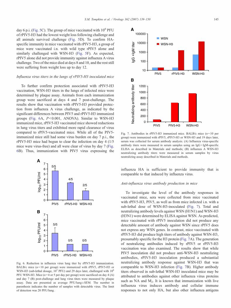
Fig. 7. Antibodies in rPIV5-H3 immunized mice. BALB/c mice (n=10 pergroup) were immunized with rPIV5, rPIV5-H3 or WSN-H3 and 19 days later,serum was collected for serum antibody analysis. (A) Influenza virus-specificantibody titers were measured in serum samples using an IgG+IgM-specificELISA as described in Materials and methods; (B) influenza A WSN-H3neutralizing antibody titers were measured in serum samples by virus
145S.M. Tompkins et al. / Virology 362 (2007) 139–150
day 6 p.i. (Fig. 5C). The group of mice vaccinated with 106 PFUof rPIV5-H3 had the lowest weight loss following challenge andall animals survived challenge (Fig. 5D). To confirm HA-specific immunity in mice vaccinated with rPIV5-H3, a group ofmice were vaccinated i.n. with wild type rPIV5 alone andsimilarly challenged with WSN-H3 (Fig. 5F). As expected,rPIV5 alone did not provide immunity against influenza Aviruschallenge. Two of themice died at days 8 and 10, and the rest stillwere suffering from weight loss up to day 12.
Influenza virus titers in the lungs of rPIV5-H3 inoculated mice
To further confirm protection associated with rPIV5-H3vaccination, WSN-H3 titers in the lungs of infected mice weredetermined by plaque assay. Animals from each immunizationgroup were sacrificed at days 4 and 7 post-challenge. Theresults show that vaccination with rPIV5-H3 provided protec-tion from influenza A virus challenge, as indicated by thesignificant differences between PIV5 and rPIV5-H3 immunizedgroups (Fig. 6A, P<0.001, ANOVA). Similar to WSN-H3immunized mice, rPIV5-H3 vaccinated mice showed reductionsin lung virus titers and exhibited more rapid clearance of viruscompared to rPIV5-vaccinated mice. While all of the PIV5-immunized mice still had some virus burden on day 7 p.i., therPIV5-H3 mice had begun to clear the infection on day 4 (1/5mice were virus-free) and all were clear of virus by day 7 (Fig.6B). Thus, immunization with PIV5 virus expressing the
Fig. 6. Reduction in influenza virus lung titer by rPIV5-H3 immunization.BALB/c mice (n=10 per group) were immunized with rPIV5, rPIV5-H3 orWSN-H3 (sub-lethal dosage, 103 PFU) and 29 days later, challenged with 106
PFU WSN-H3. Mice (n=4 or 5 per day per group) were sacrificed on day 4 (A)and day 7 (B) post-challenge and lung virus titers were measured by plaqueassay. Data are presented as average PFU/lung±SEM. The number inparenthesis indicates the number of samples with detectable virus. The limitof detection was 20 PFU/lung.
neutralizing assay described in Materials and methods.
influenza HA is sufficient to provide immunity that iscomparable to that induced by influenza virus.
Anti-influenza virus antibody production in mice
To investigate the level of the antibody responses invaccinated mice, sera were collected from mice vaccinatedwith rPIV5-H3, PIV5, as well as from mice infected i.n. with asub-lethal dose of WSN-H3-inoculated (Fig. 7). Total andneutralizing antibody levels against WSN (H1N1) andWSN-H3(H3N1) were determined by ELISA against WSN. As predicted,mice vaccinated with rPIV5 inoculation did not produce anydetectable amount of antibody against WSN since rPIV5 doesnot express any WSN genes. In contrast, mice vaccinated withrPIV5-H3 did produce high titers of antibody against WSN-H3,presumably specific for the H3 protein (Fig. 7A). The generationof neutralizing antibodies induced by rPIV5 or rPIV5-H3vaccination was also examined. The results show that whilePIV5 inoculation did not produce anti-WSN-H3 neutralizingantibodies, rPIV5-H3 inoculation produced a substantialneutralizing antibody response against WSN-H3 that wascomparable to WSN-H3 infection (Fig. 7B). Higher antibodytiters observed in sub-lethal WSN-H3 inoculated mice may beattributed to antibodies against other influenza virus proteinssuch as NA and M2. It is known that immunization with liveinfluenza virus induces antibody and cellular immuneresponses to not only HA, but also other influenza antigens

146 S.M. Tompkins et al. / Virology 362 (2007) 139–150
including neuraminidase, matrix, and nucleoprotein, which cancontribute to protection and in some cases are sufficient toprotect against virus challenge (reviewed in Epstein, 2003).
Discussion
The development of reverse genetics technology for therecovery of NNSVs from their cDNAs, has permitted theexpression of many foreign genes using these viruses (Bukreyevet al., 2005; Palese et al., 1996; Park et al., 2006; Pekosz et al.,1999; Roberts and Rose, 1998; Veits et al., 2006). Here werecovered PIV5-H3, which contains an HA gene from influenzaA/Udorn/72 virus inserted between the HN and L genes of PIV5.rPIV5-H3 was viable and the insertion of the HA gene appearedto be stable and the virus grew well in tissue culture cells. Asingle dose inoculation of the rPIV5-H3 provided immunity tomice against influenza virus infection.
The HA protein was incorporated into PIV5 virions despitethe absence of specific PIV5 sequences that may be importantfor incorporation into virions such as PIV5-specific cytoplasmictails. The HA protein was detected in purified virions by bothSDS-PAGE and electron microscopy (Fig. 2). Additionally, theamount of HN incorporated into rPIV5-H3 virions was reducedcompared to rPIV5. It is perhaps surprising that HN was notpreferentially incorporated into virions over HA. The efficientrelease of PIV5 from infected cells requires the cytoplasmic tailof HN (Schmitt et al., 1999) and it is thought that the cytoplasmictail of HN is important for formation of budding virus and/orpulling viral components into virions. Despite having acytoplasmic tail dissimilar to the HN, HA was incorporatedinto the PIV5 virions and presumably filled space at the expenseof HN.
A single dose inoculation of rPIV5-H3 provided immunity tomice against influenza virus infection. The gradient of protectionprovided by the virus indicates that effective immunity wasdependent on the dosage of the virus used and the protection wasprovided by rPIV5-H3 inoculation but not by rPIV5. Althoughthe effectiveness of rPIV5-H3 in preventing death by influenzaAvirus infection is evident, all mice lost body weight, indicatingthat protection is not optimal. This is partially due to the handlingof mice as even mock inoculated and mock challenged micesuffered about 5%weight loss. To increase potency of rPIV5-H3against influenza virus challenge, one possible remedy is to usemultiple inoculations (i.e. prime and boost) instead of a singleimmunization prior to challenge. Alternatively, it may bepossible to increase expression of HA by inserting the HAgene closer to the leader sequence of PIV5, the only de factopromoter of PIV5. Negative stranded RNAviruses such as PIV5initiate transcription from the 3′ end leader sequence, andtranscription levels of the viral genes are affected by the distanceof the gene from the leader sequence (Tokusumi et al., 2002). Forexample, the NP gene of PIV5, which is proximal to the leadersequence is the most abundantly transcribed, whereas the Lgene, which is distal to the leader sequence is least transcribed.To increase the expression level of the HA gene, the HA genecan be inserted immediately downstream of the leader sequenceand upstream of the NP gene.
Safety is a paramount concern for vaccine development.Expression of a foreign glycoprotein has the potential to expandthe host range beyond the original virus, therefore creating apotential safety hazard. However, because PIV5 already infectsall cell types that have been tested, it is less likely that adding anadditional receptor-binding gene to its genome will change itstropism. PIV5 encodes two major glycoproteins F and HN thatare important for PIV5 entry. The PIV5 F protein promotesvirus to cell fusion at the plasma membrane independent of pH.In some cells, F alone is sufficient to promote cell-to-cell fusionwithout PIV5’s receptor binding protein, HN (Paterson et al.,2000). The HN protein of PIV5 mediates virus attachment bybinding to sialic acid, which is added to surface proteins post-translationally. Unlike influenza virus protein, the HN protein ofPIV5 has no preference as to what kind of sialic acid it binds.For example, PIV5 may infect primary chicken cells (Peluso etal., 1977), which has mainly N-acetylneuraminic acid α 2,3-galactose (NeuAcα2,3Gal) and may infect human cells (Lamband Kolakofsky, 2001), which has mainly NeuAcα2,6Gal (Itoet al., 1997). Thus, it is unlikely that expressing influenza HAwill expand the host range of PIV5. Nevertheless, to address theconcern that enhanced pathogenicity may be caused by PIV5-H3, we examined the pathogenicity of the viruses in nude miceby injecting 107 PFU of each virus into the tail vein. NeitherPIV5 nor PIV5-H3 caused signs of illness or weight loss ateither early or late time-points post-infection, indicating that theviruses are not pathological even in immune deficient mice (Fig.4C). Examination of organs of the mice indicated that there wasno difference in pathological scoring between PIV5 and rPIV5-H3-infected mice (Fig. 4D). Thus, as expected, insertion of theHA gene into PIV5 genome did not cause increasedpathogenicity in mice. In the event that expressing a functionalHA is a concern, a mutant HA without HA1/HA2 cleavage sitecan be used for influenza virus vaccine development.
Unlike icosahedral viruses, PIV5 virions have many formsand shapes. This pleomorphic virion structure providesflexibility to accommodate changes in sizes of PIV5’s genome.This is an advantage for insertion of foreign genes as thechanges to the viral genome will probably not be tightlyrestricted by the virion structure. While we do not know theupper limit for the size of insertion in PIV5, in our hands,insertion of 1700 nt, an increase of about 11% of genome size,does not affect virus growth or integrity of virions. We havenow inserted and expressed a gene as large as 2.3 kb into PIV5genome without difficulty (data not shown).
The origin and natural host of PIV5 is not clear. PIV5was firstisolated from monkey cells as a contaminant in 1956, hence theoriginal name SV5 (Hull et al., 1956). However, subsequentserological testing of monkeys in the wild indicated no exposureto this virus. In contrast, monkeys in captivity at an animalfacility rapidly sero-converted, suggesting they contacted thevirus in captivity (Atoynatan and Hsiung, 1969; Tribe, 1966).Thus, considerable evidence indicates that PIV5 is not a simianvirus. PIV5 dose cause kennel cough in canines (Azetakaand Konishi, 1988; Binn et al., 1967; Cornwell et al., 1976;McCandlish et al., 1978; Rosenberg et al., 1971) and killedPIV5 is a component of the commercial vaccine “Vanguard”

147S.M. Tompkins et al. / Virology 362 (2007) 139–150
(Pfizer, Inc.) for dogs, which includes canine distemper virusvaccine. There is no convincing evidence that PIV5 causesdiseases in humans, despite much speculation in the 1970sthat PIV5 might be associated with a number of illnessesincluding multiple sclerosis (MS), subacute sclerosingpanencepalitis (SSPE), Creutzfeldt-Jakob disease (CJD),pemphigus, athero-sclerosis, Paget’s disease, hepatitis andthe common cold. Subsequent studies have ruled out PIV5 asthe etiological agent for these diseases (Chatziandreou et al.,2004; Hsiung, 1972; Vandvik and Norrby, 1989). Inretrospect, two possible explanations exist for why PIV5may have been linked to these diseases. One reason is basedon the conditions used for virus isolation in the humanstudies, i.e. the labs used monkey cell lines which can bepersistently infected with PIV5, and these cells often showno detectable cytopathic effects (Chatziandreou et al., 2004;Hsiung, 1972). Another reason is that there is known antigencross-reactivity of PIV5 to ubiquitous paramyxoviruses suchas human parainfluenza virus 2 and mumps virus, which areclosely related to PIV5 and have almost 100% exposure inhuman population (Komada et al., 1991; Randall and Young,1988; Tsurudome et al., 1989). Given the possibility of PIV5antigen cross-reactivity, it may be a concern to use PIV5 as avaccine vector. However, there is no indication from studiesin mice that antibodies can prevent PIV5 infection (Young etal., 1990). As shown in Fig. 1B and others (Arimilli et al.,2006), PIV5 can infect human primary cells, suggesting thatit is likely that PIV5 can infect humans.
While live virus vectors hold great promise, there are manypotential pitfalls, as has been demonstrated in studies using thetwo most studied virus vectors: adenovirus and VSV. Forinstance, adenovirus is a DNAvirus and thus has the potential tointegrate its genome. An additional problem is that there is avery high incidence of adenovirus sera-positive people. Also, apatient in a clinical trial died as a result of being treated with ahigh dose adenovirus-based gene vector. The other viral vectornoted, VSV, is neurotrophic, thus its potential impact onimmune comprised patients is of an important concern.Nonetheless, it is important to explore the potential use ofthese viral vectors due to the many advantages of live virusvectors over alternative vaccination strategies. As an RNAvirusthat infects respiratory epithelium, PIV5 offers an importantalternative vector with properties that make it worthy of specialconsideration and further study, especially for developing aninfluenza virus vaccine. Thus, although several NNSVs havebeen used previously to express foreign viral antigens and toprotect mice for virus challenge, PIV5 offers an interestingalternative vector with properties that make it worthy of specialconsideration and further study.
Materials and methods
Plasmid construction and RT-PCR
HA gene from pTF7.5-HA was amplified by PCR usingoligomers BH291 (5′-ATTTCCTAGGTAATTTTTAAGAAA-AAAACGATAGGACCGAACCTATCGATGCCGGCAAA-
CATGAAGACTATCATTGCTTTG-3′) and BH292 (5′-AAATTTGCATGCCATGGCCTAGGATATTCAAATG-CAAATGTTGCACCT-3′). The PCR fragment was insertedinto HincII site of pGEM2 (Promega) to generate pBH374. Thecoding region of HA gene in pBH374 was sequenced and foundto be identical to the HA gene in pTF7.5-HA. The HA gene wasexcised out of the plasmid by digestion with ClaI and NcoI. Thefragment was then filled-in by the Klenow fragment andinserted into filled-in NotI site of a plasmid (pBH311) (He et al.,1997) which contains a GFP gene between the HN and L genesin PIV5 genome to generate pBH412 which contains the PIV5genome with the HA gene as an extra gene. Standard protocolsof molecular cloning were followed. Oligomers were purchasedfrom Macromolecular Resources (Colorado State University).All sequences of the plasmids are on file and available onrequest. Plasmids pTF7.5-HA, pT7-L, pT7-P and pT7-NP weredescribed before (He et al., 1997; Murphy and Parks, 1997).
To sequence the viral RNA genome, total RNA from CV-1cells infected by virus in 6 cm plate was purified using RNeasykit (Qiagen®) according to the manufacture’s instruction. 19 μlout of 50 μl RNAwas used with 20 ng of oligomer BH195 (5′-TTCAGATTGTCCCATTTATCCGTCAGG-3′) for cDNAsynthesis at 37 °C for 1 h in a total volume of 40 μl. 10 μl ofcDNA was used in PCR with oligomers BH195 and BH196(TGTAATGGACCTAAATCGTCAAGGTCG-3′). The PCRproducts were resolved in 1% agarose gel.
Viruses and cells
Virus recovery was carried out as described before (He et al.,1997). 5 μg of plasmid pBH412 which contains a PIV5 genomewith HA gene insertion was transfected with 3 μg of pT7-L,0.2 μg of pT7-P and 2 μg of pT7-NP into A549 cells in 6-wellplates which were infected with the modified vaccinia virusAnkara strain (MVA) containing a bacteriophage T7 RNApolymerase gene for 1 h. Transfection media were changed toDMEM containing 2% fetal calf serum (FCS) 24 h posttransfection. Virus released into the media was amplified in CV-1 cells. Recovery of virus is indicated by syncytia formation inBHK 21F cells on incubation with the media from the CV-1cells. The virus was then purified as a single plaque from BHK21F cells. Human primary cells were obtained from PBMC. Thehuman primary cells (mostly macrophages) from PBMCs wereprepared as described (Lee et al., 2001) and were seeded in 6-well plate with coverslips for 6 days before used for infection.
Viruses were grown in MDBK cells for 5 to 7 days usingDMEM containing 2% FCS until their hema-absorption titersplateaued. Media were collected and pelleted at 3000 rpm usinga Sorvall tabletop centrifuge for 10 min. BSAwas added to thecleared supernatant to a final concentration of 1%. The virusstock was quickly frozen in dry ice and stored at −70 °C. Topurify the viruses, viruses in the cleared supernatant werepelleted in a Beckman ultracentrifuge Type 45Ti rotor at19,000 rpm for 1 h. The pellet was then resuspended in NTEbuffer and loaded onto 15% to 60% sucrose gradient andcentrifuged in a SW 41Ti rotor for 2 h at 24,000 rpm. The virusband was collected and virus was pelleted in a Type 70Ti rotor

148 S.M. Tompkins et al. / Virology 362 (2007) 139–150
for 1 h at 35,000 rpm (Paterson and Lamb, 1993). Virus titerswere determined by plaque assay on BHK-21F cells aspreviously described (He et al., 1997).
MDBK cells, A549 cells and CV-1 cells were cultured inDMEM with 10% FCS. BHK-21F cells were grown in DMEMwith 10% FCS and 10% tryptone phosphatase broth.
Immunoprecipitation of polypeptides
CV-1 cells in 6 cm plates were infected with rPIV5 andrPIV5-H3 at a m.o.i. of about 5 pfu/cell and labeled with100 μCi/ml 35S-Met and 35S-Cys 20 h p.i. (Paterson and Lamb,1993). Half of the plates of CV-1 cells were treated with0.01 mg/ml L-(tosylamido-2-phenyl) ethyl chloromethyl ketone(TPCK)-treated trypsin for 1 h before the cells were lysed inRIPA buffer (Paterson and Lamb, 1993). Lysates wereimmunoprecipitated using mAb Pk specific for a shared regionof the V and P proteins and rabbit polyclonal antibody specificfor Udorn HA protein. Polypeptides were analyzed by 10%SDS-PAGE and their radioactivity was detected using a FujiBioImager 1000.
Fluorescence and electron microscopy
CV-1 cells were grown on glass cover slips and infected withrPIV5 or rPIV5-H3 at a m.o.i. of about 5 PFU/cell. At 20 h p.i.,the infected cells were washed with PBS and then fixed with1% formaldehyde for 10 min at room temperature. The cellswere washed three times with PBS and incubated for 30 minwith 1:500 dilution of antibodies against PIV5 F or HA. Thecells were washed extensively to remove unbound antibodiesand secondary antibodies labeled FITC were added for30 min. The cells were further washed and placed on slides.Fluorescence was examined using a Zeiss 410 confocalmicroscope.
To examine HA in virions, purified virions were absorbedonto parlodion-coated nickel grids for 30 s. Grids were floatedon a drop of Tris-buffered saline (TBS), pH 7.4 for 5 min, afterwhich they were incubated by floating on drops of 3%ovalbumin in TBS for 1 h with HA-specific mouse mAbascites fluid 6D/1 diluted 1:300 in 1% ovalbumin in TBS.Following three successive washes with TBS for 10 min each,samples were incubated for 1 h with goat anti-mouse IgGcoupled to 10 nm gold particles diluted 1:10 in 1% ovalbumin inTBS. Grids were washed in TBS as above and finally stainedwith 2% phosphotungsic acid, pH 6.6. Before viewing, a thinlayer of carbon was evaporated over the grids for stability. Thegrids were then examined using JEOL 1230 transmissionelectron microscope (JEOL, Tokyo, Japan).
Virus titers in vivo and pathology of infection with PIV5 orrPIV5-H3
Six-week-old BALB/c mice were first anesthetized withAvertin i.p. and then infected intranasally (i.n.) with 106 PFU ofPIV5 or rPIV5-H3 in 50 μl PBS. At various times post-infection, mice were euthanized and lungs collected for either
lung virus titer or histopathologic analysis. For virus titers,lungs were homogenized in 1 ml PBS and the homogenatescleared by centrifugation. Cleared homogenates were seriallydiluted and virus titers determined by plaque assay on BHK-21Fcells (He et al., 1997). For histopathology, lungs were perfusedwith 10% buffered formalin via the trachea and then placed informalin for fixation. Sections of all lung lobes were embeddedin paraffin, sectioned at 3 μm, stained with hematoxylin andeosin, and examined by light microscopy. For each animal, fourmajor pulmonary changes (peribronchiolar infiltrations; peri-vascular infiltrations; parenchymal infiltrations; subpleuralinfiltrations) were subjectively scored 0–3 with 0=no changeand 3=greatest change and a total score determined. Forpathology studies in nude mice, 107 PFU of each virus in a200 μl volume was injected into the tail veins of nude mice. Themice were weighed and sacrificed at 3, 7 and 14 days postinjection. The organs of the mice (lung, heart, liver, kidney andspleen) were sectioned and processed for H&E staining.
Influenza A virus challenge experiments
Six-week-old BALB/c mice were first anesthetized and theninoculated intranasally by dropping 50 μl PBS containing 0,102, 104 or 10 6 PFU of viruses. Six weeks later, the mice wereinoculated intranasally with 50 μl PBS containing 106 PFU ofan influenza virus 7+1 reassortant WSN-H3 that contains allsegments from WSN strain except the HA segment which isderived from A/Udorn/72. Body weights of the mice wererecorded everyday thereafter.
Antibody measurement
Influenza virus-specific serum antibody titers were mea-sured using an IgG+IgM ELISA. Enzyme immunoassay plates(Corning Costar, Inc.) were coated with 50 μl/well of WSN orWSN-H3 containing approximately 105 PFU of virus. Viruseswere UV inactivated and the plates washed. The wells wereblocked with 200 μl/well Starting Block Buffer (Pierce, Inc.)twice for 1 min. After washing, dilutions of sera were added(50 μl/well) and incubated at room temperature for 2 h. Plateswere washed and 100 μl/well of a 1:1000 dilution of alkalinephosphate-labeled rat goat-mouse IgG+IgM, IgG2a, IgG2band/or IgA (KPL, Inc.) was added. After incubation for 1 h atroom temperature, the plates were washed, 100 μl/well pNPPphosphatase substrate (KPL, Inc.) was added, and theenzymatic reaction was allowed to develop at room tempera-ture. O.D. was measured at 405 nm on a BioTek plate reader.The ELISA titer was the lowest serum dilution with an OD 2standard deviations above the OD of the same dilution of naïveserum.
Influenza neutralizing antibody titers were measured inserum samples by virus neutralizing assay. Sera were seriallydiluted in 50 μl DMEM+5% FBS. 2000 TCID50 of WSN-H3virus was added to diluted sera and incubated for 60 min at37 °C. Serum and virus were added to 96-well microtiter platescontaining 80–90% confluent MDCK cells and incubatedovernight at 37 °C. The wells were washed with PBS and the

149S.M. Tompkins et al. / Virology 362 (2007) 139–150
medium replaced with 0.2 ml of MEM+0.25 μg/ml TPCK-treated trypsin (Worthington) and incubated for 4 days at 37 °C.Individual wells were scored for CPE and supernatants assayedfor virus by hemagglutination of chicken red blood cells(cRBCs). In brief, fresh cRBCs were washed in Alsevier’ssolution, counted and resuspended in PBS at a final concentra-tion of 0.5%. In a round-bottom, 96-well microtiter plate 50 μlof 0.5% cRBCs was added to 50 μl of culture supernatant fromthe virus neutralization, MDCK plate. Plates were tapped togently mix the cells and supernatants. The plates were incubatedat 4 °C for 1 h and scored for hemagglutination. The absence ofhemagglutination indicated a lack of infectious virus in theneutralization assay, indicating the presence of influenza-neutralizing antibodies in the serum samples. The neutralizingtiter was the lowest serum dilution neutralizing 2000 TCID50 ofvirus.
Statistical analysis
Lung virus titers were compared using one-way ANOVAstatistical analysis on log-transformed data, followed bypairwise multiple comparison using the Holm–Sidak method.Lung histology scores were compared by Student’s t-test. Allstatistical analysis was done with SigmaStat Software v3.11(Systat Software, Point Richmond, CA).
Acknowledgments
We thank Amy Strasner and Dr. Andrew Henderson forproviding human primary cells. We appreciate other membersof Biao He’s lab for discussion and technical help. This workwas supported by research grant AI-23173 (R.A.L.) andAI51372 (B.H.) from the National Institute of Allergy andInfectious Diseases and a grant from the PennsylvaniaDepartment of Agriculture (B.H.). R.A.L. is an investigator ofthe Howard Hughes Medical Institute.
References
Arimilli, S., Alexander-Miller, M.A., Parks, G.D., 2006. A simian virus 5 (SV5)P/V mutant is less cytopathic than wild-type SV5 in human dendritic cellsand is a more effective activator of dendritic cell maturation and function.J. Virol. 80, 3416–3427.
Atoynatan, T., Hsiung, G.D., 1969. Epidemiologic studies of latent virusinfections in captive monkeys and baboons: II. Serologic evidence ofmyxovirus infections with special reference to SV5. Am. J. Epidemiol. 89,472–479.
Azetaka, M., Konishi, S., 1988. Kennel cough complex: confirmation andanalysis of the outbreak in Japan. Nippon Juigaku Zasshi 50, 851–858.
Binn, L.N., Eddy, G.A., Lazar, E.C., Helms, J., Murnane, T., 1967. Virusesrecovered from laboratory dogs with respiratory disease. Proc. Soc. Exp.Biol. Med. 126, 140–145.
Bukreyev, A., Huang, Z., Yang, L., Elankumaran, S., St. Claire, M., Murphy,B.R., Samal, S.K., Collins, P.L., 2005. Recombinant Newcastle diseasevirus expressing a foreign viral antigen is attenuated and highlyimmunogenic in primates. J. Virol. 79, 13275–13284.
Chatziandreou, N., Stock, N., Young, D., Andrejeva, J., Hagmaier, K.,McGeoch, D.J., Randall, R.E., 2004. Relationships and host range ofhuman, canine, simian and porcine isolates of simian virus 5 (parainfluenzavirus 5). J. Gen. Virol. 85, 3007–3016.
Choppin, P.W., 1964. Multiplication of a myxovirus (SV5) with minimalcytopathic effects and without interference. Virology 23, 224–233.
Cohn, M.L., Robinson, E.D., Thomas, D., Faerber, M., Carey, S., Sawyer, R.,Goswami, K.K., Johnson, A.H., Richert, J.R., 1996. T cell responses to theparamyxovirus simian virus 5: studies in multiple sclerosis and normalpopulations. Pathobiology 64, 131–135.
Cornwell, H.J., McCandlish, I.A., Thompson, H., Laird, H.M., Wright, N.G.,1976. Isolation of parainfluenza virus SV5 from dogs with respiratorydisease. Vet. Rec. 98, 301–302.
Didcock, L., Young, D.F., Goodbourn, S., Randall, R.E., 1999. The V protein ofsimian virus 5 inhibits interferon signalling by targeting STAT1 forproteasome-mediated degradation. J. Virol. 73, 9928–9933.
Epstein, S.L., 2003. Control of influenza virus infection by immunity toconserved viral features. Expert Rev. Anti Infect. Ther. 1, 627–638.
He, B., Lamb, R.A., 1999. Effect of inserting paramyxovirus simian virus 5 genejunctions at the HN/L gene junction: analysis of accumulation of mRNAstranscribed from rescued viable viruses. J. Virol. 73, 6228–6234.
He, B., Paterson, R.G., Ward, C.D., Lamb, R.A., 1997. Recovery of infectiousSV5 from cloned DNA and expression of a foreign gene. Virology 237,249–260.
He, B., Paterson, R.G., Stock, N., Durbin, J.E., Durbin, R.K., Goodbourn, S.,Randall, R.E., Lamb, R.A., 2002. Recovery of paramyxovirus simian virus 5with a V protein lacking the conserved cysteine-rich domain: themultifunctional V protein blocks both interferon-beta induction andinterferon signaling. Virology 303, 15–32.
Hsiung, G.D., 1972. Parainfluenza-5 virus. Infection of man and animal. Prog.Med. Virol. 14, 241–274.
Hull, R.N., Minner, J.R., Smith, J.W., 1956. New viral agents recovered fromtissue cultures of monkey kidney cells: 1. Origin and properties of cytopathicagents S.V.1, S.V.2, S.V.4, S.V.5, S.V.6, S.V.11, S.V.12, and S.V.15. Am. J. Hyg.63, 204–215.
Ito, T., Suzuki, Y., Mitnaul, L., Vines, A., Kida, H., Kawaoka, Y., 1997.Receptor specificity of influenza A viruses correlates with the agglutinationof erythrocytes from different animal species. Virology 227, 493–499.
Jin, H., Subbarao, K., Bagai, S., Leser, G.P., Murphy, B.R., Lamb, R.A., 1996.Palmitylation of the influenza virus hemagglutinin (H3) is not essential forvirus assembly or infectivity. J. Virol. 70, 1406–1414.
Komada, H., Klippmark, E., Orvell, C., Randall, R.E., Ito, Y., Norrby, E., 1991.Immunological relationships between parainfluenza virus type 4 and otherparamyxoviruses studied by use of monoclonal antibodies. Arch. Virol. 116,277–283.
Lamb, R.A., Kolakofsky, D., 2001. Paramyxoviridae: the viruses and theirreplication. In: Knipe, D.M., Howley, P.M. (Eds.), Fields Virology, 4th ed.Lippincott, Williams and Wilkins, Philadelphia.
Lee, E.S., Zhou, H., Henderson, A.J., 2001. Endothelial cells enhance humanimmunodeficiency virus type 1 replication in macrophages through aC/EBP-dependent mechanism. J. Virol. 75, 9703–9712.
Lin, G.Y., Lamb, R.A., 2000. The paramyxovirus simian virus 5 V protein slowsprogression of the cell cycle. J. Virol. 74, 9152–9166.
Lin, Y., Bright, A.C., Rothermel, T.A., He, B., 2003. Induction of apoptosis byparamyxovirus simian virus 5 lacking a small hydrophobic gene. J. Virol. 77,3371–3383.
Lin, Y., Horvath, F., Aligo, J.A., Wilson, R., He, B., 2005. The role of simianvirus 5 V protein on viral RNA synthesis. Virology 338, 270–280.
McCandlish, I.A., Thompson, H., Cornwell, H.J., Wright, N.G., 1978. A studyof dogs with kennel cough. Vet. Rec. 102, 293–301.
Murphy, S.K., Parks, G.D., 1997. Genome nucleotide lengths that are divisibleby six are not essential but enhance replication of defective interfering RNAsof the paramyxovirus simian virus 5. Virology 232, 145–157.
Nakaya, T., Cros, J., Park, M.S., Nakaya, Y., Zheng, H., Sagrera, A., Villar, E.,Garcia-Sastre, A., Palese, P., 2001. Recombinant Newcastle disease virus asa vaccine vector. J. Virol. 75, 11868–11873.
Neumann, G., Whitt, M.A., Kawaoka, Y., 2002. A decade after the generation ofa negative-sense RNA virus from cloned cDNA-what have we learned?J. Gen. Virol. 83, 2362–2635.
Palese, P., Zheng, H., Engelhardt, O.G., Pleschka, S., Garcia-Sastre, A., 1996.Negative-strand RNA viruses: genetic engineering and applications. Proc.Natl. Acad. Sci. U.S.A. 93, 11354–11358.

150 S.M. Tompkins et al. / Virology 362 (2007) 139–150
Park, M.S., Steel, J., Garcia-Sastre, A., Swayne, D., Palese, P., 2006. From thecover: engineered viral vaccine constructs with dual specificity: Avianinfluenza and Newcastle disease. Proc. Natl. Acad. Sci. U.S.A. 103,8203–8208.
Paterson, R.G., Lamb, R.A., 1993. The molecular biology of influenzaviruses and paramyxoviruses. In: Davidson, A., Elliott, R.M. (Eds.),Molecular Virology: A Practical Approach. IRL Oxford Univ. Press, Oxford,pp. 35–73.
Paterson, R.G., Russell, C.J., Lamb, R.A., 2000. Fusion protein of theparamyxovirus SV5: destabilizing and stabilizing mutants of fusionactivation. Virology 270, 17–30.
Pekosz, A., He, B., Lamb, R.A., 1999. Reverse genetics of negative-strandRNA viruses: closing the circle. Proc. Natl. Acad. Sci. U.S.A. 96,8804–8806.
Peluso, R.W., Lamb, R.A., Choppin, P.W., 1977. Polypeptide synthesis insimian virus 5-infected cells. J. Virol. 23, 177–187.
Randall, R.E., Young, D.F., 1988. Comparison between parainfluenza virus type2 and simian virus 5: monoclonal antibodies reveal major antigenicdifferences. J. Gen. Virol. 69, 2051–2060.
Roberts, A., Rose, J.K., 1998. Recovery of negative-strand RNA viruses fromplasmid DNAs: a positive approach revitalizes a negative field. Virology247, 1–6.
Roberts, A., Kretzschmar, E., Perkins, A.S., Forman, J., Price, R., Buonocore,L., Kawaoka, Y., Rose, J.K., 1998. Vaccination with a recombinantvesicular stomatitis virus expressing an influenza virus hemagglutininprovides complete protection from influenza virus challenge. J. Virol. 72,4704–4711.
Rosenberg, F.J., Lief, F.S., Todd, J.D., Reif, J.S., 1971. Studies of caninerespiratory viruses: I. Experimental infection of dogs with an SV5-likecanine parainfluenza agent. Am. J. Epidemiol. 94, 147–165.
Schmitt, A.P., He, B., Lamb, R.A., 1999. Involvement of the cytoplasmicdomain of the hemagglutinin-neuraminidase protein in assembly of theparamyxovirus simian virus 5. J. Virol. 73, 8703–8712.
Schmitt, A.P., Leser, G.P., Morita, E., Sundquist, W.I., Lamb, R.A., 2005.Evidence for a new viral late-domain core sequence, FPIV, necessary forbudding of a paramyxovirus. J. Virol. 79 (5), 2988–2997.
Schnell, M.J., Mebatsion, T., Conzelmann, K.-K., 1994. Infectious rabiesviruses from cloned cDNA. EMBO J. 13, 4195–4203.
Sun, M., Rothermel, T.A., Shuman, L., Aligo, J.A., Xu, S., Lin, Y., Lamb, R.A.,He, B., 2004. Conserved cysteine-rich domain of paramyxovirus simianvirus 5 V protein plays an important role in blocking apoptosis. J. Virol. 78,5068–5078.
Tokusumi, T., Iida, A., Hirata, T., Kato, A., Nagai, Y., Hasegawa, M., 2002.Recombinant Sendai viruses expressing different levels of a foreign reportergene. Virus Res. 86, 33–38.
Tribe, G.W., 1966. An investigation of the incidence, epidemiology and controlof Simian virus 5. Br. J. Exp. Pathol. 47, 472–479.
Tsurudome, M., Nishio, M., Komada, H., Bando, H., Ito, Y., 1989. Extensiveantigenic diversity among human parainfluenza type 2 virus isolates andimmunological relationships among paramyxoviruses revealed by mono-clonal antibodies. Virology 171, 38–48.
Vandvik, B., Norrby, E., 1989. Paramyxovirus SV5 and multiple sclerosis.Nature 338, 769–771.
Veits, J., Wiesner, D., Fuchs, W., Hoffmann, B., Granzow, H., Starick, E.,Mundt, E., Schirrmeier, H., Mebatsion, T., Mettenleiter, T.C., Romer-Oberdorfer, A., 2006. Newcastle disease virus expressing H5 hemagglutiningene protects chickens against Newcastle disease and avian influenza. Proc.Natl. Acad. Sci. U.S.A. 103, 8197–8202.
Wilson, R.L., Fuentes, S.M., Wang, P., Taddeo, E.C., Klatt, A., Henderson, A.J.,He, B., 2006. Function of small hydrophobic proteins of paramyxovirus. J.Virol. 80, 1700–1709.
Young, D.F., Randall, R.E., Hoyle, J.A., Souberbielle, B.E., 1990. Clearance ofa persistent paramyxovirus infection is mediated by cellular immuneresponses but not by serum-neutralizing antibody. J. Virol. 64, 5403–5411.
Yuan, P., Thompson, T.B., Wurzburg, B.A., Paterson, R.G., Lamb, R.A.,Jardetzky, T.S., 2005. Structural studies of the parainfluenza virus 5hemagglutinin-neuraminidase tetramer in complex with its receptor,sialyllactose. Structure 13, 803–815.
Zakstelskaya, L.Y., Zhdanov, V.M., Yakhno, M.A., Gushchin, B.V., Klimenko,S.M., Demidova, S.A., Konovalova, N.G., Gushchina, E.A., 1976.Persistent SV5 virus infection in continuous cell cultures. Acta Virol. 20,506–511.