receptor antibody - pnas · (hormoneaction/receptor ... *centrodeinvestigaciones endocrinol6gicas,...
TRANSCRIPT

Proc. Natl. Acad. Sci. USAVol. 80, pp. 3986-3990, July 1983Cell Biology
Receptor aggregation induced by antilutropin receptor antibodyand biological response in rat testis Leydig cells
(hormone action/receptor capping/steroidogenesis/cAMP/adenylate cyclase)
ERNESTO J. PODESTA*t, ANGELA R. SOLANO*, RICARDO ATTARt, MERCEDES L. SANCHEZ*, ANDLuis MOLINA Y VEDIAt*Centro de Investigaciones Endocrinol6gicas, Hospital de Nifios Ricardo Gutierrez, 1425 Buenos Aires, Argentina; and 'Programa de Ingenieria Genktica, Institutode Investigaciones Bioquimicas, Fundaci6n Campomar, Buenos Aires, Argentina
Communicated by Luis F. Leloir, February 15, 1983
ABSTRACT Antibodies against the lutropin receptor have beenobtained by the monoclonal antibody technique. Mice were im-munized with luteal membrane from ovaries from pseudopreg-nant rats, containing high lutropin receptor concentration. Hy-bridoma cells were obtained by fusing mouse myeloma cells withspleen cells from the immunized animal. Five clones were pro-duced that secreted monoclonal antibodies that specifically inhib-ited lutropin binding to its receptor in a competitive fashion. An-tibodies from three clones were capable of blocking biologicalresponse to lutropin (e.g., testosterone production by isolated ratLeydig cells). Antibodies secreted by two other clones, however,were capable of acting as Leydig cell stimulators. Immunofluo-rescence studies demonstrated the presence of receptor cappingwhich may be associated with receptor-mediated testosterone pro-duction. Antagonist antibodies could be transformed into agonistby the addition of a second crosslinking anti-mouse IgG. The dis-covery of agonist antibodies against the receptor molecule provesthat the biological information of the lutropin-receptor complexresides in the receptor and not in the hormone.
Advances in purification, molecular characterization, and ac-tivation of hormone receptor have been difficult because of theextremely low concentration of physiologically relevant recep-tor molecules in tissue. Antibodies to insulin receptors havebeen valuable tools in elucidating the subunit structure of theinsulin receptor (1, 2) and the mechanism of receptor activation(3, 4). However, because the antibodies are probably poly-clonal, their use in associating discrete epitopes with variousfunctions of the receptor may be difficult (5).
The production of monoclonal antibodies (Mabs) directedagainst a single antigenic determinant of the receptor moleculewill allow study of receptor purification as well as its structureand function.
Antibodies against hormone receptors have been shown tobe antagonist or agonist, or both, with respect to the hormone(5). These results have led to speculation that dimerization andlocal aggregation induced by binding of these antibodies to sur-face receptors plays an important role in the induction of theimmediate biologic responses to peptide hormones (6). Mabswith the agonist characteristic have been described for the ep-idermal growth factor (EGF), which can induce early and de-layed effects of the factor (7).
Antibodies to insulin and thyrotropin receptors have also beenreported to be agonist (8, 9). These antibodies have been de-tected in certain diseases. Mouse Mabs against the thyrotropinreceptor are inactive as a thyroid stimulator (10). Recently, hu-
The publication costs of this article were defrayed in part by page chargepayment. This article must therefore be hereby marked "advertise-ment" in accordance with 18 U.S.C. §1734 solely to indicate this fact.
man Mabs with agonist and antagonist characteristic have beendescribed (11).
Stimulation of DNA synthesis in fibroblasts by EGF de-pends on the persistent occupation of a relatively small fractionof the total measurable, externally located, binding sites for thehormone (12). It has been suggested that hormone-receptorcomplexes must undergo some type of cell surface distributionand self-aggregation to elicit a biological response (13). Cleav-age of EGF with cyanogen bromide results in a unique hor-mone derivative which is virtually devoid of biological activitybut nevertheless retains substantial binding activity. The bio-logical activity of this inactive analogue can be restored bycrosslinking with specific bivalent antibodies to EGF (13).
In the present paper we describe two populations of bivalentlutropin receptor (LH-R) antibodies: one blocks binding but isactive by itself as a Leydig cell stimulator, producing the syn-thesis of testosterone; the other blocks binding and biologicalactivity (e.g., testosterone synthesis in isolated rat testis Leydigcells). The latter can be transformed into agonist by the additionof a second crosslinking anti-mouse IgG. Immunofluorescencestudies demonstrated the presence of receptor capping whichmay be associated with testosterone synthesis, suggesting thesame activation mechanism for the peptide hormone receptor,coupled or uncoupled with the adenylate cyclase complex. Thefinding of agonist and antagonist antibodies against the samereceptor molecule could be a valuable tool in studies of themechanism of hormone action, receptor aggregation, activa-tion, structure, and function.
MATERIAL AND METHODSThe preparation of LH-Rs followed published procedures (14).Immature (21 days old) Wistar rats were injected subcutane-ously with 50 international units (IU) of human follitropin (Per-gonal; Serono, Italy) and then, 65 hr later, with a single doseof 25 IU of human chorionic gonadotropin (Pregnyl; Organon).Forty-eight hours after the last treatment the rats were killed,and the ovaries were excised, homogenized in phosphate-buff-ered saline (pH 7.4), and centrifuged at 400 x g for 5 min. Thesupernatant was centrifuged at 150,000 X g for 25 min. Thepellet was resuspended in phosphate-buffered saline to a finalprotein concentration of 100 ,g/13 ul.
Cells. All cells were maintained in Dulbecco modified Eagleculture medium (DME medium; GIBCO) containing 15% heat-inactivated fetal calf serum. The nonsecreting murine myelomaX63Ag8.6.5.3 was a generous gift from Denis Monard (Fried-
Abbreviations: hLH, human lutropin; DME medium, Dulbecco mod-ified Eagle culture medium; LH-R, LH receptor; EGF, epidermal growthfactor; Mab, monoclonal antibody; IU, international unit(s).
3986

Proc. Natl. Acad. Sci. USA 80 (1983) 3987
rich Miescher Institut, Basel, Switzerland).We produced lymphocyte hybridomas by the method of Galfre
et al. (15) with minor modifications according to Hawkes et al.(16). Six-week-old BALB/c mice received two intraperitonealinjections of ovarian membrane, 200 tug/26 A.l emulsified in anequal volume of Freund complete adjuvant. Three days afterthe last immunization, the animals were killed, and 1 x 10⁸single spleen cells were fused with 107 myeloma cells in thepresence of 30% (vol/vol) polyethyleneglycol 1500 (16). Afterfusion, the cells were incubated and fed selective medium asdescribed (16), in 200 2.0-ml culture wells (Costar, Cambridge,MA). The cells were maintained at 370C in 95% air/5% CO2and the culture medium was replaced every 3 days. The cellswere grown in DME medium containing 15% fetal calf serumand 50% conditioned medium (medium from confluent non-secreting murine myeloma cells). Hybridoma colonies ap-peared in about 80% of the wells. After 15 days the culture su-pernatants were tested for anti-ovarian membrane activity bythe peroxidase technique using a peroxidase-labeled anti-mouseIg, and the crude ovarian membrane preparation was adsorbedon a Millipore filter as antigen. Positive wells were subclonedby limiting dilution (17). After subcloning the culture super-natants were tested for anti-LH-R activity by their capacity toinhibit l25I-labeled LH (125I-LH) binding to the receptor in atypical competitive binding assay.
125I-LH Binding Studies. "2I-Labeled human LH (hLH) wasobtained by enzymatic radioiodination (14, 18); the binding as-say followed published procedures (14).The source of LH-R was a rat testis homogenate (one testis
per 4 ml of phosphate-buffered saline); 100 til of homogenatewas incubated with culture supernatant for 3 hr at 23TC andthen 100 1,u of 125I-LH (20,000-200,000 dpm = 0.1-1 ng of LH)was added with and without excess unlabeled LH. Incubationwas continued for 2 hr at 34°C. Bound and free hormone wereisolated by centrifugation at 2,000 X g for 15 min at 4°C.
In all assays, the controls were culture supernatant from my-eloma hybridoma cell cultures achieved by fusion of spleen cellsfrom nonimmunized mice or from hybridoma cells obtained byfusion of spleen cells from mice immunized with unrelated an-tigen (Trypanosoma cruzi protein membrane). 'UHI-LI bindingwas similar for each of the individual control culture super-natants used.
Selection of Hybridomas. Culture supernatants from five0.2-ml culture wells were able to decrease 125I-LH binding tothe receptor when compared with controls. Activity was elim-inated from the culture supernatant by precipitation (4 hr at4°C followed by centrifugation for 20 min at 2,500 x g) with ananti-mouse IgG.The specificity of the binding inhibitory properties of the
antibodies was shown by the finding that the culture super-natants with capacity to inhibit 125I-LH binding were not ableto interfere with a "2I-labeled prolactin binding present in rattestis homogenate. In addition, '2I-LI binding inhibition wasobserved when a membrane preparation different from the oneused for immunization (rat testis vs. rat ovary) was used as areceptor source. Culture supernatants containing 125IULH bind-ing inhibitory activity also were able to stain purified Leydigcells specifically in the peroxidase-antiperoxidase (PAP) andimmunofluorescence techniques.
Testosterone Production. Isolated rat testis Leydig cells wereobtained by collagenase dispersion as described (19, 20). De-capsulated testes were incubated with collagenase (type I, Wor-thington) at 0.25 mg/ml per testis for 10 min at 34°C with con-tinuous shaking at 100 cycles per min. The final suspensioncontained 8 x 10⁵cells per ml in medium 199 with 0.1% bovineserum albumin. Cell suspension (1.5 ml) was incubated with
hybridoma-secreting medium or control medium for 3 hr at 230C;then hLH (LER-907, NIH) or medium 199, and 100 A.l of 3-methylisobutylxanthine (to a final concentration of 10 AuM) wereadded. Incubation was continued for 2 hr at 34WC under (02/C02), 95:5 (vol/vol), with continuous shaking. Testosteroneproduction was measured by radioimmunoassay after the 2-hrincubation.
RESULTSThe Mabs against LH-R were raised in an ovarian membranepreparation containing high levels of LH-R. Hybridoma clonessecreting anti-LH-R were identified by using another well-knownsource of LH-R, the testis homogenate. One hundred forty-fourhybridomas grew in 0.2-ml culture wells. Five culture super-natants were able to block '"I-LH binding to its receptors (des-ignated Mab I2, 19, IV12, V1o, and V20). All of them have re-tained that capacity, and they have been subcloned repeatedly.The binding characteristic of the interaction between re-
ceptor and anti-LH-R antibodies was studied by constructinga typical saturation curve for 125I-LH binding, with and withoutculture supernatant from Mabs I2, I9, IV12, V1O, and V2o. In or-der to observe competitive binding, the saturation curve wasconstructed by increasing the mass of 125L-LH without chang-ing its specific activity, to avoid the low 125I-LH binding activitythat occurs when nonlabeled LH is used for displacement. Thedata were plotted according to the method of Lineweaver andBurk (Fig. 1). The characteristic pattern described by Best-Bol-pomme and Dessen (21) was apparent. According to the equa-tion derived by these authors, the apparent Ki (Mab IV12) was1012 M-1, 1,000 times higher than the affinity constant for LH-R binding interaction.The competitive characteristic of the Mab IV12 anti-LH-R
was supported by immunofluorescence studies. Collagenase-dispersed rat testis Leydig cells (106 cells per ml) were incu-bated for 3 hr at 230C with culture supernatant from hybri-domas secreting anti-LH-R and then with and without hLH (100mIU/ml) for 2 hr at 340C. After the second incubation, the cellpellets were stained for direct immunofluorescence by usingfluorescein- or rhodamine-conjugated anti-mouse IgG.. Theplasma membrane location of LH-R is well known. Thus, it was
1/total, nM-1
FIG. 1. Binding of LH. Testis homogenate (100 Ad) was incubatedwith culture supernatant (100 1.l containing 2 gg of mouse immuno-globulin) for 3 hr at 230C followed by the addition of 100 Al of 1251-LH(20,000 dpm/0.1 ng of LH) and incubation for 3 hr at 3400. Bound andfree LH were isolated by centrifugation. The values are mean ± SD; n= 3.
Cell Biology: Podesta' et al.

3988 Cell Biology: Podesta' et al.
FIG. 2. Fluorescence labeling studies. Culture supernatants containing Mab V1o were incubated with rat testis Leydig cells for 3 hr at 230Cand then without (A, C, and D) and with 10 mIU of hLH (B) for 2 hr at 340C. After the second incubation, the cell pellets were isolated, washed,and incubated with a fluorescein-conjugated anti-mouse IgG. The cells were analyzed under fluorescent microscopy with a x 60 immersion objective.
possible to label the receptor by using culture supernatant frompositive clones showing a typical pattern for plasma membranereceptor stain (Fig. 2A). The specificity of the labeling and thecompetitive characteristic of anti-LH-R antibodies was sup-ported by the displacement in the fluorescence observed in thecell pellets incubated with hLH before the addition of fluo-rescein conjugated anti-mouse IgG (Fig. 2B).
In addition to a uniform plasma membrane receptor staining,it also was possible to observe fluorescent patches of receptor(Fig. 2A) and a typical "receptor capping" (Fig. 2 C and D).
Fluorescent patches were observed with the five culture su-pernatants that were able to block 1"I-LH binding.
The observation of receptor capping as mediated by anti-LH-R antibodies led us to study the biological properties of the Mabto LH-R.
U,
a)Q
CD
aza)
U,4
a)1
0.156 0:31 0.62 1.25 2.5 5.0 50
hLH (LER 907), mUI/ml
FIG. 3. Biological action of Mabs. Rat testis Leydig cells were in-cubated with media.(100 Ml containing 2 Mgofmouse immunoglobulin)from controls (x) and from hybridomas with LH-binding inhibitory ac-
tivity (e, A, oi, o, A: Mab V10, V20, I2, I9, and IV12). After incubation for3 hr at 230C, hLH (0-50'mIU/ml) was added and incubation was con-
tinued at 340C for 2 hr. Testosterone production was measured by ra-dioimmunoassay. The values are mean ± SD; n = 3.
Biological Properties of the Mab to'LH-R. Isolated rat testisLeydig cells were incubated for 3 hr at 230C with Mabs I2, I9,IV12, V1o, and V2o and control medium. Incubation was fol-lowed by the addition of hLH (LER-907) or medium 199 and0.1 mM 3-methylisobutylxanthine.Mab I2, 19, and IV12 were able to block the LH-stimulated
testosterone production by isolated Leydig cells. The inhibitoryaction of the anti-LH-R antibody was overcome by increasingthe LH concentration (Fig. 3).Two hybridomas clones, Mab V1o and V20, were able to trig-
ger specific LH-mediated biological effect (Fig. 3). Testoster-one production was observed upon addition of the antibody inabsence of LH. In addition, the dose-response relationship be-tween testosterone formation and LH concentration shows anincrease in the sensitivity of Leydig cells to gonadotropin in thepresence of anti-LH-R.
Concentration, time, and temperature dependences for theanti-LH-R with LH agonist characteristic are shown in Fig. 4.A lag period of 30-60 min was observed when incubation was
U)a)U
bD
6a0a)
EU
0 30 60 90 120 25 50 100Incubation, min Medium, ,ul
FIG. 4. Time, dose, and temperature dependences of antibody ac-tions with LH agonist characteristic. Rat testis Leydig cells were in-cubated with medium from MAB V10 (100l Ml corresponding to 2 Ag ofmouse immunoglobulins) at 23°C (o) and 340C () for up to 120 min (A)or with various amounts of medium for 3 hr at 34°C (B). After the in-cubation, testosterone production was measured by radioimmunoas-say.
Proc. Natl. Acad. Sci. USA 80 (1983)
Proc. Natl. Acad. Sci. USA 80 (1983) 3989
Table 1. Effect of a second crosslinking antibody on Mab and LH-dependent testosterone production
Testosterone,ng/106 cells
ControlMab I2Mab IV12hLH (0.8 mIU/ml)Mab I2 + hLHMab IV12 + hLHAnti-IgGMab I2 + anti-IgGMab IV12 + anti-IgG1251-hLH (0.8 mIU/ml)Anti-hLH125I-hLH + anti-hLH
20 ± 1.521 ± 1.018 ± 1.8185 ± 4.568 ± 3.050 ± 2.820 ± 1.076 ± 2.383 ± 1.0119 ± 3.422 ± 2.0
220 ± 1.8
Leydig cells were incubated for 3 hr at 2300 with Mab or 125I-hLH orboth and then in some cases a second crosslinking antibody was addedfor an additional 1-hr incubation at 3400. Results are means ± SD; n= 4.
at 37TC. On the other hand, there was no agonist activity whenincubation was performed at 23TC for 3 hr. In spite of that, Ley-dig cell response to LH stimulation was less sensitive but pos-sible at 23TC (data not shown), and binding of the antibodiesat 23TC also was evident (antibodies were incubated at 23TC be-fore the addition of LH and thev were able to block 1"I-LHbinding; see Fig. 1).
It is known that both bivalent and monovalent antibodies tothe Fab fragment are competitive antagonists of insulin bind-ing, but only the bivalent antibodies stimulate the transport andoxidation of glucose. The inactive Fab fragment regains insulin-mimicking activity by the addition of a second crosslinking an-
tibody directed against this monovalent Fab (5). In order to studythe effect of a second crosslinking antibody on the antagonistactivity of Mab, Leydig cells were incubated for 3 hr at 23TCwith the antagonist Mab IV12, followed by a second incubationwith and without anti-mouse IgG. Testosterone production wasincreased 2- to 3-fold with Mab plus anti-mouse IgG. Anti-mouseIgG had no effect in the absence of Mab during the first in-cubation (Table 1).
It is known that changes in the protein component of hLHsuch as iodination or modification of the carbohydrate side chain(22, 23) can produce some loss in the biological properties ofthe hormone (e.g., testosterone production) without affectingthe binding of the hormone to the receptor. This led us to studythe effect of a second crosslinking antibody, anti-hLH (Table 1).
It was possible to enhance the biological activity of the hLH byaddition of the second crosslinking antibodies. The anti-hLHwas not active by itself.
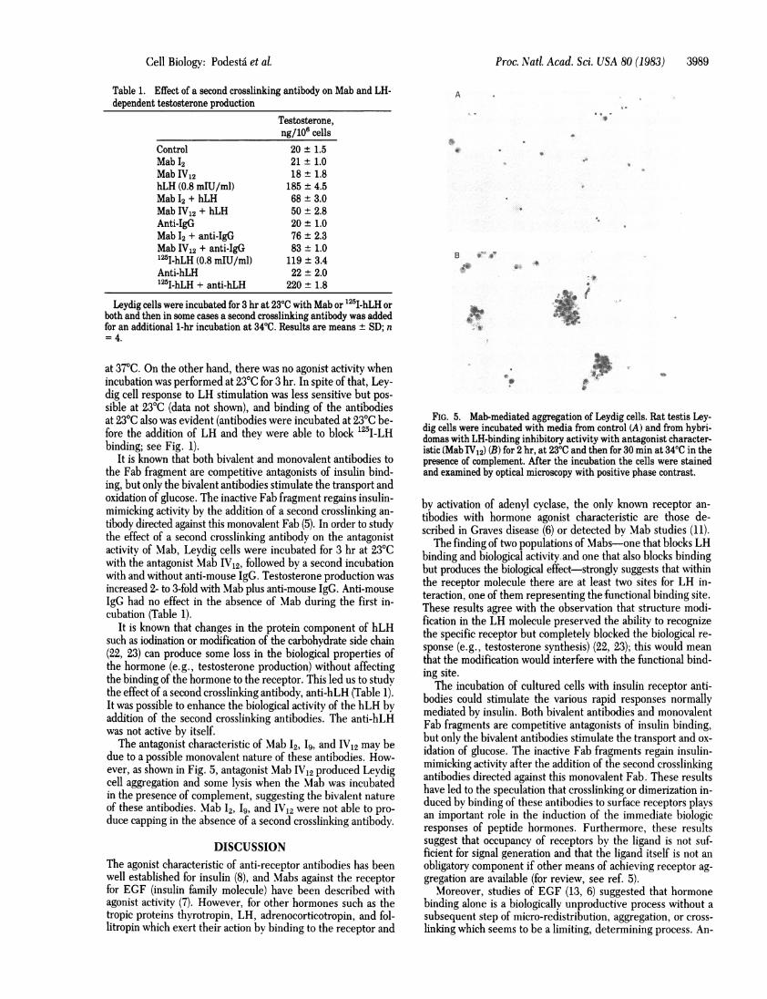
The antagonist characteristic of Mab I2, 19, and IV12 may bedue to a possible monovalent nature of these antibodies. How-ever, as shown in Fig. 5, antagonist Mab IV12 produced Levdigcell aggregation and some lysis when the Mab was incubatedin the presence of complement, suggesting the bivalent natureof these antibodies. Mab I2, I9, and IV12 were not able to pro-
duce capping in the absence of a second crosslinking antibody.
DISCUSSIONThe agonist characteristic of anti-receptor antibodies has beenwell established for insulin (8), and Mabs against the receptorfor EGF (insulin family molecule) have been described withagonist activity (7). However, for other hormones such as thetropic proteins thyrotropin, LH, adrenocorticotropin, and fol-litropin which exert their action by binding to the receptor and
FIG. 5. Mab-mediated aggregation of Leydig cells. Rat testis Ley-dig cells were incubated with media from control (A) and from hybri-domas with LH-binding inhibitory activity with antagonist character-istic (Mab TV12) (B) for 2 hr, at 23°C and then for 30 min at 340C in thepresence of complement. After the incubation the cells were stainedand examined by optical microscopy with positive phase contrast.
by activation of adenyl cyclase, the only known receptor an-tibodies with hormone agonist characteristic are those de-scribed in Graves disease (6) or detected by Mab studies (11).The finding of two populations of Mabs-one that blocks LH
binding and biological activity, and one that also-blocks bindingbut produces the biological effect-strongly suggests that withinthe receptor molecule there are at least two sites for LH in-teraction, one of them representing the-functional binding site.These results agree with the observation that structure modi-fication in the LH molecule preserved the ability to recognizethe specific receptor but completely blocked the biological re-sponse (e.g., testosterone synthesis) (22, 23); this would meanthat the modification would interfere with the functional bind--ing site.
The incubation of cultured cells with insulin receptor anti-bodies could stimulate the various rapid responses normallymediated by insulin. Both bivalent antibodies and monovalentFab fragments are competitive antagonists of insulin binding,but only the bivalent antibodies stimulate the transport and ox-idation of glucose. The inactive Fab fragments regain insulin-mimicking activity after the addition of the second crosslinkingantibodies directed against this monovalent Fab. These resultshave led to the speculation that crosslinking or dimerization in-duced by binding of these antibodies to surface receptors playsan important role in the induction of the immediate biologicresponses of peptide hormones. Furthermore, these resultssuggest that occupancy of receptors by the ligand is not suf-ficient for signal generation and that the ligand itself is not an
obligatory component if other means of achieving receptor ag-gregation are available (for review, see ref. 5).
Moreover, studies of EGF (13, 6) suggested that hormonebinding alone is a biologically unproductive process without asubsequent step of micro-redistribution, aggregation, or cross-linking which seems to be a limiting, determining process. An-
A
B *.
B 0
S..
f
Cell Biology: Podesta' et al.

3990 Cell Biology: Podesta etalP
tibody enhancement of EGF activity in sensitive and resistantcells has been described (13). Anti-insulin antibodies have nowalso been shown to enhance substantially the biological effectsof this hormone in fibroblasts (24).
In close agreement with the above results, in the presentstudy it was also possible to enhance the biological activity ofthe hLH by addition of the second crosslinking antibodies.
Our results support the above hypothesis suggesting the sameactivation mechanism for the peptide hormone receptor, cou-pled or uncoupled with the adenylate cyclase complex. Theseresults are also in agreement with the idea that the biologicalinformation of the LH-receptor complex resides in the recep-tor and not in the hormone. Furthermore, these two popula-tions of antibodies offer a powerful tool for studying the struc-ture from a functional point of view.
We thank Drs. Eduardo Charreau and Violeta Chiazzi for the pro-lactin binding determination. We are very grateful to Mrs. Noemi Bar-cala for her expert technical assistance, to Dr. Eduardo Charreau forhis helpful comments and discussion on the manuscript, and to Dr. EvelynNiday for her helpful discussion on the monoclonal antibody technique.
1. Lang, U., Kahn, C. R. & Harrison, L. C. (1980) Biochemistry 19,64-70.
2. Jacobs, S., Hazum, E. & Cuatrecasas, P. (1980)J. Biol. Chem. 255,6937-6940.
3. Heinrich, J., Pilch, P. E. & Czech, M. P. (1980)J. Biol Chem. 255,1732-1737.
4. Strosberg, A. D., Couraud, P. 0. & Schreiber, A. B. (1981) Im-munol. Today 2, 75-79.
5. King Christed, A. & Cuatrecasas, P. (1981) N. Engl. J. Med. 305,77-78.
6. Cuatrecasas, P. (1982) Membrane Recycling (Pitman, London), pp.96-108.
7. Schreiber, A. B., Lax, I., Yarden, Y., Eshhar, Z. & Schlessinger,J. (1981) Proc. Natl. Acad. Sci. USA 78, 1535-1539.
8. Kahn, C. R., Baird, K. L., Flier, J. S. & Jarret, D. B. (1977) J.Clin. Invest. 60, 1094-1106.
9. Smith, B. R. & Hall, R. (1974) FEBS Lett. 42, 301-304.10. Yavin, E., Yavin, Z., Schneider, M. D. & Kohn, L. D. (1981) Proc.
Natl. Acad. Sci. USA 78, 3180-3184.11. Valente, W. A., Vitti, P., Yavin, Z., Yavin, E., Rotella, C. M.,
Grollman, E. F., Toccafondi, R. S. & Kohn, L. (1982) Proc. Nati.Acad. Sci. USA 79, 6680-6684.
12. Shechter, Y., Hernaez, Z. & Cuatrecasas, P. (1978) Proc. Nati Acad.Sci. USA 75, 5788-5791.
13. Shechter, Y., Hernaez, Z., Schlessinger, J. & Cuatrecasas, P. (1979)Nature (London) 278, 835-838.
14. Dufau, M. L., Podestd, E. J. & Catt, K. J. (1975) Proc. Natl. Acad.Sci. USA 72, 1272-1275.
15. Galfre, G., Howe, S. C., Milstein, C., Butcher, G. W. & How-ard, J. C. (1977) Nature (London) 266, 550-552.
16. Hawkes, R., Niday, E. & Matus, A. (1982) Proc. Natl. Acad. Sci.USA 79, 2410-2414.
17. Lernahardt, W. J., Anderson, A., Coutinho, A. & Melchers, F.(1978) Exp. Cell Res. 3, 309-316.
18. Thorell, J. K. & Johnasson, B. A. (1971) Biochim. Biophys. Acta251, 363-369.
19. Mendelson, C., Dufau, M. & Catt, K. J. (1975)J. Biol. Chem. 250,8818-8823.
20. Podesta, E. J., Dufau, M. L., Solano, A. -R. & Catt, K. J. (1978)J. Biol Chem. 253, 8994-9001.
21. Best-Belpomme, M. & Dessen, P. (1973) Biochimie 55, 11-16.22. Bahl, 0. P., Marz, L. & Moyle, W. R. (1974) Hormone Binding
and Target Cell Activation in Testis, eds. Dufau, M. & Means, A.R. (Plenum, New York), pp. 125-144.
23. Liu, W. K., Yang, K. F., Burleigh, B. C. & Ward, D. M. (1974)Hormone Binding and Target Cell Activation in Testis, eds. Du-fau, M. & Means, A. R. (Plenum, New York), pp. 89-108.
24. Shechter, Y., Chang, K. J., Jacobs, S. & Cuatrecasas, P. (1979) Proc.Nati Acad. Sci. USA 76, 2720-2724.
Proc. Natl. Acad. Sci. USA 80 (1983)