reactive oxygen species and p38 mapk activate bax to induce
TRANSCRIPT
MOL 30718 1
REACTIVE OXYGEN SPECIES AND P38 MAPK ACTIVATE BAX TO INDUCE
MITOCHONDRIAL CYTOCHROME C RELEASE AND APOPTOSIS IN RESPONSE
TO MALONATE
Gomez-Lazaro M., Galindo MF., Melero-Fernandez de Mera RM, Fernandez-
Gómez FJ., Concannon CG., Segura MF, Comella JX, Prehn JHM. and Jordan J.
Grupo de Neurofarmacología. Departamento de Ciencias Médicas. Facultad de
Medicina. Universidad Castilla-La Mancha. M. G-L, M.F G, R.M. M-FM; F.J. F-G., J.J.
Department of Physiology and RCSI Neuroscience Research Centre, Royal College
of Surgeons in Ireland, 123 St Stephen’s Green, Dublin 2, Ireland. CGC, JHMJ
Cell Signaling and Apoptosis group. Departament de Ciències Mèdiques Bàsiques.
Univeristy of Lleida and Hospital Arnau de Vilanova. Lleida (Spain). MF S, JXC.
Centro Regional de Investigaciones Biomedicas. Albacete. Spain M. G-L, M.F G,
R.M. M-FM; F.J. F-G., J.J.
Molecular Pharmacology Fast Forward. Published on December 15, 2006 as doi:10.1124/mol.106.030718
Copyright 2006 by the American Society for Pharmacology and Experimental Therapeutics.
This article has not been copyedited and formatted. The final version may differ from this version.Molecular Pharmacology Fast Forward. Published on December 15, 2006 as DOI: 10.1124/mol.106.030718
at ASPE
T Journals on D
ecember 26, 2018
molpharm
.aspetjournals.orgD
ownloaded from

MOL 30718 2
Running title: Pathways involved in malonate-induced Bax translotacion.
To whom all correspondence should be sent at the following address:
Joaquin Jordán, Grupo de Neurofarmacología. Departamento de Ciencias Médicas.
Facultad de Medicina. Universidad Castilla-La Mancha. 02006-Albacete. Spain. Tel
34-967599200. Fax 34-967599327. E-Mail [email protected]
Number of text pages: 21 Number of tables: 0 Number of figures: 7 Number of references: 40 Number of words in the Abstract: 155 Number of words in the Introduction: 470 Number of words in the Discussion: 553
Abbreviations:
∆Ψm, mitochondrial transmembrane potential; CM-H2DCFDA, 2´,7´-
dichlorodihydrofluorescein diacetate; DIV, days in vitro;GFP, Green fluorescent
protein; MDA, Malondialdehyde; MEF, mouse embryonic fibroblasts; ROS , reactive
oxygen species; TMRE, tetramethylrhodamine ethyl ester.
This article has not been copyedited and formatted. The final version may differ from this version.Molecular Pharmacology Fast Forward. Published on December 15, 2006 as DOI: 10.1124/mol.106.030718
at ASPE
T Journals on D
ecember 26, 2018
molpharm
.aspetjournals.orgD
ownloaded from
MOL 30718 3
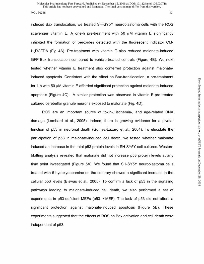
ABSTRACT
Malonate, an inhibitor of mitochondrial complex II, is a widely used toxin to study
neurodegeneration in Huntington’s disease and ischemic stroke. We have previously
shown that malonate increased reactive oxygen species (ROS) production in human
SH-SY5Y neuroblastoma cells, leading to oxidative stress, cytochrome-c release,
and apoptotic cell death. Expression of a Green Fluorescent Protein-Bax fusion
protein in SH-SY5Y neuroblastoma cells demonstrated a Bax redistribution from the
cytosol to mitochondria after 12 – 24 h of malonate treatment that coincided with
mitochondrial potential collapse and chromatin condensation. Inhibition of Bax
translocation using furosemide, as well as Bax gene deletion afforded significant
protection against malonate-induced apoptosis. Further experiments revealed that
malonate induced a prominent increase in the level of activated p38 MAP kinase, and
that treatment with the p38 MAP kinase inhibitor SKF86002 potently blocked
malonate-induced Bax translocation and apoptosis. Treatment with vitamin E
diminished ROS production, reduced the activation status of p38 MAP kinase,
inhibited Bax translocation and protected against malonate-induced apoptosis. Our
data suggest that malonate-induced ROS production and subsequent p38 MAP
kinase activation mediates the activation of the pro-apoptotic Bax protein to induce
mitochondrial membrane permeabilisation and neuronal apoptosis.
This article has not been copyedited and formatted. The final version may differ from this version.Molecular Pharmacology Fast Forward. Published on December 15, 2006 as DOI: 10.1124/mol.106.030718
at ASPE
T Journals on D
ecember 26, 2018
molpharm
.aspetjournals.orgD
ownloaded from

MOL 30718 4
Compromised mitochondrial function has been observed in several
neurodegenerative disorders, and inhibitors of mitochondrial respiration are
frequently used to mimic neurodegenerative disorders (Browne and Beal, 2002).
Intrastriatal injection of the mitochondrial complex II inhibitor malonate induces
striatal lesions similar to those described in cerebral ischemia and Huntington's
disease (Brouillet et al. 1995). The mechanisms that account for these neurotoxic
effects remain to be fully elucidated (Ferger et al., 1999). It has been proposed that
malonate toxicity involves depletion of striatal ATP (Beal et al., 1993), resulting in
neuronal depolarization and a secondary excitotoxic neuron loss (Beal et al 1994). It
has also been reported that malonate is capable of inducing a caspase-dependent
apoptotic cell death (Schulz et al., 1998).
Mitochondria are being considered a main link between cellular stress signals
activated during acute and chronic nerve cell injury and the execution of apoptotic
and necrotic cell death (Jordan et al. 2004; Mattson and Kroemer, 2003). ATP
depletion, pathophysiological increases in intracellular calcium (Ca2+) and enhanced
reactive oxygen species (ROS) production frequently occur during necrotic cell death
and can trigger an increase in the permeability of the inner mitochondrial membrane.
This process is believed to involve the formation of a multiprotein channel referred to
as mitochondrial permeability transitory pore (MPTP) (Bernardi, 1999) which triggers
the release of solutes up to 1500 Da from the mitochondrial matrix into the
cytoplasm. Apoptotic events in contrast can cause an increase in the permeability of
the outer mitochondrial membrane (Green, 2006). This process triggers the release
of intermembrane space proteins into the cytoplasm, including cytochrome c,
Smac/DIABLO, and apoptosis-inducing factor. Cytochrome-C and Smac/DIABLO are
capable of activating a family of cytosolic cysteine proteases, the caspases, while
This article has not been copyedited and formatted. The final version may differ from this version.Molecular Pharmacology Fast Forward. Published on December 15, 2006 as DOI: 10.1124/mol.106.030718
at ASPE
T Journals on D
ecember 26, 2018
molpharm
.aspetjournals.orgD
ownloaded from

MOL 30718 5
AIF has been implicated in caspase-independent forms of apoptosis (Martinou et al.,
2002; Goldstein et al., 2002).
The Bax protein has been identified as a key pro-apoptotic Bcl-2 family protein
during neuronal apoptosis. Bax normally resides in the cytosol and translocates to
mitochondria in response to a variety of apoptotic stimuli, including cerebral ischemia
(Cao et al., 2001; Putcha et al., 1999; Wolter et al., 1997). The pro-apoptotic action of
Bax is believed to be mediated by its insertion into the outer mitochondrial membrane
where it might directly form channels or regulate the activity of pre-existing channels
(Goping et al., 1998; Sharpe et al., 2004). This step requires a conformational
change in the Bax protein. The upstream events that induce this conformational
change are still largely unknown. Previously, we have demonstrated that malonate
causes apoptosis of human SH-SY5Y neuroblastoma cells, involving increased
reactive oxygen species production, oxidative stress and mitochondrial cytochrome C
release (Fernandez-Gomez et al., 2005). In the present study we were interested in
studying the requirement of Bax for malonate-induced apoptosis and in elucidating
the signalling pathways involved in malonate-induced Bax activation.
MATERIALS AND METHODS
Cell culture and drug treatment procedures- SH-SY5Y cultures were grown as
previously described (Jordan et al., 2004) (in Dulbecco’s modified Eagle’s medium
(DMEM) supplemented with 2 mM L-glutamine, penicillin (20 units/mL), streptomycin
(5 µg/mL), and 15% (v/v) fetal bovine serum (Gibco, Gaithersburg, MD, USA). Cells
were grown in a humidified cell incubator at 37ºC under a 5% CO2 atmosphere. For
GFP-Bax translocation and viability experiments, cells were plated on glass coverslip
at 2,9 x 10⁵ cells/cm2 and allowed to attach overnight. Immediately before malonate
This article has not been copyedited and formatted. The final version may differ from this version.Molecular Pharmacology Fast Forward. Published on December 15, 2006 as DOI: 10.1124/mol.106.030718
at ASPE
T Journals on D
ecember 26, 2018
molpharm
.aspetjournals.orgD
ownloaded from

MOL 30718 6
addition, dilutions of malonate were made in phosphate-buffered saline (PBS) and
added to fresh cell culture medium to achieve the required concentration.
Primary cultures of cerebellar granule neurons were obtained from dissociated
cerebella of 7-8-day-old rats (Fernandez-Gomez et al., 2006). Dissection and
dissociation were carried out in Basal Medium Eagle (BME; Life Technology).
Tissues were incubated with trypsin for 20 min at 37°C and dissociated by trituration
in a medium containing DNase and trypsin. Cells were plated on 60-mm plastic Petri
dishes pre-coated with poly-L-lysine (10 g/ml) at a concentration of 8 × 10⁶ cells /ml
in BME containing 25 mM KCl, 10% de-complemented fetal calf serum (FCS; Life
Technology), glutamine, and antibiotics. Cytosine- -D-arabino-furanoside (Ara-C) (10
M) was added at 3 days in vitro (DIV) to prevent the growth of non-neuronal cells.
All experiments were carried out after 7 days in culture.
Transfection- Cells were plated 24h before transfection at a density of 5,3 x
104 cells/cm2, on poly D-lysine-coated glass slides. Transfection was achieved using
LipofectamineTM reagent (Invitrogen, Carlsbad, CA) according to the manufacturer’s
protocol. Cells were transfected with de following plasmids encoding caspase-9
dominant-negative mutant caspase-9 (C287A; casp 9DN, a gift from Ding HF,
Medical College of Ohio, Toledo, Ohio), Bcl-2, Bcl-xL and p35 (Dr J Merino
Universidad de Santander), GFP (pGFP-C1; CLONTECH Laboratories, Inc.), GFP-
Bax (Poppe et al., 2002). After 4h incubation the transfection mixture was removed
and replaced with fresh complete medium.
Confocal microscopy- For time-lapse analysis, cells were grown in 24mm poly
D-lysine-coated glass slides and mounted in a chamber for confocal microscopy with
Krebs HEPES buffer with the following ionic composition (in mM): NaCl 140, KCl 5.9,
MgCl2 1.2, HEPES 15, glucose 10, CaCl2 2.5, pH 7.4. Images were captured with a
This article has not been copyedited and formatted. The final version may differ from this version.Molecular Pharmacology Fast Forward. Published on December 15, 2006 as DOI: 10.1124/mol.106.030718
at ASPE
T Journals on D
ecember 26, 2018
molpharm
.aspetjournals.orgD
ownloaded from

MOL 30718 7
Leica microscope using a 63X 1.4 NA objective. The excitation wavelengths for GFP
and TMRE were 488 and 543nm respectively. Images were taken for an hour every 5
minutes for control cells, and treated cells were injured with malonate after the first
photograph.
Mitochondrial potential - The cationic, lipophilic dye tetramethylrhodamine
ethyl ester (TMRE; Molecular Probes) enters cell in a form of an ester which is
subsequently hydrolysed and the product, tetramethylrhodamine, is accumulated in
mitochondria due to a high membrane potential. Cell cultures were washed in K-H,
and incubated at 37°C for 30 minutes in with TMRE (0.1 µM). Cells were then
washed with K-H and resuspended in K-H. Cell fluorescence was analyzed by
confocal microscopy as it has been described above.
Detection of peroxides / reactive oxygen species – We used the oxidation-
sensitive fluorescent dye 2´,7´-dichlorodihydrofluorescein diacetate (CM-H2DCFDA)
to measure the production of ROS, mainly hydrogen peroxide and hydroxyl radicals.
DCFH-DA is deacetylated by esterases to dichlorofluorescein (DCFH). This
nonfluorescent product is then converted by reactive species into DCF, which can
easily be visualized by fluorescence at 530 nm when excited at 485 nm. SH-SY5Y
cells seeded in 96-well culture plates were incubated with DCFC-DA (10 µg/ml) for 5
min, and fluorescence intensity was measured in a Spectra Max Gemini XS
(Molecular Devices). Average ROS production (relative to level of vehicle-treated
controls) was calculated from four individual wells in at least three independent
platings.
Assessment of apoptotic cell death - SH-SY5Y cells or cerebellar granule cells
were plated on poly D-lysine-coated glass slides. For cell death assay, nuclei were
This article has not been copyedited and formatted. The final version may differ from this version.Molecular Pharmacology Fast Forward. Published on December 15, 2006 as DOI: 10.1124/mol.106.030718
at ASPE
T Journals on D
ecember 26, 2018
molpharm
.aspetjournals.orgD
ownloaded from

MOL 30718 8
stained with 0.5 µg/ml of Hoechst 33258. Uniformly stained nuclei were scored as
healthy, viable neurons. Condensed or fragmented nuclei were scored as apoptotic.
Western blot - SH-SY5Y cell cultures were washed with cold PBS twice and
then collected by mechanical scraping with 1 ml of PBS per tissue culture dish. The
suspension was centrifuged at 12,000 –14,000 rpm for 5 min. The supernatant was
discarded, and the pellet was brought up in 150 µl of sample buffer. The protein from
each condition was quantified spectrophotometrically (Micro BCA Protein Reagent
Kit, Pierce, Rockford, IL), and an equal amount of protein (30 µg) was loaded onto 10
% SDS-PAGE gels. After electrophoresis, proteins were transferred to Immobilon
PVDF membranes. Non-specific protein binding was blocked with Blotto [4% w/v
non-fat dried milk, 4% bovine serum albumin (Sigma) and 0.1% Tween 20 Sigma)] in
PBS for 1 h. The membranes were incubated with anti-p53 [1:1000 dilution of anti-
mouse monoclonal (Pab240) sc-99 Santa Cruz], anti-pan p38 and anti-phospho-p38
(1:1000 dilution of polyclonal) overnight at 4 ºC. After washing with Blotto, the
membranes were incubated with a secondary antibody (1:5000 dilution of
peroxidase-labeled anti-mouse, Promega, Madison, WI) in Blotto. The signal was
detected using an enhanced chemiluminescence detection kit (Amersham ECL RPN
2106 Kit). Immunoblots were developed by exposure to x-ray film (Eastman-Kodak,
Rochester, NY).
Lipid peroxidation - Lipid peroxidation was measured by determining
malondialdehyde (MDA) levels. Each sample (8 x 10⁶ cells) was collected in 100µL of
ice-cold 20 mM BTris–HCl buffer, pH 7.4, and sonicated. Amounts of MDA were
determined in the cellular extracts using a Lipid Peroxidation Assay Kit from
This article has not been copyedited and formatted. The final version may differ from this version.Molecular Pharmacology Fast Forward. Published on December 15, 2006 as DOI: 10.1124/mol.106.030718
at ASPE
T Journals on D
ecember 26, 2018
molpharm
.aspetjournals.orgD
ownloaded from

MOL 30718 9
Calbiochem (No. 437634) based on the condensation reaction of the chromogene 1-
methyl-2-phenylindole with either MDA. The stable chromophores were determined
at 586 nm. Results are expressed as percent of ng MDA per mg protein found in
untreated cell cultures.
Statistics - The results were expressed at the mean +/- SD of at least three
independent experiments. Student’s two-tailed, unpaired t test was used, and values
of P<0.05 were considered to be significant. When comparing more than two
conditions statistically significant differences between groups were determined by
ANOVA followed by a Newman–Keuls post hoc analysis. The level of statistical
significance was set at P<0.05.
RESULTS
Malonate induces cell death through the mitochondrial apoptosis pathway - In
the mitochondrial apoptosis pathway, the release of cytochrome-C triggers the
formation of a caspase-3 activating complex, the apoptosome. Caspase-3 was
activated in SH-SY5Y neuroblastoma cells challenged with malonate as evidenced
by Western blot analysis. As shown in Figure 1A, the addition of 50 mM malonate to
SH-SY5Y cell cultures resulted in the activation of caspase-3 after 12 – 24 h of
treatment. To analyse whether the mitochondrial pathway participated in malonate-
induced cell death we co-transfected SH-SY5Y cells with a GFP expression vector
and Bcl-2 or Bcl-xl expression vectors. Bcl-2 and Bcl-xL are known to inhibit
mitochondrial cytochrome-C release by neutralizing the pro-apoptotic activity of Bax
and Bak. 24 h after transfection, cell cultures were treated with malonate (50 mM, 12
This article has not been copyedited and formatted. The final version may differ from this version.Molecular Pharmacology Fast Forward. Published on December 15, 2006 as DOI: 10.1124/mol.106.030718
at ASPE
T Journals on D
ecember 26, 2018
molpharm
.aspetjournals.orgD
ownloaded from

MOL 30718 10
h) and the effect of malonate on the viability of the GFP-positive SH-SY5Y cells was
determined. As shown in figure 1B, the overexpression of Bcl-2 or Bcl-xL potently
abrogated the cytotoxic effect of malonate in the SH-SY5Y cells. Similar results were
found in cell cultures transfected with the baculoviral broad spectrum caspase
inhibitor p35. The apoptosome is comprised of APAF-1, cytochrome-C and caspase-
9. Potent inhibition of malonate-induced apoptosis was also observed when we
inhibited the function of endogenous caspase-9 by overexpression of a dominant-
negative mutant form (Caspase-9 DN) (Figure 1B). Together, these results indicate
that malonate activates the mitochondrial apoptosis pathway in human SH-SY5Y
neuroblastoma cells.
Malonate induces Bax translocation to mitochondria- By using a Green
fluorescent protein (GFP)-Bax fusion protein, we addressed the question whether
Bax translocation was involved in malonate-induced apoptosis. As shown in Figure 2,
confocal imaging studies revealed that in untreated SH-SY5Y cells, GFP-Bax was
distributed evenly in the cytosolic compartment. After 12 h of malonate treatment, we
observed a marked change in GFP-Bax fluorescence from a diffuse, cytosolic to a
clustered, mitochondrial pattern. Approximately 35 % of the malonate-challenged
cells displayed a GFP-Bax translocation after 12 h. Use of the mitochondrion-
selective dye Mito-Tracker Red (Figure 2 A-C) demonstrated that the clustered,
punctuate GFP-Bax distribution colocalized with mitochondria. Disruption of
mitochondrial transmembrane potential (∆Ψm) has been demonstrated to occur
downstream of mitochondrial cytochrome-C release (Waterhouse et al., 2001). By
analyzing tetramethylrhodamine ethyl ester fluorescence intensities we studied ∆Ψm
changes in cells exhibiting either a clustered or diffuse GFP-Bax distribution. By 12 h
This article has not been copyedited and formatted. The final version may differ from this version.Molecular Pharmacology Fast Forward. Published on December 15, 2006 as DOI: 10.1124/mol.106.030718
at ASPE
T Journals on D
ecember 26, 2018
molpharm
.aspetjournals.orgD
ownloaded from

MOL 30718 11
after malonate treatment, we found that clustered punctuated GFP-Bax cells showed
an approximately 75 % decrease in TMRE fluorescence compared to those cells with
cytoplasmic diffuse GFP-Bax distribution (n = 114 cells). In malonate-treated cells
where Bax translocation had not yet occurred, mitochondrial potential remained at
the level observed in untreated cells (Figure 2D). Epifluorescence observation
suggested that cells with a clustered GFP-Bax fluorescence also exhibited a nuclear
apoptotic morphology, and analyzed by chromatin using Hoechst 33342 (Figure 2 E).
Bax is required for malonate-induced apoptosis- To determine the Bax
involvement in malonate-induced apoptosis, we blocked Bax translocation using the
chloride channel inhibitor furosemide (Karpinich et al., 2002). As shown in Figure 3A,
a three h pre-treatment with 10 µM furosemide significantly reduced Bax
translocation and afforded significant protection to the SH-SY5Y cells treated for 12 h
with 50 mM malonate.
In the next set of experiments, we addressed the question whether Bax was
required for malonate-induced apoptosis. Western blotting analysis of lysates
prepared from mouse embryonic fibroblasts (MEF) cultures revealed that malonate
failed to activate caspase-3 in cells derived from Bax-deficient mice (Figure 3B).
Indeed, the lack of Bax protein conferred resistance against malonate toxicity, as
Bax-/- MEFs were protected against malonate-induced apoptosis (Figure 3C).
Inhibition of ROS production inhibits malonate-induced Bax translocation and
apoptosis - We have previously showed that ROS production is increased in SH-
SY5Y cells cultures challenged with malonate (Fernandez-Gomez et al., 2005). To
investigate whether this increased ROS production is functionally linked to malonate-
This article has not been copyedited and formatted. The final version may differ from this version.Molecular Pharmacology Fast Forward. Published on December 15, 2006 as DOI: 10.1124/mol.106.030718
at ASPE
T Journals on D
ecember 26, 2018
molpharm
.aspetjournals.orgD
ownloaded from
MOL 30718 12
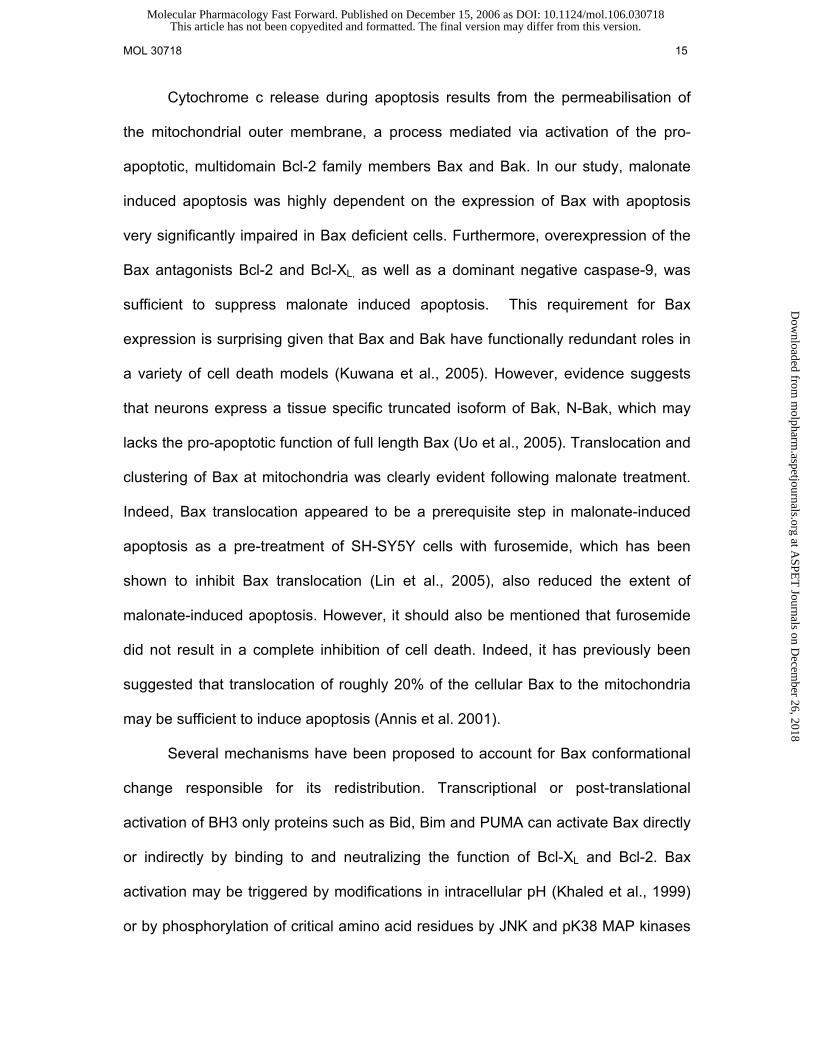
induced Bax translocation, we treated SH-SY5Y neuroblastoma cells with the ROS
scavenger vitamin E. A one-h pre-treatment with 50 µM vitamin E significantly
inhibited the formation of peroxides detected with the fluorescent indicator CM-
H2DCFDA (Fig 4A). Pre-treatment with vitamin E also reduced malonate-induced
GFP-Bax translocation compared to vehicle-treated controls (Figure 4B). We next
tested whether vitamin E treatment also conferred protection against malonate-
induced apoptosis. Consistent with the effect on Bax-translocation, a pre-treatment
for 1 h with 50 µM vitamin E afforded significant protection against malonate-induced
apoptosis (Figure 4C). A similar protection was observed in vitamin E-pre-treated
cultured cerebellar granule neurons exposed to malonate (Fig. 4D).
ROS are an important source of toxin-, ischemia-, and age-related DNA
damage (Lombard et al., 2005). Indeed, there is growing evidence for a pivotal
function of p53 in neuronal death (Gomez-Lazaro et al., 2004). To elucidate the
participation of p53 in malonate-induced cell death, we tested whether malonate
induced an increase in the total p53 protein levels in SH-SY5Y cell cultures. Western
blotting analysis revealed that malonate did not increase p53 protein levels at any
time point investigated (Figure 5A). We found that SH-SY5Y neuroblastoma cells
treated with 6-hydoxydopamine on the contrary showed a significant increase in the
cellular p53 levels (Biswas et al., 2005). To confirm a lack of p53 in the signaling
pathways leading to malonate-induced cell death, we also performed a set of
experiments in p53-deficient MEFs (p53 -/-MEF). The lack of p53 did not afford a
significant protection against malonate-induced apoptosis (Figure 5B). These
experiments suggested that the effects of ROS on Bax activation and cell death were
independent of p53.
This article has not been copyedited and formatted. The final version may differ from this version.Molecular Pharmacology Fast Forward. Published on December 15, 2006 as DOI: 10.1124/mol.106.030718
at ASPE
T Journals on D
ecember 26, 2018
molpharm
.aspetjournals.orgD
ownloaded from

MOL 30718 13
p38 MAP kinase triggers Bax translocation and apoptotic cell death in
response to malonate - The p38 MAP kinase participates in several apoptosis
pathways, mediating Bax activation and translocation (Ghatan et al., 2000). To
analyze whether p38 MAPK was activated by malonate, SH-SY5Y cells were
challenged with malonate and cytoplasmic extracts were assayed by Western
blotting using a phospho-specific antibody recognizing active p38 MAP kinase
(Sanchez-Prieto et al., 2002). Protein extracts from SH-SY5Y cells challenged with
50 mM malonate showed a marked increase in the phosphorylation status of p38
MAPK (Figure 6A). Maximal increases in p38 MAPK phosphorylation occurred after 1
h of treatment. Phosphorylation levels slowly declined subsequently, returning to
basal levels after 12 h. Next, we employed the p38 MAP kinase inhibitor SKF86002,
to study the relevance of p38 MAP kinase activation in malonate-induced Bax
translocation. At 10 µM, SKF86002 has been shown to inhibit all p38 MAPK isoforms,
α, β, δ, and γ. SH-SY5Y cell cultures were pre-treated for 12 h with 10 µM SKF86002.
As shown in Figure 6 B-C, SKF86002 potently prevented the appearance of cells
showing a clustered GFP-Bax fluorescence. Importantly, SKF86002 pre-treatment
also prevented apoptosis of SH-SY5Y cells treated with malonate (Figure 6D). A
similar, potent protection was observed in SKF86002 pre-treated cultured cerebellar
granule neurons exposed to malonate (Fig. 4D).
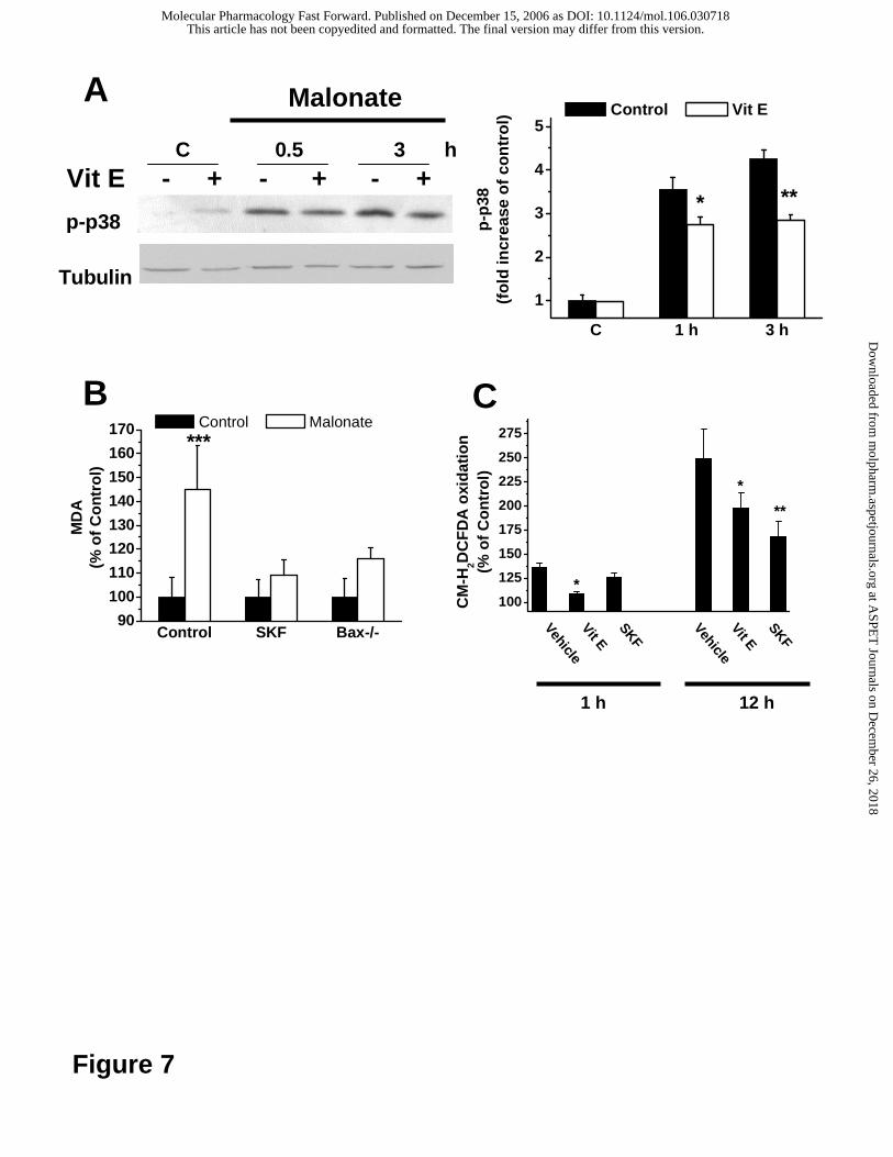
Finally, we determined whether ROS participated in the activation of p38 MAP
kinase in response to malonate. Interestingly, a pre-treatment with the anti-oxidant
vitamin E reduced the early accumulation of active p38 MAP kinase (Fig. 7A) after
0.5 and 3 h of malonate treatment, suggesting that ROS are required for malonate-
induced p38 MAP kinase activation.
This article has not been copyedited and formatted. The final version may differ from this version.Molecular Pharmacology Fast Forward. Published on December 15, 2006 as DOI: 10.1124/mol.106.030718
at ASPE
T Journals on D
ecember 26, 2018
molpharm
.aspetjournals.orgD
ownloaded from

MOL 30718 14
It has also been reported that a secondary increase in ROS production can
occur late during apoptosis and downstream of mitochondrial cytochrome c release
(Luetjens et al., 2000; Düßmann et al., 2003). In agreement with these findings, the
levels of malondialdehyde (MDA), a marker for oxidative stress (Esterbauer et al.,
1991), were reduced at a later stage (12 h after addition of malonate) in MEF cells
treated with the p38 kinase inhibitor SKF86002 compared to vehicle-treated controls
(Figure 7B). The levels of MDA in MEFs challenged with 50 mM malonate were also
significantly reduced in cells derived from Bax-deficient mice (Figure 7B).
Finally, to confirm the existence of a functionally separated primary and
secondary increase in ROS production in more detail, we investigated the effects of
vitamin E and SKF86002 pre-treatment on ROS production in SH-SY5Y
neuroblastoma cells both at 1 h (early increase) and 12 h (late increase). As
expected, pre-treatment with the p38 kinase inhibitor significantly reduced the late
increase in ROS production, but had no effect on the early increase (Fig. 7C). In
contrast, pre-treatment with vitamin E reduced ROS formation at both time points.
DISCUSSION
In this study we provide evidence that malonate-induced apoptosis of SH-
SY5Y neuroblastoma cells is mediated via the pro-apoptotic Bcl-2 family protein Bax.
We also demonstrate that both ROS and p38 MAP kinase are required to activate
Bax and apoptosis in SH-SH5Y cells and primary cerebellar granule neurons in
response to malonate, and that this may occur through functionally interacting
signalling pathways.
This article has not been copyedited and formatted. The final version may differ from this version.Molecular Pharmacology Fast Forward. Published on December 15, 2006 as DOI: 10.1124/mol.106.030718
at ASPE
T Journals on D
ecember 26, 2018
molpharm
.aspetjournals.orgD
ownloaded from
MOL 30718 15
Cytochrome c release during apoptosis results from the permeabilisation of
the mitochondrial outer membrane, a process mediated via activation of the pro-
apoptotic, multidomain Bcl-2 family members Bax and Bak. In our study, malonate
induced apoptosis was highly dependent on the expression of Bax with apoptosis
very significantly impaired in Bax deficient cells. Furthermore, overexpression of the
Bax antagonists Bcl-2 and Bcl-XL, as well as a dominant negative caspase-9, was
sufficient to suppress malonate induced apoptosis. This requirement for Bax
expression is surprising given that Bax and Bak have functionally redundant roles in
a variety of cell death models (Kuwana et al., 2005). However, evidence suggests
that neurons express a tissue specific truncated isoform of Bak, N-Bak, which may
lacks the pro-apoptotic function of full length Bax (Uo et al., 2005). Translocation and
clustering of Bax at mitochondria was clearly evident following malonate treatment.
Indeed, Bax translocation appeared to be a prerequisite step in malonate-induced
apoptosis as a pre-treatment of SH-SY5Y cells with furosemide, which has been
shown to inhibit Bax translocation (Lin et al., 2005), also reduced the extent of
malonate-induced apoptosis. However, it should also be mentioned that furosemide
did not result in a complete inhibition of cell death. Indeed, it has previously been
suggested that translocation of roughly 20% of the cellular Bax to the mitochondria
may be sufficient to induce apoptosis (Annis et al. 2001).
Several mechanisms have been proposed to account for Bax conformational
change responsible for its redistribution. Transcriptional or post-translational
activation of BH3 only proteins such as Bid, Bim and PUMA can activate Bax directly
or indirectly by binding to and neutralizing the function of Bcl-XL and Bcl-2. Bax
activation may be triggered by modifications in intracellular pH (Khaled et al., 1999)
or by phosphorylation of critical amino acid residues by JNK and pK38 MAP kinases
This article has not been copyedited and formatted. The final version may differ from this version.Molecular Pharmacology Fast Forward. Published on December 15, 2006 as DOI: 10.1124/mol.106.030718
at ASPE
T Journals on D
ecember 26, 2018
molpharm
.aspetjournals.orgD
ownloaded from

MOL 30718 16
(De Chiara et al., 2006). Malonate produced a marked increase in the
phosphorylation of p38 MAP kinase in SH-SY5Y cells. Furthermore, use of the p38
MAP kinase inhibitor, SKF86002, potently inhibited Bax translocation and offered
significant protection against malonate-induced apoptosis. Indeed, there is a
significant body of evidence suggesting that p38 MAPK activation plays an important
role during excitototoxic and neurodegenerative processes (Cao et al., 2004). In
addition to the p38 MAPK inhibitor, treatment of SH-SY5Y cells with the antioxidant
vitamin E also inhibited Bax translocation resulting in decreased levels of apoptosis.
Interestingly, the inhibition of ROS levels ameliorated the effect of malonate on p38
MAP kinase phosphorylation, suggesting that malonate activates ROS which
subsequently activate p38 kinase (Ghatan et al., 2000; Choi et al., 2004).
In summary we demonstrate that malonate is able to induce mitochondrial
cytochrome c releases through the redistribution of Bax, and that this event is
mediated by ROS and the p38 MAP kinase pathway. Interfering with signalling
pathways activated either by modulating the accumulation of ROS or by
pharmacological inhibition of p38 MAP kinase may afford protection against malonate
toxicity. These findings may have important therapeutic implications for the treatment
of disorders such as HD and ischaemic stroke.
Acknowledgments
We are grateful to Remedios Sanchis for technical assistance.
This article has not been copyedited and formatted. The final version may differ from this version.Molecular Pharmacology Fast Forward. Published on December 15, 2006 as DOI: 10.1124/mol.106.030718
at ASPE
T Journals on D
ecember 26, 2018
molpharm
.aspetjournals.orgD
ownloaded from

MOL 30718 17
REFERENCES
Annis MG, Zamzami N, Zhu W, Penn LZ, Kroemer G, Leber B, and Andrews DW
(2001) Endoplasmic reticulum localized Bcl-2 prevents apoptosis when redistribution
of cytochrome c is a late event. Oncogene 20:1939-1952.
Beal MF, Brouillet E, Jenkins B, Henshaw R, Rosen B, and Hyman BT (1993) Age-
dependent striatal excitotoxic lesions produced by the endogenous mitochondrial
inhibitor malonate. J Neurochem 61:1147-1150.
Beal MF, Henshaw DR, Jenkins BG, Rosen BR, and Schulz JB (1994) Coenzyme
Q10 and nicotinamide block striatal lesions produced by the mitochondrial toxin
malonate. Ann Neurol 36:882-888.
Bernardi P (1999) Mitochondrial transport of cations: channels, exchangers, and
permeability transition. Physiol Rev 79:1127-1155.
Biswas SC, Ryu E, Park C, Malagelada C, and Greene LA (2005) . Puma and p53
play required roles in death evoked in a cellular model of Parkinson disease.
Neurochem Res 30:839-845.
Brouillet E, Hantraye P, Ferrante RJ, Dolan R, Leroy-Willig A, Kowall NW, and Beal
MF (1995) Chronic mitochondrial energy impairment produces selective striatal
degeneration and abnormal choreiform movements in primates. Proc Natl Acad Sci
USA 92:7105-7109.
Browne SE, and Beal MF (2002) Toxin-induced mitochondrial dysfunction. Int Rev
Neurobiol 53:243-279.
This article has not been copyedited and formatted. The final version may differ from this version.Molecular Pharmacology Fast Forward. Published on December 15, 2006 as DOI: 10.1124/mol.106.030718
at ASPE
T Journals on D
ecember 26, 2018
molpharm
.aspetjournals.orgD
ownloaded from

MOL 30718 18
Cao G, Minami M, Pei W, Yan C, Chen D, O'Horo C, Graham SH, and Chen J (2001)
Intracellular Bax translocation after transient cerebral ischemia: implications for a role
of the mitochondrial apoptotic signaling pathway in ischemic neuronal death. J Cereb
Blood Flow Metab 21:321-333.
Cao J, Semenova MM, Solovyan VT, Han J, Coffey ET, and Courtney MJ (2004)
Distinct requirements for p38alpha and c-Jun N-terminal kinase stress-activated
protein kinases in different forms of apoptotic neuronal death. J Biol Chem
279:35903-35913.
Choi WS, Eom DS, Han BS, Kim WK, Han BH, Choi EJ, Oh TH, Markelonis GJ, Cho
JW, Oh YJ (2004) Phosphorylation of p38 MAPK induced by oxidative stress is linked
to activation of both caspase-8- and -9-mediated apoptotic pathways in dopaminergic
neurons J Biol Chem 279:20451-20460.
De Chiara G, Marcocci ME, Torcia M, Lucibello M, Rosini P, Bonini P, Higashimoto
Y, Damonte G, Armirotti A, Amodei S, Palamara AT, Russo T, Garaci E, and
Cozzolino F (2006) Bcl-2 Phosphorylation by p38 MAPK: identification of target sites
and biologic consequences J Biol Chem 79:14016-14023.
Düßmann H, Rehm M, Kögel D, and Prehn JHM (2003) Mitochondrial membrane
permeabilization and superoxide production during apoptosis. A single-cell analysis
J. Biol Chem 278:12645-12649.
Esterbauer H, Schaur RJ, and Zollner H (1991) Chemistry and biochemistry of 4-
hydroxynonenal, malonaldehyde and related aldehydes Free Radic Biol Med 11:81-
128.
This article has not been copyedited and formatted. The final version may differ from this version.Molecular Pharmacology Fast Forward. Published on December 15, 2006 as DOI: 10.1124/mol.106.030718
at ASPE
T Journals on D
ecember 26, 2018
molpharm
.aspetjournals.orgD
ownloaded from
MOL 30718 19
Ferger B, Eberhardt O, Teismann P, de Groote C, and Schulz JB (1999) Malonate-
induced generation of reactive oxygen species in rat striatum depends on dopamine
release but not on NMDA receptor activation. J Neurochem 73:1329-1332.
Fernandez-Gomez FJ, Galindo MF, Gomez-Lazaro M, Yuste VJ, Comella JX, Aguirre
N, and Jordan J (2005) Malonate induces cell death via mitochondrial potential
collapse and delayed swelling through an ROS-dependent pathway. Br J Pharmacol
144:528-537.
Fernandez-Gomez FJ, Pastor MD, Garcia-Martinez EM, Melero-Fernandez de Mera
R, Gou-Fabregas M, Gomez-Lazaro M, Calvo S, Soler RM, Galindo MF, Jordan J.
(2006) Pyruvate protects cerebellar granular cells from 6-hydroxydopamine-induced
cytotoxicity by activating the Akt signaling pathway and increasing glutathione
peroxidase expression. Neurobiol Dis 24:296-307.
Ghatan S, Larner S, Kinoshita Y, Hetman M, Patel L, Xia Z, Youle RJ, and Morrison
RS (2000) p38 MAP kinase mediates bax translocation in nitric oxide-induced
apoptosis in neurons. J Cell Biol 150:335-347.
Goldstein JC, Waterhouse NJ, Juin P, Evan GI, and Green DR (2002) The
coordinate release of cytochrome c during apoptosis is rapid, complete and
kinetically invariant. Nat Cell Biol 2:156-162.
Gomez-Lazaro M, Fernandez-Gomez FJ, and Jordan J (2004) p53: twenty five years
understanding the mechanism of genome protection. J Physiol Biochem 60:287-307.
This article has not been copyedited and formatted. The final version may differ from this version.Molecular Pharmacology Fast Forward. Published on December 15, 2006 as DOI: 10.1124/mol.106.030718
at ASPE
T Journals on D
ecember 26, 2018
molpharm
.aspetjournals.orgD
ownloaded from

MOL 30718 20
Goping IS, Gross A, Lavoie JN, Nguyen M, Jemmerson R, Roth K, Korsmeyer SJ,
and Shore GC (1998) Regulated targeting of BAX to mitochondria. J Cell Biol
143:207-215.
Green DR (2006) At the gates of death. Cancer Cell 9:328-330.
Jordan J, Cena V, and Prehn JH (2003) Mitochondrial control of neuron death and its
role in neurodegenerative disorders. J Physiol Biochem 59:129-141.
Jordan J, Galindo MF, Tornero D, Gonzalez-Garcia C, and Cena V (2004) Bcl-xL
blocks mitochondrial multiple conductance channel activation and inhibits 6-OHDA-
induced death in SH-SY5Y cells. J Neurochem 89:124-133.
Karpinich NO, Tafani M, Rothman RJ, Russo MA, and Farber JL (2002) The course
of etoposide-induced apoptosis from damage to DNA and p53 activation to
mitochondrial release of cytochrome c. J Biol Chem 277:16547-16552.
Khaled AR, Kim K, Hofmeister R, Muegge K, and Durum SK (1999) Withdrawal of IL-
7 induces Bax translocation from cytosol to mitochondria through a rise in
intracellular pH. Proc Natl Acad Sci USA 96:14476-14481.
Kuwana T, Bouchier-Hayes L, Chipuk JE, Bonzon C, Sullivan BA, Green DR, and
Newmeyer DD (2005) BH3 domains of BH3-only proteins differentially regulate Bax-
mediated mitochondrial membrane permeabilization both directly and indirectly Mol
Cell 17:525-535.
Lin CH, Lu YZ, Cheng FC, Chu LF, and Hsueh CM (2005) Bax-regulated
mitochondria-mediated apoptosis is responsible for the in vitro ischemia induced
neuronal cell death of Sprague Dawley rat. Neurosci Lett 387:22-27.
This article has not been copyedited and formatted. The final version may differ from this version.Molecular Pharmacology Fast Forward. Published on December 15, 2006 as DOI: 10.1124/mol.106.030718
at ASPE
T Journals on D
ecember 26, 2018
molpharm
.aspetjournals.orgD
ownloaded from

MOL 30718 21
Lombard DB, Chua KF, Mostoslavsky R, Franco S, Gostissa M, and Alt FW (2005)
DNA repair, genome stability, and aging. Cell 120:497-512.
Luetjens CM, Bui NT, Sengpiel B, Münstermann G, Poppe M, Krohn AJ, Bauerbach
E, Krieglstein J, and Prehn JHM (2000) Delayed mitochondrial dysfunction in
excitotoxic neuron death: cytochrome c release and a secondary increase in
superoxide production J Neurosci 20:5715-5723.
Martinou JC, Desagher S, and Antonsson B (2002) Cytochrome c release from
mitochondria: all or nothing. Nat Cell Biol 2:E41-43.
Mattson MP and Kroemer G (2003) Mitochondria in cell death: novel targets for
neuroprotection and cardioprotection. Trends Mol Med 9:196-205.
Poppe M, Reimertz C, Munstermann G, Kogel D, and Prehn JH (2002) Ceramide-
induced apoptosis of D283 medulloblastoma cells requires mitochondrial respiratory
chain activity but occurs independently of caspases and is not sensitive to Bcl-xL
overexpression J Neurochem 82:482-494.
Putcha GV, Deshmukh M, and Johnson EMJr (1999) BAX translocation is a critical
event in neuronal apoptosis: regulation by neuroprotectants, BCL-2, and caspases. J
Neurosci 19:7476-7485.
Sanchez-Prieto R, Sanchez-Arevalo VJ, Servitja JM, and Gutkind JS (2002)
Regulation of p73 by c-Abl through the p38 MAP kinase pathway. Oncogene 21:974-
979.
This article has not been copyedited and formatted. The final version may differ from this version.Molecular Pharmacology Fast Forward. Published on December 15, 2006 as DOI: 10.1124/mol.106.030718
at ASPE
T Journals on D
ecember 26, 2018
molpharm
.aspetjournals.orgD
ownloaded from

MOL 30718 22
Schulz JB, Weller M, Matthews RT, Heneka MT, Groscurth P, Martinou JC,
Lommatzsch J, von Coelln R, Wullner U, Loschmann PA, Beal MF, Dichgans J, and
Klockgether T (1998) Extended therapeutic window for caspase inhibition and
synergy with MK-801 in the treatment of cerebral histotoxic hypoxia. Cell Death Differ
5:847-857.
Sharpe JC, Arnoult D, and Youle RJ (2004) Control of mitochondrial permeability by
Bcl-2 family members. Biochim Biophys Acta 1644:107-113.
Uo T, Kinoshita Y, and Morrison RS (2005) Neurons exclusively express N-Bak, a
BH3 domain-only Bak isoform that promotes neuronal apoptosis J Biol Chem
280:9065-9073.
Waterhouse NJ, Goldstein JC, von Ahsen O, Schuler M, Newmeyer DD, and Green
DR (2001) Cytochrome c maintains mitochondrial transmembrane potential and ATP
generation after outer mitochondrial membrane permeabilization during the apoptotic
process. J Cell Biol 153:319-328.
Wolter KG, Hsu YT, Smith CL, Nechushtan A, Xi XG, and Youle RJ (1997)
Movement of Bax from the cytosol to mitochondria during apoptosis. J Cell Biol
139:1281-1292.
Wong S, McLaughlin J, Cheng D, and Witte ON (2003) Cell context-specific effects of
the BCR-ABL oncogene monitored in hematopoietic progenitors Blood 101:4088-
4097.
This article has not been copyedited and formatted. The final version may differ from this version.Molecular Pharmacology Fast Forward. Published on December 15, 2006 as DOI: 10.1124/mol.106.030718
at ASPE
T Journals on D
ecember 26, 2018
molpharm
.aspetjournals.orgD
ownloaded from
MOL 30718 23
FOOT NOTES
*This study was supported by SAF2002-04721 and SAF2005-07919-C02-01 from
CICYT and 04005-00 Consejería de Sanidad from Junta de Comunidades de Castilla
La Mancha to J.J. Ministerio de Sanidad y Consumo, Fundació La Caixa and
Generalitat de Catalunya to J.X.C. and Science Foundation Ireland grant
03/RP1/B344 to JHMP. M. G-L and F.J. F-G are fellows from JCCM.
Person to receive reprint request
Joaquin Jordán, Grupo de Neurofarmacología. Departamento de Ciencias Médicas.
Facultad de Medicina. Universidad Castilla-La Mancha. 02006-Albacete. Spain. Tel
34-967599200. Fax 34-967599327. E-Mail [email protected]
This article has not been copyedited and formatted. The final version may differ from this version.Molecular Pharmacology Fast Forward. Published on December 15, 2006 as DOI: 10.1124/mol.106.030718
at ASPE
T Journals on D
ecember 26, 2018
molpharm
.aspetjournals.orgD
ownloaded from

MOL 30718 24
Legends for figures
Fig.1. Malonate induces apoptosis and activates mitochondrial apoptosis pathway in
SH-SY5Y cells. A. Kinetics of malonate treatment on caspase-3 protein cleavage.
Cells cultures were challenged with 50 mM malonate for the indicated times. Cells
were then collected and total protein was extracted. Caspase-3 protein levels were
determined by Western blot analysis. B. Cell cultures were co-transfected with GFP
and Bcl-2, Bcl-XL, p35 or Casp9 DN 24 h before malonate exposures. Cell viability
was performed by studying the state of chromatin using Hoechst 33342 staining in
GFP-positive cells 12 h after malonate addition. Each column represents the average
obtained from four independent experiments.
Fig. 2. Malonate induces GFP-Bax translocation to mitochondria. A. SH-SY5Y cells
were transfected with GFP-Bax using LipofectamineTM as described in Materials and
Methods and were incubated for 24 h to allow for sufficient GFP-Bax expression and
treated with 50 mM malonate. By 12 h after insults cells were fixed in 4 %
paraformaldehyde. Confocal images were captured using a 63 X oil immersion lens.
GFP-Bax demonstrated primarily diffuse staining in control (upper panel), while by 12
h after 50 mM malonate treatments a punctate pattern is evident (lower panel). Mito
Track red staining was used to study the mitochondrial distribution. The images
shown are representative of results obtained in four separate experiments, each
performed in triplicate. Histograms represent the values of GFP-Bax fluorescence
standard deviation (B) and GFP-Bax distribution patterns (C) in SH-SY5Y cells. D.
Bax mediates mitochondrial transmembrane potential disruption. By 12 h after 50
mM treatment TMRE fluorescence intensities were analyzed to study ∆Ψm changes
from cells with either a punctuate or diffuse GFP-Bax distribution challenged or not
This article has not been copyedited and formatted. The final version may differ from this version.Molecular Pharmacology Fast Forward. Published on December 15, 2006 as DOI: 10.1124/mol.106.030718
at ASPE
T Journals on D
ecember 26, 2018
molpharm
.aspetjournals.orgD
ownloaded from

MOL 30718 25
with malonate. E. At 12 hours of 50 mM malonate treatment, GFP-Bax cells with a
punctuate pattern after malonate present fragmented chromatin (arrows). Hoechst
33342 dye was added to study the state of chromatin. GFP-Bax was captured in the
FITC channel (upper panel) and Hoechst 33342 was captured in the DAPI channel
(lower panel). Results are presented as mean ± S.D.; they are representative of at
least three experiments, each performed in triplicate. **p < 0.01 and ***p < 0.001
versus control conditions.
Fig.3. Bax is required for malonate-induce apoptosis. A-B. Three h before malonate
(50mM) addition cells were treated with furosemide (10 µM) and by 12 h after insult
fixed in 4 % paraformaldehyde. A. Confocal images were captured and GFP-Bax
fluorescence distribution patterns were analyzed. B. Percent of apoptotic nuclei from
SH-SY5Y cells cultures were determined by analyzing morphological state of the
chromatin stained with Hoechst. C. Kinetics of malonate treatment on caspase-3
protein cleavage. Cell cultures were challenged with 50 mM malonate for the
indicated times. Cells from MEF wild type (WT) and Bax-/- mice (KO) cultures were
then collected and total protein was extracted. Caspase-3 protein levels were
determined by Western blot analysis at the indicated times. D. Bax protein
expression is required for malonate-induced cell death. MEF wt and Bax-/- cells were
treated with 50 mM malonate. By 12 h after addition the percent of apoptotic nuclei
was determined analyzing the state of the chromatin using Hoechst 33348. Results
are presented as mean ± S.D.; they are representative of at least three experiments,
each performed in triplicate. ***p < 0.001 versus control conditions.
This article has not been copyedited and formatted. The final version may differ from this version.Molecular Pharmacology Fast Forward. Published on December 15, 2006 as DOI: 10.1124/mol.106.030718
at ASPE
T Journals on D
ecember 26, 2018
molpharm
.aspetjournals.orgD
ownloaded from

MOL 30718 26
Fig. 4. Vitamin E inhibits ROS induction, GFP-BAX translocation and cell death in
response to malonate. Cells were pre-treated with 50 µM vitamin E for one h and
then 50 mM malonate was added to the culture media. A. ROS production was
determined by measuring DCF fluorescence in a Spectra Max Gemini XS microplate
reader. For GFP-BAX translocation and cell death experiments, cells were fixed with
4 % paraformaldehyde and confocal images of GFP-Bax fluorescence distribution
patterns were captured with an epifluorescence microscope. Punctuated GFP-bax
distribution was determined and expressed as percent of GFP-Bax transfected cells
(B). C-D. Percent of apoptotic nuclei from SH-SY5Y (C) or cerebellar granular (D)
cells cultures were determined by analyzing morphological state of the chromatin
stained with Hoechst. SH-SY5Y cell cultures were pre-treated for 12 h with 10 µM
SKF86002 before malonate addition. Results are presented as mean ± S.D.; they are
representative of at least three experiments, each performed in triplicate. *p<0.05; **,
p<0.01, ***p < 0.001 versus control conditions.
Fig. 5. Malonate-induced apoptosis is p53 independent. A. Whole-cell extracts from
SH-SY5Y cells treated with or without malonate (50 µM) were subjected to Western
blotting technique and probed with an anti-p53 antibody. Cell cultures challenged for
6h with 100 µM 6-hydroxydopamine (6OD) were used as positive control. Similar
results were achieved in three independent experiments. B. MEF wt and p53-/- cells
were treated with 50 mM malonate. By 12 h after addition the percent of apoptotic
nuclei was determined by analyzing the state of the chromatin using Hoechst 33348.
Results are presented as mean ± S.D.; they are representative of at least three
experiments, each performed in triplicate.
This article has not been copyedited and formatted. The final version may differ from this version.Molecular Pharmacology Fast Forward. Published on December 15, 2006 as DOI: 10.1124/mol.106.030718
at ASPE
T Journals on D
ecember 26, 2018
molpharm
.aspetjournals.orgD
ownloaded from

MOL 30718 27
Fig. 6. Malonate-induced apoptosis is mediated via p38 MAPK. A. Immunoblot
showing phospho-p38 MAPK levels in 50 mM malonate-challenged SH-SY5Y cell
extracts. B. SH-SY5Y cells were transfected with GFP-Bax and were incubated for
24 h to allow for sufficient GFP-Bax expression and treated with 50 mM malonate. By
12 h before insults cells were treated with SKF86002 (10 µM) and by 12 h after
malonate addition fixed in 4 % paraformaldehyde. Confocal images were captured in
a 63 oil immersion lens and GFP-Bax distribution patterns (B) and GFP-bax
fluorescence standard deviation (C) were analyzed. D. Cell viability was performed
by studying the state of chromatin using Hoechst 33342 staining 12 h after malonate
addition. Results are presented as mean ± S.D.; they are representative of at least
three experiments, each performed in triplicate. **p< 0.01; ***p < 0.001 versus control
conditions.
Fig. 7. A. Immunoblot showing phospho-p38 MAPK levels from 50 mM
malonate-challenged SH-SY5Y cell extracts pretreated with 50 µM vitamin E (Vit E)
for the indicated times. Controls were treated with vehicle. B. Levels of MDA in cell
cultures were determined after 12 h after 50 mM malonate addition. SH-SY5Y cell
cultures were pre-treated for 12 h with 10 µM SKF86002 before malonate addition.
Bax-deficient MEFs were included to study the effect of bax gene deletion on MDA
formation. C. Effects of pre-treatment with vitamin E (1h, 50 µM) and SKF86002 (12
h, 10 µM) on ROS production in SH-SY5Y neuroblastoma cells 1 h (early increase)
and 12 h (late increase) after malonate addition. Results are presented as mean ±
S.D.; they are representative of at least three experiments, each performed in
triplicate. *p<0.05; **, p<0.01, ***p < 0.001.
This article has not been copyedited and formatted. The final version may differ from this version.Molecular Pharmacology Fast Forward. Published on December 15, 2006 as DOI: 10.1124/mol.106.030718
at ASPE
T Journals on D
ecember 26, 2018
molpharm
.aspetjournals.orgD
ownloaded from

GFP
GFP
Casp-9DNBcl-2Bcl-xLp35
0
5
10
15
20
25
30Malonate
Ap
op
toti
c n
ucl
ei (
%)
C 1 6 12 24 h
MalonateA
B
Pro-casp-3
Casp-3 active14 kDa —
37 kDa —
Figure 1
This article has not been copyedited and formatted. The final version may differ from this version.Molecular Pharmacology Fast Forward. Published on December 15, 2006 as DOI: 10.1124/mol.106.030718
at ASPE
T Journals on D
ecember 26, 2018
molpharm
.aspetjournals.orgD
ownloaded from

GFP-Bax Mito Track Red Overlay
Control
Malonate
A
Control Malonate0
10
20
30
40
50
60
***
Pu
nct
ated
Bax
dis
trib
uti
on
(% c
ell t
ran
sfec
ted
wit
h B
ax)
Control 3 6 120
102030405060708090
**
Time (h)
50 mM Malonate
GF
P-B
ax F
luo
resc
ence
(S.D
.)
B C
Control Malonate0
2500
5000
7500
10000
12500
15000
***
***
TM
RE
Flu
ore
scen
ce (
AF
U)
Difuse PunctateD E
Figure 2
This article has not been copyedited and formatted. The final version may differ from this version.Molecular Pharmacology Fast Forward. Published on December 15, 2006 as DOI: 10.1124/mol.106.030718
at ASPE
T Journals on D
ecember 26, 2018
molpharm
.aspetjournals.orgD
ownloaded from

Control Furosemide0
10
20
30
40
50
60
***
Pu
nct
ated
Bax
dis
trib
uti
on
(% c
ell t
ran
sfec
ted
wit
h B
ax)
Vehicle Malonate
Bax+/+ Bax-/-0
10
20
30
40
50
60
***Ap
op
toti
c N
ucl
ei(%
)
Vehicle Malonate
A
D
WTKO WTKO WTKO WTKO
Malonate
Pro-casp-3
Casp-3 active
C 6 12 24 h
Vehicle Furosemide0
10
20
30
40
50
60
***
Ap
op
toti
c N
ucl
ei(%
)
Vehicle Malonate
C
14 kDa —
37 kDa —
B
Figure 3
This article has not been copyedited and formatted. The final version may differ from this version.Molecular Pharmacology Fast Forward. Published on December 15, 2006 as DOI: 10.1124/mol.106.030718
at ASPE
T Journals on D
ecember 26, 2018
molpharm
.aspetjournals.orgD
ownloaded from

Control Vitamin E0
10
20
30
40
50
60
***Ap
op
toti
c N
ucl
ei(%
)
Vehicle Malonate
A
C
Control Vitamin E0
10
20
30
40
50
60
***
Pu
nct
ated
Bax
dis
trib
uti
on
(% c
ell t
ran
sfec
ted
wit
h B
ax)
Vehicle Malonate
0 1 3 9 12
100
150
200
250
300
cc
b
dd
a
c
ab
aCM
-H2D
CF
DA
oxi
datio
n(%
of C
ontr
ol)
Time (h)
Control VitE
B
D
Control
Vehicle
Vit E
SKF
0
10
20
30
40
50
100 mM Malonate
*
**
Ap
op
toti
c N
ucl
ei(%
)
Figure 4
This article has not been copyedited and formatted. The final version may differ from this version.Molecular Pharmacology Fast Forward. Published on December 15, 2006 as DOI: 10.1124/mol.106.030718
at ASPE
T Journals on D
ecember 26, 2018
molpharm
.aspetjournals.orgD
ownloaded from

p53 +/+ p53 -/-0
10
20
30
40
50
60
Ap
op
toti
c N
ucl
ei(%
)
Vehicle Malonate
C 3 6 12 h 6OD
Malonate
A
B
p53
Tubulin
Figure 5
This article has not been copyedited and formatted. The final version may differ from this version.Molecular Pharmacology Fast Forward. Published on December 15, 2006 as DOI: 10.1124/mol.106.030718
at ASPE
T Journals on D
ecember 26, 2018
molpharm
.aspetjournals.orgD
ownloaded from

Control Malonate40
50
60
70
80
**
GF
P-B
ax F
luo
resc
ence
(S.D
.)
Control SKF 86002
Control Malonate0
20
40
60
80
***
Pu
nct
ated
Bax
dis
trib
uti
on(%
cel
l tra
nsf
ecte
d w
ith
Bax
)
Control SKF 86002
Control Malonate0
10
20
30
40
50
***
Ap
op
toti
c N
ucl
ei(%
) Control SKF 86002
p-p38
Malonate
Tubulin
A B
DC
0 0.5 3 6 12 h
Figure 6
This article has not been copyedited and formatted. The final version may differ from this version.Molecular Pharmacology Fast Forward. Published on December 15, 2006 as DOI: 10.1124/mol.106.030718
at ASPE
T Journals on D
ecember 26, 2018
molpharm
.aspetjournals.orgD
ownloaded from
C 1 h 3 h
1
2
3
4
5
* **
p-p
38(f
old
incr
ease
of
con
tro
l)
Control Vit E
Vit E - + - + - +
Malonate
p-p38
Tubulin
C 0.5 3 h
A
B
Control SKF Bax-/-90
100
110
120
130
140
150
160
170***
MD
A(%
of
Co
ntr
ol)
Control Malonate
VehicleVit E
SKF
VehicleVit E
SKF
100
125
150
175
200
225
250
275
**
*
*
CM
-H2D
CF
DA
oxi
dat
ion
(% o
f C
on
tro
l)
1 h 12 h
C
Figure 7
This article has not been copyedited and formatted. The final version may differ from this version.Molecular Pharmacology Fast Forward. Published on December 15, 2006 as DOI: 10.1124/mol.106.030718
at ASPE
T Journals on D
ecember 26, 2018
molpharm
.aspetjournals.orgD
ownloaded from