feedback inhibition of creb signaling by p38 mapk … · 2018-03-28 · feedback inhibition of creb...
TRANSCRIPT
RESEARCH Open Access
Feedback inhibition of CREB signaling byp38 MAPK contributes to the negativeregulation of steroidogenesisJiaxin Li1†, Qian Zhou1†, Zhuang Ma1, Meina Wang1, Wen-Jun Shen2,3, Salman Azhar2,3, Zhigang Guo1
and Zhigang Hu1*
Abstract
Background: Steroidogenesis is a complex, multi-steps biological process in which, cholesterol precursor isconverted to steroids in a tissue specific and tropic hormone dependent manner. Given that steroidogenesisis achieved by coordinated functioning of multiple tissue specific enzymes, many steroids intermediates/metabolitesare generated during this process. Both the steroid products as well as major lipoprotein cholesterol donor, high-density lipoprotein 3 (hHDL3) have the potential to negatively regulate steroidogenesis via increased oxidative stress/reactive oxygen species (ROS) generation.
Methods: In the current study, we examined the effects of treatment of a mouse model of steroidogenesis, Y1-BS1adrenocortical tumor cells with pregnenolone, 22(R)-Hydroxycholesterol [22(R)-diol] or hHDL3 on ROS production,phosphorylation status of p38 MAPK and cAMP response element-binding protein (CREB), CREB transcriptional activityand mRNA expression of StAR, CPY11A1/P450scc and antioxidant enzymes, superoxide dismutases [Cu,ZnSOD (SOD1),MnSOD (SOD2)], catalase (CAT) and glutathione peroxidase 1 (GPX1). We also detected the steroid product in p38MAPK inhibitor treated Y1 cells by HPLC-MS / MS.
Results: Treatment of Y1 cells with H2O2 greatly enhanced the phosphorylation of both p38 MAPK and CREB protein.Likewise, treatment of cells with pregnenolone, 22(R) diol or hHDL3 increased ROS production measured with theoxidation-sensitive fluorescent probe 2′,7′-Dichlorofluorescin diacetate (DCFH-DA). Under identical experimentalconditions, treatment of cells with these agents also increased the phosphorylation of p38 MAPK and CREB. Thisincreased CREB phosphorylation however, was associated with its decreased transcriptional activity. The stimulatoryeffects of pregnenolone, 22(R)-diol and hHDL3 on CREB phosphorylation was abolished by a specific p38 MAPKinhibitor, SB203580. Pregnenolone, and 22(R) diol but not hHDL3 upregulated the mRNA expression of SOD1, SOD2and GPX1, while down-regulated the mRNA levels of StAR and CYP11A1. The p38 inhibitor SB203580 could increasethe steroid production in HDL3, 22(R)-diol or pregnenolone treated cells.
Conclusion: Our data demonstrate induction of a ROS/p38 MAPK -mediated feedback inhibitory pathway by oxy-cholesterol and steroid intermediates and products attenuates steroidogenesis via inhibition of CREB transcriptionalactivity.
Keywords: Steroidogenesis, p38 MAPK, CREB, Steroids intermediates, Feedback regulation
* Correspondence: [email protected]†Equal contributors1Jiangsu Key Laboratory for Molecular and Medical Biotechnology, College ofLife Sciences, Nanjing Normal University, 1 WenYuan Road, Nanjing 210023,ChinaFull list of author information is available at the end of the article
© The Author(s). 2017 Open Access This article is distributed under the terms of the Creative Commons Attribution 4.0International License (http://creativecommons.org/licenses/by/4.0/), which permits unrestricted use, distribution, andreproduction in any medium, provided you give appropriate credit to the original author(s) and the source, provide a link tothe Creative Commons license, and indicate if changes were made. The Creative Commons Public Domain Dedication waiver(http://creativecommons.org/publicdomain/zero/1.0/) applies to the data made available in this article, unless otherwise stated.
Li et al. Reproductive Biology and Endocrinology (2017) 15:19 DOI 10.1186/s12958-017-0239-4
BackgroundSteroidogenesis is a multi-step process by which thecholesterol is converted to parent steroid, pregnenolone,which is further metabolized into other steroids in a tis-sue specific manner [1, 2]. The cholesterol required forsteroid hormone synthesis can be theoretically obtainedfrom several different potential sources including denovo synthesis from acetate, cholesteryl esters stored inthe form of lipid droplets or can be obtained from circu-lating lipoproteins via low-density lipoprotein (LDL)receptor/endocytic pathway or SR-BI (for high-densitylipoprotein or HDL)/selective uptake pathway [3, 4]. ste-roidogenic process is subjected to a dual regulation—acuteand chronic regulation [5], although both are under thecontrol of tissue-specific tropic hormone. Adrenocortico-tropin hormone (ACTH) increases glucocorticoid (cortisolin humans and corticosterone in rodents) synthesis in ad-renal fasciculata cells, ACTH, K+ or angiotensin II (AngII)control mineralocorticoid (aldosterone) synthesis inadrenal glomerulosa cells, follicle-stimulating hormone(FSH) controls the female sex steroid (progesterone andestrogen) synthesis in ovarian granulosa cells, whereasluteinizing hormone (LH) regulates progesterone synthesisin luteinized ovarian granulosa-luteal cells, androgen pro-duction in ovarian theca-interstitial cells and testosteronesynthesis in testicular Leydig cells [2].ROS, such as H2O2 and superoxide anion (O2
●–) areproduced by the mitochondrial electron transport chainas a byproduct of oxidative phosphorylation [6]. In ste-roidogenic cells, there is a secondary source for ROSproduction. During steroidogenesis, ROS is produced bycytochrome P450 enzymes catalyzing the steroid hydrox-ylation steps, particularly by CYP11B1 and to a lesserextent by CYP11A1 [7]. Moreover, tropic hormones,ACTH and LH not only stimulate steroid synthesis insteroidogenic cells of their target tissues but also pro-mote ROS production, which in turn can cause DNAdamage, protein oxidation and membrane lipid peroxi-dation in steroidogenic cells [8, 9]. We have previouslyreported that age-related decline in steroid synthesis iscaused by excessive oxidative damage to cellular machin-ery involved in steroidogenesis. In addition, we demon-strated that aging leads to a significant reduction inenzymatic and non-enzymatic antioxidant systems andincreased membrane lipid peroxidation in steroidogeniccells of adrenal gland and testis [10].Extensive evidence now suggests that low (physio-
logical) but not high concentrations of ROS such asH2O2 contribute to the regulation of multiple cellularsignaling pathways [11–13]. Given this, we consideredthe possibility that ROS generated during steroidogene-sis may have a negative feedback effect on steroid hor-mone synthesis. Indeed, there is evidence that ROS suchas H2O2 interferes with the normal transport of
mobilized cytoplasmic cholesterol to and within the mito-chondria for side chain cleavage in steroidogenic cells[14–16]. Current evidence suggests that one mechanismby which ROS may influence cellular signaling pathways,is via increased phosphorylation and activation of oxidantsensitive p38 MAPKs [17–19]. p38 MAPKs are activatedby a wide variety of cellular stresses, such as cytokines,ultraviolet irradiation, heat shock, and osmotic shock, andin turn regulate a number of metabolic processes includ-ing cell proliferation, differentiation, apoptosis and othernumbers of different biological effects [20–22]. Our ownstudies have shown that oxidant mediated activation ofp38 MAPK inhibits steroidogenesis via down-regulationof StAR gene transcription [22, 23].Besides oxidants, there is ample evidence to suggest that
tropic hormones (e.g., FSH and ACTH) themselves canstimulate the phosphorylation and activation of p38MAPK. The hormonal activation of p38 MAPK, is pre-sumably required to terminate the hormone-stimulatedacute steroidogenesis. When considered in this context, itis likely that p38 MAPK serves as a negative regulator ofsteroidogenesis via a feedback dependent mechanism. Inthe current study, we sought to determine whether thesteroid intermediates or precursors can stimulate p38MAPK phosphorylation/activation. We provide evidencethat the steroid substrate/precursors and intermediates,HDL3, 22(R)-diol and pregnenolone, can promote thephosphorylation and activation of p38 MAPK. In addition,phosphorylation and activity of CREB protein, a key tran-scription factor involved in the transcriptional regulationof steroidogenic enzymes, is also regulated by these me-tabolites. The mRNA expression of StAR and CYP11A1(P450scc), which mediate the first steps in steroidogenesis,were detected and regulated by these steroid precursorsand intermediates. Finally, we demonstrate that expressionof antioxidant enzymes, SODs and GPX is also subjectedto regulation by the steroid substrate/precursors andintermediates. In summary, this study provides first evi-dence that treatment of a model rodent adrenal cell linewith steroid precursors/ metabolites promote ROS pro-duction, stimulate (activate) p38 MAPK and CREB phos-phorylation and changed gene transcription of StAR,CYP11A1 and three major antioxidant enzymes. TheROS/p38 MAPK-mediated increased phosphorylation ofCREB, however, is associated with a reduction in its tran-scriptional activity. This attenuation in CREB transcrip-tional activity in turn results in inhibition of mRNAexpression of key steroidogenic enzymes/proteins such asStAR via a feedback mechanism.
MethodsReagents and antibodies22(R)-Hydroxycholesterol [22(R)-diol], 21-Acetoxypregnenolone and p38 MAP kinase inhibitor (SB 203580)
Li et al. Reproductive Biology and Endocrinology (2017) 15:19 Page 2 of 13
were obtained from Sigma–Aldrich (St. Louis, MO).Promega Dual-Luciferase Reporter Kit (E1980) waspurchased from Promega Corporation (Madison, WI).pCRE-Luc cis-reporter plasmid and pLuc-MCS vectorwere obtained from Agilent Technologies (Santa Clara,CA, USA). 20α-hydroxyprogesterone was purchasedfrom Chemsky International Co., Ltd. (Shanghai, China).Anti-phospho-CREB (Ser133), anti-CREB, anti-p38MAPK, and anti-phospho-p38 MAPK (Thr180/Tyr182) antibodies were supplied by Cell SignalingTechnology, Inc. (Danvers, MA, USA). Anti-GAPDHwas purchased from Santa Cruz Biotechnology (SantaCruz, CA, USA). IRDye® 800CW Goat anti-MouseIgG (H + L) and IRDye® 800CW Goat anti-rabbit IgG(H + L) were purchased from LI-COR Biosciences(Lincoln, NE, USA). SYBR Green Master Mix wassupplied by TAKARA Biotechnology (Dalian) Co.LTD (Dalian, China). Lipofectamine® 2000 wasobtained from Invitrogen (Life Technologies, GrandIsland, NY, USA). Tissue culture supplies were fromLife Technologies through its Gibco Cell CultureMedia Division (Grand Island, NY, USA).
Cell culture and treatmentSR-BI enriched Y1-BS1 cells were initially kindly suppliedby late Dr David Williams (State University at StonyBrook, Stony Brook, NY, USA). Y1-BS1 cells, which isresponse to hormone and lipoprotein with increased ste-roidogenesis, is a stable subclone of the Y1 mouse adreno-cortical tumor cell line isolated by Yasumura et al. 1966[24, 25]. Y1-BS1 has been proven to be robust in steroido-genesis and retains responsiveness to hormone [24]. Incontrast to mouse adrenocortical cells/adrenal gland thatproduce corticosterone as the major steroid product,cultured Y1-BS1 cells, both under basal conditions and inresponse to trophic hormone stimulation, synthesize andsecrete 20α-hydroxy-Δ4-pregnene-3-one (20α-hydroxy-progesterone) and 11β,20α-dihydroxy-Δ4-pregnene-3-one(11β, 20α-hydroxyprogesterone) [26]. Y1-BS1 cells alsosecrete small quantities of progesterone. Y1-BS1 cells werecultured in F-12K medium supplemented with 10%fetal bovine serum (FBS), and 100 unit/ml penicillinand 100 μg/ml streptomycin. Cell cultures were main-tained at 37 °C in a humidified incubator in the pres-ence of 5% CO2/95% air. When required, cells wereincubated with H2O2 (100 nM) or Bt2cAMP(2.5 mM) for an appropriate time [27]. For othertreatment, cells were plated and cultured in 6-well or12-well plates with 10% bovine (b) lipoprotein-deficient serum (LPDS), and subsequently treated withhuman (h) apoE-free high density lipoproteins(hHDL3, 30 μg/ml), 22(R)-diol (10 μM) or pregneno-lone (10 μM) for varying incubation time. hHDL3 andbLPDS were isolated as previously described [28, 29].
Measurement of intracellular reactive oxygen species(ROS) productionThe levels of intracellular ROS were quantified using theReactive Oxygen Species Assay Kit purchased fromBeyotime Biotechnology (Shanghai, China). In brief, trip-licate dishes of cultured Y1-BS1 cells (5X10⁵) cells/wellwere pre-loaded with 10 μM DCFH-DA (2,7-Dichlorodi-hydrofluorescein diacetate) for 30 min. Subsequently,dishes were washed and incubated with HDL3 (30 μgprotein/ml), 22(R)-diol (10 μM) or pregnenolone(10 μM) for indicated time. At the end of incubation,the culture medium was removed from dishes and cellswere immediately lysed and centrifuged. DCFH-DAfluorescence of the cell lysates was measured usingTecan Infinite® 200 Pro Microplate Reader with excita-tion and emission wavelengths of 480 and 525 nmrespectively.
Luciferase assaysLuciferase assays were carried out using cell extractsfrom transfected Y1-BS1 cells. Groups of cultured disheswith 60% confluent Y1-BS1 cells were transfected with1 μg pCRE- luc plasmid or pLuc-MCS vector per well ina 24-well plate using Lipofectamine® 2000 as a transfec-tion reagent (Life Technologies, Grand Island, NY,USA). Twenty-four hours after transfection, the cellswere re-cultured in F-12K medium supplemented with10% LPDS overnight followed by treatment with HDL3,22(R)-diol or pregnenolone for an additional 5 h. Cellswere harvested, lysed and cell extracts luciferaseassayed with dual-luciferase reporter assay system(Promega) according to the manufacturer’s protocoland light signal (bioluminescence) was quantifiedusing a Tecan Infinite® 200 Pro luminometer. Renillaluciferase was used as a normalization control. Theresults are expressed as relative luciferase activity(ratio of Firefly/Renilla luciferase activity), and datashown are the mean (±SD) of triplicate valuesobtained from a representative experiment that wasindependently repeated for at least three times.
RNA isolation and quantitative PCRTotal RNA was extracted from Y1-BS1 cells using amiRNeasy mini kit (Qiagen Inc., Valencia, CA) followingmanufacturer’s instructions. The isolated RNA was tran-scribed to first strand cDNA at 42 °C for 1 h in an incu-bation medium containing 2 μg of total RNA andsuperscript II reverse transcriptase (Life Technologies,Grand Island, NY). Amplification of cDNAs was per-formed with an ABI StepOneplus system according tomanufacturer’s instructions. Each sample contained 2 μlcDNA (1:10 dilution of the original cDNA), 500 nMeach of sense and antisense primer,10 μl 2x SYBERGreen premix and 0.4 μl Rox (qPCR kit, cat. no.
Li et al. Reproductive Biology and Endocrinology (2017) 15:19 Page 3 of 13

RR420A; TAKARA Biotechnology, Dalian, China) in afinal volume of 20 μl. 36B4 was used as an internal con-trol. All primer sequences used for qPCR are presentedin Table 1.
Western blot analysisFor Western blotting, Y1-BS1 cell lysates were mixedwith equal volumes of 5 × Laemmli sample buffer[120 mM Tris-HCl, pH 6.8, 2% SDS (w/v), 10% su-crose (w/v), and 1% 2-mercaptoethanol] and subjectedto 12% SDS-PAGE. For each sample, a constantamount of protein (10–20 μg) was loaded on the gel.Protein markers were also loaded on the gel. Afterelectrophoretic separation, the proteins were trans-ferred to Immobilon PVDF membranes (MilliporeCorp., Bedford, MA) using standard techniques. Theprotein blots were incubated with first antibody for2 h at room temperature, then probed with IRDye in-frared secondary mouse or rabbit anti-rabbit IgG andvisualized using the odyssey® infrared imaging system(LI-COR Biosciences, Lincoln, NE, USA).
Hormone analysisY1-BS1 cells were pre-loaded into 12-well plate (5X10⁵
cells/well) and then cultured in F-12K medium supple-mented with 10% LPDS ± p38 MAPK inhibitorSB203580 overnight. Subsequently, dishes were washedand incubated with hHDL3 (30 μg protein/ml), 22(R)-diol (10 μM) or pregnenolone (10 μM) for 5 h. Themedium samples were collected for hormone analysisusing isotope dilution high performance liquidchromatography-tandem mass spectrometry (ID-HPLC-MS / MS). HPLC-MS / MS was performed by ShanghaiDian Medical Testing Institute (Shanghai, China) aspreviously escribed [30].
Statistical analysisData are expressed as mean ± SEM for at least threeindependent experiments. Statistical analyses were per-formed using ANOVA followed by the Bonferroni’s post-test using GraphPad Prism Software, Prism 6 (GraphPadSoftware, Inc., San Diego, CA, USA). A statistical differ-ence of p < 0.05 was considered significant.
ResultsH2O2 stimulates the phosphorylation of p38 MAPK andcAMP responsive CREB transcription factorIncreasing evidence suggests that ROS such as •O2
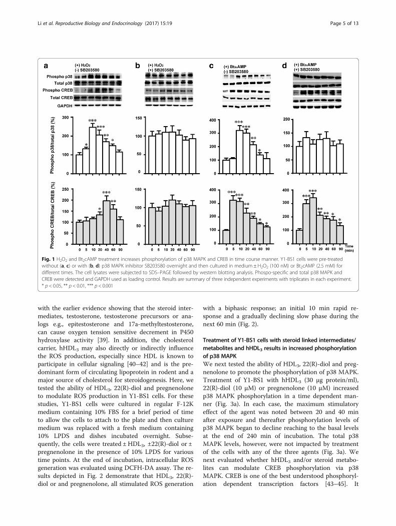
- , •OHand H2O2, promote the phosphorylation and activationof MAPK, among which p38 MAPK is highly responsiveto oxidant stress which has important roles in cell sig-naling and homeostasis [31–34]. We previously demon-strated that treatment of Y1-BS1 cells with superoxide,H2O2 or a lipid peroxidation product, 4-hydroxy-2-nonenal (HNE) reciprocally inhibited steroid productionand increased the phosphorylation and activation of p38MAPK [23]. CREB is a key transcription factor that playsa pivotal role in the regulation of steroidogenic enzymesat their gene transcription level. It is stimulated by anumber of agents including cAMP, growth factors andUV exposure [35–37]. The results presented in Fig. 1aand b demonstrate that treatment of Y1-BS1 cells with100 nM H2O2 for 5 min caused a robust phosphoryl-ation of p38 MAPK and a modest but significant stimu-lation of phosphorylated form of CREB for 20 min,thereafter the phosphorylation of p38 and CREB de-creased. Pretreatment of cells with a specific p38MAPK inhibitor, SB203580 abolished the stimulatoryeffects of H2O2 on p38 MAPK and CREB phosphoryl-ation. These results not only complement the previ-ously published studies from our laboratory [22] butalso unequivocally establish that CREB is a down-stream critical target of p38 MAPK in steroidogeniccells. Meanwhile, we detected that Bt2cAMP couldstimulated both the phosphorylation of p38 MAPKand CREB (Fig. 1c). While SB203580 abolished thestimulatory phosphorylation of p38 MAPK byBt2cAMP, phosphorylation of CREB was not dimin-ished (Fig.1d). These results are consistent with thereported finding that cAMP stimulated the phosphor-ylation of CREB through PKA pathway [36, 38].
Steroid intermediates, 22(R)-diol and pregnenolone andcirculating cholesterol carrier hHDL3 promote intracellularROS productionSteroidogenesis being a multi-step process results ingeneration and/or accumulation of a number of inter-mediary metabolites such as 22(R)-diol and pregneno-lone, which could potentially further enhance theintracellular ROS production. This possibility is in line
Table 1 Primers used for quantitative real-time PCR
Primers for qPCR
Mouse StAR 5′-CGGAGCAGAGTGGTGTCATC-3′-F5′-TGAGTTTAGTCTTGGAGGGACTTC-3′-R
Mouse CYP11A1 5′-ACTGTGAACTGAAGGCTGG-3′-F5′-GGGAAAGAGGGAAAGAGGATG-3′-R
Mouse SOD1 5′-AAGACTGGAAATGCTGGGAG-3′-F5′-GGTTTGAGGGTAGCAGATGAG-3′-R
Mouse SOD2 5′-TGCTCTAATCAGGACCCATTG-3′-F5′-CATTCTCCCAGTTGATTACATTCC-3′-R
Mouse CAT 5′-TCACCTGTAATCAACGCTGG-3′-F5′-AGCCCTAACCTTTCATTTCCC-3′-R
Mouse GPX1 5′-CAGGAGAATGGCAAGAATGAAG-3′-F5′-GAAGGTAAAGAGCGGGTGAG-3′-R
Mouse 36B4 5′-TTTGGGCATCACCACGAAAA-3′-F5′-GGACACCCTCCAGAAAGCGA-3′-R
Li et al. Reproductive Biology and Endocrinology (2017) 15:19 Page 4 of 13
with the earlier evidence showing that the steroid inter-mediates, testosterone, testosterone precursors or ana-logs e.g., epitestosterone and 17a-methyltestosterone,can cause oxygen tension sensitive decrement in P450hydroxylase activity [39]. In addition, the cholesterolcarrier, hHDL3 may also directly or indirectly influencethe ROS production, especially since HDL is known toparticipate in cellular signaling [40–42] and is the pre-dominant form of circulating lipoprotein in rodent and amajor source of cholesterol for steroidogenesis. Here, wetested the ability of HDL3, 22(R)-diol and pregnenoloneto modulate ROS production in Y1-BS1 cells. For thesestudies, Y1-BS1 cells were cultured in regular F-12Kmedium containing 10% FBS for a brief period of timeto allow the cells to attach to the plate and then culturemedium was replaced with a fresh medium containing10% LPDS and dishes incubated overnight. Subse-quently, the cells were treated ± HDL3, ±22(R)-diol or ±pregnenolone in the presence of 10% LPDS for varioustime points. At the end of incubation, intracellular ROSgeneration was evaluated using DCFH-DA assay. The re-sults depicted in Fig. 2 demonstrate that HDL3, 22(R)-diol or and pregnenolone, all stimulated ROS generation
with a biphasic response; an initial 10 min rapid re-sponse and a gradually declining slow phase during thenext 60 min (Fig. 2).
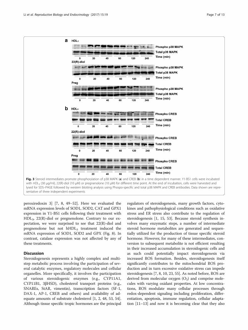
Treatment of Y1-BS1 cells with steroid linked intermediates/metabolites and hHDL3 results in increased phosphorylationof p38 MAPKWe next tested the ability of HDL3, 22(R)-diol and preg-nenolone to promote the phosphorylation of p38 MAPK.Treatment of Y1-BS1 with hHDL3 (30 μg protein/ml),22(R)-diol (10 μM) or pregnenolone (10 μM) increasedp38 MAPK phosphorylation in a time dependent man-ner (Fig. 3a). In each case, the maximum stimulatoryeffect of the agent was noted between 20 and 40 minafter exposure and thereafter phosphorylation levels ofp38 MAPK began to decline reaching to the basal levelsat the end of 240 min of incubation. The total p38MAPK levels, however, were not impacted by treatmentof the cells with any of the three agents (Fig. 3a). Wenext evaluated whether hHDL3 and/or steroid metabo-lites can modulate CREB phosphorylation via p38MAPK. CREB is one of the best understood phosphoryl-ation dependent transcription factors [43–45]. It
Fig. 1 H2O2 and Bt2cAMP treatment increases phosphorylation of p38 MAPK and CREB in time course manner. Y1-BS1 cells were pre-treatedwithout (a, c) or with (b, d) p38 MAPK inhibitor SB203580 overnight and then cultured in medium ± H2O2 (100 nM) or Bt2cAMP (2.5 mM) fordifferent times. The cell lysates were subjected to SDS–PAGE followed by western blotting analysis. Phospo-specific and total p38 MAPK andCREB were detected and GAPDH used as loading control. Results are summary of three independent experiments with triplicates in each experiment.* p < 0.05, ** p < 0.01, *** p < 0.001
Li et al. Reproductive Biology and Endocrinology (2017) 15:19 Page 5 of 13

activates transcription of the target genes in response toa diverse array of stimuli including peptide hormones,growth factors and neuronal activity. Figure 3b showsthat HDL3, 22(R)-diol or pregnenolone treatment, likeH2O2, increased the phosphorylation of CREB with max-imal stimulation occurring around 20 min. These resultsled us to conclude that HDL3, 22(R)-diol and pregneno-lone promote CREB phosphorylation via upstream p38MAPK signaling pathway. To provide additional evi-dence for this conclusion, we treated cells with thesethree agents in the presence and absence of a specificp38 MAPK inhibitor, SB203580 and reassessed the phos-phorylation status of both p38 MAPK and CREB pro-teins. The results presented in Fig. 4 demonstrate thatco-treatment of cells with SB203580 blocked the abilityof HDL3, 22(R)-diol and pregnenolone to stimulate thephosphorylation of both p38 MAP and CREB, furtherconfirming the p38 MAPK catalyzed phosphorylation ofCREB.
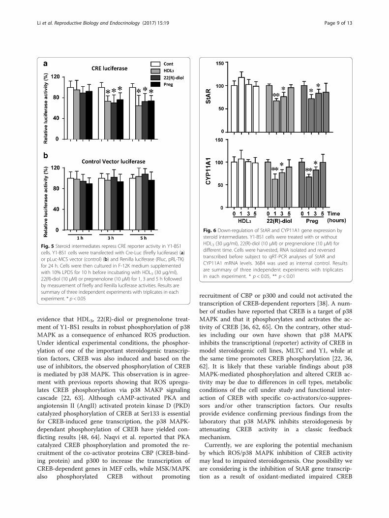
Steroid intermediates repress CREB activityThe above results indicate that phosphorylated forms ofp38 MAPK and CREB were significantly increased inY1-BS1 exposed to HDL3, 22(R)-diol or pregnenolonecompared to vehicle control (Figs. 3 and 4). We nextevaluated the impact of these agents on CREB activityusing a CRE/CREB Reporter kit designed for monitoringthe activity of the cAMP/PKA signaling in cultured cells.As shown in Fig. 5, CREB activity was slightly but sig-nificantly decreased in response to treatment with thesethree agents for 3 to 5 h. These data open up the
possibility that overproduction/increased accumulationof steroid intermediates or sustained activation of cellsto HDL leads to enhanced ROS production and conse-quently loss of CREB activity.
Regulation of StAR and CYP11A1 gene expressionStAR protein mediates rate-limiting step of steroidogen-esis in which cholesterol was translocated from cyto-plasm to mitochondria [46]. Then, P450scc (CYP11A1)catalyzes the conversion of StAR-delivered cholesterol topregnenolone [47]. The transcription of both StAR andCYP11A1 is also regulated by tropic hormones involvingcAMP signaling pathway in multiple transcription fac-tors, including CREB [48]. Since we observed that CREBactivity is repressed by HDL3, 22(R)-diol and pregneno-lone, we measured the mRNA levels of its downstreamtargets, StAR and CYP11A1 following treatment of cellswith these three agents. As shown in Fig. 6, the mRNAexpression of both StAR and CYP11A1 was down-regulated by 22(R)-diol and pregnenolone treatment butnot by hHDL3 for 1 or 3 h’ treatment, while the mRNAexpression recovered after a longer time treatment.
Inhibition of p38 MAPK phosphorylation upregulatesteroidogenesisWe have demonstrated that inhibition of p38 MAPKactivity with SB203580 potentiated the Bt2cAMP andBt2cAMP + hHDL3-stimulated steroid production in Y1-BS1 adrenal cells [23]. Although, oxidants-induced acti-vation p38 MAPK activity critically mediates the oxidantinhibition of steroid production, we also evaluated thepotential effects of steroid metabolites and intermediates–induced activation of p38 MAPK on steroid synthesis.Y1-BS1 cells were treated with three steroid intermedi-ates in the presence and absence of SB203580 andmedium samples analyzed for steroid production usingHPLC-MS/ MS. As shown in Fig. 7, although additionalsubstrates (HDL3, 22(R)-diol or pregnenolone) loadingincreased the steroid production, inclusion of SB203580to inhibit the p38 activity could increase the steroid pro-duction in HDL3, 22(R)-diol or pregnenolone treatedcells. These findings are consistent with previous resultsshowing that inhibition of p38 MAPK by SB203580could ameliorate the oxidative stress-induced repressionof steroidogenesis [23].
Upregulation of anti-oxidant enzymes gene expressionROS-induced oxidative stress is defined as a disturb-ance in the balance between production of ROS andantioxidants defenses, particularly the reduced func-tioning of the antioxidant enzymes (e.g., SOD1 [cyto-solic CuZn-SOD], SOD2 [mitochondrial Mn-SOD],GPX1 [cytosolic/mitochondrial glutathione peroxidase 1],CAT [peroxisomal catalase] and PRDX3 [mitochondrial
Fig. 2 Steroid intermediates stimulate reactive oxygen species (ROS)production and oxidative stress in Y1-BS1 cells. Y1-BS1 cells werepre-loaded with 10 μM DCFH-DA for 30 min, washed and incubatedwith HDL3 (30 μg/ml), 22(R)-diol (10 μM) or pregnenolone (10 μM)for different time point. At the end of incubation, the media were re-moved and cells were immediately lysed and centrifuged. DCFH-DAfluorescence of the cell lysates was measured using a microplatereader with excitation and emission wavelengths of 480 and 525 nmrespectively. Results are summary of three independent experimentswith triplicates in each experiment. * p < 0.05, **p < 0.01, ***p < 0.001
Li et al. Reproductive Biology and Endocrinology (2017) 15:19 Page 6 of 13
peroxiredoxin 3] [7, 8, 49–52]. Here we evaluated themRNA expression levels of SOD1, SOD2, CAT and GPX1expression in Y1-BS1 cells following their treatment withHDL3, 22(R)-diol or pregnenolone. Contrary to our ex-pectation, we were surprised to see that 22(R)-diol andpregnenolone but not hHDL3 treatment induced themRNA expression of SOD1, SOD2 and GPX (Fig. 8). Incontrast, catalase expression was not affected by any ofthese treatments.
DiscussionSteroidogenesis represents a highly complex and multi-step metabolic process involving the participation of sev-eral catalytic enzymes, regulatory molecules and cellularorganelles. More specifically, it involves the participationof various steroidogenic enzymes (e.g., CYP11A1,CYP11B1, 3βHSD), cholesterol transport proteins (e.g.,SNAREs, StAR, vimentin), transcription factors (SF-1,DAX-1, AP-1, CREB and others) and availability of ad-equate amounts of substrate cholesterol [1, 2, 48, 53, 54].Although tissue specific tropic hormones are the principal
regulators of steroidogenesis, many growth factors, cyto-kines and pathophysiological conditions such as oxidativestress and ER stress also contribute to the regulation ofsteroidogenesis [1, 15, 53]. Because steroid synthesis in-volves many enzymatic steps, a number of intermediatesteroid hormone metabolites are generated and sequen-tially utilized for the production of tissue specific steroidhormone. However, for many of these intermediates, con-version to subsequent metabolite is not efficient resultingin their increased accumulation in steroidogenic cells andas such could potentially impact steroidogenesis viaincreased ROS formation. Besides, steroidogenesis itselfsignificantly contributes to the mitochondrial ROS pro-duction and in turn excessive oxidative stress can impedesteroidogenesis [7, 8, 10, 23, 55]. As noted before, ROS arederived from molecular oxygen (O2) and comprise mole-cules with varying oxidant properties. At low concentra-tions, ROS modulate many cellular processes throughredox-dependent signaling including proliferation, differ-entiation, apoptosis, immune regulation, cellular adapta-tion [11–13] and now it is becoming clear that they also
Fig. 3 Steroid intermediates promote phosphorylation of p38 MAPK (a) and CREB (b) in a time dependent manner. Y1-BS1 cells were incubatedwith HDL3 (30 μg/ml), 22(R)-diol (10 μM) or pregnenolone (10 μM) for different time point. At the end of incubation, cells were harvested andlysed for SDS–PAGE followed by western blotting analysis using Phospo-specific and total p38 MAPK and CREB antibodies. Data shown are repre-sentative of three independent experiments
Li et al. Reproductive Biology and Endocrinology (2017) 15:19 Page 7 of 13

participate in the (negative) regulation of steroidogenesis[7, 8, 10, 23, 55, 56]. In contrast, overproduction or highlevels of ROS results in oxidative stress, a deleteriousprocess that can be an important mediator of damage tocellular macromolecular structures including lipids andmembranes, proteins and DNA [57, 58]. Thus, mamma-lian cells and more specifically, steroidogenic cells possessa potent anti-oxidant network to scavenge and neutralizethe excessively produced ROS. Under normal physio-logical conditions, a balance between ROS productionand elimination maintains optimal homeostasis. How-ever, disturbances of such equilibrium under patho-physiological conditions can alter the functioning ofnormal cellular process with detrimental metabolicconsequences.In this study we made a novel observation that en-
dogenous steroid intermediates such as 22(R)-diol andpregnenolone as well as a donor of cholesterol substrate,hHDL3 can promote ROS production and ensuingoxidative stress in Y1-BS1 adrenal cells. Our observa-tions are in line with previous studies showing superoxide/ H2O2 produced during ACTH-stimulated adrenalsteroid production is involved in the termination of thesteroidogenic response [8]. They demonstrated thatH2O2 produced by P450 enzymes during steroidogenesisinactivates potent mitochondrial antioxidant enzyme,peroxiredoxin 3, which in turn triggers a sequence ofevents including increased accumulation of H2O2,
activation of p38 MAPK, suppression of StAR mRNA/protein expression and inhibition of steroidogenesis [8].Our results suggest that it is not only the H2O2 pro-duced during Mn-SOD catalyzed dismutation of super-oxide anions and ensuing inactivation of peroxiredoxin 3that inhibits steroidogenesis, but also steroid metabolitessuch as 22(R)-diol and pregnenolone possess similarcapabilities in promoting ROS production and interfer-ing with normal steroidogenesis. However, we weresurprised to find that hHDL3 a cholesterol donor forsteroidogenesis also promotes ROS production. This iseven more surprising given that HDL is considered anantioxidant with relevance to cardiovascular disease [59].At present we are unable to provide exact explanationby which HDL treatment of Y1 BS1 cells results inincreased ROS production, but it may be that hHDL3being a cholesterol donor, delivers excessive amounts ofcholesterol to cells for its catabolism and this leads toexaggerated production of ROS and excessive oxidativestress. Obviously, more experimental studies are neededto sort out among these various possibilities.Previous data suggest that hydrogen peroxide is a
potent activator for p38 MAPK [23, 60]. Other reportsindicate that tropic hormones such as FSH and ACTHalso induce the phosphorylation and activation of p38MAPK [8, 61]. Several investigators also reported thatp38 MAPK is one of the regulators of CREB phosphoryl-ation and its activity [22, 36, 62]. Our data provide
Fig. 4 Inhibition of p38 MAPK blocked the phosphorylation of CREB by steroid intermediates. Y1-BS1 cells were pre-treated without (a) or with(b) p38 MAPK inhibitor (10 μM) overnight. Cells were then incubated with HDL3 (30 μg/ml), 22(R)-diol (10 μM) or pregnenolone (10 μM) for20 min. At the end of the incubation, cell lysates were subjected to SDS–PAGE followed by western blotting analysis. Data shown are representativeof three independent experiments
Li et al. Reproductive Biology and Endocrinology (2017) 15:19 Page 8 of 13
evidence that HDL3, 22(R)-diol or pregnenolone treat-ment of Y1-BS1 results in robust phosphorylation of p38MAPK as a consequence of enhanced ROS production.Under identical experimental conditions, the phosphor-ylation of one of the important steroidogenic transcrip-tion factors, CREB was also induced and based on theuse of inhibitors, the observed phosphorylation of CREBis mediated by p38 MAPK. This observation is in agree-ment with previous reports showing that ROS upregu-lates CREB phosphorylation via p38 MAKP signalingcascade [22, 63]. Although cAMP-activated PKA andangiotensin II (AngII) activated protein kinase D (PKD)catalyzed phosphorylation of CREB at Ser133 is essentialfor CREB-induced gene transcription, the p38 MAPK-dependant phosphorylation of CREB have yielded con-flicting results [48, 64]. Naqvi et al. reported that PKAcatalyzed CREB phosphorylation and promoted the re-cruitment of the co-activator proteins CBP (CREB-bind-ing protein) and p300 to increase the transcription ofCREB-dependent genes in MEF cells, while MSK/MAPKalso phosphorylated CREB without promoting
recruitment of CBP or p300 and could not activated thetranscription of CREB-dependent reporters [38]. A num-ber of studies have reported that CREB is a target of p38MAPK and that it phosphorylates and activates the ac-tivity of CREB [36, 62, 65]. On the contrary, other stud-ies including our own have shown that p38 MAPKinhibits the transcriptional (reporter) activity of CREB inmodel steroidogenic cell lines, MLTC and Y1, while atthe same time promotes CREB phosphorylation [22, 36,62]. It is likely that these variable findings about p38MAPK-mediated phosphorylation and altered CREB ac-tivity may be due to differences in cell types, metabolicconditions of the cell under study and functional inter-action of CREB with specific co-activators/co-suppres-sors and/or other transcription factors. Our resultsprovide evidence confirming previous findings from thelaboratory that p38 MAPK inhibits steroidogenesis byattenuating CREB activity in a classic feedbackmechanism.Currently, we are exploring the potential mechanism
by which ROS/p38 MAPK inhibition of CREB activitymay lead to impaired steroidogenesis. One possibility weare considering is the inhibition of StAR gene transcrip-tion as a result of oxidant-mediated impaired CREB
Fig. 5 Steroid intermediates repress CRE reporter activity in Y1-BS1cells. Y1-BS1 cells were transfected with Cre-Luc (firefly luciferase) (a)or pLuc-MCS vector (control) (b) and Renilla luciferase (Rluc, pRL-TK)for 24 h. Cells were then cultured in F-12K medium supplementedwith 10% LPDS for 10 h before incubating with HDL3 (30 μg/ml),22(R)-diol (10 μM) or pregnenolone (10 μM) for 1, 3 and 5 h followedby measurement of firefly and Renilla luciferase activities. Results aresummary of three independent experiments with triplicates in eachexperiment. * p < 0.05
Fig. 6 Down-regulation of StAR and CYP11A1 gene expression bysteroid intermediates. Y1-BS1 cells were treated with or withoutHDL3 (30 μg/ml), 22(R)-diol (10 μM) or pregnenolone (10 μM) fordifferent time. Cells were harvested, RNA isolated and reversedtranscribed before subject to qRT-PCR analyses of StAR andCYP11A1 mRNA levels. 36B4 was used as internal control. Resultsare summary of three independent experiments with triplicatesin each experiment. * p < 0.05, ** p < 0.01
Li et al. Reproductive Biology and Endocrinology (2017) 15:19 Page 9 of 13

activity. This is in line with the observation that StARexpression is sensitive to both physiological and patho-physiological levels of ROS. Moreover, earlier studieshave shown that CREB protein in cooperation with SF1is a major regulator of StAR protein gene transcription[66, 67]. Finally, we have earlier shown that oxidants-p38 MAPK cause inhibition of StAR promoter activityprimarily by interfering with CREB activity [22]. Otherinvestigators reported that HDL2, very-low-density lipo-protein (VLDL) and glyco-oxidized VLDL can induceCyp11B2 expression and stimulate steroid production ina human adrenocortical carcinoma cell line, NCI H295R[68–70]. Saha et al [70] also reported slight increases inStAR expression by native VLDL and glycol-oxidizedVLDL but not by oxidized VLDL. The data presentedhere show that 22(R)-diol and pregnenolone but notHDL3 repress the gene expression of StAR andCYP11A1. We further provided the evidence that 22(R)-diol and pregnenolone-mediated repression of StAR andCYP11A1 gene expression is achieved through excessiveoxidative stress and associated p38 MAPK signaling cas-cade. Our steroid hormone production data provide add-itional support to the notion that SB203580 inhibition ofp38 MAPK augments the steroids production in Y1-BS1cells treated with steroid metabolites/intermediates. Anumber of studies have implicated p38 MAPK signalingcascade in the regulation of steroidogenesis [71], al-though p38 MAPK regulation of steroidogenesis is
complex and extent of p38 MAPK varies with steroido-genic cell types. For example, inhibition of p38 MAPKactivity by SB203580 in IL-1α-stimulated immature ratLeydig cells leads to downregulation of StAR gene ex-pression and attenuation of steroid production. [72].
Fig. 8 Up-regulation of anti-oxidant enzymes gene expression bysteroid intermediates. Y1-BS1 cells were treated with or withoutHDL3 (30 μg/ml), 22(R)-diol (10 μM) or pregnenolone (10 μM) forvarious hours. RNA were isolated and reversed transcribed beforesubject to qRT-PCR analyses of SOD1, SOD2, GPX1 and CAT mRNAlevels. 36B4 was used as internal control. Results are summary ofthree independent experiments with triplicates in each experiment.* p < 0.05, ** p < 0.01
Fig. 7 Inhibition of p38 MAPK activity by SB203580 augmentssteroid production. Y1-BS1 cells were pre-treated with or withoutSB203580 (10 μM) overnight. Cells were then incubated with HDL3(30 μg/ml), 22(R)-diol (10 μM) or pregnenolone (10 μM) for 5 h.At the end of the incubation, the medium sampless werecollected for hormone analysis using HPLC-MS / MS. Results aremean of three independent experiments with triplicates in eachexperiment. * p < 0.05, ** p < 0.01
Li et al. Reproductive Biology and Endocrinology (2017) 15:19 Page 10 of 13

Other studies have shown that inhibition of p38 MAPKactivity by SB203580 in ovarian granulosa cells is accom-panied by increased inhibition of LH/hCG/FSH medi-ated StAR expression and progesterone synthesis [61,73]. Likewise, in primary cultures of rat adrenal glomer-ulosa cells, Angiotensin II activates the p38 MAPK andresults in increases in StAR expression and steroid syn-thesis [71, 74]. Interestingly, our previous studies haveshown that inhibition of p38 MAPK by either SB203580or SB202190 in adrenocortical from old rats restorescorticosterone synthesis to the levels seen in cells fromyoung animals [23]. In this study, inhibition ofp38 MAPK activity by SB203580 enhanced 20α-hydroxyprogesterone production in mouse Y1-BS1 adre-nocortical tumor cell cotreated with HDL3, 22(R)-diol orpregnenolone. In addition, we will examine whetherROS/p38 MAPK modulate the expression of some ofthe critical SNARE proteins. In a recent publication, weidentified several SNAREs, whose expression is essentialfor cholesterol transport to outer mitochondrial mem-brane for optimal steroid production [54].Another surprising finding from our studies was that
treatment of Y1-BS1 with 22(R)-diol and pregnenolonebut not HDL3 leads to increased mRNA expression ofthree antioxidant enzymes, SOD1, SOD2 and CAT. Incontrast, studies by Kil et al [8] observed no changesin the expression of levels of SOD1, SOD2, CAT, andGPX1 in intact adrenals following treatment of micewith ACTH. Interestingly, a recently published studyreported that p38 MAPKα causes the induction ofantioxidant enzymes SOD2 and catalase by two dis-tinct mechanisms [18]. Obviously, more studies areneeded to sort out molecular mechanisms involved inthe transcriptional/posttranscriptional and/or post-translational regulation of these antioxidant enzymesin vitro and in vivo.
ConclusionIn conclusion, our studies provide evidence that expos-ure of adrenal cells to steroid intermediates/metabolites,22(R)-diol and pregnenolone, and hHDL3 led toincreased ROS production and associated enhancedphosphorylation of p38 MAPK and CREB. This oxidant-mediated up-regulation of CREB phosphorylation ismediated by p38 MAPK. The increased CREB phosphor-ylation however, was accompanied by a significant lossof CREB’s transcriptional activity. Furthermore, treat-ment of cells with 22(R)-diol and pregnenolone andensuing oxidative stress resulted in decreased mRNAlevels of StAR and CYP11A1 and increased mRNA levelsof antioxidant enzymes SOD1, SOD2 and CAT. Fromthese studies we conclude that ROS/p38 MAPK inhib-ition of CREB transcriptional activity is likely responsiblefor ROS-induced feedback inhibition of steroidogenesis.
Abbreviations22(R)-diol: 22(R)-Hydroxycholesterol; ACTH: Adrenocorticotropin hormone;AngII: Angiotensin II; CaM II: Calcium-calmodulin kinase II; CAT: Catalase;CREB: cAMP response element-binding protein; DCFH-DA: 2′,7′-Dichlorofluorescin diacetate; FBS: Fetal bovine serum; FSH: Follicle-stimulating hormone; GPX: Glutathione peroxidase; HDL: High-densitylipoprotein; HNE: 4-hydroxy-2-nonenal; IMM: Inner mitochondrial membrane;LDL: Low-density lipoprotein; LH: Luteinizing hormone; LPDS: Lipoprotein-deficient serum; MSK: Mitogen- and stress-activated kinase; OMM: Outermitochondrial membrane; PI3K: Phosphatidylinositol 3-kinase; PKA: Proteinkinase A; PKB: Protein kinase B; PKC: Protein kinase C; PKD: Protein kinase D;pp90 RSK: pp 90 ribosomal S6 kinase; ROS: Reactive oxygen species;SF1: Steroidogenic factor 1; SOD: Superoxide dismutase; SR-BI: ScavengerReceptor Class B, Type I; VLDL: Very-low-density lipoprotein
AcknowledgementNone
FundingThe work is supported by National Natural Science Foundation of China(31400659), Jiangsu Provincial Natural Science Foundation (BK20140920),Priority Academic Program Development of Jiangsu Higher EducationInstitutions, Natural Science Fund of Colleges and Universities in JiangsuProvince (14KJB180012), and National institute of Health (NIH/NHLBI2R01HL033881 and VA Merit Review 5I01BX001923, SA).
Availability of data and materialsNot applicable.
Authors’ contributionsConceived and designed the experiments: JL, ZG and ZH. Performed theexperiments: JL, QZ, ZM and ZH. Analyzed the data: JL, QZ, ZM, MWand ZH. Wrote the paper: ZH, SA. Edited the manuscript: ZH, WJS, ZGand SA. All authors read and approved the final manuscript.
Competing interestsThe authors declare that they have no competing interests.
Consent for publicationNot applicable.
Ethics approval and consent to participateNot applicable.
Publisher’s NoteSpringer Nature remains neutral with regard to jurisdictional claims inpublished maps and institutional affiliations.
Author details1Jiangsu Key Laboratory for Molecular and Medical Biotechnology, College ofLife Sciences, Nanjing Normal University, 1 WenYuan Road, Nanjing 210023,China. 2Geriatric Research, Education and Clinical Center, VA Palo Alto HealthCare System, Palo Alto, CA 94304, USA. 3Stanford University School ofMedicine, Palo Alto, CA 94304, USA.
Received: 2 December 2016 Accepted: 6 March 2017
References1. Miller WL, Auchus RJ. The molecular biology, biochemistry, and
physiology of human steroidogenesis and its disorders. Endocr Rev.2010;32:81–151.
2. Hu J, Zhang Z, Shen W-J, Azhar S. Cellular cholesterol delivery, intracellularprocessing and utilization for biosynthesis of steroid hormones. Nutr Metab.2010;7:1.
3. Azhar S, Reaven E. Scavenger receptor class BI and selective cholesterylester uptake: partners in the regulation of steroidogenesis. Mol CellEndocrinol. 2002;195:1–26.
4. Hattangady NG, Olala LO, Bollag WB, Rainey WE. Acute and chronicregulation of aldosterone production. Mol Cell Endocrinol. 2012;350:151–62.
Li et al. Reproductive Biology and Endocrinology (2017) 15:19 Page 11 of 13
5. Lehoux J-G, Fleury A, Ducharme L. The acute and chronic effects ofadrenocorticotropin on the levels of messenger ribonucleic acid andprotein of steroidogenic enzymes in rat adrenal in vivo 1. Endocrinology.1998;139:3913–22.
6. Raha S, Robinson BH. Mitochondria, oxygen free radicals, disease andageing. Trends Biochem Sci. 2000;25:502–8.
7. Hanukoglu I. Antioxidant protective mechanisms against reactive oxygenspecies (ROS) generated by mitochondrial P450 systems in steroidogeniccells. Drug Metab Rev. 2006;38:171–96.
8. Kil IS, Lee SK, Ryu KW, Woo HA, Hu M-C, Bae SH, Rhee SG. Feedbackcontrol of adrenal steroidogenesis via H 2 O 2-dependent, reversibleinactivation of peroxiredoxin III in mitochondria. Mol Cell. 2012;46:584–94.
9. Beattie MC, Chen H, Fan J, Papadopoulos V, Miller P, Zirkin BR. Aging andluteinizing hormone effects on reactive oxygen species production andDNA damage in rat Leydig cells. Biol Reprod. 2013;88:100.
10. Abidi P, Leers‐Sucheta S, Cortez Y, Han J, Azhar S. Evidence that age‐related changes in p38 MAP kinase contribute to the decreased steroidproduction by the adrenocortical cells from old rats. Aging Cell. 2008;7:168–78.
11. Dröge W. Free radicals in the physiological control of cell function. PhysiolRev. 2002;82:47–95.
12. Ray PD, Huang B-W, Tsuji Y. Reactive oxygen species (ROS) homeostasis andredox regulation in cellular signaling. Cell Signal. 2012;24:981–90.
13. Sena LA, Chandel NS. Physiological roles of mitochondrial reactive oxygenspecies. Mol Cell. 2012;48:158–67.
14. BEHRMAN HR, ATEN RF. Evidence That Hydrogen Peroxide Blocks Hormone-Sensitive Cholesterol Transport into Mitochondria of Rat Luteal Cells*.Endocrinology. 1991;128:2958–66.
15. Stocco DM, Wells J, Clark BJ. The effects of hydrogen peroxide onsteroidogenesis in mouse Leydig tumor cells. Endocrinology. 1993;133:2827–32.
16. Diemer T, Allen JA, Hales KH, Hales DB. Reactive oxygen disruptsmitochondria in MA-10 tumor Leydig cells and inhibits steroidogenic acuteregulatory (StAR) protein and steroidogenesis. Endocrinology. 2003;144:2882–91.
17. McClung JM, Judge AR, Powers SK, Yan Z. p38 MAPK links oxidative stressto autophagy-related gene expression in cachectic muscle wasting. Am JPhys Cell Phys. 2010;298:C542–9.
18. Gutiérrez-Uzquiza Á, Arechederra M, Bragado P, Aguirre-Ghiso JA, Porras A.p38α Mediates Cell Survival in Response to Oxidative Stress via Induction ofAntioxidant Genes EFFECT ON THE p70S6K PATHWAY. J Biol Chem. 2012;287:2632–42.
19. Yamada T, Egashira N, Bando A, Nishime Y, Tonogai Y, Imuta M,Yano T, Oishi R. Activation of p38 MAPK by oxidative stress underlyingepirubicin-induced vascular endothelial cell injury. Free Radic Biol Med.2012;52:1285–93.
20. Cuenda A, Rousseau S. p38 MAP-kinases pathway regulation, function androle in human diseases. Biochimica et Biophysica Acta (BBA)-Molecular CellRes. 2007;1773:1358–75.
21. Cuadrado A, Nebreda AR. Mechanisms and functions of p38 MAPKsignalling. Biochem J. 2010;429:403–17.
22. Zaidi SK, Shen W-J, Bittner S, Bittner A, McLean MP, Han J, Davis RJ, KraemerFB, Azhar S. p38 MAPK regulates steroidogenesis through transcriptionalrepression of STAR gene. J Mol Endocrinol. 2014;53:1–16.
23. Abidi P, Zhang H, Zaidi SM, Shen W-J, Leers-Sucheta S, Cortez Y, Han J,Azhar S. Oxidative stress-induced inhibition of adrenal steroidogenesisrequires participation of p38 mitogen-activated protein kinase signalingpathway. J Endocrinol. 2008;198:193–207.
24. Rainey WE, Saner K, Schimmer BP. Adrenocortical cell lines. Mol CellEndocrinol. 2004;228:23–38.
25. Yasumura Y, Buonassisi V, Sato G. Clonal analysis of differentiated functionin animal cell cultures. Cancer research. 1966;26(3 Part 1):529-35.
26. Temel RE, Trigatti B, DeMattos RB, Azhar S, Krieger M, Williams DL.Scavenger receptor class B, type I (SR-BI) is the major route for the deliveryof high density lipoprotein cholesterol to the steroidogenic pathway incultured mouse adrenocortical cells. Proc Natl Acad Sci U S A. 1997;94:13600–5.
27. Hu Z, Li J, Kuang Z, Wang M, Azhar S, Guo Z. Cell-Specific Polymorphismand Hormonal Regulation of DNA Methylation in Scavenger Receptor ClassB, Type I. DNA Cell Biol. 2016;35:280–9.
28. Reaven E, Tsai L, Azhar S. Intracellular events in the “selective” transport oflipoprotein-derived cholesteryl esters. J Biol Chem. 1996;271:16208–17.
29. Hu Z, Hu J, Zhang Z, Shen W-J, Yun CC, Berlot CH, Kraemer FB, Azhar S.Regulation of expression and function of scavenger receptor class B, type I(SR-BI) by Na+/H+ exchanger regulatory factors (NHERFs). J Biol Chem. 2013;288:11416–35.
30. Zhou Y, Wang Y, Jiaping YU. Research on the determination of serumsteroid hormones by isotope dilution HPLC-MS/MS. 2015.
31. Kyriakis JM, Avruch J. Mammalian mitogen-activated protein kinase signaltransduction pathways activated by stress and inflammation. Physiol Rev.2001;81:807–69.
32. McCubrey JA, LaHair MM, Franklin RA. Reactive oxygen species-inducedactivation of the MAP kinase signaling pathways. Antioxid Redox Signal.2006;8:1775–89.
33. Son Y, Kim S, Chung H-T, Pae H-O. Reactive oxygen species in the activationof MAP kinases. Methods Enzymol. 2013;528:27–48.
34. Devasagayam T, Tilak J, Boloor K, Sane KS, Ghaskadbi SS, Lele R. Free radicalsand antioxidants in human health: current status and future prospects. Japi.2004;52:4.
35. Xing J, Kornhauser JM, Xia Z, Thiele EA, Greenberg ME. Nerve growth factoractivates extracellular signal-regulated kinase and p38 mitogen-activatedprotein kinase pathways to stimulate CREB serine 133 phosphorylation. MolCell Biol. 1998;18:1946–55.
36. Delghandi MP, Johannessen M, Moens U. The cAMP signalling pathwayactivates CREB through PKA, p38 and MSK1 in NIH 3 T3 cells. Cell Signal.2005;17:1343–51.
37. Iordanov M, Bender K, Ade T, Schmid W, Sachsenmaier C, Engel K, GaestelM, Rahmsdorf H, Herrlich P. CREB is activated by UVC through a p38/HOG‐1‐dependent protein kinase. EMBO J. 1997;16:1009–22.
38. Naqvi S, Martin KJ, Arthur JS. CREB phosphorylation at Ser133 regulatestranscription via distinct mechanisms downstream of cAMP and MAPKsignalling. Biochem J. 2014;458:469–79.
39. Quinn P, Payne A. Steroid product-induced, oxygen-mediated damage ofmicrosomal cytochrome P-450 enzymes in Leydig cell cultures. Relationshipto desensitization. J Biol Chem. 1985;260:2092–9.
40. Grewal T, de Diego I, Kirchhoff MF, Tebar F, Heeren J, Rinninger F, Enrich C.High density lipoprotein-induced signaling of the MAPK pathway involvesscavenger receptor type BI-mediated activation of Ras. J Biol Chem. 2003;278:16478–81.
41. Pan B, Ma Y, Ren H, He Y, Wang Y, Lv X, Liu D, Ji L, Yu B, Wang Y.Diabetic HDL is dysfunctional in stimulating endothelial cell migrationand proliferation due to down regulation of SR-BI expression. PLoSOne. 2012;7:e48530.
42. Mineo C, Shaul PW. Regulation of signal transduction by HDL. J Lipid Res.2013;54:2315–24.
43. Shaywitz AJ, Greenberg ME. CREB: a stimulus-induced transcription factoractivated by a diverse array of extracellular signals. Annu Rev Biochem.1999;68:821–61.
44. Mayr B, Montminy M. Transcriptional regulation by the phosphorylation-dependent factor CREB. Nat Rev Mol Cell Biol. 2001;2:599–609.
45. Sakamoto KM, Frank DA. CREB in the pathophysiology of cancer:implications for targeting transcription factors for cancer therapy. ClinCancer Res. 2009;15:2583–7.
46. Stocco DM. StAR protein and the regulation of steroid hormonebiosynthesis. Annu Rev Physiol. 2001;63:193–213.
47. Simpson ER, Miller DA. Cholesterol side-chain cleavage, cytochrome P450,and iron-sulfur protein in human placental mitochondria. Arch BiochemBiophys. 1978;190:800–8.
48. Lavoie HA, King SR. Transcriptional regulation of steroidogenic genes:STARD1, CYP11A1 and HSD3B. Exp Biol Med. 2009;234:880–907.
49. Chen H, Irizarry RA, Luo L, Zirkin BR. Leydig cell gene expression: effects ofage and caloric restriction. Exp Gerontol. 2004;39:31–43.
50. Fu H, Wada-Hiraike O, Hirano M, Kawamura Y, Sakurabashi A, ShiraneA, Morita Y, Isono W, Oishi H, Koga K. SIRT3 positively regulates theexpression of folliculogenesis-and luteinization-related genes andprogesterone secretion by manipulating oxidative stress in humanluteinized granulosa cells. Endocrinology. 2014;155:3079–87.
51. Indo HP, Yen H-C, Nakanishi I, Matsumoto K-i, Tamura M, Nagano Y,Matsui H, Gusev O, Cornette R, Okuda T. A mitochondrial superoxidetheory for oxidative stress diseases and aging. J Clin Biochem Nutr.2015;56:1.
Li et al. Reproductive Biology and Endocrinology (2017) 15:19 Page 12 of 13

52. Candas D, Li JJ. MnSOD in oxidative stress response-potential regulation viamitochondrial protein influx. Antioxid Redox Signal. 2014;20:1599–617.
53. Stocco DM, Wang X, Jo Y, Manna PR. Multiple signaling pathwaysregulating steroidogenesis and steroidogenic acute regulatory proteinexpression: more complicated than we thought. Mol Endocrinol. 2005;19:2647–59.
54. Lin Y, Hou X, Shen WJ, Hanssen R, Khor VK, Cortez Y, Roseman AN, Azhar S,Kraemer FB. SNARE-mediated cholesterol movement to mitochondriasupports steroidogenesis in rodent cells. Mol Endocrinol. 2016;30(2):234-47.
55. Vitale G, Salvioli S, Franceschi C. Oxidative stress and the ageing endocrinesystem. Nat Rev Endocrinol. 2013;9:228–40.
56. Azhar S, Cao L, Reaven E. Alteration of the adrenal antioxidant defensesystem during aging in rats. J Clin Investig. 1995;96:1414.
57. Valko M, Leibfritz D, Moncol J, Cronin MT, Mazur M, Telser J. Free radicalsand antioxidants in normal physiological functions and human disease.Int J Biochem Cell Biol. 2007;39:44–84.
58. Ye Z-W, Zhang J, Townsend DM, Tew KD. Oxidative stress, redox regulationand diseases of cellular differentiation. Biochimica et Biophysica Acta(BBA)-General Subjects. 2015;1850:1607–21.
59. Karlsson H, Kontush A, James RW. Functionality of HDL: antioxidationand detoxifying effects. In High Density Lipoproteins. Springer; 2015:207-228. https://link.springer.com/chapter/10.1007%2F978-3-319-09665-0_5.
60. Kulisz A, Chen N, Chandel NS, Shao Z, Schumacker PT. Mitochondrial ROSinitiate phosphorylation of p38 MAP kinase during hypoxia incardiomyocytes. Am J Phys Lung Cell Mol Phys. 2002;282:L1324–9.
61. Yu F-Q, Han C-S, Yang W, Jin X, Hu Z-Y, Liu Y-X. Activation of the p38MAPK pathway by follicle-stimulating hormone regulates steroidogenesisin granulosa cells differentially. J Endocrinol. 2005;186:85–96.
62. Butler MP, Hanly JA, Moynagh PN. Pellino3 is a novel upstream regulator ofp38 MAPK and activates CREB in a p38-dependent manner. J Biol Chem.2005;280:27759–68.
63. Jinlian L, Yingbin Z, Chunbo W. p38 MAPK in regulating cellular responsesto ultraviolet radiation. J Biomed Sci. 2007;14:303–12.
64. Olala LO, Choudhary V, Johnson MH, Bollag WB. AngiotensinII-induced protein kinase D activates the ATF/CREB family oftranscription factors and promotes StAR mRNA expression.Endocrinology. 2014;155:2524–33.
65. Saha B, Singh SK, Sarkar C, Bera R, Ratha J, Tobin DJ, Bhadra R. Activation ofthe Mitf promoter by lipid‐stimulated activation of p38‐stress signalling toCREB. Pigment Cell Res. 2006;19:595–605.
66. Manna P, Eubank D, Lalli E, Sassone-Corsi P, Stocco D. Transcriptionalregulation of the mouse steroidogenic acute regulatory protein gene bythe cAMP response-element binding protein and steroidogenic factor 1.J Mol Endocrinol. 2003;30:381–97.
67. Clem BF, Hudson EA, Clark BJ. Cyclic adenosine 3′, 5′-monophosphate(cAMP) enhances cAMP-responsive element binding (CREB) proteinphosphorylation and phospho-CREB interaction with the mousesteroidogenic acute regulatory protein gene promoter. Endocrinology. 2005;146:1348–56.
68. Cherradi N, Bideau M, Arnaudeau S, Demaurex N, James RW, Azhar S,Capponi AM. Angiotensin II promotes selective uptake of high densitylipoprotein cholesterol esters in bovine adrenal glomerulosa and humanadrenocortical carcinoma cells through induction of scavenger receptorclass B type I. Endocrinology. 2001;142:4540–9.
69. Xing Y, Cohen A, Rothblat G, Sankaranarayanan S, Weibel G, Royer L,Francone OL, Rainey WE. Aldosterone Production in Human AdrenocorticalCells Is Stimulated by High-Density Lipoprotein 2 (HDL2) through IncreasedExpression of Aldosterone Synthase (CYP11B2). Endocrinology. 2011;152:751–63.
70. Saha S, Bornstein SR, Graessler J, Kopprasch S. Very-low-density lipoproteinmediates transcriptional regulation of aldosterone synthase in humanadrenocortical cells through multiple signaling pathways. Cell Tissue Res.2012;348:71–80.
71. Manna PR, Stocco DM. The Role of Specific Mitogen-Activated ProteinKinase Signaling Cascades in the Regulation of Steroidogenesis. J SignalTransduction. 2011;2011:821615.
72. Svechnikov K, Stocco DM, Söder O. Interleukin-1alpha stimulatessteroidogenic acute regulatory protein expression via p38 MAP kinase inimmature rat Leydig cells. J Mol Endocrinol. 2003;30:59–67.
73. Tajima K, Dantes A, Yao Z, Sorokina K, Kotsuji F, Seger R, Amsterdam A.Down-regulation of steroidogenic response to gonadotropins in humanand rat preovulatory granulosa cells involves mitogen-activated proteinkinase activation and modulation of DAX-1 and steroidogenic factor-1.J Clin Endocrinol Metab. 2003;88:2288–99.
74. Otis M, Campbell S, Payet MD, Gallo-Payet N. Angiotensin II stimulatesprotein synthesis and inhibits proliferation in primary cultures of rat adrenalglomerulosa cells. Endocrinology. 2005;146:633–42.
• We accept pre-submission inquiries
• Our selector tool helps you to find the most relevant journal
• We provide round the clock customer support
• Convenient online submission
• Thorough peer review
• Inclusion in PubMed and all major indexing services
• Maximum visibility for your research
Submit your manuscript atwww.biomedcentral.com/submit
Submit your next manuscript to BioMed Central and we will help you at every step:
Li et al. Reproductive Biology and Endocrinology (2017) 15:19 Page 13 of 13