rbm online vol5 no3 printed - redlara
TRANSCRIPT

Reviews
328
IntroductionTwo interrelated challenges face embryologists in IVF clinics.A current clinical need is to find out why human embryos havesuch low implantation rates that several treatment cycles ormultiple embryo transfers are needed for assistedreproduction. Finding the cause demands a deeper scientificunderstanding of the regulation of the formation and growth ofoocytes and embryo development, and knowledge on thesetopics forms the first part of this manuscript.
Clinical practice aimed at improving implantation rates will
then be described in the second section of this paper. Initially,investigators believed human embryos had implantationpotentials typical of other mammals, i.e. 80–90% per embryoin many species. Instead, these rates are approximately 20%even in couples wishing to achieve conception, and similar toimplantation rates achieved with single embryo transfersduring assisted human conception.
Detailed attention will therefore be given to the culture ofhuman embryos and selection for those embryos with highimplantation potential. This quality is not studied in anyanimal species, and it is apparently entirely a clinical problem.
Fundamentals of human embryonic growth invitro and the selection of high-quality embryos for transfer
Dr Irene Boiso was born in Buenos Aires, Argentina. She received her BSc degree inBiological Sciences from the Universidad Central de Venezuela. In 1993 she was awardeda 3-year fellowship from the Spanish Instituto de Cooperación Iberoamericana and movedto Barcelona, Spain. She wrote her thesis on the effect of cryopreservation on the structureof the meiotic spindle of mature and immature human oocytes, and received a PhD degree(cum laude) from the Universidad Autónoma de Barcelona. She joined the team of theServicio de Medicina de la Reproducción from the Institut Universitari Dexeus in Barcelonaas an embryologist in 1994. She has been a researcher at the Fundación Santiago DexeusFont since 1999. Her areas of interest include assisted reproductive technologies, earlyembryonic development, oocyte and ovarian tissue cryopreservation and oocyte in-vitromaturation.
Irene Boiso1, Anna Veiga1, Robert G Edwards2,3
1Reproductive Medicine Service, Department of Obstetrics and Gynaecology, Institut Universitari Dexeus, PaseoBonanova 89–91, Barcelona, 08017, Spain2Editor, Reproductive BioMedicine Online, Duck End Farm, Dry Drayton, Cambridge CB3 8DB, UK
3Correspondence: e-mail: [email protected]
Dr Irene Boiso
AbstractKnowledge of the nature of embryo growth, and the handling and scoring of quality in human embryos are significantaspects for embryologists in IVF clinics. This review describes the formation, growth and maturation of human oocytes,many aspects of fertilization in vitro, embryonic transcription during preimplantation stages, and the formation of polarities,timing controls, role of mitochondria and functions of endocrine and paracrine systems. Modern concepts are fullydiscussed, together with their significance in the practice of IVF. This knowledge is essential for the correct clinical care ofhuman embryos growing in vitro, especially in view of their uncharacteristic tendency to vary widely in implantationpotential. Underlying causes of such variation have not been identified. Stringent tests must be enforced to ensure humanembryos develop under optimal conditions, and are scored for quality using the most advanced techniques. Optimal methodsof culture are described, including methods such as co-culture introduced to improve embryo quality but less importanttoday. Detailed attention is given to quality as assessed from embryonic characteristics determined by timers, polarities,disturbed embryo growth and anomalous cell cycles. Methods for classification are described. Approaches to single embryotransfers are described, including the use of sequential media to produce high-quality blastocysts. These approaches, andothers involved in surgical methods to remove fragments, transfer ooplasm or utilize newer approaches such aspreimplantation diagnosis of chromosomal complements in embryos are covered. New outlooks in this field aresummarized.
Keywords:embryo surgery, embryonic regulation, human embryos, pregnancy rates, selection for implantation potential
RBMOnline - Vol 5. No 3. 328–350 Reproductive BioMedicine Online; www.rbmonline.com/Article/555 on web 11 September 2002
This review will also be published in a Spanish textbook entitled ModernAssisted Conception.
329
Reviews - Fundamentals of human embryonic growth in vitro - I Boiso et al.
Why some embryos implant, whereas most do not, will be acommon theme running through both sections of thismanuscript.
Part 1. The human embryo in thelaboratoryDevelopmental systems discussed in this section include cellstructure, transcription, maternal and embryonic geneexpression, polarity, timing and integration, mitochondria andembryonic cytokines. Understanding these factors fromearliest stages of oocyte formation in humans depends partiallyon analysing homologous systems in flies, nematodes andamphibians. Clear gene homologies have been identifiedbetween these disparate organisms (Edwards, 2001).
Formation, growth and maturation of theoocyte
Follicle formation and growth
Differentiating primordial germ cells produce gametogeniccells of both sexes in mammals. Multiplying mitotically inboth sexes, they diverge and differentiate into male and femalegermline cells. Female germ cells form oogonia, and thenoocytes as they enter their first meiotic division. This step isdelayed in male embryos until the testis has formed. In mostmammals, meiosis in primordial oocytes arrests at thediplotene stage of meiosis 1, when a germinal vesicle forms.Several ovarian cells, probably arising from rete tubules,enclose each oocyte to form the primary follicle.
All human follicles are formed before birth to form a pool,which is utilized progressively from the moment of theirformation, through puberty and during successive reproductivecycles in the adult (Kably and Barroso, 2000). The end ofreproductive life in women, and in certain mouse strains, issignified as the pool declines to very few or no remainingfollicles.
Follicles migrate from the pool at a constant rate over much ofthe reproductive lifespan in women, with a sudden increaseduring perimenopausal years (Faddyet al., 1992). Theydevelop at astonishingly regular rates through the stages offolliculogenesis. The great majority of growing follicles andoocytes never attain ovulation or fertilization, many becomingatretic during each stage of differentiation. Folliclescontinuing to grow develop an antral cavity and becomesensitive to gonadotrophins as FSH and, later, LH receptorsdifferentiate on theca and granulosa cells. Pituitarygonadotrophins are now essential, their high levels duringovarian stimulation preventing atresia so that many folliclescontinue to grow (Kably and Barroso, 2000).
In natural cycles, one follicle becomes dominant. Internalparacrine systems and external secretions from the ovary to thehypothalamus co-operate in selecting the dominant follicle,which then impairs challenger follicles (Baker and Spears,1999; Mikkelsenet al., 2001). This significant phase of folliclegrowth occurs as follicles reach >10 mm diameter and thedominant follicle withstands slight decreases in FSHconcentrations typical of the late follicular phase of the
menstrual cycle. It is also important if the aspiration ofimmature oocytes is desired, since a dominant follicle andoestradiol output improves their chances of maturing in vitroand establishing pregnancy (Mikkelsen et al., 2001). Ovarianstimulation also overcomes atresia, so many follicles dodevelop to ovulation (Risquez, 2001; Ulloa-Aguirreet al.,2000).
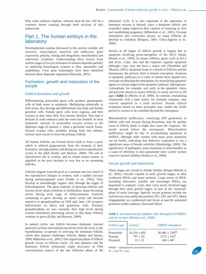
Atresia at all stages of follicle growth is largely due toapoptosis involving proto-oncogenes of the Bcl-2 family(Hsuehet al., 1994). In mouse follicles, genes such as bcl-2and bclxs, c-myc, bax and the caspasesregulate apoptosisalthough c-myc may not have a major role (Nandekar andDharma, 2001). Apoptosis leads to a dearth of follicles and themenopause, the primary limit to female conception. Analysesof apoptotic pathways as a cause of atresia have opened newconcepts on delaying the menopause, by neutralizing apoptoticagents at various stages in the apoptotic pathway. Sphingosine-1-phosphate, for example, acts early in the apoptotic chain,and prevents atresia in mouse follicles, so many survive to oldages (Table 1) (Morita et al. 2000). In contrast, neutralizingcompounds with a later action, for example caspases, mayconvert apoptosis to a form necrosis. Distant clinicaltreatments based on these principles may enable the fertileperiod in women to be extended beyond the menopause.
Mitochondrial inefficiency, restricting ATP generation infollicle cells and oocytes during dictyotene, may be anothercause of follicle death. It might also cause the ‘oopause’, thesterile period before the menopause. Mitochondrialinefficiency might be due to accumulating mutations inmtDNA, although older women with many point mutationscan be fertile, indicating that defective respiration is not asignificant cause of female infertility (Shoubridge, 2000). Thesignificance of pathogenic point mutations in mitochondria asa cause of infertility is also questioned since carrier womenconceive normal children (Schonet al., 2000).
Oocyte growth and maturation
These stages are crucial to female fertility (Brugo-Olmedaetal., 2001). Oocytes expand in early growth stages, as theysynthesize RNAs and many proteins. Large stores of RNA,including ribosomal, transfer and messenger RNAs, aredeposited in ooplasm. Later, they carry newly fertilized eggsthrough their early growth stages, as part of the ‘maternal’phase of early cleavage. Specific oocyte proteins include thewell-known zona pellucida proteins ZP1, ZP2 and ZP3. Manyoligopeptides are synthesized and locate at specific polarizedpositions within ooplasm, discussed below.
Table 1. Increased oocyte numbers after disruption of SMPD1activity in mice (Moritaet al., 2000).
Follicle type Control SMPD1 disruption
Primordial 19,210 ± 602 30,480 ± 2397b
Primary 707 ± 93 1573 ± 141b
Preantral 13 ± 13 160 ± 46a
SPMD1 = sphingomyelin phosphodiesterase 1.aP < 0.05; bP < 0.01.

330
Reviews - Fundamentals of human embryonic growth in vitro - I Boiso et al.
During their growth stages, oocytes co-operate with granulosacells, and might control most if not all aspects of follicularmetabolism. A ‘production line’ in the formation and migrationof primordial follicles from the pool may also be oocyte-regulated. Expanding oocytes develop a cytoarchitecture andsynthesize various proteins concerned with metabolicprocesses and structural features. Preparations for theirmaturation phase are indicated as the germinal vesiclemigrates from the centre of the oocyte to the cortex.Maturation divisions are triggered by the LH surge, meiosisresuming at diakinesis and the second meiotic divisionprogress until an arrest at metaphase II and an extruded firstpolar body. Meiotic resumption in maturing oocytes isregulated by the genes MPF (maturation promoting factor) andc-mos which encodes mos protein. MPF is a protein-serine/threonine kinase formed through the association ofcyclin B2 and cdc2, its activity being regulated byphosphorylation. Metaphase II is reached at 37 h after theonset of the LH surge in women, and signifies the moment ofovulation and entry of oocytes into the ampulla of the oviduct.
Maturation phases of meiosis II have been described in detail(Edwards and Brody, 1995). Chromosome misalignment onthe meiotic spindle, premature centromere separation, and thepresence of translocations can disturb their normal segregationinto oocyte and first polar body. This can lead to monosomy,trisomy and aneuploidy in resulting embryos. Defects incytoplasm during maturation are an equally common cause ofimpaired development as chromosomal anomalies (Eppigetal., 1994).
Mitochondria are important in follicle, oocyte and embryonicgrowth. They reproduce asexually, according to their owncycle of multiplication and death. They display high mutationrates perhaps driven by their own free radicals. Each somaticcell possesses between 1 and 10,000 mtDNA copies, allusually identical (homoplasmic), although two or more typesarise in some cells (heteroplasmy). Primordial germ cells inyolk sac each contain perhaps <10 mitochondria, and numbersincrease as these cells migrate to genital ridge, to reach 200 inoogonia at week 9 (Cummins, 2002). Mitochondria associatewith nuclear pores and cytoplasmic bridges linkingneighbouring oogonia, which may permit some form ofcytoplasmic flow (Mottaet al., 1997). In growing oocytes,they cluster with organelles close to the nucleus to formBalbiani’s body and nuage (Mottaet al., 1997). Nuage, amatrix of ribosomes, mitochondria and tubules, may representgermplasm, and local mitochondrial stores of ATP mightsustain a need for high metabolic activity. Mitochondriabecome inert in fully grown and in maturing human oocytes,possess a dense matrix and few transverse cristae, and are
usually absent from cortical cytoplasm. They translocate alongthe outer surface of the nuclear membrane during germinalvesicle breakdown and early meiotic prophase, and associatewith microtubule organizing centres in long arrays (Figure 1).Those associated with pronuclei segregate into individualblastomeres less stringently than peripheral mitochondria.Mitochondrial inefficiency and low ATP concentrations mayinfluence follicle growth and embryonic development,especially in older women, and may be one causechromosomal non-disjunction (Van Blerkomet al., 1997). Theamount of oxygen dissolved in follicular fluid may depend onblood supply to individual follicles, and could be a majordeterminant in the quality of human embryos for implantation.
Oocytes in many if not all vertebrates establish theiranterior/posterior (A/P) and dorso–ventral (D/V) axes even asthey are formed. This step seems to be essential in all speciesfrom earliest formative stages. It presumably forms a scaffoldfor numerous transcripts and proteins synthesized duringsuccessive stages of oocyte growth. Numerous genesdetermine axes in oocytes and surrounding follicle cells,including gurken, EGF and staufen in Drosophila. Theiractions are very well known, even to fine details ofRNA:protein interactions. Several of them share sequencehomologies with mammalian genes or regulate conservedsystems in mammals, e.g. ooplasmic rotation at fertilization(Table 2) (Edwards, 2001). Little is known about these earlyoocyte stages in mammals although close parallels areidentified in lower animals. The mammalian oocyte iscertainly polarized during its growth phases with well-definedanimal and vegetal poles (Antczak and Van Blerkom, 1997),the animal pole in mature oocytes being marked to somedegree by the first polar body.
Fertilization and the fertilized egg
Sperm binding to the zona pellucida and oolemma
Fertilization is a highly significant moment in conception.Virtually impossible to measure in vivo, its failure could be amajor cause of human infertility. Knowledge on itscomponents has accumulated from studies in vitro (e.g.Edwards and Brody, 1995). Spermatozoa ascending the femalereproductive tract may briefly attach to oviductal epithelium,then be liberated in groups ready for fertilization. Good spermmotility is essential for their passage through cumulus cellsand the zona pellucida. Abnormal forms of spermatozoa canbind to the zona pellucida but may be incapable of passingthrough it. Spermatozoa binding to the zona pellucida haveundergone the acrosome reaction, when the outer acrosomalmembrane fuses with the outer sperm membrane. This changeis induced as spermatozoa bind to ZP3, and later to ZP2, in thezona pellucida They are activated to release intracellularcalcium, leading to the formation of multiple vesiculations inthe acrosomal vesicle, and the release of its contents includinghyaluronidase, acrosin, arylsulphatase and other enzymes.These enzymes, and the characteristic sperm hyperactivity atthis stage might assist spermatozoa to pass through the zonapellucida to the perivitelline space surrounding the oocyte(Croxatto, 2002).
Spermatozoa rapidly migrate through the zona pellucida,although in human eggs many remain trapped there,
Table 2. Some homologies between human, mouse andDrosophilagenes in early embryos. Based on Edwards (2001).
Human Mouse Drosophila
Premature ovarian failure Formins diaphanousHS6ST – egalitarianstaufen – staufenOct-4 Oct-4 –α integrins α integrins –

331
Reviews - Fundamentals of human embryonic growth in vitro - I Boiso et al.
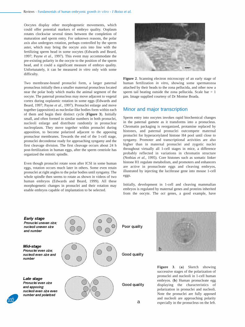
apparently unable, or too late, to bind to the oolemma. Thefertilizing spermatozoon can bind instantly to receptors inoolemma, via its posterior acrosomal membrane, even as itstail still protrudes outside the zona (Figure 2). The entirespermatozoon is ultimately drawn into ooplasm. The oocyte isnow activated, and discharges the contents of cortical granulesinto perivitelline space. A ‘tanning’ effect of these dischargeson inner layers of the zona pellucida apparently stops otherspermatozoa from further penetration of the zona, even ifalready half-way through it. Nevertheless, two or morespermatozoa do occasionally penetrate into human oocytes.
Very few sperm mitochondria enter fertilized eggs(approximately 75), and may be overlooked among the100,000 or more in the oocyte. They are unlikely to have along-term role, since even a small paternal leakage is notcompatible with detailed characteristics of mitochondrialinheritance. Sperm mitochondria are probably degraded byubiquitin mediated systems or segregated to trophectodermand its derivatives (Ludwiget al., 2001).
Oocyte responses to sperm binding
Sperm attachment activates the oocyte, stimulating thecompletion of meiosis II through anaphase and telophase, andextrusion of the first polar body. This process is facilitated asoocytes produce calpain, which inactivates mos protein and
MPF and so removes the metaphase II block. Somewhatsurprisingly, human oocytes are also activated duringintracytoplasmic sperm injection (ICSI). This contrasts withoocytes of several animals which require an electrical or someother stimulus for activation. Morphologically abnormalspermatozoa can be used successfully in ICSI, withoutapparent harm to resulting embryos (Ludwiget al., 2001). Mosprotein can be inactivated experimentally in maturing mouseoocytes by targeting it using RNA interference or by knocking-out the gene in transgenic mice. Removal of this metaphase IIblock permits maturing oocytes to pass through metaphase II,anaphase and telophase to spontaneously expel their secondpolar body. They form rudimentary parthenogenetic embryos,without any form of sperm involvement (Tavernakiset al.,2000).
Rotations of the meiotic spindle during anaphase and telophaseexpel half of the meiotic chromosomes into the second polarbody, leaving the remainder in ooplasm. Rotation is easilyimpaired by colchicine or similar agents and in certain clinicalconditions such as recurrent hydatidiform mole. This conditiondisturbs normal spindle rotation, so it moves entire intoooplasm or polar body (Edwards and Brody, 1995). Either nochromosomes or a diploid number remain in oocytes, sofertilization then results in androgenetic haploid or triploidembryos.
Figure 1. Successive stages of oocyte formation in the fetal human ovary. (a) Aprimary meiotic oocyte in a sexual cord at 14 weeks gestation. Its large sphericalnucleus (N) contains chromosomes in the prophase of meiosis and is surroundedby many mitochondria (M). Original magnification SEM ×7500. (b) Primarymeiotic oocyte in a 14-week ovary. Mitochondria (M) are assembled around thenucleus (N) and are associated with microtubules (arrow). Sphericalmitochondria contain sparse cristae. N indicates the outer surface of the nuclearenvelope. Original magnification SEM ×15,000. (c) A human fetal ovary at 22weeks of gestation showing mitochondria (M) intermingled with microtubules(arrows). Mitochondria are present in a cytoplasmic area closely associated withthe nucleus (N). Original magnification SEM ×12,000. (d) A primary oocyteexamined by transmission electron microscopy and showing mitochondria (M)in close apposition with the nuclear membrane. N, nucleus. Originalmagnification ×18,000. Reproduced by courtesy of PM Motta and S Makabe,Rome and Tokyo Universities.
a
d
b c
M
NM
N
M
N
MN
332
Reviews - Fundamentals of human embryonic growth in vitro - I Boiso et al.
Oocytes display other morphogenetic movements, whichcould offer potential markers of embryo quality. Ooplasmrotates clockwise several times between the completion ofmaturation and sperm entry. For unknown reasons, the polaraxis also undergoes rotation, perhaps controlled by the spermaster, which may bring the oocyte axis into line with thefertilizing sperm head in some oocytes (Edwards and Beard,1997; Payneet al., 1997). This event may accommodate thepre-existing polarity in the oocyte to the position of the spermhead, and it could a significant measure of embryo quality.Unfortunately, it can be measured in vitro only with somedifficulty.
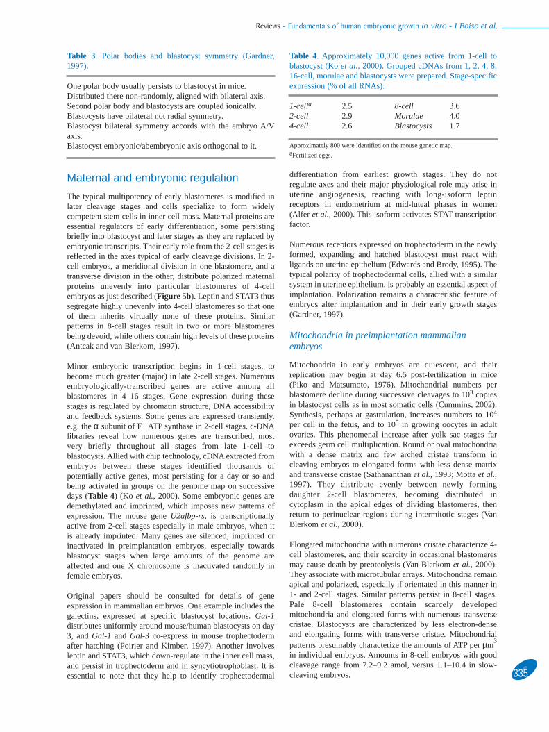
Two membrane-bound pronuclei form, a larger paternalpronucleus initially then a smaller maternal pronucleus locatednear the polar body which marks the animal segment of theoocyte. The paternal pronucleus may move adjacent to vegetalcortex during ooplasmic rotation in some eggs (Edwards andBeard, 1997; Payneet al., 1997). Pronuclei enlarge and movetogether (apposition) as nucleolar-like bodies form within eachof them and begin their distinct cycle (Figure 3). Initiallysmall, and often formed in similar numbers in both pronuclei,nucleoli enlarge and distribute randomly in pronuclearnucleoplasm. They move together within pronuclei duringapposition, to become polarized adjacent to the apposingpronuclear membranes. Towards the end of the 1-cell stage,pronuclei decondense ready for approaching syngamy and thefirst cleavage division. The first cleavage occurs about 24 hpost-fertilization in human eggs, after the sperm centriole hasorganized the mitotic spindle.
Even though pronuclei rotate soon after ICSI in some humaneggs, rotation occurs much later in others. Some even retainpronuclei at right angles to the polar bodies until syngamy. Thewhole spindle then seems to rotate as shown in videos of twohuman embryos (Edwards and Beard, 1999). All thesemorphogenetic changes in pronuclei and their rotation mayenable embryos capable of implantation to be selected.
Minor and major transcription
Sperm entry into oocytes invokes rapid biochemical changesin the paternal gamete as it transforms into a pronucleus.Chromatin packaging is reorganized, protamine replaced byhistones, and paternal pronuclei outcompete maternalpronuclei for hyperacetylated histone H4 pool until close tosyngamy. Promoter and transcriptional activities are alsohigher than in maternal pronuclei and zygotic nucleithroughout virtually all 1-cell stages in mice, a differenceprobably reflected in variations in chromatin structure(Nothiaset al., 1995). Core histones such as somatic linkerhistone H1 regulate metabolism, and promoters and enhancersare active in pronucleate eggs and cleaving embryos,illustrated by injecting the luciferase gene into mouse 1-celleggs.
Initially, development in 1-cell and cleaving mammalianembryos is regulated by maternal genes and proteins inheritedfrom the oocyte. The oct genes, a good example, have
Figure 2. Scanning electron microscopy of an early stage ofhuman fertilization in vitro, showing some spermatozoaattached by their heads to the zona pellucida, and other now asperm tail beating outside the zona pellucida. Scale bar = 1µm. Image supplied courtesy of Dr Montse Boada.
Figure 3. (a) Sketch showingsuccessive stages of the polarization ofpronuclei and nucleoli in 1-cell humanembryos. (b) Human pronucleate eggdisplaying the characteristics ofpolarization in pronuclei and nucleoli.Note the pronuclei are fully apposedand nucleoli are approaching polarityespecially in the pronucleus on the left.
b
a

333
Reviews - Fundamentals of human embryonic growth in vitro - I Boiso et al.
numerous functions in cleaving embryos and blastocysts,interacting with transcription factors and regulatory elements.Maternal effects become less evident as developmentproceeds. Many such situations are discussed below, onespecific example being maternal expression of SCF (stem cellfactor) and tumour necrosis factor (TNF) α until 4–6 cellstages in mice (Sharkeyet al., 1995). Some zygotic genes areexpressed transiently in late 1-cell and 2-cell stages, known asthe period of ‘minor’ embryonic transcription. Examplesinclude the translation initiation factor e1F-4C, transcriptionrequiring complex (TRC), some sex-determining genes carriedon the Y chromosome and the major class 1 H-2D gene(Edwards and Beard, 1997). Major zygotic transcriptionbegins later, in the 2–4 cell stage.
Shape, size and topography of mitochondria change aftersperm entry. Some concentrate in ooplasm near the spermaster, then in peri-pronuclear regions as they are depleted fromcortex. This may cause the ‘halo’ effect, valuable in scoringhuman embryos for quality. Those around pronuclei or inooplasm may associate with ooplasmic architecture, especiallymicrotubules (Mottaet al., 1997), and display a clear polarizeddistribution on outfacing surfaces of apposed pronuclei. Inlater pronuclear stages, mitochondria surround pronuclei in aconcentrated elliptical array, perhaps producing ATP inassociation with microtubules (Van Blerkomet al., 2000). ATPis generated by oxidative phosphorylation in cleavingembryos, shifting to glycolysis at compaction. In human eggswith pronuclei and a halo, together with polarizing nucleoli,numerous mitochondria concentrate in perinuclear ooplasm,and few are present in regions of cytoplasmic flare. Areasdevoid of mitochondria in ooplasm found in some oocytes canbe transmitted to blastomeres (Van Blerkomet al., 2000).
Cleavage stages, morulae andblastocysts
Patterns of cell division
Early blastomeres are relatively undifferentiated, althoughsizes of 2-cell blastomeres differ slightly. Four-cell embryosare products of a meridional division in one 2-cell blastomere,and a transverse division in the other (Figure 4). Threecleavage divisions by 2.5 days post-fertilization produce 8-cellhuman embryos in their initial stages of differentiation. Manyblastomeres have a normal morphology. Others displayfragments, some adjacent to the site of cytokinesis, andpossibly caused by membrane ruffling. Large fragmentscharacterize some blastomeres, perhaps indicative of celldeath, discussed in detail below. Apoptosis in oocytes, polarbodies and blastomeres might impair growth and causeblastomere fragmentation and programmed cell death asmeasured by terminal deoxynucleotidyl transferase-mediateddUDP nick-end labelling (TUNEL)-positive staining (Takaseet al., 1995). Cell death is widespread in preimplantationhuman embryos, arresting embryos with various degrees ofapoptosis, including cytoplasmic, nuclear and DNAfragmentation. Scattered cells in morulae and blastocystsdisplay apoptosis, which may eliminate abnormal cells (Hardy,1997).
Cellular polarity, as opposed to embryonic polarity, becomesimportant from 8-cell stages. Gap junctions and zonularadherents-type junctions form in membranes of 8-cell or olderembryos. Established and maintained by microfilaments andmicrotubules in outer cells of 16-cell mouse embryos, itinvolves a specific outer location of carbohydrates, alkalinephosphate and 5-nucleotidase, actin and myosin (Ziomek andJohnson, 1980).
Figure 4. Sketch showing possible animal(darker shading) and vegetal (light) poles,forming an axis across the unfertilized egg.Note the meridional first cleavage plane bisectsthe oocyte from the metaphase II spindle (blackarrow), so its 2-cell daughter blastomeres havea polarity resembling the oocyte. In 2-cellembryos, one blastomere cleaves meridionallyagain (black arrow), producing two similardaughters in the 4-cell stage. The other 2-cellblastomere cleaves transversely (speckledarrow), so one daughter inherits mostly animaland the other mostly vegetal cytoplasm. Basedon Edwards (2001).

334
Reviews - Fundamentals of human embryonic growth in vitro - I Boiso et al.
Morphologically distinct populations of inner cells (inner cellmass precursors) and outer cells (trophoblast precursors)segregate at the 16 cell stage, as compaction occurs, morulaeare formed and these cell lineages now begin their own geneactivity. Morulae and blastocysts have typical structures, theformer arising after compaction and the latter within a day orso. The inner cell mass is located eccentrically, a hint ofpolarity, its embryonic/abembryonic axis being typical ofearlier polarities in the embryo as revealed from the permanentattachment of a polar body in its original position in cleavingembryos and blastocysts (Gardner, 1997). Inner cell masscontains numerous stem cells already allocated to varioustissues. Outer cells of morulae and blastocysts producetrophoblast, the first tissue to differentiate ready for embryoattachment to uterine epithelium at implantation.
Tightly packed gap junctions and microvilli characterize theouter surface of trophectoderm, intermittent punctate regionsforming between adjacent cells in human blastocyts (Hardy,1997). Trophectoderm divides into polar (overlying inner cellsmass) and mural regions. Its growth is tightly regulated. Cellsin central polar trophectoderm displace murally in expandingmouse blastocysts whereas periperhal cells move either way(Gardner, 1996). A subset of trophectoderm differentiates intogiant cells expressing cyclin B1, which is inhibited in non-giant cell precursors (Palazonet al., 1998).
Many early twentieth century investigators believed in axespassing transversely across the mammalian embryo, just as inlower animal forms. These have now been identified,especially that extending from the metaphase II spindle (theanimal pole) to the opposite segment of oolemma (vegetalpole). Direct evidence arose in studies fixing polar bodiesfixed in one position, marking the point of sperm entry, and thedistribution of ooplasmic proteins in oocytes, fertilized andcleaving embryos and blastocysts (Table 3) (Edwards and
Beard, 1997; Gardner, 1997). Polarity is apparent long beforeovulation, even in oocytes with 1–2 layers of follicle cells(Antczak and van Blerkom, 1997). It is also shown by themovement of germinal vesicles to cortex in growing oocytes asmaturation approaches. A leaflet of a unique myosin A attachesthe second metaphase spindle to oolemma at the point of thisaxis, its only known function in the entire lifespan (Hewitsonet al., 1999).
Oocyte polarity apparently rotates after sperm entry, toaccommodate it with the position of the fertilizingspermatozoon (Edwards and Beard, 1997; Payneet al., 1997).Polarities then dominate chromatin structure in pronuclei,movements of nucleoli, pronuclear rotation and the axes ofcleavage (Figure 4). Pronuclei in some eggs rotate to polaritysoon after fertilization, yet in others they persist at right anglesto polarity until syngamy. Several genes have been identified atspecific positions at the poles, the equator or radially distributedin fertilized eggs (Figure 5a). Planes of successive cell divisionsrelate to this polar axis, distributing maternal proteins such asleptin and STAT3 to specific loci in preimplantation embryos(Antczak and van Blerkom, 1997) (Figure 5b). A single 4-cellblastomere with high levels of these proteins could betrophectoderm precursor, where these proteins concentrate inblastocysts. Another blastomere, virtually devoid of theseproteins, could be germline precursor (Edwards and Beard,1999). Other proteins display similar distributions as leptin andSTAT3 in oocytes, described above, including bax, Bcl-x, TGF-2, VEGF, c-kit and c-erb (Antczak and Van Blerkom, 1999). Incontrast, leptin receptor locates in ooplasm at fertilization, andin pronuclei (Antczak and van Blerkom, 1997) while the α1 andα3 subunits of Na/K-ATPase, and Dazla protein locate radiallyin cortex of mouse oocytes (Edwards, 2001). The nuclearmultipotency factor oct-4 is not polarized. Embryos with highimplantation potential may display characteristics typical of anearly polarization after fertilization.
Figure 5. (a) Diagram showing the distribution of various genes in animal and vegetal poles of fertilized mouse and human eggs,one gene with a variable equatorial distribution and several expressed radially. (b) Model indicating the cortical location of leptinand STAT3 in unfertilized oocytes, fertilized eggs, and cleaving embryos. Notice how cleavage planes shown in Figure 4 distributethese maternal proteins unequally by the 4-cell stage. Shading represents animal–vegetal gradient. Modified from Edwards (2001).
a b
335
Reviews - Fundamentals of human embryonic growth in vitro - I Boiso et al.
Maternal and embryonic regulation
The typical multipotency of early blastomeres is modified inlater cleavage stages and cells specialize to form widelycompetent stem cells in inner cell mass. Maternal proteins areessential regulators of early differentiation, some persistingbriefly into blastocyst and later stages as they are replaced byembryonic transcripts. Their early role from the 2-cell stages isreflected in the axes typical of early cleavage divisions. In 2-cell embryos, a meridional division in one blastomere, and atransverse division in the other, distribute polarized maternalproteins unevenly into particular blastomeres of 4-cellembryos as just described (Figure 5b). Leptin and STAT3 thussegregate highly unevenly into 4-cell blastomeres so that oneof them inherits virtually none of these proteins. Similarpatterns in 8-cell stages result in two or more blastomeresbeing devoid, while others contain high levels of these proteins(Antcak and van Blerkom, 1997).
Minor embryonic transcription begins in 1-cell stages, tobecome much greater (major) in late 2-cell stages. Numerousembryologically-transcribed genes are active among allblastomeres in 4–16 stages. Gene expression during thesestages is regulated by chromatin structure, DNA accessibilityand feedback systems. Some genes are expressed transiently,e.g. the α subunit of F1 ATP synthase in 2-cell stages. c-DNAlibraries reveal how numerous genes are transcribed, mostvery briefly throughout all stages from late 1-cell toblastocysts. Allied with chip technology, cDNA extracted fromembryos between these stages identified thousands ofpotentially active genes, most persisting for a day or so andbeing activated in groups on the genome map on successivedays (Table 4) (Ko et al., 2000). Some embryonic genes aredemethylated and imprinted, which imposes new patterns ofexpression. The mouse gene U2afbp-rs, is transcriptionallyactive from 2-cell stages especially in male embryos, when itis already imprinted. Many genes are silenced, imprinted orinactivated in preimplantation embryos, especially towardsblastocyst stages when large amounts of the genome areaffected and one X chromosome is inactivated randomly infemale embryos.
Original papers should be consulted for details of geneexpression in mammalian embryos. One example includes thegalectins, expressed at specific blastocyst locations. Gal-1distributes uniformly around mouse/human blastocysts on day3, and Gal-1 and Gal-3 co-express in mouse trophectodermafter hatching (Poirier and Kimber, 1997). Another involvesleptin and STAT3, which down-regulate in the inner cell mass,and persist in trophectoderm and in syncytiotrophoblast. It isessential to note that they help to identify trophectodermal
differentiation from earliest growth stages. They do notregulate axes and their major physiological role may arise inuterine angiogenesis, reacting with long-isoform leptinreceptors in endometrium at mid-luteal phases in women(Alfer et al., 2000). This isoform activates STAT transcriptionfactor.
Numerous receptors expressed on trophectoderm in the newlyformed, expanding and hatched blastocyst must react withligands on uterine epithelium (Edwards and Brody, 1995). Thetypical polarity of trophectodermal cells, allied with a similarsystem in uterine epithelium, is probably an essential aspect ofimplantation. Polarization remains a characteristic feature ofembryos after implantation and in their early growth stages(Gardner, 1997).
Mitochondria in preimplantation mammalianembryos
Mitochondria in early embryos are quiescent, and theirreplication may begin at day 6.5 post-fertilization in mice(Piko and Matsumoto, 1976). Mitochondrial numbers perblastomere decline during successive cleavages to 103 copiesin blastocyst cells as in most somatic cells (Cummins, 2002).Synthesis, perhaps at gastrulation, increases numbers to 104
per cell in the fetus, and to 105 in growing oocytes in adultovaries. This phenomenal increase after yolk sac stages farexceeds germ cell multiplication. Round or oval mitochondriawith a dense matrix and few arched cristae transform incleaving embryos to elongated forms with less dense matrixand transverse cristae (Sathananthanet al.,1993; Mottaet al.,1997). They distribute evenly between newly formingdaughter 2-cell blastomeres, becoming distributed incytoplasm in the apical edges of dividing blastomeres, thenreturn to perinuclear regions during intermitotic stages (VanBlerkomet al., 2000).
Elongated mitochondria with numerous cristae characterize 4-cell blastomeres, and their scarcity in occasional blastomeresmay cause death by preoteolysis (Van Blerkomet al., 2000).They associate with microtubular arrays. Mitochondria remainapical and polarized, especially if orientated in this manner in1- and 2-cell stages. Similar patterns persist in 8-cell stages.Pale 8-cell blastomeres contain scarcely developedmitochondria and elongated forms with numerous transversecristae. Blastocysts are characterized by less electron-denseand elongating forms with transverse cristae. Mitochondrialpatterns presumably characterize the amounts of ATP per µm
3
in individual embryos. Amounts in 8-cell embryos with goodcleavage range from 7.2–9.2 amol, versus 1.1–10.4 in slow-cleaving embryos.
Table 3. Polar bodies and blastocyst symmetry (Gardner,1997).
One polar body usually persists to blastocyst in mice.Distributed there non-randomly, aligned with bilateral axis.Second polar body and blastocysts are coupled ionically.Blastocysts have bilateral not radial symmetry.Blastocyst bilateral symmetry accords with the embryo A/Vaxis.Blastocyst embryonic/abembryonic axis orthogonal to it.
Table 4. Approximately 10,000 genes active from 1-cell toblastocyst (Koet al., 2000). Grouped cDNAs from 1, 2, 4, 8,16-cell, morulae and blastocysts were prepared. Stage-specificexpression (% of all RNAs).
1-cella 2.5 8-cell 3.62-cell 2.9 Morulae 4.04-cell 2.6 Blastocysts 1.7
Approximately 800 were identified on the mouse genetic map.aFertilized eggs.

336
Reviews - Fundamentals of human embryonic growth in vitro - I Boiso et al.
Endocrine and paracrine systems inpreimplantation embryos
An astonishing group of endocrine and paracrine factors areactive in early mammalian embryos, sources of autocrine andparacrine systems and preparing for implantation. Theircomplex interactions are essential in establishing embryoquality. Some systems resemble those in thehypothalamic/pituitary axis, e.g. the synthesis of transcripts ofgonadotrophin releasing hormone (GnRH) and GnRH receptorin 8-cell, morulae and expanded blastocysts (Casalfiet al.,1999), and for human chorionic gonadotrophin (HCG) in 2-cell stage in human embryos and then in trophectoderm andtrophoblast (Juriscovaet al., 1999), while the hormone itself isfirst released from day 7–8 human embryos (Fishelet al.,1984).
Embryonic cytokines include leukaemia inhibitory factor(LIF), integrins, epidermal growth factor (EGF), transforminggrowth factor (TGF), platelet-derived growth factor (PDGF)and insulin-like growth factor (IGF). Many display an initialmaternal inheritance. LIF is produced in human trophectodermand in endometrium during the mid- and late secretory phases,and its receptor LIF-R in inner cell mass. LIF-Rβ and gp130are characteristically heterodimerized between maternal andembryonic proteins from 8-cell stages to provide high affinitycell signalling perhaps involved in maintaining pluripotency ininner cell mass and ES cells (Charnock-Joneset al., 1994).Gene knockout showed how the maternal expression of LIFwas essential for human implantation (Stewartet al., 1992).
Several integrins are active during the implantation phase,some inherited maternally (Campbellet al., 1995). Humanoocytes express two α subunits, α3 and αv, and three βsubunits, β1, β3 andβ4, with β1 located in cortex displaying amarked polarity and still being weakly expressed in mouseblastocysts. Integrins α5β1, α6β1, α5β3 are producedcontinuously, while αvβ3, and α1β1 are regulateddevelopmentally (Sutherlandet al., 1993).
c-kit and its ligand, Steel factor (Sl, or stem cell factor, SCF)co-express with EGF and TGFα and EGF receptor (EGFR),their common receptor in cleaving human embryos. A smallvariant of SCF, of 821 bp lacking exon 6, displays maternaland later embryonic expression in 2-cell human embryos andmorulae (Sharkeyet al., 1995). Smaller and larger variantforms with additional exons characterize cleaving embryos.The relative significance of these isoforms is unknown.
EGF, TGF and their common receptor EGFR co-express inhuman preimplantation embryos (Chiaet al., 1995).Homologous with Drosophila Notch, EGF is involved inintracellular signalling in follicles, expressed duringmorula/blastocyst transition, then restricted to trophectoderm.It may have growth factor activity at 8-cell stages andpromotes HCG secretion by syncytiotrophoblast (Smotrichetal., 1996). The extracellular domain of EGFR, expressedweakly in 2–4 cell mouse but not human embryos, localizes inapical cell surface membranes in blastomeres. Maternal TGFαpersists to late cleavage stages, promotes blastocyst expansionin mice if added to culture medium and binds to EGFR. TGFβ,a homologue of activin A and a mesodermal inducer, shifts inexpression from maternal to embryonic in blastocysts and
early stem cells (Rappoleeet al., 1988). Its isoforms, TGFβ1β2 β3, are transcribed in 4-cell mouse embryos, inner cellmass and trophectoderm. A and B subunits of PDGF, initiallymaternal, form dimeric AA, AB and BB disulphide-bondedpolypeptide chains which bind to receptors. Its receptorsPDGFRα and PDGFRβ are expressed from 4–8 cell stages.
Maternal forms of insulin, IGF-II and their receptors persistuntil 2-cell stages and reappear in inner cell mass andtrophectoderm (Liuet al., 1997). Released into and sensitive toionic changes in culture medium, it may provide a marker ofembryo quality. Its synthesis in 8-cell human embryos isenhanced by platelet activating factor (PAF) or by culturingembryos in groups. IGF-1R is expressed moderately in human4-cell embryos. IGF-IIR is synthesized throughoutpreimplantation phases and may protect against apoptosis.
Among other cytokines, TNFα is released from 2- to 4-cellmouse and human embryos, and its receptor, TNFRp60, mightform in 6-cell and older human embryos. PAF might assistembryonic growth, but PAF-R is not expressed (Sharkeyet al.,1995). PAF may sustain IGF-II synthesis and induce calciumoscillations at 2-cell stages. Cytokines secreted in later stagesinclude interleukins IL-6 and IL-6R. fgf4, a growth factor ininner cell mass, is a target gene for Sox 2and oct4.
The implantation process must be understood to appreciate thenature of embryo-uterine interactions. Brief mention can onlybe made here on the endometrium. Essential aspects ofattachment and invasion of the embryo involve detailedinteractions of markers of uterine activity including LIF,integrins, and many of the cytokines discussed above (Lessey,2001). Some integrins date the endometrium through itsvarious stages, but may be insufficient to provide exactmarkers. Mucins, uteroglobins and pinopods may also providegood indications of implantation potential, although muchmore knowledge is needed.
Timing and integration in developmentalprocesses
Close timing and integration characterize morphogeneticmovements in mammalian oocytes and early embryos asdescribed earlier. Integrative systems are essential fromearliest stages of oocyte formation, to regulate oocytedifferentiation, polarities, microtubule activity and otheraspects of cellular architecture, movement and mitochondrialorganization. These essential regulators coordinate manyaspects of growth, cleavage divisions and morphogeneticmovements such as blastulation, and could provide markers ofnormal growth.
Several timing systems in embryos cannot be switched offonce started. Individual cells contain several clocks, somedescribed above, affecting various cellular processes. Clocksactive in fertilized eggs and embryos continue relentlesslythroughout cleavage, blastulation and possibly implantation.Clear examples are found in mammals, e.g. exact timings ofsuccessive maturational stages in oocytes resulting in the exact37 h interval between an HCG injection and ovulation in allwomen treated for IVF (Steptoe and Edwards, 1970).Cytokines also regulate timing, e.g. granulocyte–macrophagecolony-stimulating factor (GMCSF) promotes growth of

337
Reviews - Fundamentals of human embryonic growth in vitro - I Boiso et al.
human embryos to blastocysts, perhaps by enhancing celldivision (Sjoblomet al., 1999).
While highly regular, cleavage times can be influenced byexternal agents, shown by the rapid cleavage of male mouse,cow and human embryos, although this concept has beenchallenged. The Ped (preimplantation embryo development)gene, located in the MHC complex in mice, influences timingof preimplantation growth. Transcribed embryonically from 2-cell stages, the protein locates to inner cell mass andtrophectoderm (Warneret al., 1998). Its fast-cleaving (Qa-2+)allele stimulates embryos to produce more inner cell mass andtrophectoderm cells than those with the slow-cleaving allele(Qa-2–). Ped may be homologous with, or closely linked tohuman HLA-G (Warneret al., 1998), although this gene wasnot identified active in cDNA libraries prepared fromindividual human embryos (Verlinskyet al., 1998).
Circadian rhythms in reproductive cycles are well known toregulate the master clock in the hypothalamic suprachiasmaticnucleus. Similar local systems in individual cells organizetheir own clock. It depends on light stimuli from retinareaching the suprachiasmatic nucleus. Peripheral clocks runwith a few hours delay. Feedback systems in Drosophilaregulate themPer genes, i.e. mPer1, 2 and 3, and twocryptochrome genes mCry1 and mCry2. Similar geneticsystems in mammals include suprachiasmatic signalsinvolving the Perand Cry genes, and immortalized embryonicfibroblasts utilize the same system if they do not carry Cry1and Cry2 mutants. Master clocks tick constantly; peripheralclocks damp down after some days (Yagitaet al., 2001).Diurnal zygotic clocks are illustrated by the biphasictranscription of RNA polymerase I and III, so expression ofthis enzyme can be time related and stage-dependent (Nothiaset al., 1996). Perhaps intra- or intercellular conflicts in timingbetween individual blastomeres influences embryonic growthwhen blastomeres are highly independent.
Timers integrate with other developmental controls, operativethrough multifunctional regulatory genes throughoutpreimplantation stages. Present in multipotential cellsthroughout cleavage stages, the Oct 4 gene, a transcriptionalregulator, down-regulates in trophectoderm, continuesexpression in inner cell mass, but is finally restricted toprimordial germ cells (Ovitt and Scholer, 1998). Its expressionrelates to an undifferentiated cellular phenotype, and onefunction is to silence HCGβ gene in inner cell mass (Liu andRoberts, 1996).
Classic examples of integration include the Par genes, wellknown in establishing and integrating polarities and germlinein C. elegansoocytes. Several interact when axes and cell fateare determined. Six or more distinct oocyte proteins distributeunequally among successive blastomeres to help impose theirdistinct embryonic fates (Guo and Kemphues, 1996). par-1and par-4 are primary organizers of protein and transcriptlocalization in cytoplasm; par-2, 3, 5 influence spindleorientation. Par-1 characterizes their actions, encoding aserine-threonine protein kinase to interact with a protein kinase(PKC) and Par-2 at restricted sites over particular time periods.Similar systems exist in Drosophila and mammals, but areknown only in outline (Edwards, 2001).
Telomeres locate on terminal regions of homologueschromosomes, and may measure longevity. They are expressedin immortal cell lines and many tumours. Their length ismaintained by telomerase, and shortening is characteristic ofageing cells. Composed of repeated DNA sequences,telomerase replenishes them by adding to DNA synthesizedduring the cell cycle. This enzyme reaches high levels in fetal,newborn and adult gonads, but not in oocytes or spermatozoa(Wright et al., 1996). Resynthesized in early embryos, highlevels are attained in blastocysts but then decline indifferentiating tissues (Eisenhaueret al., 1997). Telomeresmay stabilize chromosome ends to prevent potentialrecombinations. Shortened telomeres in Dolly the sheepindicated premature ageing as compared with normal sheep ofthe same age.
Part 2. The human embryo in theclinic
Introduction
Human embryos have typically low implantation rates whethergrown in vivo or in vitro. Overall clinical pregnancy rates of<30% with two or more replaced embryos decline further withincreasing maternal age, certain aetiologies of infertility andwith various clinical parameters. Establishing singletonpregnancies has proved to be an elusive target, compromisedby the need to replace several embryos to attain sufficientlyhigh pregnancy rates. Improved implantation rates will only beachieved by selecting for transfer those embryos with highimplantation potential. Selection will depend on currentknowledge on fundamentals of embryonic growth, yet ifsuccessful it should enable single transfers of selected embryosbetween 1–8 cells or blastocysts. Either approach couldtransform the infertility treatment and the diagnosis of geneticdiseases in preimplantation stages. Transferring cleavingembryos avoids prolonged culture periods and the death invitro of many cleaving embryos capable of implantation unlessleft too long in culture. Transferring blastocysts demands allthe skills of the IVF clinic but those that survive have passed astringent test of quality. It is essential that users of eitherapproach relate numbers of pregnancies to numbers oftransferred and started embryos, the only verifiable parametersof success.
General principles of embryo culture andtransfer
Laboratory conditions
The IVF laboratory must provide a non-toxic, pathogen freeand stable environment, with suitable equipment andtechniques. Suboptimal culture conditions can be expressed asembryonic death in vitro, or loss of viability after implantation.Environmental pollutants must be monitored to avoid thecontamination of media.
The importance of good culture methods, and highly suitablemedia cannot be overstressed. Acidity is maintained around7.4, usually with bicarbonate buffer under 5% CO2 in air, aphysiologically based system of low toxicity. CO2 in liquidphases must be equilibrated with gas phases, often achieved byplacing media in CO2 incubators before use. Some laboratories

338
Reviews - Fundamentals of human embryonic growth in vitro - I Boiso et al.
use 5% O2, 5% CO2 and 90% N2. HEPES buffer maintains pHstability in the bicarbonate buffer system when workingoutside the CO2 incubator, e.g. during oocyte recovery.
Osmotic pressure of ~285 mOsm/kg must be maintainedstringently, and needs close care since it can shift with time,temperature and atmospheric humidity. Evaporation is aserious problem, demanding constant air humidity inincubators. Open culture systems include small plastic tubescontaining 1 ml of medium, now largely replaced by wells inplastic dishes. One closed system utilizes microdroplets ofmedium under paraffin or mineral oil. It protects oocytes andembryos from contamination, and from swings in pH,temperature and gas phases. This overlayer also counteractsevaporation, provides a physical barrier to infections andseparate droplets permit rapid and easy examination of singleembryos in vitro. Paraffin oil must be constantly equilibratedwith CO2, with steps to maintain it when working outside theincubator.
Embryo transfer to the mother is the final stage of culture.Several transfer catheters each offer different benefits. Goodtransfer technique is crucial to IVF success, since otherwiseimplantation is jeopardized, to literally destroy all that went onbefore. Nurses have replaced doctors for transfers in someclinics, with similar success. Catheter technology, a significantelement in embryo replacement, was assessed by Edwards andBrody (1995).
Media
Media used for oocyte retrieval, oocyte recovery and embryoculture must meet minimal nutritional requirements, providestable pH through effecting buffering, and have an appropriateosmolarity. Quality control must ensure they are endotoxinfree, especially with media prepared in house. Pre-testedcommercial media are suitable, the majority having similarquality standards as media made within the clinic.
Many media are based on well-known physiological saltsolutions, such as Krebs, Waymouth’s, and Earle’s, fortifiedwith human albumin, serum or its synthetic substitutes. Animalproteins must be excluded. More complex media includeHam’s F10 and T6, widely used in IVF although less sorecently. Ménézo’s B2 medium contains amino acids,vitamins, nucleic acid precursors and saline, but includesbovine serum albumin. HTF is a balanced salt solution basedon human tubal fluid, free of amino acids, with a higher K+
content than other media and an ionic environment similar tooviduct (Quinnet al., 1985).
Sequential media offer changing requirements for embryos indifferent developmental stages. They have been used sinceIVF was introduced 30 years ago when a physiological saline,e.g. Earle’s or Krebs’ fortified by 8% v/v of serum or humanalbumin, sustained fertilization, while complex media, e.g.Ham’s F10 fortified with higher serum or albumin levels andsometimes with glutamine, sustained after pronucleate stages.A recent, widely used system began as three distinct mediadesigned for successive growth stages. The first, with littleglucose, sustained for fertilization. The second containedlactate, and nonessential amino acids to culture cleavingembryos. Amino acids promote the development of human
blastocysts in vitro (Devreker et al., 2001). Pyruvate wasincluded and glucose was omitted, being considered toxic forembryos aged days 1 to 3. Care was needed with amino acids,which might spontaneously break down to ammonium at 37ºC,and harm embryo growth. The third medium for growth toblastocysts contained glucose, essential and non-essentialamino acids, vitamins and nucleic acids precursors amongother components (Gardner and Lane, 1997). These were theforerunners of G1 and G2 media, widely used today, and ofother less-used sequential media. Merits of sequential mediaand harmful effects of glucose have been questioned in favourof one medium, KSOM, over the 5-day period (Biggers, 2000).
Longer-term cultures using sequential media, e.g. G1 and G2,produce high-quality blastocysts, so few are transferred and somultiple pregnancies are rare (Gardner and Schoolcraft, 1998;Gardner et al., 2000). Only one-half of embryos reachblastocysts, others dying between days 3 and 5, which is not verydifferent to media available in the 1980s (Fishelet al., 1985).
Embryo selection and transfer on days 2–3 is also widelypractised. Fortunately, in humans if not in animals, the uterineenvironment tolerates early embryos and transfer procedures.Three or even more embryos have been transferred, riskinghigh multiple pregnancies. Most clinics abhor low pregnancyrates, often associated with few replaced embryos, so modelsof embryo quality became paramount. These were devised toselect the best single embryos for transfer and so avoidmultiple births (Table 5) (Steeret al., 1992; Staessenet al.,1993). Today, embryos are selected between 1–8 cell stages forkey characteristics, such as early polarization, timing ofcleavage divisions, the degree of blastomere fragmentationand good morphology, hopefully several times daily. Earlierselection equates with early transfer, shorter culture periodsand the use of cryopreservation to store remaining high-qualityembryos.
Co-culture
Co-culture has been widely popular in some clinics. Feedercell monolayers may remove heavy metal divalent cations andmetabolic inhibitors, reduce high oxygen levels, and providesmall embryotrophic metabolites and growth factors.However, exact mechanisms are not understood. Initiallyapplied 1965, then in the 1990s, cellular feeder layerscombined with sequential media were used for longer-termcultures to blastocysts. Today, IVF clinics utilize Vero cells(kidney epithelial cells from African monkeys), oviductalepithelial cells of different species (Ménézoet al., 1986;
Table 5. Cumulative embryo score predicting optimal numbersof embryos to replace (Steeret al., 1992).
Initial classification
Grade 1: Total fragmentation or embryos still pronucleate.Grade 2: Serious blastomere fragmentation of 10–50%.Grade 3: Uneven blastomeres with <10% fragmentation.Grade 4: Equal-sized symmetrical blastomeres.
Multiply grade by numbers of blastomeres to produce a qualityscore.

339
Reviews - Fundamentals of human embryonic growth in vitro - I Boiso et al.
Bongsoet al., 1989), bovine fetal uterine fibroblasts (Wiemeret al., 1994), autologous granulosa cells (Plachotet al., 1993)and homologous endometrial cells. Beneficial effects areneither species-specific nor specific to the female reproductivetract, which raised concern about their value (Edwards andBrody, 1995). Cell lines are grown under standard cell culturemethods with specific modifications. Vero cells, commerciallyavailable in frozen cryotubes, are checked for bacteria, virusesand other possible contaminants.
Co-cultures were credited with sustaining more embryo cellsand better inner cell masses (Ménézoet al., 1998). Initially, co-cultured human embryos produced 61% blastocyst versus 3%in controls. Such low rates in controls implied defects inculture methods. Co-cultures with Vero cells led to slightlyhigher pregnancy rates than in controls (23% co-culture versus16% in controls), as clinical trials yielded 57% blastocysts and44% pregnancies among 62 patients given a mean of 1.65blastocysts (Ménézoet al., 1992). Wide variations between co-cultures and controls were reported (Plachotet al., 1993;Wiemer et al., 1994), and some patients failed to reachtransfer. Claimed benefits included reduced blastomerefragmentation and more pregnancies. Other investigatorsdemurred. A prospective randomized study using Vero cellsproduced no significant advantages for growth to blastocysts(Van Blerkom, 1993), nor did benefits accrue in numbers ofpregnancies for first-time IVF patients. Many co-culturestudies were not controlled statistically, or involvedcomparisons with previous transfers in the same patient. Someimplantation rates in controls were desperately low eventhough patients were subfertile.
Co-cultures offer a restricted future, and are now largelyreplaced by sequential media. Some experience with co-cultures has nevertheless been positive for patients withprevious failed transfers or advanced age. Our own work(Boiso and Vega, unpublished) produced two-thirds ofembryos reaching blastocysts. Pregnancy rates were not highat 12.5% pregnancies per transfer, but many of the patientssuffered from a low pregnancy potential.
Cryopreservation
Many human embryos are cryopreserved after those selectedfor high quality have been replaced. Many thousands now liein store. Slow-freezing, slow-thawing systems using glycerol,dimethyl sulphoxide, propanediol and others have been usedover many years (Edwards and Brody, 1995). In some clinics,pregnancy rates with thawed embryos were only just belowthose gained using fresh embryos. Some clinics produced verypoor results, a risk to be considered when assessing the valueof cryopreservation.
Slow methods are now being replaced with faster methodssuch as vitrification or combinations of high concentrations ofseveral cryopreservative agents (Rall and Fahy, 1985).Overall, several investigators produced results equivalent tothose gained with well-established slow forms ofcryopreservation. Improvements in oocyte quality are claimedwith better straw technology, and human births have beenreported using vitrified embryos (Kuleshovaet al., 1999). Thisbetter approach to cryopreservation cannot be long delayed.
Evaluation of oocytes and 1-cell embryos
Oocyte quality is essential in determining embryo viability. Itis measured non-invasively in oocytes first polar bodies, andzonae pellucidae by scoring visible characteristics during IVFand ICSI procedures. Properties of granulosa cells may also behelpful.
The freshly collected oocyte
Follicular fluids and oocytes arriving in the laboratory shouldbe quickly scanned and examined for maturity such as anexpanded cumulus mass, coronal cells surrounding the oocyteand its nuclear status. A visible polar body indicates the oocyteis in metaphase II. Granulosa cells form large viscous massessurrounding oocytes. One classification of oocytes accordingto their maturity is germinal vesicle stage (prophase I),metaphase I, metaphase II, very mature or post-maturemetaphase II, and atretic or degenerative (Veeck, 1999).
Morphology
Cumulus and coronal cells obscure oocytes duringconventional IVF. More information is gained duringmicromanipulative techniques such as ICSI, which requiretheir complete removal. Nuclear maturation and cytoplasmicmorphology can then be assessed and handling facilitated.Morphological defects in oocytes include an enlargedperivitelline space, cytoplasmic granularity and inclusions.Variations are not uncommon in oocyte morphologygranularity, such as areas of necrosis, organelle clustering,vacuoles, clustering of smooth endoplasmic reticulum,anomalies in the first polar body and zona pellucida, and non-spherical shape of the oocytes (Figure 6), and have beenlinked with aneuploidy, abnormal fertilization, poor embryoquality, and low pregnancy and implantation rates (reviewedby Alikani et al.1995). Results are controversial. Aneuploidyin oocytes and embryos may be linked to several dysmorphicforms arising before metaphase I, perhaps up to one-half ofcases (Van Blerkom and Henry, 1992). Embryos originatingfrom dysmorphic oocytes suffered higher rates of pregnancyloss (Alikaniet al., 1995). Similar conclusions were drawn inother studies, but without effects on fertilization rates andembryo quality. Persistent cytoplasmic granulation in oocytesof patients during repeated treatment cycles did not affectfertilization and pregnancy rates, except for large, deepgranular zones which were linked with aneuploidy andabortion (Kahramanet al., 2000).
First polar bodies have received much attention, with similardisagreements about their predictive value. Positive examplesinclude low fertilization rates and embryo quality when it isfragmented and the perivitelline space is enlarged (Xia, 1997).Fragmented or huge polar bodies may also signify disorderedfertilization rates. Many negative reports have been published.
More controlled trials are needed to confirm all these findingson oocytes. Many parameters are subjective. Attention shouldbe given to the aspiration of atretic or endometriotic follicles,which may produce oocytes with many of the variousdysmorphisms.

340
Reviews - Fundamentals of human embryonic growth in vitro - I Boiso et al.
Normal and abnormal pronuclei
Cytoplasm in 1-cell embryos can be scored visibly alterationsfor the presence of ‘haloes’ around cortical regions orsurrounding pronuclei (Figure 7). These may be invokedduring mitochondrial aggregation in perinuclear regions, andcould be associated with good quality embryos.
Two pronuclei found at 16–18 h post-insemination is usuallytaken as evidence of fertilization. They should enlarge, andtheir membranes should be polarized at membranal sites ofpronuclear apposition (Payneet al., 1997). Pronuclei differ intheir form of growth, paternal pronuclei being larger untilclose to syngamy. Pronuclear diameters differing by at least 4microns are associated with more developmental arrest thansmaller differences.
Unusual numbers of pronuclei may form after fertilization, i.e.none, one, three or more. If neither pronuclei nor one largepolar body is observed, the meiotic spindle might have passedoutside the egg as occurs in patients with recurrenthydatidiform mole (Edwards and Brody, 1995). The presenceof two polar bodies only does not necessarily indicatefertilization failure, since pronuclear formation may havefailed after sperm entry. This condition arises afterexperimental treatments on oocytes, e.g. heavy X-irradiation,but is very rare.
Some eggs possess a single pronucleus. It can have a variedorigin. It may be due to parthenogenetic activation, perhaps thecause in 25–30% of cases (Staessenet al., 1993), and morecommon after ICSI (Munné and Cohen, 1998). Uniformhaploid embryos can also arise and proceed normally througha few cleavages. Delayed formation of one pronucleus, or thefusion of two pronuclei, are alternative causes (Nagyet al.,1994). Meiotic errors in chromosomal segregation could havethe same consequence. Sometimes, two pronuclei seem tofuse, to produce one pronucleus. If correctly identified, theseembryos can probably be transferred safely, perhaps afterscoring their growth to blastocysts.
More than two pronuclei imply polyspermic penetration orretention of the second polar in the oocyte. Fertilizationinvolving two spermatozoa (dispermy) produces eggs withthree pronuclei and two polar bodies. This frequent anomaly inhuman eggs can result in the formation of two sperm asters andtripolar spindles at syngamy, leading to complex segregationalanomalies (Figure 8). The worst cases presumably arrest afterirregular cleavage and fragmentation. Others develop aschromosome mosaics, and a few as pure triploids (Munné andCohen, 1998). While eggs with three or more pronuclei areusually not transferred, it is still possible to excise the extramale pronucleus and restore diploidy to the embryo.
Digyny arises when the second polar body is retained in theegg. This results in three pronuclei, two being maternal. Thiscondition (monospermic digyny) affects 4% or fewer fertilizedeggs. Most develop as uniform triploids, indicating thatsyngamy involved three pronuclei with a single bipolarspindle.
Figure 6. Anomalies in human oocytes. (a) Non-spherical shape. (b) Cytoplasmic granularity. (c) Cytoplasmic inclusions. (d)Vacuolated oocyte. (e) Fragmented polar body. (f) Clustering of organelles in the perinuclear region in a fertilized egg with threepronuclei; note that all three pronuclei are apposed (image supplied courtesy of Dr Montse Boada).
Figure 7. Presence of a local halo in ooplasm of a humanoocyte. Notice the pronuclei are conjoined and nucleoli in onepronucleus are apposing.
a b c
d e f

341
Reviews - Fundamentals of human embryonic growth in vitro - I Boiso et al.
IVF and ICSI
Benefits of ICSI and IVF have been widely explored. ICSI isclearly far superior for severe forms of male infertility, butmore IVF embryos develop to blastocysts (Schröderet al.,2001). Perhaps IVF is more ‘natural’, with sperm entrystimulating normal responses of oocyte activation with itsmajor consequences such as induced Ca
2+oscillations, cortical
granule discharge, decondensation of the sperm head andnatural forms of ooplasmic rotation. The delayed dissolutionof the acrosomal vesicle in monkey oocytes after ICSI trapsareas of DNA in early forms of decondensation (Hewitsonetal., 1999). Incomplete sperm injection, oolemmal breakage,apparent incomplete oocyte activation and rapid formation ofpronuclei are included among other consequences of ICSI,although effects on embryonic growth do not seem to besignificant.
Curiosity about ICSI, and the existence of polarities inmammalian eggs, has led to analyses on embryo quality whenspermatozoa are placed at specific points in oocytes (reviewedby Ludwig 2001). Despite many reports, there is no overallagreement about an optimal site. The significance of placingspermatozoa at widely differing sites may be nullified throughthe rotation of ooplasm to accommodate to sperm entry fromany site.
Polarities and selection in 1-cell stages
Polarity characteristics in human oocytes and embryos offersome of the best chances of deciding embryo quality. They aremeasured using various parameters. These include pronuclearrotation at intervals after sperm entry, pronuclear appositionand chromatin coarsening, nucleolar growth and polarizationin pronuclei, and an early first cleavage division.
In this context, Garelloet al.(1999) measured angles betweenthe pronuclear axis and the axis of pronuclei with polar bodies,to calculate the degree of rotation. Wider angles wereassociated with low embryo quality, and a low angular
variation with higher implantation rates. Care is needed withthis form of analysis because first polar bodies can bedisplaced from their original position.
Nuclear chromatin also displays polarity characteristics,coarsening at points of pronuclear apposition. This parameterhas not been used to score embryos. Nucleolar migration andalignment, and pronuclear apposition, are clear markers ofgrowth, and best achieved before human embryos arecryopreserved (Wrightet al., 1990). These data were amongthe earliest on polarities. Structure, size and position ofnucleoli were employed by Tesariket al. (2000) to classifyfertilized human eggs, measuring discordance between the twopronuclei, well scattered nucleoli, and small and large nucleolipolarized adjacent to pronuclear apposition (Figure 3).Chosen embryos were selected again later from their day 3morphology. Implantation rates reached 50% with one embryohaving optimal patterns, this estimate being reviseddownwards later but still above 30% (Tables 6, 7) (Tesariketal., 2000). Greater precision might be gained if eggs are scoredseveral times daily. Detailed trials are needed to confirm theseconclusions.
A scoring system based on pronuclear size and the alignmentand number, size and distribution of nucleoli within thepronuclei produced four embryo categories (Table 6) (Scottetal., 2000). High quality embryos were identified (Table 7),scores on nucleoli being more effective than those onmorphology. Implantation rates of 31% agree closely with30% found by Tesariket al.(2000). They challenge the higherrates obtained with blastocysts, descibed below, which areobtained after many embryos capable of implantation diebetween days 3 and 5.
The cleaving embryo
Embryo selection based on multinucleated andfragmented blastomeres
Most embryos have successive and timely blastomeres ofapproximately even size during early cleavages. Multiplenuclei (MNB) in blastomeres are not infrequent in humanembryos (Figure 9). They are important signals of reducedembryo viability, used widely, and among the best parametersfor scoring embryo quality. Multinucleation and mosaicism inembryos are more frequent with dysmorphic pronuclei(Sadowyet al., 1998).
Two-thirds of multinucleated blastomeres involve defectivecleavage in a mononucleated parent cell, such as flawedchromosome migration, nuclear fragmentation, failure todivide, and/or errors in cell packaging (Pickeringet al., 1995).Multinucleation in early cleavage stages, but not at 8-cell andlater, compromises implantation and diploidy (Hardy, 1997;Alikani et al., 2000; Sandalinaset al., 2001). Disorderedchromosome separation at anaphase and telophase results inmosaic embryos (Kligmanet al., 1996), although progeny ofmultinucleated blastomeres are not always abnormal (Staessenet al., 1998). This characteristic is usually included with otherswhen constructing models of embryo quality.
Blastomere fragmentation is very frequent, and may be amongthe most significant defect in cleaving embryos whether innate
Figure 8. Formation of a tripolar spindle in a pronucleatehuman egg that had earlier contained three pronuclei. Noticethe three sets of chromosomes, stained red, and the threedistinct poles of the spindle apparatus.

342
Reviews - Fundamentals of human embryonic growth in vitro - I Boiso et al.
or due to external factors. It has been studied over many years(Plachot and Mandelbaum, 1990). Classifications areexpressed as the percentage of embryo volume occupied byfragments. Spindle errors may result in lagging or brokenchromosomes, which are expelled into fragments.Sophisticated analyses have largely replaced the earlier habitof simply classifying embryos as free of fragments orotherwise (Alikaniet al., 1999). Loss of polarized proteins intofragments could affect blastomeres unequally and impairdevelopmental potential in embryos, particularly 1–2 cellstages (Antczak and Van Blerkom, 1999). The percentage offragmentation is positively correlated with mosaicism, but notto aneuploidy and fragmentation (Munné and Cohen, 1998).Fragments can be excised surgically, using micromanipulation,to improve embryo quality.
Such sophisticated analyses now classify fragments into fivegroups according to size and distribution (Table 8; Figure 10)(Alikani et al., 1999). This model assesses the degree andpattern of fragmentation on day 3 and reliably predictsimplantation rates. Type IV is serious, even in minorquantities, shown in transfers consisting uniquely of theseembryos (Alikaniet al., 1999). Exceptions occur, e.g. the 5%of embryos which implant despite fragments sized betweenone-tenth and one-half of embryo volume at day 2. Overall,blastocyst formation is impaired with 15% fragmentation(Alikani et al., 2000). Not all patterns are detrimental, exceptfor loss of large cytoplasmic volumes.
Examples of data gained with transfers including fragmentedembryos are given in Table 9. This parameter is often includedwith others in more complex models, as shown below. Scoringand selecting embryos can be done without difficulty, whichadds to its wide use as a method for scoring human embryos inso many clinics.
Embryo selection based on timing and polarities
Knowledge on timing and polarity in human oocytes and eggshas produced models designed to measure the ability ofindividual human embryos to achieve implantation (Edwardsand Beard, 1999). This widely used model for scoring embryoquality is often combined in complex models. Embryos withearly first and second cleavages have implantation rates wellabove 30% per embryo (Edwardset al., 1984) and many formblastocysts (Sakkaset al.,1998). Several groups have includedthis marker as a major parameter helping to identify high-quality embryos (e.g. Scottet al., 2000). Selecting singleembryos with early cleavage and other characteristics mightestablish pregnancy rates surpassing 30%, compared with 20%if only one embryo was available for transfer (Vilskaet al.,1999).
Early cleavage is also measured by counting number ofblastomeres at specific times after insemination or ICSI. Thismethod is also in wide use in numerous models.
Table 6. Numbers, size and polarization of human nucleoli inpronucleate eggs (Scottet al.,2000).
Z-1: equal numbers and size of nucleoli (between 3 and 7 pernucleus) aligned at pronuclear junction.
Z-2: equal numbers of nucleoli not yet aligned at pronuclearjunction.
Z-3: unequal sized and numbers of nucleoli, unequalalignment at pronuclear junction.
Z-4: grossly abnormal embryos, different sized nuclei, nucleinot aligned in central ooplasm, small misplaced nuclei.
Figure 9. (a) Presence ofthree multiple nuclei in oneblastomere of a human 2-cell embryo. (b) Uneven-sized blastomeres in a 4-cellhuman embryo.
a
b
Table 7. Results with selection for polarized nucleoli andmorphology.
A. Data of Tesarik et al. (2000). Embryos were selected for
nucleoli and good morphology.
Polarized Non-polarized
Transfers 98 77No. embryos transferred 189 161Mean no. transferred 1.9 2.1Pregnancies (%) 44 (45) 17 (22)Embryo sacs 57 18Implantation rate 30 11
B. Data of Scottet al. (2000). Results in relation to type ofclassification and day of transfer.
Nucleoli Plus morphology
Day 3 Day 5 Day 3 Day 5
No. transfers 94 117 134 87Clinical pregnancies 57 73 33 58(%)Mean no. transferred 2.8 1.9 3.3 2.2embryosImplantation rate (%) 31 52 19 39
Growth to day 5 blastocysts according to classification: Z-1 = 50%; Z-2 =25%; Z-3 = 15%; Z-4 = 15%.

343
Reviews - Fundamentals of human embryonic growth in vitro - I Boiso et al.
Measuring multiple criteria in embryos
It is now time to combine several parameters of quality in 1-cell and cleaving embryos to select embryos of very highimplantation potential. This process began when modelspredicting the relative importance of embryo and uterus wereproposed 20 years ago.
One model involved an embryo score, based on blastomerenumbers and fragmentation to achieve a cumulative embryoscore, and moderate scores gave modest implantation rates andfew multiple pregnancies (Table 5; Figure 11) (Steeret al.,1992). Weighting coefficients were then applied to alignmentsof pronuclei and nucleoli, presence of an ooplasmic halo, earlyfirst cleavage and good morphology, all being summed toscore individual embryos (Scottet al., 2000). Embryos withhigh scores implanted in far greater numbers than the others,more than 90% of embryos that implanted having high scores.High embryo scores were also associated with steadily risingoestrogens during follicular phases, implying that high embryoquality is a consequence of good follicle physiology.
‘Strict embryo criteria’ were formulated and applied to day 2or day 3 cleavage stage embryos to select single embryos withhigh implantation potential (Gerriset al., 1999; Van Royenetal., 1999). The ‘top’ quality embryo was characterized
examining retrospectively all 23 double transfers resulting inongoing twins. Top-quality embryos possessed nomultinucleated blastomeres, four or five blastomeres on day 2and seven or more on day 3, with <20% anucleate fragments.Initial studies utilized this data to begin a policy of electivesingle embryo transfers (SET) in a prospective randomizedtrial comparing single and double embryo transfers. Singletransfers of selected embryos had implantation rates of 42.3%with ongoing pregnancy rates of 38.5%. Double electedtransfers produced similar implantation rates (48%) andongoing pregnancy rates of 74.1% (Gerriset al., 1999).Extension of this trial involved group cultures of embryos withtwo clear pronuclei on day 1, scoring each one for multiplenuclei. Blastomere numbers and fragments were counted onday 2. A final selection on day 3 chose embryos which had 4–5blastomeres on day 2, and 7 or more on day 3, combined with<20% fragments and total lack of multinucleated blastomereson days 2 and 3. Implantation rates with single selectedembryos reached 50%, with ongoing pregnancies of 33%(Table 9) (Gerris et al., 2001). Multiple pregnancies werevirtually absent in the group of 118 pregnant patients aftersingle embryo transfers.
The blastocyst
Embryo quality and culture in defined medium
Blastocyst transfer depends on advanced technologies andconsiderable skill in laboratories. It may represent the strictestform of embryo selection. The first clinical pregnancy arosefrom a transferred blastocyst, and initial analyses of embryogrowth in vitro showed how 50% of human embryosdeveloped to blastocyst stages in fortified Ham’s F10 (Fishelet al., 1985). Transfers of cryopreserved blastocysts were onlymoderately successful, but in this study the best-qualityembryos had previously been used for transfer (Fehillyet al.1985; Hartshorneet al., 1991). Disappointing results using asingle medium to culture blastocysts led to the introduction ofco-cultures, producing 50% blastocyst formation andimplantation rates of 25% with many multiple pregnancies(Ménézoet al., 1998). Blastocyst transfer was also utilized forpatients with repeated implantation failures (Veigaet al., 1995,1999a).
Sequential media were then applied in detail to blastocystculture. It gave similar rates of success as co-culture (Ménézo
Table 8. Model for classifying blastomere fragmentation(Alikani et al., 1999).
Type I: minimal volume, fragments associated with oneblastomere.
Type II: fragments localized and predominantly in perivitellinespace.
Type III: small scattered fragments, between blastomeres,peripheral or both.
Type IV: large fragments, resembling whole blastomeres,distributed randomly, associated with uneven blastomeres.
Type V: necrotic fragments, characteristic granularity andcytoplasmic contraction.
Other types are grouped as having no distinct pattern.
Implantation rates for Types I–IV declined from 37.9 to 18.2%. Selectivehatching was applied to all transferred embryos.
Table 9. Elective single embryo transfers (SET) on day 3 (Gerriset al., 2001).
All ages SET/TOP SET/nonTOP All non-SET Total
No. of transfers 207 56 810 1073No. of conceptions (%) 102 (49) 16 (29) 399 (49) 517 (48)Biochemical pregnancy 18 4Clinical miscarriages 10 1Ongoing pregnancies (%) 71 (34) 11 (20) 279 (34) 361 (34)Singleton pregnancies 70 11 174 255Ectopic pregnancy 3 0Multiple pregnancies 1 0 105 106
SET/TOP: single top-quality embryo transfer.SET/nonTOP: single non-top-quality embryo transfer.

344
Reviews - Fundamentals of human embryonic growth in vitro - I Boiso et al.
et al., 1998). High implantation rates using sequential mediaarose with selected groups of patients responding well toovarian stimulation, with sufficient high-quality embryos onday 3 or with previous IVF cycles or pregnancies (Table 10)(Gardner et al., 2000; Schoolcraft and Gardner, 2000).Comparisons between day 3 embryos grown in fortified Ham’sF10 (implantation rates of 37%) and those grown in sequentialmedia (55%) did not seem to be very different, considering thedegree of embryonic death between days 3 and 5 is 33%(Sandalinaset al., 2001). Implantation rates of highly selectedembryos reached 70%, and multiple pregnancies werereduced. Prolonged culture has not helped patients in their firstIVF cycle. Randomized studies are needed to establish the useof blastocyst transfers in relation to types of patients whowould benefit.
Timing of blastocyst formation is significant. Expandedblastocysts frozen on day 5 or 6 provided almost 40% ofpregnancies when thawed, versus 6.2% of those frozen on day7. The best blastocysts are those formed on day 5 (Shoukiretal., 1998).
Scoring blastocyst morphologies
Wide variation in morphology arises in long-term blastocystcultures. Blastocyst quality was measured in terms of innercell mass size, areas of necrosis and other factors (Fehilly etal., 1985; Hartshorneet al., 1991). Compactness and volume
of inner cell mass was also used to evaluate blastocyst quality(Ménézoet al., 1998). Variations are found with compactionand blastulation, optimal values involving compaction by day4, cavitation on days 5–7 and hatching on days 7–8. Optimalmean cell numbers in blastocysts are 50–100 on day 5 and100–200 on day 6 (Figure 11).
Blastocyst scoring by a numerical score has been based onblastocoel expansion, development of inner cell mass,development of trophectoderm and state of the zona pellucida(Table 11) (Gardneret al., 2000). A novel scoring systemclassified embryos with numbers, and inner cell mass andtrophectoderm by letters.
A top-scoring blastocyst is thereby classified ≥3AA. Applyingthis score pregnancy outcome gave high implantation rateswith two high scoring blastocysts (Table 10). If only one highscoring blastocyst was available, and a low-scorer was added,transfers produced lower implantation rates. Low-scoringblastocysts gave low implantation rates (Gardneret al., 2000).
Figure 10. Two examples offragmentation in a humanembryo. (a) Fragments in acleaving embryo; note thefragments are differentsizes. (b) A totallyfragmented embryo.
a
b
Figure 11. Top-quality humanembryos. (a) 4-cell. (b)blastocyst.
a
b
Table 10. Results with blastocyst transfers (Gardneret al.,1998; Schoolcraft and Gardner, 2000).
A. Transfers on day 3 versus day 5.
Day 3 Day 5
Stage 7.9 cellsa Blastocystsb
Mean no. fertilized eggs 10.9 11.6Mean no. transferred 3.7 2.2Mean no. cryopreserved 3.0 3.2Implantation rate (%) 37.0 55.4Pregnancy rate (%) 66 71
aGrown in Ham’s F10, assisted hatching.bGrown in sequential media.
B. Number of blastocyst transferred on day 5.
Stage Blastocysts BlastocystsMean no. fertilized eggs 10.9 11.6Mean no. transferred 2 3Implantation rate (%) 52 49Pregnancy rate (%) 68 87
C. Pregnancies according number of good quality blastocystson day 5.
No. good quality blastocysts 2 1 0Patients 64 21 15Implantation rate 70 50 28Pregnancy rate 87 70 44Twins 61 50 29
All values in percentages.
345
Reviews - Fundamentals of human embryonic growth in vitro - I Boiso et al.
Other approaches to improving embryoquality
Surgery on embryos
New research often brings new ideas to alleviate infertility.Some are perhaps too complicated for large-scale benefits.IVF is complex enough for most couples, and invasivemethods interfering with embryos make further large demandson resources. They may be welcome, even essential, forspecific cases or conditions. The recent use of somatic cells intheir G1 phase of the cell cycle is a case in point; it may bringunexpected benefits to some families where both parents lackgametes. So far, it has encountered similar difficulties tocloning, in that embryos develop very poorly. Nevertheless,clinical methods of establishing fertilization (if that is thecorrect term) are now available (Lacham-Kaplanet al., 2001;Tesariket al., 2001).
Zona drilling has also had a somewhat chequered history. Itoffers a clear basis for research, since hatching might be aproblem for some blastocysts. Some earlier studiesrecommended it for older mothers, or those embryos with apoor prognosis of implantation (Cohenet al., 1992). Moves tonon-contact infrared laser systems simplified the process, butprospective controlled studies have been rare. One has nowbeen reported, utilizing two 1.48 IR based systems, focusingthe laser beam through microscopic optics to the plane of thespecimen (Malteret al., 2001). Despite technical difficulties,and a weaker growth in some treated embryos using onesystem, clinical pregnancy rates of 42–49% were obtainedwith three replaced embryos, itself not an improvement onstandard acid drilling.
Other visible characteristics typical of polarity may also behelpful in identifying high-quality embryos includingmitochondrial clouds, cortical granules, Balbiani’s body,microgranules, an opaque rim of ooplasm, thickness of thezona pellucida and perhaps even germ plasma. Othercharacteristics may be revealed using more invasive methods,e.g. video recordings every few hours or staining or usingspecial microscopical methods to check for the distribution ofgene products.
Mitochondrial and ooplasmic transfers
Mitochondrial mutations in individual oocytes may indicatereproductive senescence, and impair transcription andregulation of mtDNA in early embryos. Mitochondrial transferused among different primates revealed congruences betweenmitochondrial and nuclear genes, indicating that care will beneeded about mitochondrial transfer techniques (Nagaoet al.,1998).
Accumulating mtDNA rearrangements might reduce oxidativephosphorylation, especially if age-related mtDNArearrangements accumulate in ovarian oocytes. In one study,the frequency of point mutation at T414G located in themtDNA control region increased from 4.4% to 39.5% inoocytes of older women, i.e. from one oocyte among 11patients aged 26–36 compared with 17 among ten women aged37–42 years (Barrittet al., 2000). Donating ooplasm fromyoung to older oocytes may improve cytoplasmic andmitochondrial function in recipient embryos, and a humanchild has been born carrying donor mitochondria (Barrittet al.,2001).
Preimplantation genetic diagnosis and embryoquality
Recent developments in preimplantation genetic diagnosis(PGD) have led to human embryos being classified for theirchromosomal content. It is clear that enormous numbers ofpreimplantation embryos suffer from various chromosomaldisorders, some arising through malsegregation, and othersthrough cellular and embryological systems (Table 12)(Márquez et al.,2000). Detailed knowledge now being gainedthrough fluorescence in-situ hybridization (FISH) and othermethods has provided information on numerous forms ofchromosomal disorder.
PGD enables many of these errors to be identified, usingseveral chromosomes to score embryos. The majordisadvantage of this approach is the need to remove one or twoblastomeres from embryos for diagnosis, which mustunderestimate values obtained if all blastocysts could beassessed, and might compromise embryonic growth. Earlydevelopmental blocks characterize embryos withchromosomal abnormalities, illustrated by data from PGDrevealing more abnormal cells in arrested embryos (54%) ascompared with those reaching blastocysts (17.1%) (Veigaetal., 1999b). Nor does extended culture modify the proportionof blastocysts carrying numerical chromosomal abnormalities(Sandalinaset al., 2001).
Combining PGD with chromosomal analysis now helps toclarify exact survival rates in embryos with varyingchromosomal inheritance (Table 12). Some forms of damage
Table 11. Model for grading of human blastocysts in vitro(Gardner and Schoolcraft, 1999; Gardneret al., 2000).
Blastocyst
Grade 1: early blastocyst, blastocoel less than half embryovolume.
Grade 2: blastocoel size half or more of embryo volume.
Grade 3: full blastocyst, blastocoel completely fills embryovolume.
Grade 4: expanded blastocyst, blastocoel volume larger thanin early embryo, thinning zona.
Grade 5: hatching, trophectoderm herniating through zona.
Grade 6: hatched blastocyst completely escaped from zona.
Inner cell mass scoring for embryos graded 3–6
Grade A: tightly packed, many cells.
Grade B: loosely grouped, several cells.
Grade C: very few cells.
Trophectoderm scoring for embryos graded 3–6
Grade A: many cells forming a cohesive epithelium.
Grade B: few cells forming a loose epithelium.
Grade C: very few, large cells.

346
Reviews - Fundamentals of human embryonic growth in vitro - I Boiso et al.
are compatible over several cleavages, but on the other hand,embryos classified as normal succumb in large numbers beforereaching blastocysts. Even so, improved possibilities ofpregnancy are emerging. A study in women older than 40 yearsinvolved 23 patients in whom 17 cycles with onechromosomally normal embryo was transferred. Sixestablished ongoing pregnancies (35%), although somepatients had no normal embryos (Obasajuet al., 2001).Pregnancies arose from single normal embryo transfers in sixothers.
ConclusionsNumerous regulatory systems control embryonicdevelopment. Some have been well studied, includingpolarities, transcription, metabolism and mitochondria. Othersare still under-analysed, e.g. timing, integration, and cell cyclesystems. Benefits could accrue from simple systems, such asavoiding apoptosis, measuring HLA antigens, and payingmuch more attention to timing systems. Several clocks exist insingle cells, and the possibilities for disturbing embryonicgrowth would seem to be considerable. It is even possible thaterrors known in cytokinesis or fragmentation involve timingerrors causing conflicts within and between specificblastomeres. Other emerging characteristics of humanembryos seem to be far more difficult to utilize simply.Analysing gene systems, histones or maternal proteins seemsto be far too difficult. There is no need in a sense to analyse
zona proteins any further from a clinical standpoint in view ofthe effectiveness of ICSI.
Selecting quality in human embryos is also leading IVF intonew social issues. If relatively few embryos can implant, thenmany patients may not have any high-quality embryos fortransfer. This has already arisen in some clinics (Gerriset al.,2001). Perhaps a model will be devised where one first class,two second class embryos, and more are transferred withprogressive declines in embryo quality. It certainly seemsethically wooden to pass laws restricting transfers to oneembryo in fertile women, and two if the patient is older orsubfertile. Several embryos may have to be transferred, even inyounger women, if all their embryos have a poor prognosis forimplantation.
Finally, all major efforts at improving IVF success have beenaimed at producing more follicles or better media for growingoocytes and embryos. Much debate concerns the advantages oftransferring embryos on day 3 or day 5 (Table 13). Yet it seemsthat most human oocytes are already of low quality before theywere even aspirated for IVF. Knowledge on polarities placesnew approaches in a different framework. Axes are almostcertainly formed in newly forming oocytes in mammals, aswith other organisms. This almost certainly occurs in the fetalovary, and be inaccessible to study, or in resting primordialhuman follicles or those about to leave the pool. If the latter isthe case, they might just be available for analysis on polarities,
Table 12. Frequency of chromosomal anomalies in human embryos in vitro, and the proportions deveoping tospecific preimplantation stages Márquez et al., 2000; Sandalinaset al., 2001).
A. Chromosome analyses on 363 cleaving embryos (Márquezet al., 2000).
Condition Maternal age (years)
20–34 35–39 40–47 AllAneuploidfor XY, 13, 16, 18, 21 13.5 13.9 25.5 15.9Mosaic, haploid, polyploid 32.6 32.6 37.3 33.9Total abnormal 43.3 44.7 53.3 46.3
B. Proportions of selected groups of embryos arresting at various developmental stages until blastocyst(Sandalinas et al., 2001).
Number Arrest Live blastocysts (%)
Day 3 Day 4
Normal
Uniform 9 3Mosaic 2n/4n 23 3 2 18 (78)Total 9 2 21 (66)
Aneuploids
Monosomies 23 14 7 2 (9)Trisomies 35 15 7 13 (37)
Diploid mosaics
2N/4N 9 3 3 3 (33)2N/chaotic 36 36 8 6 (12)Polyploids 14 11 0 3 (21)Haploids 5 4 1 0

347
Reviews - Fundamentals of human embryonic growth in vitro - I Boiso et al.
gene expression and other factors. Such approaches may beessential since modern gonadotrophins, for all their expense,merely forestall apoptosis in fully-grown oocytes and havelittle effect on oocyte quality. Different media ormanipulations on embryos try to improve embryos whosequality was lost months earlier. Similar questions might arisewith spermatogenesis, where aberrant axes may cause thedesperately low sperm quality typical of most men.
References
Alfer J, Muller-Schottle F, Classen-Link Iet al.2000 Theendometrium as a novel target of leptin: differences in fertilityand subfertility. Molecular Human Reproduction 15, 595–601.
Alikani M, Palermo G, Adler Aet al. 1995 Intracytoplasmic sperminjection in dysmorphic human oocytes. Zygote3, 283–288.
Alikani M, Cohen J, Tomkin G et al.1999 Human embryofragmentation in vitro and its implications for pregnancy andimplantation. Fertility and Sterility71, 836–842.
Alikani M, Calderón G, Tomkin G et al.2000 Cleavage anomalies inearly human embryos and survival after prolonged culture in-vitro. Human Reproduction15, 2634–2643.
Antczak M, Van Blerkom J 1997 Oocyte influences on earlydevelopment: leptin and STAT3 are polarized in mouse andhuman oocytes and differentially distributed within the cells ofthe preimplantation embryo. Molecular Human Reproduction3,1067–1086.
Antczak M, Van Blerkom J 1999 Temporal and spatial aspects offragmentation in early human embryos: possible effects ondevelopmental competence and association with the differentialelimination of regulatory proteins from polarized domains.Human Reproduction14, 429–447.
Baker SJ, Spears N 1999 The role of intra-ovarian interactions in theregulation of follicle dominance. Human Reproduction Update5,153–165.
Barritt JA, Cohen J, Brenner CA 2000 Mitochondrial point mutationin human oocytes is associated with maternal age. ReproductiveBioMedicine Online1, 96–100.
Barritt JA, Brenner CA, Malter HE, Cohen J 2001 Mitochondria inhuman offspring derived from ooplasmic transplantation: briefcommunication. Human Reproduction 16, 513–516.
Biggers JD 2000 Ethical issues and the commercialization of embryoculture media. Reproductive BioMedicine Online1, 74–76.
Bongso A, Ng SC, Sathanathan Het al.1989 Improved quality ofhuman embryos when cocultured with human ampullary cells.Human Reproduction4, 706–713.
Brugo-Olmedo S, Chilik C, Kopelman S 2001 Definition and causesof infertility. Reproductive BioMedicine Online2, 41–53.
Campbell S, Swann HR, Seif MWet al.1995 Cell adhesionmolecules on the oocyte and preimplantation human embryo.
Molecular Human Reproduction1, see Human Reproduction10,1571–1578.
Casalfi EM, Raga F, Polan ML 1999 GnRH mRNA and proteinexpression in human preimplantation embryos. Molecular HumanReproduction5, 234–239.
Charnock-Jones DS, Sharkey AM, Fenwick P, Smith SK 1994Leukemia inhibitory factor mRNA concentration peaks in humanendometrium at the time of implantation and the blastocystcontains receptors mRNA for the receptor at this time. J.Reproduction and Fertility101, 421–426.
Chia CM, Winston RM, Handyside AH 1995 EGF, TGF-α andEGFR expression in human preimplantation embryos.Development121, 299–307.
Cohen J, Alikani M, Trowbridge J, Rosenwaks J 1992 Implantationenhancement by selective assisted hatching using zona drilling ofembryos with poor prognosis. Human Reproduction7, 685–691.
Croxatto H 2002 Physiology of gamete and embryo transport throughthe Fallopian tube. Reproductive BioMedicine Online 4, 160–169.
Cummins J 2002 The role of maternal mitochondria duringoogenesis, fertilization and embryogenesis. ReproductiveBioMedicine Online4, 176–182.
Devreker F, Hardy K, Van den Bergh M et al.2001 Amino acidspromote human blastocyst development in vitro.HumanReproduction16, 749–756.
Edwards RG 2001 Oocyte differentiation and human embryo quality.1. Molecular and morphogenetic homologies between oocytesand embryos in Drosophila, C. elegans, Xenopus and mammals.Reproductive BioMedicine Online3, 138–160.
Edwards RG, Beard H 1997 Oocyte polarity and cell determinationin early mammalian embryos. Molecular Human Reproduction3,863–905.
Edwards RG, Beard HK 1999 Is the success of human IVF more amatter of genetics and evolution than growing blastocysts.Human Reproduction14, 1–4.
Edwards RG, Brody SA 1995 Principles and Practice of AssistedHuman Reproduction.WB Saunders, Philadelphia, USA.
Edwards RG, Fishel SB, Cohen Jet al.1984 Factors affecting thesuccess of in vitro fertilization for alleviating human infertility.Journal of In Vitro Fertilization and Embryo Transfer1, 3–23.
Eisenhauer KM, Gerstein R, Chiu Cet al.1997 Telomerase activityin female and male rat germ cells undergoing meiosis and in earlyembryos. Biology of Reproduction56, 1120–1126.
Eppig JJ, Schultz RM, O’Brien M, Chesnel P 1994 Relationshipbetween the developmental programs controlling nuclear andcytoplasmic maturation of mouse oocytes. DevelopmentalBiology164, 1–9.
Faddy MJ, Gosden RG, Gougeon Aet al. 1992 Accelerateddisappearance of ovarian follicles in mid life: implications forforecasting menopause. Human Reproduction7, 1342–1346.
Fehilly CB, Cohen J, Simons RF et al.1985 Cryopreservation ofcleaving embryos and expanded blastocysts in the human: acomparative study. Fertility and Sterility44, 638–644.
Fishel SB, Edwards RG, Evans CJ 1984 Human chorionicgonadotrophin secreted by preimplantation embryos cultured invitro. Science223, 816–818.
Fishel SB, Cohen J, Fehilly Cet al.1985 Factors influencing humanembryonic development in vitro. Annals of the New YorkAcademy of Science442, 342–356.
Gardner DK, Lane M 1997 Culture and selection of viableblastocysts: a feasible proposition for human IVF? HumanReproduction Update3, 367–382.
Gardner DK, Schoolcraft WB 1998 Human embryo viability: whatdetermines developmental potential, and can it be assessed?Journal of Assisted Reproduction and Genetics15, 455–458.
Gardner DK, Lane M, Stevens J et al.2000 Blastocyst score affectsimplantation and pregnancy outcome: towards single blastocysttransfer. Fertiltiy and Sterility73, 1155–1158.
Gardner RL 1996 Clonal analyses of growth of the polartrophectoderm in the mouse. Human Reproduction11,1979–1984.
Gardner RL 1997 The early blastocyst is bilaterally symmetrical and
Table 13. Advantages of day 3 versus day 5 selection.
Day 3 Day 5
Easier culture Longer culture
Embryos
More for transfer Fewer for transfer30–35% implantation rate 50% implantation rateMore cryopreserved Fewer cryopreserved
Pregnancies
More? Fewer?Replace 1–2 Replace 1–245–50% pregnancy rate 50–65% pregnancy rate

348
Reviews - Fundamentals of human embryonic growth in vitro - I Boiso et al.
its axis of symmetry with the animal-vegetal axis of the zygote inthe mouse. Development124, 289–301.
Garello C, Baker H, Rai J, Montgomery Set al.1999 Pronuclearorientation, polar body placement, and embryo quality aftercytoplasmic sperm injection and in-vitro fertilization: furtherevidence for polarity in human oocytes? Human Reproduction14,2588–2595.
Gerris J, De Neubourg D, Mangelschots Ket al.1999 Prevention oftwin pregnancy after in-vitro fertilization or intracytoplasmicsperm injection based on strict embryo criteria: a prospectiverandomized clinical trial. Human Reproduction14, 2581–2587.
Gerris J, Van Royen E, De Noubourg Det al.2001 Impact of singleembryo transfer on the overall and twin-pregnancy rats of anIVF/ICSI programme. Reproductive BioMedicine Online2,172–177.
Guo S, Kemphues KJ 1996 Molecular genetics of asymmetriccleavage in the early Caenorhabditis elegansembryo. CurrentOpinion in Genetics and Development6, 408–415.
Hardy K 1997 Cell death in the mammalian blastocyst. MolecularHuman Reproduction3, 919–925.
Hartshorne GM, Elder K, Crow J et al.1991 The influence of in-vitro development upon post-thaw survival and implantation ofcryopreserved human blastocysts. Human Reproduction6,136–141.
Hewitson L, Dominko T, Takahashi Det al.1999 Birth of ICSImonkeys: unique checkpoints during the first cell cycle offertilization. Nature Medicine5, 431–433.
Hsueh AJW, Billig H, Tsafriri A 1994 Ovarian follicle atresia: ahormonally controlled apoptotic process. Endocrine Reviews15,707–724.
Juriscova A, Antenos M, Kapasi Ket al.1999 Variability in theexpression of trophectodermal markers β-human chorionicgonadotrophin, human leukocyte antigen-G and specific β-1glycoprotein by the human blastocyst. Human Reproduction14,1852–1858.
Kably A, Barroso G 2000 Introduction to the anatomy andphysiology of human conception. Reproductive BioMedicineOnline1, 109–121.
Kahraman S, Yakin K, Dönmez Eet al.2000 Relationship betweengranular cytoplasm of oocytes and pregnancy outcome followingintracytoplasmic sperm injection. Human Reproduction15,2390–2393.
Kligman I, Benadiva C, Alikani M, Munné S 1996 The presence ofmultinucleated blastomeres in human embryos is correlated withchromosomal abnormalities. Human Reproduction11,1492–1498.
Ko MSH, Kitchen JR, Wang Xet al.2000 Large-scale cDNAanalysis reveals phased gene expression patterns duringpreimplantation mouse development. Development127,1737–1749.
Kuleshova LL, Giamoroli L, Ferrarrati A, Trounson A 1999 Birthfollowing vitrification of a small number of human oocytes.Human Reproduction14, 3077–3079.
Lacham-Kaplan O, Daniels R, Trounson A 2001 Fertilization ofmouse oocytes using somatic cells as male germ cells.Reproductive BioMedicine Online3, 205–211.
Lessey B 2001 The use of biomarkers for the assessment of uterinereceptivity. In: DK Gardneret al. (eds) Textbook of AssistedReproductive Techniques. Martin Dunitz, London, pp. 353–366.
Liu L, Roberts RM 1996 Silencing of the gene for the beta subunit ofhuman chorionic gonadotrophin by the embryonic transcriptionfactor Oct3/4. Journal of Biological Chemistry271,16683–16689.
Liu HC, He Z-Y, Mele C et al. 1997 Hormonal regulation ofexpression of messenger mRNA encoding insulin-like growthfactor binding proteins in human endometrial stromal cellscultured in vitro. Molecular Human Reproduction3, 21–26.
Ludwig M 2001 Impact of intracytoplasmic sperm injection on theactivation and fertilization process of oocytes. ReproductiveBioMedicine Online3, 230–240.
Malter H, Schimmel T, Cohen J 2001 Zona dissection by infraredlaser: developmental consequences in the mouse, technicalconsiderations and controlled clinical trial. ReproductiveBioMedicine Online3, 117–123.
Márquez C, Sandalinas M, Bahce Met al.2000 Chromosomeabnormalities in 1,225 cleavage-stage human embryos.Reproductive BioMedicine Online1, 17–26.
Ménézo Y, Guérin JF, Czyba JC 1986 Improvement of human earlyembryo development in vitro by co-culture on monolayers ofVero cells. Contraception, Fertilité, Sterilité42, 301–306.
Ménézo Y, Nicollet B, Herbaut N, André D 1992 Freezing coculturedhuman blastocysts. Fertility and Sterility58, 977–980.
Ménézo Y, Veiga A, Benkhalifa M 1998 Improved methods forblastocyst formation and culture. Human Reproduction13 (suppl.4), 256–265.
Mikkelsen AL, Host E, Blaabjerg J, Lindenberg S 2001 Maternalserum supplementation in culture medium benefits maturation ofimmature human oocytes. Reproductive BioMedicine Online3,112–116.
Morita Y, Perez GI, Paris Fet al.2000 Oocyte apoptosis issuppressed by disruption of the sphingomyelinase gene or bysphingosine-1-phosphate therapy. Nature Medicine 6, 1109–1114.
Motta PM, Nottola SA, Makabe S 1997 Natural history of the femalegerm cells from its origin to full maturation through prenatalovarian development. European Journal of Obstetrics andGynaecology75, 5–10.
Munné S, Cohen J 1998 Chromosome abnormalities in humanembryos. Human ReproductionUpdate 4, 842–855.
Nagao Y, Totsuka Y, Atomi Yet al.1998 Decreased physicalperformance of congenic mice with mismatch between thenuclear and mitochondrial genome. Genes and Genetic Systems73, 21–27.
Nagy ZP, Liu J, Joris H et al.1994 Time-course of oocyte activation,pronucleus formation and cleavage in human oocytes fertilized byintracytoplasmic sperm injection. Human Reproduction9,1743–1748.
Nandekar T, Dharma S 2001 Expression of bcl-xsand c-mycinatretic follicles of mouse ovary.Reproductive BioMedicineOnline3, 221–225.
Nothias JY, Majunder S, Kaneko KJ, DePamphlis ML 1995Regulation of gene expression at the beginning of mammaliandevelopment. Journal of Biological Chemistry270,22077–22080.
Obasaju M, Kadam A, Biancardi Tet al.2001 Pregnancies fromsingle normal embryo transfer in women older than 40 years.Reproductive BioMedicine Online2, 98–101.
Ovitt CE, Scholer HR 1998 The molecular biology of Oct-4 in theearly mouse embryo. Molecular Human Reproduction4,1021–1031.
Palazon LS, Davies TJ, Gardner RL 1998 Translational inhibition ofcyclin B1 and appearance of cyclin D1 very early in thedifferentiation of mouse trophoblast giant cells. MolecularHuman Reproduction4, 1013–1020.
Payne D, Flaherty SP, Barry MF, Matthews CD 1997 Preliminaryobservations on polar body extrusion and pronuclear formation inhuman oocytes using time-lapse video cinematography. HumanReproduction12, 532–541.
Pickering SJ, Taylor A, Johnson MH, Braude PR 1995 An analysis ofmultinucleated blastomere formation in human embryos. HumanReproduction10, 1912–1922.
Piko L, Matsumoto L 1976 Number of mitochondria and someproperties of mitochondrial DNA in the mouse egg.Developmental Biology49, 1–10.
Plachot M, Mandelbaum J 1990 Oocyte maturation, fertilization andembryonic growth in vitro. British Medical Bulletin46, 675–694.
Plachot M, Antoine JM, Alvarez S et al.1993 Granulosa cellsimprove human embryo development in vitro. HumanReproduction8, 2133–2140.
Poirier F, Kimber S 1997 Cell surface carbohydrates and lectins inearly development. Molecular Human Reproduction 3, 907–918.
349
Reviews - Fundamentals of human embryonic growth in vitro - I Boiso et al.
Quinn P, Kerin JF, Warnes GM 1985 Improved pregnancy rate inhuman in vitro fertilization with the use of a medium based onthe composition of human tubal fluid. Fertility and Sterility44,493–498.
Rall WF, Fahy GM 1985 Ice-free cryopreservation of mouseembryos at –196°C by vitrification. Nature 313, 573–575.
Rappolee DA, Brenner CA, Schultz R et al. 1988 Developmentalexpression of PDGF, TGFα and TGFβ genes in preimplantationembryos. Science241, 1823–1825.
Risquez F 2001 Induction of follicle growth and ovulation withurinary and recombinant goandotrophins. ReproductiveBioMedicine Online3, 54–72.
Sadowy S, Tomkin G, Munne Set al.1998 Impaired development ofzygotes with eneven pronuclear size. Zygote6, 137–141.
Sakkas D, Shoukir Y, Chardonnens Det al.1998 Early cleavage ofhuman embryos to the two-cell stage after intracytoplasmic sperminjection as an indicator of embryo viability. HumanReproduction13, 182–187.
Sandalinas M, Sadowy S, Alikani M et al.2001 Developmentalability of chromosomally abnormal human embryos to develop tothe blastocyst stage. Human Reproduction16, 1954–1958.
Sathananthan H, Ng SC, Bongso Aet al.1993 Visual Atlas of EarlyHuman Development for Assisted Reproductive Technology.National University Hospital and Serono, Singapore.
Schon EA 1994 Mitochondrial DNA and the genetics ofmitochondrial disease. In: AHV Shapira and S DiMauro (eds)Mitochondrial Disorders in Neurology. Butterworth Heinemann,Oxford, pp. 31–48.
Schoolcraft WB, Gardner DK 2000 Blastocyst culture and transferincreases the efficiency of oocyte donation.Fertility and Sterility74, 482–486.
Schröder AK, Diedrich K, Ludwig M 2001 Fertilization rate andpreimplantation development after intracytoplasmic sperminjection. Reproductive BioMedicine Online3, 226–240.
Scott L, Alvero R, Leondires M, Miller B 2000 The morphology ofhuman pronuclear embryos is positively related to blastocystdevelopment and implantation. Human Reproduction15,2394–2403.
Sharkey AM, Bellow R, Blayney M et al. 1995 Stage-specificexpression of cytokine and receptor messenger ribonucleic acidsin human preimplantation embryos. Biology of Reproduction53,974–978.
Shoubridge EA 2000 Mitochondrial DNA segregation in thedeveloping embryo. Human Reproduction15 (suppl. 2), 229–234.
Shoukir Y, Campana A, Farley T, Sakkas D 1997 Early cleavage ofin-vitro fertilized human embryos to the 2-cell stage: a novelindicator of embryo quality and viability. Human Reproduction12, 1531–1536.
Shoukir Y, Chardonnens D, Campana Aet al. 1998 The rate ofdevelopment and time of transfer play different roles ininfluencing the viability of human blastocysts. HumanReproduction13, 676–681.
Sjoblom C, Wikland M, Robertson SA 1999 Granulocyte-macrophage colony-stimulating factor promotes human blastocystdevelopment in vitro. Human Reproduction14, 3069–3076.
Smotrich DB, Stillman RJ, Widra EAet al. 1996Immunocytochemical localization of growth factors and theirreceptors in human pre-embryos and Fallopian tubes. HumanReproduction11, 184–190.
Staessen C, Van Steirteghem AC 1998 The genetic constitution ofmultinuclear balstomeres and their derivative daughterblastomeres. Human Reproduction13, 1625–1631.
Staessen C, Janssenswillen C, Van den Abbeel E et al.1993Avoidance of triplet pregnancies by elective transfer of two goodquality embryos. Human Reproduction8, 1650–1653.
Steer CV, Mills CL, Tan SLet al.1992 The cumulative embryoscore: a predictive embryo scoring technique to select the optimalnumber of embryos to transfer in an in-vitro fertilization andembryo transfer programme. Human Reproduction7, 117–119.
Steptoe PC, Edwards RG 1970 Laparoscopic recovery of
preovulatory human oocytes after priming of ovaries withgonadotrophins. Lanceti, 683–689.
Stewart CL, Kaspar P, Brunet LJet al.1992 Blastocyst implantationdepends on maternal expression of leukaemia inhibitory factor.Nature359, 76–79.
Sutherland AE, Calarco PG, Damsky CH 1993 Developmentalregulation of integrin expression at the time of implantation in themouse embryo. Development119, 1175–1186.
Takase K, Ishikawa MP, Hoshiai H et al 1995 Apoptosis in thedegenerative process of unfertilised mouse ova. Tohoku Journalof Experimental Medicine175, 69–76.
Tavernakis N, Wang SL, Doroukov M et al.2000 Heritable andinducible genetic interference by double-stranded RNA encodedby transgenes. Nature Genetics24, 180–183.
Tesarik J, Junca AM, Hazout Aet al.2000 Embryos with highimplantation potential after intracytoplasmic sperm injection canbe recognized by a simple, non invasive examination ofpronuclear morphology. Human Reproduction15, 1396–1399.
Tesarik J, Nagy ZP, Sousa Met al.2001 Fertilizable oocytesreconstructed from patient’s somatic cell nuclei and donorooplasts. Reproductive BioMedicine Online2, 160–164.
Ulloa-Aguirre A, Timossi C 2000 Biochemical and functional aspectsof gonadotrophin-releasing hormone and gonadotrophins.Reproductive BioMedicine Online1, 48–62.
Van Blerkom J 1993 Development of human embryos to the hatchedblastocyst stage in the presence or absence of a monolayer ofvero cells. Human Reproduction8, 1525–1539.
Van Blerkom J, Henry G 1992 Oocyte dysmorphism and aneuploidyin meiotically mature human oocytes after ovarian stimulation.Human Reproduction7, 379–390.
Van Blerkom J, Antczak M, Schrader R 1997 The developmentalpotential of the human oocyte is related to dissolved oxygenconcentration of follicular fluid: association with endothelialgrowth factor levels and perifollicular blood flow characteristics.Human Reproduction12, 1047–1055.
Van Blerkom J, Davis P, Alexander S 2000 Differentialmitochondrial distribution in human pronuclear embryos leads todisproportionate inheritance between blastomeres: relationship tomicrotubular organization, ATP content and competence. HumanReproduction15, 2621–2633.
Van Royen E, Mangelschots K, De Neubourg D et al.1999Characterization of a top quality embryo, a step towards single-embryo transfer. Human Reproduction14, 2345–2349.
Veeck LL 1999 An Atlas of Human Gametes and Conceptuses.Parthenon Publishing, New York, pp. 19.
Veiga A, Torelló MJ, Boiso I et al.1995 Optimization of theimplantation in the in-vitro fertilization laboratory. HumanReproduction10, 98–106.
Veiga A, Torelló MJ, Ménézo Yet al.1999a Use of co-culture onVero cells to improve clinical implantation rate. HumanReproduction14, 101–109.
Veiga A, Gil Y, Boada M et al.1999b Confirmation of diagnosis inpreimplantation genetic diagnosis (PGD) through blastocystculture: preliminary results. Prenatal Diagnosis19, 1242–1247.
Verlinsky Y, Morozov G, Verlinsky Oet al.1998 Isolation of cDNAlibraries from individual human preimplantation embryos.Molecular Human Reproduction4, 571–575.
Vilska S, Tiitinen A, Hydén-Granskog C, Hovatta O 1999 Electivetransfer of one embryo results in an acceptable pregnancy rateand eliminates the risk of multiple birth. Human Reproduction14,2392–2395.
Warner CM, McElhinny AS, Wu Let al.1998 Role of the Ped geneand apoptosis genes in control of preimplantation development.Journal of Assisted Reproduction and Genetics15, 331–337.
Wiemer K, Hu Y, Cuervo M et al.1994 The combination ofcoculture and selective assisted hatching: results from clinicalapplication. Molecular Human Reproduction61, 105–110.
Wright EW, Piatyszek MA, Rainey WEet al.1996 Telomeraseactivity in human germline and embryonic tissues and cells.Developmental Genetics18, 173–179.

350
Yagita K, Tamanini F, van der Horst GTJ, Okamura H 2001Molecular mechanisms of the biological clock in culturedfibroblasts. Science292, 278–281.
Ziomek CA, Johnson MH 1980 Cell surface interaction inducespolarization of mouse 8-cell blastomeres at compaction. Cell 21,935–942.
Wright G, Wiker S, Elsner C et al.1990 Observations on themorphology of pronuclei and nucleoli in human zygotes andimplications for cryopreservation. Human Reproduction5,109–115.
Xia P 1997 Intracytoplasmic sperm injection: correlation of oocytegrade based on polar body, perivitelline space and cytoplasmicinclusions with fertilization rate and embryo quality. HumanReproduction12, 1750–1755.
Reviews - Fundamentals of human embryonic growth in vitro - I Boiso et al.