radial artery cannulation: a comprehensive review of recent
TRANSCRIPT
Review Article
Radial Artery Cannulation: A Comprehensive Review ofRecent Anatomic and Physiologic Investigations
Marek Brzezinski, MD, PhD*†
Thomas Luisetti, MD‡
Martin J. London, MD*†
Consistent anatomic accessibility, ease of cannulation, and a low rate of complicationshave made the radial artery the preferred site for arterial cannulation. Radial arterycatheterization is a relatively safe procedure with an incidence of permanent ischemiccomplications of 0.09%. Although its anatomy in the forearm and the hand is variable,adequate collateral flow in the event of radial artery thrombosis is present in mostpatients. Harvesting of the radial artery as a conduit for coronary artery bypassgrafting, advances in plastic and reconstructive surgery of the hand, and its use as anentry site for cardiac catheterization has provided new insight into the collateral bloodflow to the hand and the impact of radial arterial instrumentation. The ModifiedAllen’s Test has been the most frequently used method to clinically assess adequacy ofulnar artery collateral flow despite the lack of evidence that it can predict ischemiccomplications in the setting of radial artery occlusion. Doppler ultrasound can be usedto evaluate collateral hand perfusion in an effort to stratify risk of potential ischemicinjury from cannulation. Limited research has demonstrated a beneficial effect ofheparinized flush solutions on arterial catheter patency but only in patients withprolonged monitoring (�24 h). Conservative management may be equally as effectiveas surgical intervention in treating ischemic complications resulting from radial arterycannulation. Limited clinical experience with the ultrasound-guided arterial cannula-tion method suggests that this technique is associated with increased success ofcannulation with fewer attempts. Whether use of the latter technique is associated witha decrease in complications has not yet been verified in prospective studies. Researchis needed to assess the safety of using the ulnar artery as an alternative to radial arterycannulation because the proximity and attachments of the ulnar artery to the ulnarnerve may potentially expose it to a higher risk of injury.(Anesth Analg 2009;109:1763–81)
Continuous arterial blood pressure monitoring viadirect radial artery cannulation along with easy accessfor blood sampling can provide the clinician with vitalinformation in the perioperative period.1 Historically,this technique can be traced to 1733 when StephenHales inserted a narrow brass pipe into an artery of ahorse and fitted a 9-foot-long vertical glass tube to the
pipe. He witnessed how the systemic pressure pushedthe blood to a height of 8 feet 3 inches. Catheterizationof arteries by surgical exposure was described byFarinas and Radner in the first half of the 20thcentury.2 In humans, continuous recording of pulsewaves and arterial blood pressure with small plasticcatheters was first described in 1949 by Peterson et al.3
The catheters were inserted percutaneously into thebrachial artery through a metal needle and usedduring the perioperative period for as long as 10 h.Percutaneous catheterization with a polyethylenecatheter through a large-bore needle in the femoralartery was first described by Peirce4,5 in 1951. Soonthereafter, Seldinger6 introduced the percutaneouscatheterization method over a guidewire. Percutane-ous cannulation of the radial artery with a tefloncatheter was described by Barr7 in 1961. However,most radial artery catheters between 1955 and 1970were inserted by surgical cutdown.
The consistent anatomic accessibility, ease of can-nulation, and a low rate of complications make theradial artery the preferred site for arterial cannula-tion.8 Although some consider the ulnar artery thelarger of the 2 arteries supplying the hand,9–11 itscannulation can be technically challenging because ofits more tortuous and deeper course.12 In 1990, the
From the *Department of Anesthesia and Perioperative Care,University of California; †Anesthesiology Service, VA MedicalCenter, San Francisco, California; and ‡Sierra Nevada MemorialHospital, Grass Valley, California.
Accepted for publication July 21, 2009.Supported in part by Departmental Funds.Martin J. London is Section Editor of Perioperative Echocardiogra-
phy and Cardiovascular Education for the Journal. The manuscriptwas handled by Charles W. Hogue, Jr., Associate Editor-in-Chief forCardiovascular Anesthesiology and Dr. London was not involved inany way with the editorial process or decision.
Supplemental digital content is available for this article. DirectURL citations appear in the printed text and are provided in theHTML and PDF versions of this article on the journal’s Web site(www.anesthesia-analgesia.org).
Reprints will not be available from the authors.Address correspondence to Marek Brzezinski, MD, PhD, Anesthe-
siology Service (129) VA Medical Center, 4150 Clement St., SanFrancisco, CA 94121. Address e-mail to [email protected].
Copyright © 2009 International Anesthesia Research SocietyDOI: 10.1213/ANE.0b013e3181bbd416
Vol. 109, No. 6, December 2009 1763
number of arterial catheters placed perioperativelywas estimated to be 8 million in the United States and2.5 million in Europe.13 An increasingly older andmedically complex patient population, together withan increase in the complexity of surgical procedures,have likely led to an increase in the perioperative use ofthis procedure. Invasive arterial monitoring, nonethe-less, is associated with risk including bleeding, hema-toma, pseudoaneurysm, infection, nerve damage, anddistal limb ischemia.14–18 The use of the radial artery asan alternative arterial conduit for coronary artery bypassgrafting,19,20 its use for newer reconstructive hand sur-geries,21,22 and as an alternative route for diagnostic andtherapeutic cardiac catheterization23–26 has providednew knowledge about the anatomy and physiology ofthis artery and perfusion of the hand.
We present a detailed review of issues important forradial artery cannulation including the anatomy of theblood supply to the hand, complications and methodsfor predicting and treating such complications, and adiscussion of the efficacy of heparinized versus nonhe-parinized solutions to maintain arterial catheter patency.
ANATOMIC CONSIDERATIONSThe radial and ulnar arteries form the arterial blood
supply to the forearm and the hand. The radial arteryoriginates from the brachial artery in the cubital fossa,medial to the biceps tendon, and continues its coursetoward the styloid process of the radius.27 Variants in theorigin or in the course of the radial artery28–39 have beenfound in up to 30% of individuals28 (Table 1). Less ana-tomic variation is found in the distal forearm, where arterialcannulation is usually performed.28 The ulnar artery origi-nates medial to the biceps tendon in the cubital fossa andgives rise to the common interosseous artery, continuing itscourse toward the lateral side of the pisiform bone.27
Anatomic variation in the origin and course of the ulnarartery is relatively infrequent (3%–5%).28
The “classic” anatomic literature views the radialartery as the smaller of the 2 major hand arter-ies,11,27,43 implying that radial artery removal is safeand better tolerated than removal of the ulnarartery. There is strong evidence that the ulnar arterydiameter is larger in the cubital fossa where botharteries arise.44,45 However, this relationship is lessclear at the wrist44,46—49 because the ulnar artery givesoff multiple branches in the forearm, whereas theradial artery serves mainly as an arterial conduit to thehand (Table 2).44,45,49–51 This view is further supportedby a recent postmortem study measuring the internaldiameters of the radial and ulnar arteries at thewrist.50 The radial artery was larger or equal to theulnar artery in 87% of arms, and the mean radialartery diameter was reported to be 26%–28% largerthan that of the ulnar artery. The radial artery diam-eter was also found to be significantly larger than theulnar artery diameter (2.45 vs 2.3 mm, P � 0.0001) ina retrospective review of duplex ultrasound findingsfrom 327 patients.54
At the level of the wrist and hand, the radial andulnar arteries create a dense anastomotic network of 4arches, which provide the arterial blood flow to thehand (Fig. 1). Three of these arches occur on thepalmar side of the hand and include the palmar carpalarch, the deep palmar arch, and the superficial palmararch. The arterial network on the dorsal side consistsof the dorsal palmar rete. The superficial palmar archis formed by the terminal part of the ulnar artery.27,43
The deep palmar arch is formed by the terminal part ofthe radial artery.27,43 The deep palmar arch gives rise to3 or 4 palmar metacarpal arteries,22,43 and the superficialpalmar arch to 3 or 4 common palmar digital arteries.56
The superficial palmar arch and deep palmar arch arethe most clinically significant arches because they pro-vide blood flow to all the digits of the hand.
Although the blood supply of the hand has beenstudied by numerous investigators,22,28,46,47,52,55,57–65
substantial variability in the anatomy of the superficialand deep palmar arches seems to be the only consis-tent finding (Fig. 1, Table 3).60 Jaschtschinski59 in 1897originally subdivided the superficial palmar arch into2 types: complete and incomplete. This classification isstill useful today to identify patients with an anasto-motic network potentially inadequate to tolerate ra-dial artery ligation, particularly the thumb (Fig. 1,Table 3).60,68 Theoretically, a patient with a completesuperficial palmar arch and deep palmar arch shouldbe able to tolerate ligation of the radial or ulnar arterybecause collateral flow will preserve perfusion to the
Table 1. Variants in the Origin or in the Course of theRadial Artery
Variants in the origin of the radial arteryHigh origin, defined as radial artery arising either from
the brachial or axillary artery proximal to theantecubital fossa, has been found in 2.4% to 14.3% ofupper extremities.28–30,38–40
Opposite origin of the radial and ulnar arteries to theusual arrangement, defined as the origin of the radialartery from the medial and of the ulnar artery fromthe lateral side of the brachial artery, has been rarelyreported.31,32
Absent radial artery with an estimated incidence of0.03% is rare.36,37,40,41 The anterior interosseous arterywas found to provide the blood supply.
Duplication of the radial artery. Two radial arteries inthe forearm have been infrequently described(0.2%),28,33,38,40 with only 1 case report of a realduplication of the radial artery with relation ofbrachial artery.40,41
Variants in the course of the radial arteryCrossing of the radial artery over the brachial artery.34
Radial artery running to the forearm in front of theaponeurosis of the biceps brachii muscle.34
Radial artery passing deep to the tendon of the bicepsbrachii muscle.35
Tortuosity of radial and brachial artery that can beassociated with a more challenging anatomy forinstrumentation (4.2%–5.2%).38,42
Superficial radial artery, i.e., radial artery with normalorigin that crosses over the tendons that define thesnuffbox (0.5%).40
1764 Radial Artery Cannulation ANESTHESIA & ANALGESIA

digits. Conversely, radial artery occlusion in a patientwith 2 incomplete arches might substantially increasethe risk for digital ischemia.60
Even though the described anatomic variationsof the superficial palmar arch and deep palmar archare numerous,22,28,46,47,52,55,57–65 few general state-ments can be made. First, a complete superficial
palmar arch is present in between 43% and 97% ofhands,11,48,49,52,55,58,60,62 with the majority of the stud-ies showing its presence in �80% of patients. Second,the incidence of a complete deep palmar arch variesbetween 67% and 100%, with most studies reporting acomplete deep palmar arch in at least 90%–95% ofhands. It is important to note that multiple techniques
Figure 1. Variations in the anatomyof the superficial palmar arch (SPA)and the deep palmar arch (DPA). A,Classic (and complete) SPA: the SPAfrom the ulnar artery (UA) suppliesthe index finger and thumb and anas-tomoses with the superficial palmarbranch of the radial artery (RA). B,Complete SPA: the SPA from the UAsupplies the thumb. Complete DPA:the distal end of the DPA anastomo-ses with the deep palmar branch ofthe UA. C, Incomplete SPA: the SPAdoes not provide a metacarpal branchto supply the thumb. D, IncompleteDPA: no continuity is found betweenthe DPA of the UA and the RA. Thedotted line represents the dorsal ar-tery. (Reproduced from Ruengsakul-rach et al.55 with permission.)
Table 2. Inner Diameter of the Radial and Ulnar Arteries Measured at the Level of the Wrist
Study Radial artery Ulnar arteryFazan et al.,49 46 hands, 25 cadavers Complete superficial palmar arch:
R, 3.1 mm � 0.2 and L,3.1 mm � 0.2
Complete superficial palmar arch:R, 2.5 mm � 0.2 and L,2.6 mm � 0.1
Incomplete superficial palmararch: R, 2.6 mm � 0.3 and L,2.7 mm � 0.2
Incomplete superficial palmararch: R, 2.6 mm � 0.2 and L,2.6 mm � 0.2
Bilge et al.,48 50 hands, 26 cadavers Complete superficial palmar arch:R, 3.50 mm � 0.64 and L, 3.42mm � 0.65
Complete superficial palmar arch:R, 3.57 mm � 0.75 and L, 3.59mm � 0.74
Incomplete superficial palmararch: R, 3.85 mm � 0.85 and L,3.55 mm � 0.61
Incomplete superficial palmararch: R, 3.42 mm � 0.30 and L,3.55 mm � 0.46
Gellman et al.,52 45 hands 2.6 mm (2.3–5 mm) 2.5 mm (1.4–4.5 mm)Haerle et al.,44 41 cadavers 3.3 mm (3.1–3.5 mm)* 2.6 mm (2.5–2.8 mm)Riekkinen et al.,50 postmortem
angiogram in 41 cadaversR, 3.2 mm* and L, 3.0 mm* R, 2.5 mm and L, 2.4 mm
Kohonen et al.,53 biplaneultrasonography, 145 patientsscheduled for elective heart surgery
Proximal diameter: 3.06 � 0.63mm (range, 1.2–5.3 mm)
Proximal diameter: 3.25 � 0.72mm (range, 1.3–5.8 mm)
Distal diameter: 2.6 � 0.46 mm(range, 0.9–3.06 mm)
Distal diameter: 2.39 � 0.49(range, 1.0–3.5 mm)
R � right; L � left.* The mean diameter of the radial artery significantly (P � 0.001) larger than the mean diameter of the ulnar artery.
Vol. 109, No. 6, December 2009 © 2009 International Anesthesia Research Society 1765
have been used in these anatomic studies (e.g., grossdissection,11 latex injection,52 or stereoscopic arterio-graphs62) each of which may result in different measure-ments. Finally, physiologic studies using noninvasivemethods reported a complete superficial palmar arch inbetween 84% and 95% of hands. Although physiologicstudies cannot necessarily identify anatomic structures,these results suggest a high physiologic adaptability ofthe hand’s dense arterial network47,56,63–65,69 (WebSupplement Table 1, see Supplemental Digital Content 1,http://links.lww.com/AA/A30).
PREVALENCE OF RADIALARTERY ATHEROSCLEROSIS
The popularity of the radial artery as a conduit forcoronary revascularization has led to an increased inter-est in assessment of the prevalence of atheroscleroticdisease of this artery. Advancing age is associated with
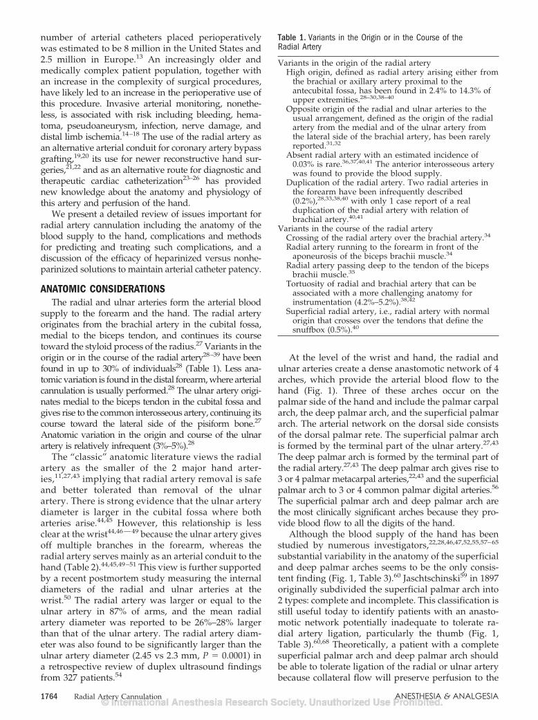
adaptive thickening of the intima.70,71 This process dif-ferentially affects various arterial beds and can varybetween relatively harmless adaptive intimal thicken-ing70 to advanced atherosclerotic lesions.71 Regardless ofintimal involvement, the media can develop calcifica-tions (Monckeberg’s calcifications).72 The incidence ofatherosclerosis demonstrated by ultrasound imaging hasbeen reported to be far less common in the radial arterythan in the common carotid artery.73 Preoperative Dopp-ler ultrasound examination in patients with cardiacdisease demonstrates atherosclerosis and calcification ofthe radial artery in 7%–9% and 8%–25% of patients,respectively74–77 (Table 4). The incidence of calcificationsin diabetic patients (Fig. 2) has been reported to be ashigh as 82%, with dense calcifications in 34%.78
Investigators using histopathologic and morpho-metric analyses reported that in patients with coro-nary artery disease, the radial artery is more likely to
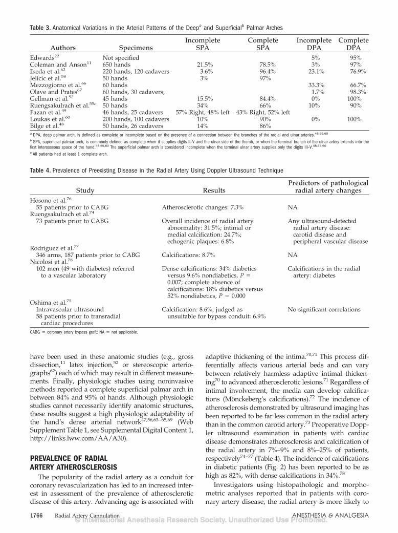
Table 3. Anatomical Variations in the Arterial Patterns of the Deepa and Superficialb Palmar Arches
Authors SpecimensIncomplete
SPAComplete
SPAIncomplete
DPAComplete
DPAEdwards22 Not specified 5% 95%Coleman and Anson11 650 hands 21.5% 78.5% 3% 97%Ikeda et al.62 220 hands, 120 cadavers 3.6% 96.4% 23.1% 76.9%Jelicic et al.58 50 hands 3% 97%Mezzogiorno et al.66 60 hands 33.3% 66.7%Olave and Prates67 60 hands, 30 cadavers, 1.7% 98.3%Gellman et al.52 45 hands 15.5% 84.4% 0% 100%Ruengsakulrach et al.55c 50 hands 34% 66% 10% 90%Fazan et al.49 46 hands, 25 cadavers 57% Right, 48% left 43% Right, 52% leftLoukas et al.60 200 hands, 100 cadavers 10% 90% 0% 100%Bilge et al.48 50 hands, 26 cadavers 14% 86%a DPA, deep palmar arch, is defined as complete or incomplete based on the presence of a connection between the branches of the radial and ulnar arteries.48,55,60
b SPA, superficial palmar arch, is commonly defined as complete when it supplies digits II–V and the ulnar side of the thumb, or when the terminal branch of the ulnar artery extends into thefirst interosseous space of the hand.48,55,60 The superficial palmar arch is considered incomplete when the terminal ulnar artery supplies only the digits III–V.48,55,60
c All patients had at least 1 complete arch.
Table 4. Prevalence of Preexisting Disease in the Radial Artery Using Doppler Ultrasound Technique
Study ResultsPredictors of pathological
radial artery changesHosono et al.76
55 patients prior to CABG Atherosclerotic changes: 7.3% NARuengsakulrach et al.74
73 patients prior to CABG Overall incidence of radial arteryabnormality: 31.5%; intimal ormedial calcification: 24.7%;echogenic plaques: 6.8%
Any ultrasound-detectedradial artery disease:carotid disease andperipheral vascular disease
Rodriguez et al.77
346 arms, 187 patients prior to CABG Calcifications: 8.7% NANicolosi et al.78
102 men (49 with diabetes) referredto a vascular laboratory
Dense calcifications: 34% diabeticsversus 9.6% nondiabetics, P �0.007; complete absence ofcalcifications: 18% diabetics versus52% nondiabetics, P � 0.000
Calcifications in the radialartery: diabetes
Oshima et al.75
Intravascular ultrasound Calcification: 8.6%; judged asunsuitable for bypass conduit: 6.9%
No significant correlations58 patients prior to transradial
cardiac proceduresCABG � coronary artery bypass graft; NA � not applicable.
1766 Radial Artery Cannulation ANESTHESIA & ANALGESIA
have intimal hyperplasia, atherosclerosis, and medialcalcification than the internal mammary artery (Table5).79–82 Intimal hyperplasia has been reported in67%–94%,79–81,83 atherosclerosis in 5%–6%,80,83 andmedial calcification in 6%–13% of radial arterysamples.80,83 Atherosclerosis of the radial artery is asegmental disease with predilection to the distal part ofthe artery.83 The most consistently reported predictors ofradial artery atherosclerosis and medial calcifications areperipheral vascular disease, smoking, age, and diabetes(Table 5).74,78,80,81,83 In contrast, radial artery specimensobtained in 59 hemodialysis patients at the time offistula surgery showed no atherosclerotic changesand an incidence of intimal hyperplasia of 76%.84
Old age and diabetes were identified as risk factorsfor the latter. The incidence of ischemic heart dis-ease was significantly greater in the group withintimal hyperplasia (48% vs 14%, P � 0.035).84
Indwelling radial artery catheters have been foundto induce local injury (e.g., intimal damage and pro-liferation).85 Even cannulation for only 6 h has been
associated with arterial wall scarring. Significant long-term structural changes have been reported aftertransradial cardiac catheterization86–90 leading to asignificant reduction in the radial artery diameter,86,89
stenosis (segmental or diffuse), or even radial arteryocclusion.87,91 The incidence of radial artery occlusions 1mo after transradial artery coronary angioplasty wasreported to be 2.8%.92
EVALUATION OF HAND CIRCULATION BEFORERADIAL ARTERY CANNULATIONAllen’s Test
Both the necessity and the optimal method to assessadequacy of collateral blood flow to the hand beforeradial artery cannulation are controversial.16 Clini-cally, the Allen’s Test is most often used to evaluatebaseline hand circulation. It was first described in 1929by Dr. Allen93,94 as a means to evaluate collateralcirculation simultaneously in both hands of patientswith thromboangiitis obliterans, and then modified byWright95,96 in the 1950s as a means to evaluate flow ina single hand. The Modified Allen’s Test has beensubsequently used to assess collateral blood flow tothe hand. With firm occlusive pressure held on boththe radial and ulnar arteries, the patient is asked toclench his or her fist several times until the palmarskin is blanched. The arteries should be compressedproximal to the expected tip of the arterial catheterbecause proximal branches of radial artery to the handcirculation could elicit falsely normal results.97,98 Thepatient is then instructed to unclench the fist, and thenulnar artery pressure is released while maintainingocclusion of the radial artery. Overextension of thehand and wide spreading of the fingers should beavoided because it can lead to falsely abnormal re-sults.99,100 The time required for palmar capillary refillis noted. The test is then repeated with the radialartery pressure released while maintaining occlusionof the ulnar artery (inverse Modified Allen’s Test).Although it is simple to perform, there are severallimitations including the primary end point (return tonormal skin color), which is prone to observer vari-ability.99 Not unexpectedly, a wide range of values forthe time required for hand reperfusion has beenreported (from 3 to 15 s).14,68,85,97,99–107 The frequencyof an abnormal Modified Allen’s Test (variously de-fined) ranges from �1% to 27%.100,108 Its clinicalreliability as a screening tool varies greatly as well.Ruengsakulrach et al.68 compared the Modified Allen’sTest (�10 s) with Doppler ultrasonography of thethumb artery in 71 patients and found the ModifiedAllen’s Test to have a sensitivity of 100% and speci-ficity of 97%. They further reported use of the Modi-fied Allen’s Test as a screening tool in 1657 radialartery harvests from 1323 patients with no ischemiccomplications.68 Others reported a similar lack ofischemic complications when the Modified Allen’sTest was used to guide suitability of radial artery
Figure 2. Ultrasound images showing longitudinal views ofnormal (A) and calcific (B) radial arteries. The normal vesselhas a thin, homogeneous wall and smooth luminal surface(A). The calcified artery (B) is characterized by multipleechogenic areas in the vessel wall (vertical arrows) and by anirregular luminal surface. The horizontal arrow indicates acalcific plaque extending into the vessel lumen. (Repro-duced from Nicolosi et al.78 with permission.)
Vol. 109, No. 6, December 2009 © 2009 International Anesthesia Research Society 1767
harvest.97,109–111 The major argument against the rou-tine use of the Modified Allen’s Test is the lack ofevidence that it can predict hand ischemia after radialartery cannulation.16,112,113 Slogoff et al.16 evaluatedthe Modified Allen’s Test in 411 cardiovascular surgi-cal patients reporting that 3.9% of patients had arecovery time of �15 s. Despite this, radial arterycannulation was performed in these patients withoutischemic complications. Abu-Omar et al.109 reportedradial artery harvesting without ischemic sequelae in38 patients with an abnormal Modified Allen’s Testbut normal Doppler ultrasound results (zero incidencein a small number of patients does not preclude aconsiderable risk of ischemic complications114). Con-sistent with these findings are those of Barbeau et
al.102 who found that 80% of patients with an abnor-mal Modified Allen’s Test scheduled for transradialcardiac instrumentation had adequate collateral per-fusion on plethysmography and oximetry tests. Ghu-ran et al.115 have even proposed that prescreeningwith the Modified Allen’s Test in the presence ofpalpable radial pulse is not required, because theyreported no ischemic sequelae in 630 patients whounderwent 662 transradial coronary interventionswithout prescreening. Conversely, hand ischemia af-ter radial artery cannulation has been reported despitea normal Modified Allen’s Test before cannula-tion.104,116–119 Mangano and Hickey118 described de-velopment of progressive ischemic injury requiringamputation of the distal segments of 2 fingers in a
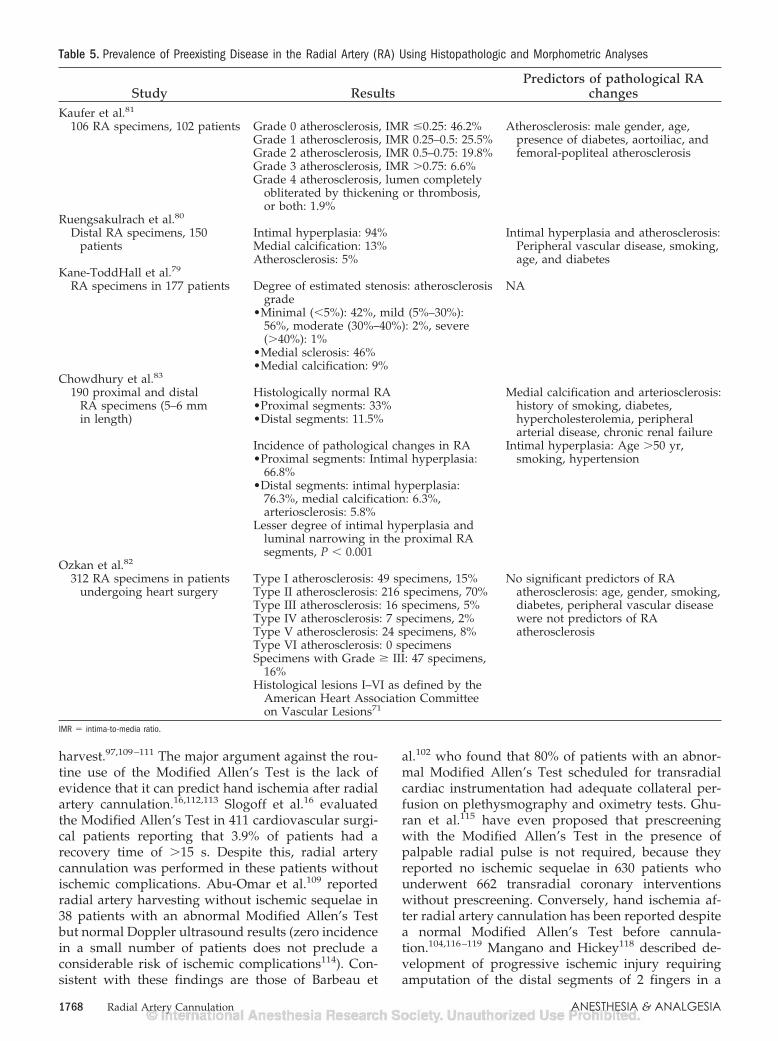
Table 5. Prevalence of Preexisting Disease in the Radial Artery (RA) Using Histopathologic and Morphometric Analyses
Study ResultsPredictors of pathological RA
changesKaufer et al.81
106 RA specimens, 102 patients Grade 0 atherosclerosis, IMR �0.25: 46.2% Atherosclerosis: male gender, age,presence of diabetes, aortoiliac, andfemoral-popliteal atherosclerosis
Grade 1 atherosclerosis, IMR 0.25–0.5: 25.5%Grade 2 atherosclerosis, IMR 0.5–0.75: 19.8%Grade 3 atherosclerosis, IMR �0.75: 6.6%Grade 4 atherosclerosis, lumen completely
obliterated by thickening or thrombosis,or both: 1.9%
Ruengsakulrach et al.80
Distal RA specimens, 150patients
Intimal hyperplasia: 94% Intimal hyperplasia and atherosclerosis:Peripheral vascular disease, smoking,age, and diabetes
Medial calcification: 13%Atherosclerosis: 5%
Kane-ToddHall et al.79
RA specimens in 177 patients Degree of estimated stenosis: atherosclerosisgrade
NA
•Minimal (�5%): 42%, mild (5%–30%):56%, moderate (30%–40%): 2%, severe(�40%): 1%
•Medial sclerosis: 46%•Medial calcification: 9%
Chowdhury et al.83
190 proximal and distalRA specimens (5–6 mmin length)
Histologically normal RA•Proximal segments: 33%•Distal segments: 11.5%
Medial calcification and arteriosclerosis:history of smoking, diabetes,hypercholesterolemia, peripheralarterial disease, chronic renal failure
Incidence of pathological changes in RA Intimal hyperplasia: Age �50 yr,smoking, hypertension•Proximal segments: Intimal hyperplasia:
66.8%•Distal segments: intimal hyperplasia:
76.3%, medial calcification: 6.3%,arteriosclerosis: 5.8%
Lesser degree of intimal hyperplasia andluminal narrowing in the proximal RAsegments, P � 0.001
Ozkan et al.82
312 RA specimens in patientsundergoing heart surgery
Type I atherosclerosis: 49 specimens, 15% No significant predictors of RAatherosclerosis: age, gender, smoking,diabetes, peripheral vascular diseasewere not predictors of RAatherosclerosis
Type II atherosclerosis: 216 specimens, 70%Type III atherosclerosis: 16 specimens, 5%Type IV atherosclerosis: 7 specimens, 2%Type V atherosclerosis: 24 specimens, 8%Type VI atherosclerosis: 0 specimensSpecimens with Grade � III: 47 specimens,
16%Histological lesions I–VI as defined by the
American Heart Association Committeeon Vascular Lesions71
IMR � intima-to-media ratio.
1768 Radial Artery Cannulation ANESTHESIA & ANALGESIA
patient with a normal Modified Allen’s Test anduncomplicated perioperative course. The authors hy-pothesized that an embolic event was the mechanismfor digit ischemia. The predictive value of a normalprecannulation Modified Allen’s Test was furtherquestioned by Stead and Stirt120 who reported thatdigital perfusion was independent from the palmarperfusion as measured by the Modified Allen’s Test.Jarvis et al.103 compared the Modified Allen’s Testwith Doppler ultrasound of the princeps pollicis ar-tery in 93 hands of 47 patients before radial arteryharvest and reported it to be a poor predictor of ulnarartery collateral flow. The diagnostic accuracy of theModified Allen’s Test, compared with ultrasound,was only 80%, with a sensitivity of 76% and a speci-ficity of 82% occurring with a 5-s recovery time.103 Theauthors concluded that the Modified Allen’s Test wasunable to identify a cutoff point for determiningadequate collateral blood flow to the hand. Glavin andJones121 compared the Modified Allen’s Test withDoppler ultrasound in 75 patients (150 extremities)finding the former to have a sensitivity of 87% tocorrectly diagnose the presence of ulnar artery bloodflow and a negative predictive value of only 0.18; i.e.,80% of all abnormal Modified Allen’s Test results intheir study were incorrect.
Adjuncts to the Allen’s TestPulse oximetry has been used with the Modified
Allen’s Test to make interpretation more objec-tive102,122–130 and less dependent on the patient’s co-operation.127,129,130 The time for the oxygen saturation(measured on the thumb or finger) to return to base-line after release of the occlusion is measured for eachartery. However, this method has been found tooverdiagnose normal hand circulation compared withthe Modified Allen’s Test65,102,122,126 (Web Supplem-ent Table 2, see Supplemental Digital Content 2,http://links.lww.com/AA/A31). Cheng et al.122
reported that all patients with an indeterminate Modi-fied Allen’s Test had a normal test using pulse oximetry.However, because blood flows as low as 4%–9% ofbaseline are associated with normal pulse oximetryvalues, the demonstration of normal pulse oximetrysaturation may not ensure adequate tissue perfusion.131
Despite this theoretical concern,65 the Modified Allen’sTest using pulse oximetry has been used for selection ofpatients for radial artery harvest with no instances ofvascular compromise in a series of 401 patients.125
The incorporation of plethysmography with theModified Allen’s Test allows visualization of pulsatileflow and more objective assessment of reperfusion.132
Some consider it superior to pulse oximetry in evalu-ating the hand’s collateral perfusion.65 However,plethysmography suffers from the inability to quan-tify blood flow.133
The introduction of Doppler ultrasound in theassessment of collateral hand circulation allows for
a comprehensive examination of the hand and fore-arm arteries47,63– 65,68,69,77,100,104,134,135 (Web Supple-ment Table 3, see Supplemental Digital Content 3,http://links.lww.com/AA/A32). A Doppler ultrasoundexamination consists of 2 parts. The first evaluates the“static” anatomy and flow of the arteries77 and thesecond part incorporates the Modified Allen’s Test with“dynamic” radial and ulnar artery compressions toassess the response of the collateral circulation.68,69 It isperformed with the Doppler ultrasound probe placedover the ulnar artery, radial artery, superior palmar arch,or dorsal digital thumb artery. There are no establishedstandard criteria for Doppler ultrasound findings thatdefine abnormal hand collateral perfusion. Accordingly,multiple definitions of inadequate collateral flow havebeen reported.63,64,68,69,77,100,101,103,104,134,136 Finally, Ru-engsakulrach et al.68 suggested that no flow in the dorsaldigital thumb artery with radial artery occlusion is thesole absolute contraindication for radial artery harvest.
Other tests for arterial collateral flow assessment ofthe hand include the “snuffbox test,”136,137 “squirttest,”138 postocclusive reactive circulatory hyperaemiatest,139 measurement of the systolic thumb pres-sure,140–142 and the radial hyperemic response test.143
Even magnetic resonance angiography has been sug-gested for preoperative evaluation of hand circulation.144
Together, the literature suggests that a normalModified Allen’s Test safely selects patients for radialartery harvest.53,68,97,109–111,145–147 In contrast, there isno proof that the Modified Allen’s Test can predicthand ischemia with radial artery cannulation.
ULNAR ARTERY CANNULATIONFew studies have addressed the use of the ulnar
artery for invasive arterial blood pressure monitoringreporting a safety and efficacy profile similar to thatfor radial artery cannulation.16,148–152 In a series of 50patients, Karacalar et al.151 described a 100% successrate of cannulation in patients with strong ulnar pulseand 59% success rate in patients with a weak ulnarpulse without complications.151 Slogoff et al.16 re-ported no hand ischemia in 22 patients who had anulnar artery catheter placed after a failed radial arterycannulation. However, digital ischemia after ulnarartery cannulation after unsuccessful radial arterycatheterization has been reported.153 Hand ischemiahas been reported in pediatric patients with prolongedulnar artery cannulation in the setting of prior radialartery cannulation.149 Although there is a theoreticalconcern that ulnar artery cannulation could causeneural trauma to the ulnar nerve, the literature lacksevidence of such a complication.16,23,151,154–158 There isincreasing interest in the use of the ulnar artery as anentry site for percutaneous coronary interventionswhen there are few other portal options.23,154–157,159
This approach has been safely used in patients withadequate radial artery flow,23,152,155,159 in those withcompromised radial artery flow resulting from mul-tiple punctures,156,157 and in those with known
Vol. 109, No. 6, December 2009 © 2009 International Anesthesia Research Society 1769
chronic radial artery occlusion.154 A randomizedstudy of 431 patients found the transulnar approachfor coronary angioplasty to be as safe and effective asthe transradial artery approach.152 Similar rates ofaccess success (transulnar 93.1% vs transradial 95.5%),complications, and asymptomatic artery occlusions(transulnar 5.7% vs transradial 4.7%) were reported.Mangin et al.157 evaluated the transulnar artery ap-proach in 117 consecutive patients who underwent122 percutaneous coronary interventions reportingpuncture failure in only 9 of 122 attempts. Complica-tions were noted in 7 patients (7.5%) including local (5patients) or extended (1 patient) hematoma and falseaneurysm (1 patient). The role of the Modified Allan’sTest in risk stratification before cannulation of theulnar artery is poorly defined.154,156
RADIAL ARTERY HARVESTRadial artery harvest for coronary artery bypass
graft surgery provides a model for examination of theeffects of radial artery occlusion. Removal of the radialartery is associated with a significant increase in ulnarartery diameter and blood flow velocity.160 Most in-vestigators evaluating hand perfusion days to monthsafter surgery using various methods (e.g., photo-electric plethysmography,160 laser Doppler flow-meter,161,162 venous occlusion plethysmography,163
digital-brachial indices,164 or pulsed wave Doppler165)have reported no significant decline in hand perfus-ion relative to the nonoperated hand (Web Supple-ment Table 4, see Supplemental Digital Content 4,http://links.lww.com/AA/A33). Early postoperativeforearm blood flow has been reported to be similar topreoperative values during exercise-induced ischemicreperfusion.163 In contrast, Lee et al.166 reported asignificant decline in digital blood flow 7 days afterradial artery harvest. However, after 3 yr, blood flowincreased to levels similar to those in the controlarms.167 The long-term effects of radial artery harvestwere examined in a series of 34 asymptomatic patientsby Serricchio et al.168 who reported that ulnar arterypeak systolic velocity was greater in the operated armcompared with the control arm 5 yr after radial arteryharvest. Handgrip exercise stress led to a significantincrease in ulnar artery diameter in both arms. Despitethis increase, handgrip exercise was associated with adecrease in transcutaneous Pao2 and an increase intranscutaneous Paco2 in the operated hand.168 After10 yr, a small degree of exercise-induced transcutane-ous oxygen desaturation in the absence of symptomswas reported.169,170 Long-term follow-up data169,170
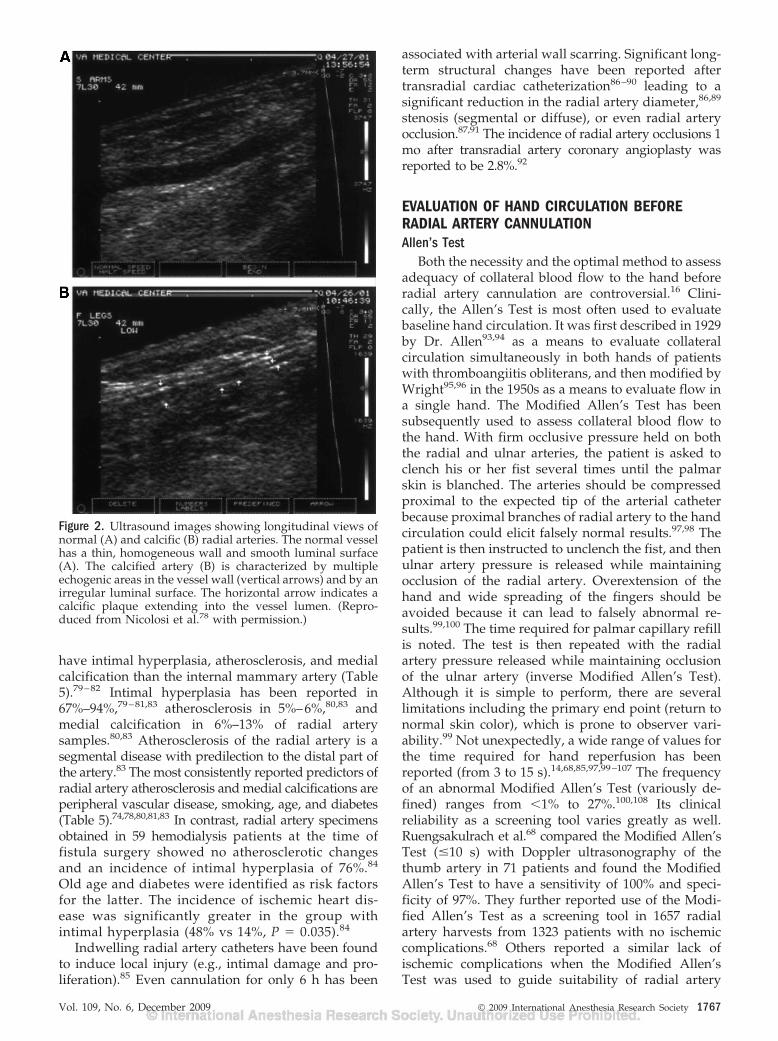
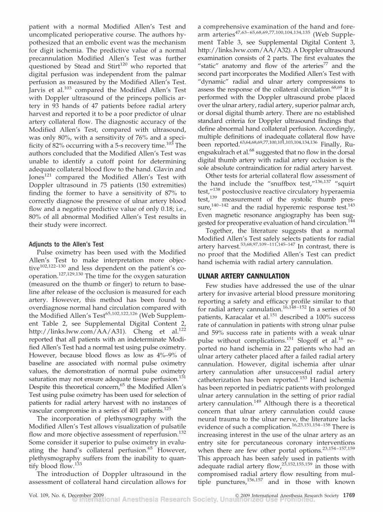
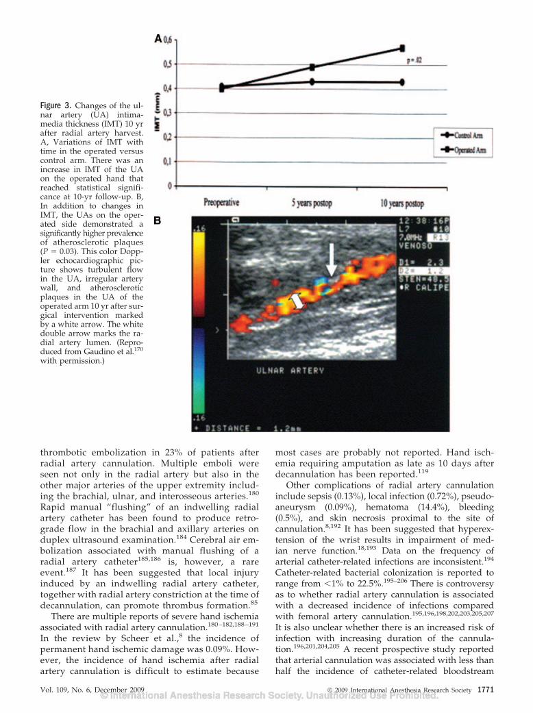
further suggest that the compensatory increase inulnar artery blood flow after radial artery harvest mayaccelerate atherosclerosis (Fig. 3). Echo-Dopplerevaluation performed in 39 patients 10 yr after radialartery harvest demonstrated greater intima-mediathickness of the ulnar artery (Fig. 3), and a higherprevalence of atherosclerotic plaques compared withthe nonoperated arm.169
A growing body of literature examining microsur-gery of radial artery flap transfer supports the long-term safety of radial artery harvest.171–175 Physiologicadaptation after radial artery harvest includes enlarge-ment in the diameter of the remaining forearm arteriesand a compensatory increase in blood flow velocity tothe hand.168,170,172,173 During rest, these adaptationsusually provide adequate perfusion, but with exerciseinsufficient perfusion can occur.169,168
Although a rare event, the most feared complica-tion of radial artery harvest is acute hand ischemia.Nunoo-Mensah176 described a patient with acute handischemia despite a normal preharvest ModifiedAllen’s Test, normal pulse oximetry saturation duringintraoperative radial artery occlusion, and good back-flow from the distal radial artery stump. The patientwas subsequently found to have a congenital absenceof the ulnar artery and a large interosseous artery. Thepatient underwent successful cephalic vein to distalradial artery revascularization. Three other patientshave been described to have experienced hand isch-emia after radial artery harvest. Tatoulis et al.177
reported postoperative fingertip ischemia in 2 patientswith scleroderma (0.08%) after radial artery harvest.Manabe et al.104 described 1 patient who, despite anormal Modified Allen’s Test, developed ischemia ofthe thumb several days after the operation.
COMPLICATIONS OF RADIAL ARTERY CANNULATIONThe reported incidence of at least temporary radial
artery occlusion after cannulation is between 1.5% and88%.178,179 In a review of 78 publications involving19,617 cannulations, Scheer et al.8 reported that theincidence of temporary radial artery occlusion was19.7%. Temporary spasm can occur in up to 57% ofradial arteries immediately after cannula insertion.148
Thrombotic occlusion has been described as early as2 h after radial artery catheter insertion or as late as aweek after catheter removal.16,180 In a study of 100surgical patients undergoing radial artery cannula-tions, of which 40 developed radial artery occlusion,Bedford and Wollman85 found that at the time ofdecannulation, only 42% of these 40 occlusions werepresent. Another 30% of all occlusions occurred within24 h of decannulation and another 28% occurred laterthan 1 day after decannulation. Symptoms of radialartery occlusion can persist for several days aftercatheter removal.16,181 Davis and Stewart,14 usingDoppler ultrasound, reported a 24% incidence ofcomplete occlusion 8 days after decannulation. Recan-nulation of an occluded radial artery as late as 75 daysafter catheter removal has been reported.85
Digital embolization, a major source of handischemia with radial artery cannulation,16,118,182,183
can lead to irreversible digital ischemia even in asetting of macroscopically and microscopically nor-mal radial, ulnar, and superficial palmar arteries.183
Downs et al.,180 in a study of 32 patients, reported
1770 Radial Artery Cannulation ANESTHESIA & ANALGESIA
thrombotic embolization in 23% of patients afterradial artery cannulation. Multiple emboli wereseen not only in the radial artery but also in theother major arteries of the upper extremity includ-ing the brachial, ulnar, and interosseous arteries.180
Rapid manual “flushing” of an indwelling radialartery catheter has been found to produce retro-grade flow in the brachial and axillary arteries onduplex ultrasound examination.184 Cerebral air em-bolization associated with manual flushing of aradial artery catheter185,186 is, however, a rareevent.187 It has been suggested that local injuryinduced by an indwelling radial artery catheter,together with radial artery constriction at the time ofdecannulation, can promote thrombus formation.85
There are multiple reports of severe hand ischemiaassociated with radial artery cannulation.180–182,188–191
In the review by Scheer et al.,8 the incidence ofpermanent hand ischemic damage was 0.09%. How-ever, the incidence of hand ischemia after radialartery cannulation is difficult to estimate because
most cases are probably not reported. Hand isch-emia requiring amputation as late as 10 days afterdecannulation has been reported.119
Other complications of radial artery cannulationinclude sepsis (0.13%), local infection (0.72%), pseudo-aneurysm (0.09%), hematoma (14.4%), bleeding(0.5%), and skin necrosis proximal to the site ofcannulation.8,192 It has been suggested that hyperex-tension of the wrist results in impairment of med-ian nerve function.18,193 Data on the frequency ofarterial catheter-related infections are inconsistent.194
Catheter-related bacterial colonization is reported torange from �1% to 22.5%.195–206 There is controversyas to whether radial artery cannulation is associatedwith a decreased incidence of infections comparedwith femoral artery cannulation.195,196,198,202,203,205,207
It is also unclear whether there is an increased risk ofinfection with increasing duration of the cannula-tion.196,201,204,205 A recent prospective study reportedthat arterial cannulation was associated with less thanhalf the incidence of catheter-related bloodstream
Figure 3. Changes of the ul-nar artery (UA) intima-media thickness (IMT) 10 yrafter radial artery harvest.A, Variations of IMT withtime in the operated versuscontrol arm. There was anincrease in IMT of the UAon the operated hand thatreached statistical signifi-cance at 10-yr follow-up. B,In addition to changes inIMT, the UAs on the oper-ated side demonstrated asignificantly higher prevalenceof atherosclerotic plaques(P � 0.03). This color Dopp-ler echocardiographic pic-ture shows turbulent flowin the UA, irregular arterywall, and atheroscleroticplaques in the UA of theoperated arm 10 yr after sur-gical intervention markedby a white arrow. The whitedouble arrow marks the ra-dial artery lumen. (Repro-duced from Gaudino et al.170
with permission.)
Vol. 109, No. 6, December 2009 © 2009 International Anesthesia Research Society 1771
infection compared with central venous catheteriza-tion (0.92 [95% confidence interval {CI}, 0.13–6.44] vs2.23 [95% CI, 1.12–4.44] per 1000 catheter days, respec-tively).205 However, both sites had the same incidenceof catheter colonization (15.71 [95% CI, 9.5–25.9] vs16.83 [95% CI, 13.3–21.3] per 1000 catheter days,respectively),205 emphasizing the importance of thearterial cannulation site as a potential source of sep-sis.194,208 However, current guidelines from the Centersfor Disease Control and Prevention209 and others194,205
do not recommend routine replacement of peripheralarterial catheters at fixed intervals to prevent infections.Immunocompromised patients, however, may benefitfrom routine catheter change every 4 days.201 An aseptictechnique for radial artery catheter placement thatincludes skin cleansing with an antiseptic alcohol con-taining chlorhexidine solution is recommended.209,210
Maximal barrier precautions did not, however, reducethe risk of arterial catheter-related bloodstream infectionin a randomized study.211
RISK FACTORS FOR ISCHEMIC HAND INJURY WITHRADIAL ARTERY CANNULATION
There remains considerable controversy over reli-able predictors of radial artery occlusion and ischemichand injury after direct cannulation.14,16,17,148,212,213 Ina seminal study of 1699 patients from the Texas HeartInstitute, Slogoff et al.16 were unable to identify anypredictors of serious ischemic complications of directradial artery blood pressure monitoring. However,analysis of the aggregate literature suggests that acombination of profound circulatory failure, hypoten-sion, and high-dose vasopressor therapy may increasethe risk of hand ischemia16,182,188,190,214 (Table 6). Signsof multiple digital emboli have been frequently re-ported in such instances.16,85,183,214,217 Hematoma atthe puncture site has been associated with an in-creased incidence of occlusion.14,16,17,212 Other factorsreported to be associated with radial artery injury are morecontroversial such as the number of puncture attempts,14,16
artery size,16,85,105,213,215 the composition of the cathe-ter (teflon versus polypropylene),14,16,148,178,180,213,215,216
catheter diameter,16,85,105,213,215 the duration of cannula-tion,14,16,85,148,218,219 and gender.17,148 The method ofpuncture (direct puncture versus transfixation tech-nique) has been reported to have no effect on risk forthrombosis,17,212 and recannulation of previously cannu-lated radial arteries did not increase the frequency ofocclusions.14 The use of large sheaths (5F or 6F) forcannulation, as used in transradial coronary interven-tions, has been associated with vessel narrowing, occlu-sion, and subsequent failure to cannulate the radialartery.220 Finally, longer catheters (�2 inches) wereassociated with higher catheter patency221 and fewerincidences of occlusion after decannulation comparedwith shorter catheters (�2 inches).222
A plethora of patient-specific (e.g., atherosclerosis),cannulation-related (e.g., thrombosis, vasospasm, em-boli), and hospital course–related (e.g., hypotension,vasopressors) risk factors emphasizes the multifacto-rial nature of ischemic complications of indwellingradial artery cannulation making precannulation riskassessment challenging (Table 6).182 Any of these riskfactors might override compensatory mechanismsprotecting hand perfusion leading to ischemia despiteadequate precannulation hand collateralization.182
Whether ultrasound-guided arterial cannulation canimprove outcomes from radial artery cannulation isnot yet clearly established.223–228 Tables 6 and 7 pro-vide a summary of risk factor assessment before radialartery cannulation (Table 6) and an algorithm foravoiding catheter-associated complications (Table 7).
HEPARIN VERSUS NONHEPARIN FLUSH SOLUTIONSFOR MAINTAINING ARTERIAL CATHETER PATENCY
Much debate has centered around the most ap-propriate solution for maintaining the patency of
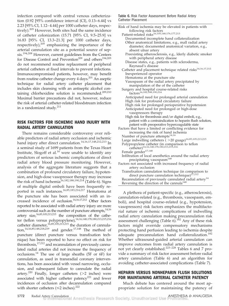
Table 6. Risk Factor Assessment Before Radial ArteryCatheter Placement
Risk of hand ischemia may be elevated in patients withfollowing risk factors
Patient-related risks16,105,148,176,177,213
Documented incomplete hand collateralizationOther anatomical limitations, e.g., mall radial artery
diameter; documented anatomical variation, e.g.,absent ulnar artery
Preexisting atherosclerosis, e.g., lderly diabetic smokerwith peripheral artery disease
Disease states, e.g., patients with scleroderma,Raynaud’s disease
Catheter and placement technique related risks14,16,17,212
Inexperienced operatorHematoma at the puncture siteVasospasm of the radial artery precipitated by
manipulation of the of the catheterSurgery and hospital course-related risks
factors16,182,188,190,214
Anticipated need for prolonged arterial cannulationHigh risk for profound circulatory failureHigh risk for prolonged perioperative hypotensionAnticipated need for prolonged or high-dose
vasopressors therapyHigh risk for thrombosis and/or digital emboli, e.g.,
patient with a contraindication to heparin flush solution,patient with preoperative hypercoagulable state
Factors that have a limited or conflicting evidence forincreasing the risk of hand ischemia
Number of puncture attempts14,16
Large indwelling catheters ( �20 guage)16,85,105,213,215
Polypropylene catheter (in comparison to tefloncatheter)14,16,148,178,180,213,215,216
Female gender17,148
Infiltration of local anesthetics around the radial arteryprecipitating vasospasm148
Factors not associated with increased frequency of radialartery occlusion
Transfixation cannulation technique (in comparison todirect puncture cannulation technique)17,212
Recannulation of previously cannulated radial artery14
Reversing the direction of the cannula148
1772 Radial Artery Cannulation ANESTHESIA & ANALGESIA
arterial catheters during continuous blood pressuremonitoring. Heparinized solutions are consideredadvantageous by some investigations, but heparinexposure might promote antibody formation lead-ing to heparin-induced thrombocytopenia.235–237
Continuous heparin flush solution has been re-ported to affect coagulation studies if drawn viaarterial access.238 –241
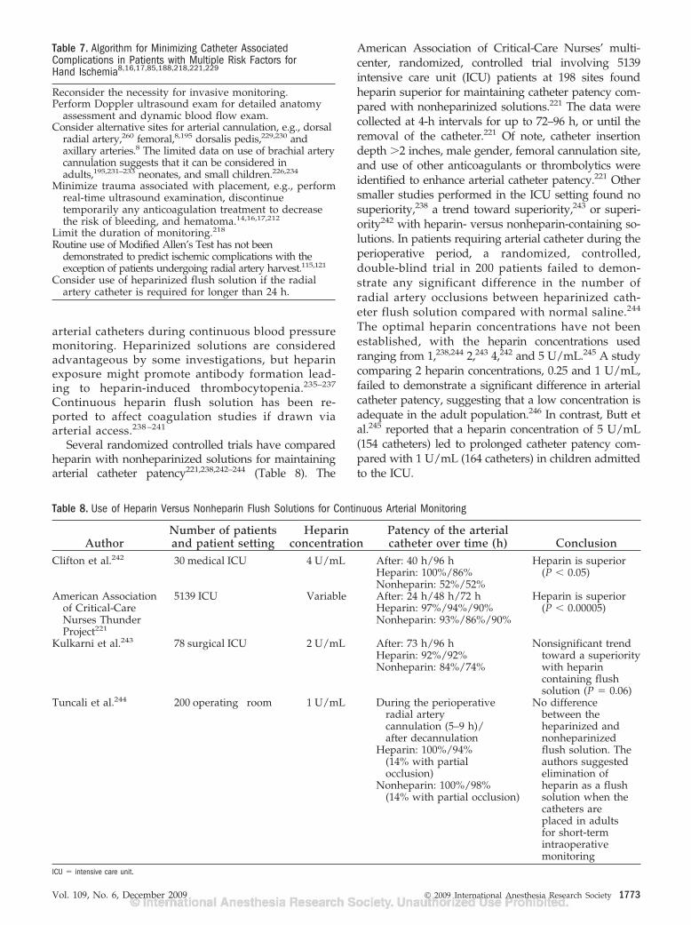
Several randomized controlled trials have comparedheparin with nonheparinized solutions for maintainingarterial catheter patency221,238,242–244 (Table 8). The
American Association of Critical-Care Nurses’ multi-center, randomized, controlled trial involving 5139intensive care unit (ICU) patients at 198 sites foundheparin superior for maintaining catheter patency com-pared with nonheparinized solutions.221 The data werecollected at 4-h intervals for up to 72–96 h, or until theremoval of the catheter.221 Of note, catheter insertiondepth �2 inches, male gender, femoral cannulation site,and use of other anticoagulants or thrombolytics wereidentified to enhance arterial catheter patency.221 Othersmaller studies performed in the ICU setting found nosuperiority,238 a trend toward superiority,243 or superi-ority242 with heparin- versus nonheparin-containing so-lutions. In patients requiring arterial catheter during theperioperative period, a randomized, controlled,double-blind trial in 200 patients failed to demon-strate any significant difference in the number ofradial artery occlusions between heparinized cath-eter flush solution compared with normal saline.244
The optimal heparin concentrations have not beenestablished, with the heparin concentrations usedranging from 1,238,244 2,243 4,242 and 5 U/mL.245 A studycomparing 2 heparin concentrations, 0.25 and 1 U/mL,failed to demonstrate a significant difference in arterialcatheter patency, suggesting that a low concentration isadequate in the adult population.246 In contrast, Butt etal.245 reported that a heparin concentration of 5 U/mL(154 catheters) led to prolonged catheter patency com-pared with 1 U/mL (164 catheters) in children admittedto the ICU.
Table 7. Algorithm for Minimizing Catheter AssociatedComplications in Patients with Multiple Risk Factors forHand Ischemia8,16,17,85,188,218,221,229
Reconsider the necessity for invasive monitoring.Perform Doppler ultrasound exam for detailed anatomy
assessment and dynamic blood flow exam.Consider alternative sites for arterial cannulation, e.g., dorsal
radial artery,260 femoral,8,195 dorsalis pedis,229,230 andaxillary arteries.8 The limited data on use of brachial arterycannulation suggests that it can be considered inadults,195,231–233 neonates, and small children.226,234
Minimize trauma associated with placement, e.g., performreal-time ultrasound examination, discontinuetemporarily any anticoagulation treatment to decreasethe risk of bleeding, and hematoma.14,16,17,212
Limit the duration of monitoring.218
Routine use of Modified Allen’s Test has not beendemonstrated to predict ischemic complications with theexception of patients undergoing radial artery harvest.115,121
Consider use of heparinized flush solution if the radialartery catheter is required for longer than 24 h.
Table 8. Use of Heparin Versus Nonheparin Flush Solutions for Continuous Arterial Monitoring
AuthorNumber of patientsand patient setting
Heparinconcentration
Patency of the arterialcatheter over time (h) Conclusion
Clifton et al.242 30 medical ICU 4 U/mL After: 40 h/96 hHeparin: 100%/86%Nonheparin: 52%/52%
Heparin is superior(P � 0.05)
American Associationof Critical-CareNurses ThunderProject221
5139 ICU Variable After: 24 h/48 h/72 hHeparin: 97%/94%/90%Nonheparin: 93%/86%/90%
Heparin is superior(P � 0.00005)
Kulkarni et al.243 78 surgical ICU 2 U/mL After: 73 h/96 hHeparin: 92%/92%Nonheparin: 84%/74%
Nonsignificant trendtoward a superioritywith heparincontaining flushsolution (P � 0.06)
Tuncali et al.244 200 operating room 1 U/mL During the perioperativeradial arterycannulation (5–9 h)/after decannulation
Heparin: 100%/94%(14% with partialocclusion)
Nonheparin: 100%/98%(14% with partial occlusion)
No differencebetween theheparinized andnonheparinizedflush solution. Theauthors suggestedelimination ofheparin as a flushsolution when thecatheters areplaced in adultsfor short-termintraoperativemonitoring
ICU � intensive care unit.
Vol. 109, No. 6, December 2009 © 2009 International Anesthesia Research Society 1773

Table 9. Clinical Reports on Characteristics, Treatment Options, and Outcome of Ischemic Complications of Radial ArteryCatheterization (RAC)a
AuthorPatients,sex/age MAT
Reason forplacement of RAC Risk factors
Duration ofRAC
Onset ofischemia Diagnosis Treatment Outcome
Baker et al.190 F/90 Equivocal Appendectomy Hypotensionvasopressor
6 d 7 d All 5 patients hadthrombi
Fluids, heparin Amputation,digit 3
M/52 Normal Intestinal surgery 56 h 40 h Dextran-40, heparin,sympathetic block,thrombectomy
Amputation, digit1–5
M/59 NA Intestinal surgery 48 h 48 h Dextran-40, heparin,reserpine
Amputation, digit1–2
M/68 Normal Pancreas cancer 78 h 72 h Dextran-40, heparin,sympathetic block
Amputation, digit1–2
M/74 Normal Major vascular 29 h 28 h Dextran-40, heparin,sympathetic block
Cold sensitivity,digit 1–3
Crossland andNeviaser 247
10 patients Normal Not specified NA NA NA Thrombosis Cannula removal Recovery in 10patients
Total of 600 RAC.Sixty patientsdevelopedhand ischemia(incidence of 10%)
50 patients NA Not specified NA NA 45 patients neededsurgicalexploration
Recovery in 37patients
Amputation in 13patients
Burrell248 F/57 NA Cardio-respiratoryarrest
NA 24 h 10 h afterdecannulation
Vasospasm Intraarterial dilutedsolution ofphentolamine
Recovery
Arthurs116 M/78 Normal Femoral aneurysmrepair
Hypotension 7 d aftercannulation
None Recovery over2 wk
Mangano andHickey118
M/54 Normal CABG None 24 h Axillary block,surgicalexploration
Digitalamputation
Gallacher117 M/67 Normal Left lowerlobectomy
Raynaud’s Intraoperativeperiod
Immediatelypostoperatively
Intraarterialverapamil 1 mg
Recovery
Sarma249 M/54 a AAA repair Hypotension Intraoperativeperiod
Immediatelypostoperatively
Intraarterialprilocaine 25 mg
Recovery
Mangar et al.119 M/35 Equivocal Femoral-tibialbypass
None 10 d postoperatively Anticoagulation Limb amputation
Bright et al.250 F/14 NA Tetralogy of Fallotrepair
Low CO Within 12 hpostoperatively
Axillary block,thrombectomy
Limb amputation
Lee et al.183 M/46 NA Septic shock Hypotensionvasopressors
8 h 8 h Complete occlusionof all commondigital arteries(thrombus versusembolus)
Intraarterialpapaverine
Gangrene/death
Cannula removalDextran-40,
nitroglycerinepatch
Lee et al.217 M/48 Normal Spine surgery None Intraoperativeperiod
8 d Thrombus Surgical exploration RecoveryPostoperatively:
heparin,Dextran-40
Scheer et al.8 4/19,617patients(0.09%)
Permanentischemicdamage
Review of 78 studieswith a total of19,617 RAC.Four patientsdeveloped handischemia(incidenceof 0.09%)
English et al.251 M/14 NA Spine fusion Hypotension Intraoperativeperiod
Immediatelypostoperatively
Warm compresses Immediateresolution
Geschwind et al.214 M/59 NA NA 2 patientsreceivedvasopressors
NA Time interval fromdecannulation tothe initiationof thrombolytictherapyaveraged 6 d(2–12 d)
Vasospasm in2 patients
Catheter-directedthrombolyticinfusion ofUrokinase
Recovery
Total of 7000 RAC(incidence ofthrombolytictherapy 0.1%)
F/46 Several hadhistory ofvasculardisease
All 7 hada combination ofthrombi andemboli
The dose rangedbetween570,000 IU and5,900,000 IU
Amputation
The total number ofpatients with handischemia was notprovided
F/49 RecoveryF/41 FailureF/65 Recovery
M/62 RecoveryM/54 Recovery
(Continued)
1774 Radial Artery Cannulation ANESTHESIA & ANALGESIA
TREATMENT OF ISCHEMIC COMPLICATIONSThere is no consensus on the optimal treatment for
ischemic injuries resulting from radial artery cannula-tion (Table 9). Early recognition is likely the mostimportant means to reduce permanent injury. Anabsent pulse, dampened waveform, blanched ormottled skin, delayed capillary refill, and painful andcold hand or fingers with motor weakness are presen-tations of hand ischemia.182,190,214 Blistering and skinulceration are late findings.
Arterial color flow Doppler ultrasound, angiogra-phy, or magnetic resonance imaging can be used toevaluate arterial flow in the arteries of the affectedlimb. Doppler ultrasound examination has the advan-tage of being noninvasive and easily performed, but itis limited by the inability to identify the mechanism ofcompromised blood flow. Immediate consultationwith a vascular surgeon is imperative.183,217 The radialartery catheter should be removed to ensure that it isnot contributing to flow obstruction if intraarterialdrug administration or arteriography is not underconsideration.
Treatment is aimed at the underlying mechanism(e.g., radial artery thrombus, ulnar artery or radialartery vasospasm, local radial artery trauma, reducedsystemic arterial perfusion, digital embolization, orpreviously unrecognized congenital inadequate collat-eral hand circulation). Different management tech-niques for radial artery occlusion have been attemptedand are summarized in Table 9.116–119,182,188–190,249–251
Aspiration of the thrombus at the catheter tip has beendescribed to restore arterial pulsation in 60% of patientswith suspected thrombosis.195 Intraarterial verapamil,prilocaine, and phentolamine have been successful toreverse ischemic symptoms.117,118,195,248,250 Other pro-posed treatments include low-molecular-weight dextranand low-dose heparin.182,183,190 Geschwind et al.214 re-ported angiographic flow restoration with �20% re-sidual thrombus leading to clinical improvement in 5 of
7 patients treated with intraarterial urokinase for radialartery occlusion due to thrombosis.
Hot compresses to the involved extremity mayresolve vasospasm251 (but could aggravate the isch-emia if applied to the hand). Sympathetic nerveblock14,252 or cervicodorsal sympathetic block190
should be considered for suspected arterial vaso-spasm.14,188,190,252,253 There is a growing body of lit-erature on prevention and management of radialartery spasm during transradial artery cardiac cathe-terization demonstrating that intraarterially adminis-tered vasodilators (e.g., nitrates, calcium channelblockers, lidocaine, and molsidomine) are safe andeffective in preventing radial artery spasm.254–256 Ra-dial artery spasm after an initial failed attempt can bereversed with subcutaneously administered nitroglyc-erin alone257 or in combination with 2% lidocaine.258
Surgical exploration is often necessary for patientswith absent radial artery blood flow and severe handischemia as a complication of radial artery cannula-tion. Despite successful cases being reported, opera-tive therapy has not been conclusively demonstratedto be superior to medical therapy.217,247 In a retrospec-tive analysis of treatment of 8 patients with handischemia after radial artery cannulation, surgical re-vascularization was attempted in 5 patients.182 Of the5, 1 patient died; all patients who survived developedgangrene of the first or second digit, with 2 patientsrequiring finger amputation. In contrast, only 1 of 3patients treated conservatively developed gangreneand underwent amputation.182 Unsuccessful surgicalexploration has been reported by others.118
Note added in proof: A crucial reference (Pyles et al.260)originally published by Anesthesia & Analgesia wasadded during the proof review process.
ACKNOWLEDGMENTSThe authors acknowledge the administrative and editorial
assistance of John Rukkila, ELS.
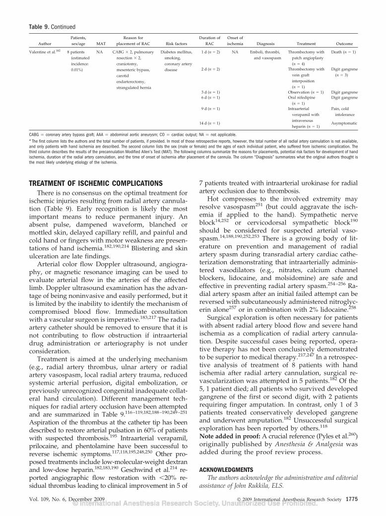
Table 9. Continued
AuthorPatients,sex/age MAT
Reason forplacement of RAC Risk factors
Duration ofRAC
Onset ofischemia Diagnosis Treatment Outcome
Valentine et al.182 8 patients(estimatedincidence:0.01%)
NA CABG � 2, pulmonaryresection � 2,craniotomy,mesenteric bypass,carotidendarterectomy,strangulated hernia
Diabetes mellitus,smoking,coronary arterydisease
1 d (n � 2) NA Emboli, thrombi,and vasospasm
Thrombectomy withpatch angioplasty(n � 4)
Death (n � 1)
2 d (n � 2) Thrombectomy withvein graftinterposition(n � 1)
Digit gangrene(n � 3)
3 d (n � 1) Observation (n � 1) Digit gangrene6 d (n � 1) Oral nifedipine
(n � 1)Digit gangrene
9 d (n � 1) Intraarterialverapamil withintravenousheparin (n � 1)
Pain, coldintolerance
14 d (n � 1) Asymptomatic
CABG � coronary artery bypass graft; AAA � abdominal aortic aneurysm; CO � cardiac output; NA � not applicable.a The first column lists the authors and the total number of patients, if provided. In most of those retrospective reports, however, the total number of all radial artery cannulation is not available,and only patients with hand ischemia are described. The second column lists the sex (male or female) and the ages of each individual patient, who suffered from ischemic complication. Thethird column describes the results of the precannulation Modified Allen’s Test (MAT). The following columns summarize the reasons for placements, potential risk factors for development of handischemia, duration of the radial artery cannulation, and the time of onset of ischemia after placement of the cannula. The column “Diagnosis” summarizes what the original authors thought isthe most likely underlying etiology of the ischemia.
Vol. 109, No. 6, December 2009 © 2009 International Anesthesia Research Society 1775

REFERENCES
1. Statement on Intravascular Catheterization Procedures. At-lanta, Georgia: ASA House of Delegates, 2005:1–2
2. Gidlund A. Development of apparatus and methods for roentgenstudies in haemodynamics. Acta Radiol Suppl 1956;130:7–70
3. Peterson LH, Dripps RD, Risman GC. A method for recordingthe arterial pressure pulse and blood pressure in man. AmHeart J 1949;37:771–82
4. Peirce EC II. Percutaneous femoral artery catheterization inman with special reference to aortography. Surg GynecolObstet 1951;93:56–74
5. Peirce EC II. Percutaneous arterial catheterization in dogs withspecial reference to aortography. Ann Surg 1951;133:544–7
6. Seldinger SI. Catheter replacement of the needle in percutaneousarteriography; a new technique. Acta Radiol 1953;39:368–76
7. Barr PO. Percutaneous puncture of the radial artery with amulti-purpose Teflon catheter for indwelling use. Acta PhysiolScand 1961;51:343–7
8. Scheer BV, Perel A, Pfeiffer UJ. Clinical review: complicationsand risk factors of peripheral arterial catheters used for hae-modynamic monitoring in anaesthesia and intensive caremedicine. Crit Care 2002;6:198–204
9. Ryan JF, Raines J, Dalton BC, Mathieu A. Arterial dynamics ofradial artery cannulation. Anesth Analg 1973;52:1017–25
10. Vogelzang RL. Arteriography of the hand and wrist. HandClin 1991;7:63–86
11. Coleman SS, Anson BJ. Arterial patterns in the hand based upona study of 650 specimens. Surg Gynecol Obstet 1961;113:409–24
12. Shah N, Bedford RF. Invasive and noninvasive blood pressuremonitoring. In: Lake CL, Hines RL, Blitt CD, eds. Clinical moni-toring practical applications for anesthesia and critical care. 1st ed.Philadelphia: W.B. Saunders Company, 2001:181–203
13. Gardner R. Direct arterial pressure monitoring. Curr AnaesthCrit Care 1990;1:239–46
14. Davis FM, Stewart JM. Radial artery cannulation. A prospec-tive study in patients undergoing cardiothoracic surgery. Br JAnaesth 1980;52:41–7
15. Hausmann D, Schulte am Esch J, Fischdick G. [Radial arterycannulation—a prospective study on its complication rate byclinical and sonographic evaluation (author’s transl)]. AnasthIntensivther Notfallmed 1981;16:269–73
16. Slogoff S, Keats AS, Arlund C. On the safety of radial arterycannulation. Anesthesiology 1983;59:42–7
17. Cederholm I, Sorensen J, Carlsson C. Thrombosis followingpercutaneous radial artery cannulation. Acta AnaesthesiolScand 1986;30:227–30
18. Chowet AL, Lopez JR, Brock-Utne JG, Jaffe RA. Wrist hyper-extension leads to median nerve conduction block: implicationsfor intra-arterial catheter placement. Anesthesiology 2004;100:287–91
19. Acar C, Jebara VA, Portoghese M, Beyssen B, Pagny JY, GrareP, Chachques JC, Fabiani JN, Deloche A, Guermonprez JL.Revival of the radial artery for coronary artery bypass grafting.Ann Thorac Surg 1992;54:652–9
20. Carpentier A, Guermonprez JL, Deloche A, Frechette C, Du-Bost C. The aorta-to-coronary radial artery bypass graft. Atechnique avoiding pathological changes in grafts. Ann ThoracSurg 1973;16:111–21
21. Rockwell WB, Smith SM, Tolliston T, Valnicek SM. Arterialconduits for extremity microvascular bypass surgery. PlastReconstr Surg 2003;112:829–34
22. Edwards EA. Organization of the small arteries of the handand digits. Am J Surg 1960;99:837–46
23. Dashkoff N, Dashkoff PB, Zizzi JA Sr, Wadhwani J, Zizzi JA Jr.Ulnar artery cannulation for coronary angiography and percu-taneous coronary intervention: case reports and anatomicconsiderations. Catheter Cardiovasc Interv 2002;55:93–6
24. Campeau L. Percutaneous radial artery approach for coronaryangiography. Cathet Cardiovasc Diagn 1989;16:3–7
25. Nagai Y, Metter EJ, Earley CJ, Kemper MK, Becker LC, LakattaEG, Fleg JL. Increased carotid artery intimal-medial thicknessin asymptomatic older subjects with exercise-induced myocar-dial ischemia. Circulation 1998;98:1504–9
26. Kamiya H, Ushijima T, Kanamori T, Ikeda C, Nakagaki C,Ueyama K, Watanabe G. Use of the radial artery graft aftertransradial catheterization: is it suitable as a bypass conduit?Ann Thorac Surg 2003;76:1505–9
27. Johnson D, Ellis H. Pectoral girdle and upper limb. In: Stan-dring S, ed. Gray’s anatomy. New York: Elsevier ChurchillLivingstone, 2005:799–942
28. McCormack LJ, Cauldwell EW, Anson BJ. Brachial and ante-brachial arterial patterns; a study of 750 extremities. SurgGynecol Obstet 1953;96:43–54
29. Sargon M, Celik HH. Proximal origins of radial and commoninterosseous arteries. Kaibogaku Zasshi 1994;69:406–9
30. Uglietta JP, Kadir S. Arteriographic study of variant arterialanatomy of the upper extremities. Cardiovasc Intervent Radiol1989;12:145–8
31. Durgun B, Yucel AH, Kizilkanat ED, Dere F. Multiple arterialvariation of the human upper limb. Surg Radiol Anat2002;24:125–8
32. Yucel AH. Unilateral variation of the arterial pattern of thehuman upper extremity with a muscle variation of the hand.Acta Med Okayama 1999;53:61–5
33. Alameddine AK, Alimov VK, Englelman RM, Rousou JA,Flack JE III, Deaton DW, Englelman DT. Anatomic variationsof the radial artery: significance when harvesting for coronaryartery bypass grafting. J Thorac Cardiovasc Surg 2004;127:1825–7
34. Sargon MF, Tanyeli E, Surucu HS, Yazar F, Arifoglu Y. Acomplicated variation of the upper extremity vascularisation.Kaibogaku Zasshi 1996;71:211–4
35. Kumar MR. Multiple arterial variations in the upper limb of aSouth Indian female cadaver. Clin Anat 2004;17:233–5
36. Porter CJ, Mellow CG. Anatomically aberrant forearm arteries:an absent radial artery with co-dominant median and ulnararteries. Br J Plast Surg 2001;54:727–8
37. Poteat WL. Report of a rare human variation: absence of theradial artery. Anat Rec 1986;214:89–95
38. Yoo BS, Yoon J, Ko JY, Kim JY, Lee SH, Hwang SO, Choe KH.Anatomical consideration of the radial artery for transradialcoronary procedures: arterial diameter, branching anomalyand vessel tortuosity. Int J Cardiol 2005;101:421–7
39. Karlsson S, Niechajev IA. Arterial anatomy of the upperextremity. Acta Radiol Diagn (Stockh) 1982;23:115–21
40. Rodriguez-Niedenfuhr M, Vazquez T, Nearn L, Ferreira B,Parkin I, Sanudo JR. Variations of the arterial pattern in theupper limb revisited: a morphological and statistical study,with a review of the literature. J Anat 2001;199:547–66
41. Kadanoff D, Balkansky G. [2 cases with rare variations ofarteries of the upper extremities]. Anat Anz 1966;118:289–96
42. Yokoyama N, Takeshita S, Ochiai M, Koyama Y, Hoshino S,Isshiki T, Sato T. Anatomic variations of the radial artery inpatients undergoing transradial coronary intervention. Cath-eter Cardiovasc Interv 2000;49:357–62
43. Moore KL, Dalley AF. Upper limb. Clinically oriented anat-omy. 4 ed. Philadelphia: Lippincott Williams & Wilkins,1999:665–810
44. Haerle M, Hafner HM, Dietz K, Schaller HE, Brunelli F.Vascular dominance in the forearm. Plast Reconstr Surg2003;111:1891–8
45. Keen JA. A study of the arterial variations in the limbs, withspecial reference to symmetry of vascular patterns. Am J Anat1961;108:245–61
46. Haerle M, Hafner HM, Schaller HE, Brunelli F. Dominances infinger arteries. J Hand Surg Br 2002;27:526–9
47. Little JM, Zylstra PL, West J, May J. Circulatory patterns in thenormal hand. Br J Surg 1973;60:652–5
48. Bilge O, Pinar Y, Ozer MA, Govsa F. A morphometric study onthe superficial palmar arch of the hand. Surg Radiol Anat2006;28:343–50
49. Fazan VP, Borges CT, Da Silva JH, Caetano AG, Filho OA.Superficial palmar arch: an arterial diameter study. J Anat2004;204:307–11
50. Riekkinen HV, Karkola KO, Kankainen A. The radial artery islarger than the ulnar. Ann Thorac Surg 2003;75:882–4
51. Tonks AM, Lawrence J, Lovie MJ. Comparison of ulnar andradial arterial blood-flow at the wrist. J Hand Surg Br1995;20:240–2
52. Gellman H, Botte MJ, Shankwiler J, Gelberman RH. Arterialpatterns of the deep and superficial palmar arches. Clin OrthopRelat Res 2001;383:41–6
53. Kohonen M, Teerenhovi O, Terho T, Laurikka J, Tarkka M. Isthe Allen test reliable enough? Eur J Cardiothorac Surg 2007;32:902–5
1776 Radial Artery Cannulation ANESTHESIA & ANALGESIA

54. Loh YJ, Nakao M, Tan WD, Lim CH, Tan YS, Chua YL. Factorsinfluencing radial artery size. Asian Cardiovasc Thorac Ann2007;15:324–6
55. Ruengsakulrach P, Eizenberg N, Fahrer C, Fahrer M, BuxtonBF. Surgical implications of variations in hand collateral circu-lation: anatomy revisited. J Thorac Cardiovasc Surg2001;122:682–6
56. Al-Turk M, Metcalf WK. A study of the superficial palmararteries using the Doppler Ultrasonic Flowmeter. J Anat1984;138(Pt 1):27–32
57. Callow A. Vascular disorders of the upper extremity. In:Jupiter J, ed. Flynn’s hand surgery. Baltimore: Williams &Wilkins, 1991:629–47
58. Jelicic N, Gajisin S, Zbrodowski A. Arcus palmaris superficia-lis. Acta Anat (Basel) 1988;132:187–90
59. Jaschtschinski S. Morphologie und Topologie des Arcus volarissublimes und profundus des Menschen. Anat Heft1897;7:161–88
60. Loukas M, Holdman D, Holdman S. Anatomical variations ofthe superficial and deep palmar arches. Folia Morphol (Warsz)2005;64:78–83
61. Ruengsakulrach P, Buxton BF, Eizenberg N, Fahrer M. Ana-tomic assessment of hand circulation in harvesting the radialartery. J Thorac Cardiovasc Surg 2001;122:178–80
62. Ikeda A, Ugawa A, Kazihara Y, Hamada N. Arterial patterns inthe hand based on a three-dimensional analysis of 220 cadaverhands. J Hand Surg Am 1988;13:501–9
63. Doscher W, Viswanathan B, Stein T, Margolis IB. Physiologicanatomy of the palmar circulation in 200 normal hands.J Cardiovasc Surg (Torino) 1985;26:171–4
64. Doscher W, Viswanathan B, Stein T, Margolis IB. Hemody-namic assessment of the circulation in 200 normal hands. AnnSurg 1983;198:776–9
65. Fuhrman TM, Pippin WD, Talmage LA, Reilley TE. Evaluationof collateral circulation of the hand. J Clin Monit 1992;8:28–32
66. Mezzogiorno A, Passiatore C, Mezzogiorno V. Anatomic varia-tions of the deep palmar arteries in man. Acta Anat (Basel)1994;149:221–4
67. Olave E, Prates JC. Deep palmar arch patterns in Brazilianindividuals. Surg Radiol Anat 1999;21:267–71
68. Ruengsakulrach P, Brooks M, Hare DL, Gordon I, Buxton BF.Preoperative assessment of hand circulation by means ofDoppler ultrasonography and the modified Allen test. J ThoracCardiovasc Surg 2001;121:526–31
69. Pola P, Serricchio M, Flore R, Manasse E, Favuzzi A, PossatiGF. Safe removal of the radial artery for myocardial revascu-larization: a Doppler study to prevent ischemic complicationsto the hand. J Thorac Cardiovasc Surg 1996;112:737–44
70. Stary HC, Blankenhorn DH, Chandler AB, Glagov S, Insull W Jr,Richardson M, Rosenfeld ME, Schaffer SA, Schwartz CJ, WagnerWD. A definition of the intima of human arteries and of itsatherosclerosis-prone regions. A report from the Committee onVascular Lesions of the Council on Arteriosclerosis, AmericanHeart Association. Circulation 1992;85:391–405
71. Stary HC, Chandler AB, Dinsmore RE, Fuster V, Glagov S,Insull W Jr, Rosenfeld ME, Schwartz CJ, Wagner WD, WisslerRW. A definition of advanced types of atherosclerotic lesionsand a histological classification of atherosclerosis. A reportfrom the Committee on Vascular Lesions of the Council onArteriosclerosis, American Heart Association. Circulation1995;92:1355–74
72. Proudfoot D, Shanahan CM. Biology of calcification in vascularcells: intima versus media. Herz 2001;26:245–51
73. Gaudino M, Tondi P, Serricchio M, Spatuzza P, SantoliquidoA, Flora R, Girola F, Nasso G, Pola P, Possati G. Atheroscleroticinvolvement of the radial artery in patients with coronaryartery disease and its relation with midterm radial artery graftpatency and endothelial function. J Thorac Cardiovasc Surg2003;126:1968–71
74. Ruengsakulrach P, Brooks M, Sinclair R, Hare D, Gordon I,Buxton B. Prevalence and prediction of calcification andplaques in radial artery grafts by ultrasound. J Thorac Cardio-vasc Surg 2001;122:398–9
75. Oshima A, Takeshita S, Kozuma K, Yokoyama N, MotoyoshiK, Ishikawa S, Honda M, Oga K, Ochiai M, Isshiki T. Intravas-cular ultrasound analysis of the radial artery for coronaryartery bypass grafting. Ann Thorac Surg 2005;79:99–103
76. Hosono M, Suehiro S, Shibata T, Sasaki Y, Kumano H, Ki-noshita H. Duplex scanning to assess radial artery suitabilityfor coronary artery bypass grafting. Jpn J Thorac CardiovascSurg 2000;48:217–21
77. Rodriguez E, Ormont ML, Lambert EH, Needleman L, HalpernEJ, Diehl JT, Edie RN, Mannion JD. The role of preoperativeradial artery ultrasound and digital plethysmography prior tocoronary artery bypass grafting. Eur J Cardiothorac Surg2001;19:135–9
78. Nicolosi AC, Pohl LL, Parsons P, Cambria RA, Olinger GN.Increased incidence of radial artery calcification in patientswith diabetes mellitus. J Surg Res 2002;102:1–5
79. Kane-ToddHall SM, Taggart SP, Clements-Jewery H, RoskellDE. Pre-existing vascular disease in the radial artery and othercoronary artery bypass conduits. Eur J Med Res 1999;4:11–4
80. Ruengsakulrach P, Sinclair R, Komeda M, Raman J, Gordon I,Buxton B. Comparative histopathology of radial artery versusinternal thoracic artery and risk factors for development ofintimal hyperplasia and atherosclerosis. Circulation 1999;100:II139–44
81. Kaufer E, Factor SM, Frame R, Brodman RF. Pathology of theradial and internal thoracic arteries used as coronary arterybypass grafts. Ann Thorac Surg 1997;63:1118–22
82. Ozkan S, Akay TH, Gultekin B, Aslim E, Arslan A, OzdemirBH, Becit N, Tasdelen A. Atherosclerosis of radial and internalthoracic arteries used in coronary bypass: atherosclerosis inarterial grafts. J Card Surg 2007;22:385–9
83. Chowdhury UK, Airan B, Mishra PK, Kothari SS, Subrama-niam GK, Ray R, Singh R, Venugopal P. Histopathology andmorphometry of radial artery conduits: basic study and clinicalapplication. Ann Thorac Surg 2004;78:1614–21
84. Kim YO, Song HC, Yoon SA, Yang CW, Kim NI, Choi YJ, LeeEJ, Kim WY, Chang YS, Bang BK. Preexisting intimal hyper-plasia of radial artery is associated with early failure ofradiocephalic arteriovenous fistula in hemodialysis patients.Am J Kidney Dis 2003;41:422–8
85. Bedford RF, Wollman H. Complications of percutaneousradial-artery cannulation: an objective prospective study inman. Anesthesiology 1973;38:228–36
86. Madssen E, Haere P, Wiseth R. Radial artery diameter andvasodilatory properties after transradial coronary angiogra-phy. Ann Thorac Surg 2006;82:1698–702
87. Nagai S, Abe S, Sato T, Hozawa K, Yuki K, Hanashima K,Tomoike H. Ultrasonic assessment of vascular complications incoronary angiography and angioplasty after transradial ap-proach. Am J Cardiol 1999;83:180–6
88. Wakeyama T, Ogawa H, Iida H, Takaki A, Iwami T, MochizukiM, Tanaka T. Intima-media thickening of the radial artery aftertransradial intervention. An intravascular ultrasound study.J Am Coll Cardiol 2003;41:1109–14
89. Edmundson A, Mann T. Nonocclusive radial artery injuryresulting from transradial coronary interventions: radial arteryIVUS. J Invasive Cardiol 2005;17:528–31
90. Goldberg S. There’s no free lunch? J Invasive Cardiol2005;17:532
91. Hall JJ, Arnold AM, Valentine RP, McCready RA, Mick MJ.Ultrasound imaging of the radial artery following its use forcardiac catheterization. Am J Cardiol 1996;77:108–9
92. Stella PR, Kiemeneij F, Laarman GJ, Odekerken D, Slagboom T,van der Wieken R. Incidence and outcome of radial arteryocclusion following transradial artery coronary angioplasty.Cathet Cardiovasc Diagn 1997;40:156–8
93. Cable DG, Mullany CJ, Schaff HV. The Allen test. Ann ThoracSurg 1999;67:876–7
94. Allen E. Thromboangiitis obliterans: methods of diagnosis ofchronic occlusive arterial lesions distal to the wrist withillustrative cases. Am J Med Sci 1929;178:237–44
95. Ejrup B, Fischer B, Wright IS. Clinical evaluation of blood flowto the hand. The false-positive Allen test. Circulation1966;33:778–80
96. Wright IS. Vascular diseases in clinical practice. 2 ed. Chicago:The Year Book Publishers Inc., 1952
97. Asif M, Sarkar PK. Three-digit Allen’s test. Ann Thorac Surg2007;84:686–7
98. Gandhi SK, Reynolds AC. A modification of Allen’s test todetect aberrant ulnar collateral circulation. Anesthesiology1983;59:147–8
Vol. 109, No. 6, December 2009 © 2009 International Anesthesia Research Society 1777
99. Greenhow DE. Incorrect performance of Allen’s test—ulnar-artery follow erroneously presumed inadequate. Anesthesiol-ogy 1972;37:356–7
100. Kamienski RW, Barnes RW. Critique of the Allen test forcontinuity of the palmar arch assessed by doppler ultrasound.Surg Gynecol Obstet 1976;142:861–4
101. Agrifoglio M, Dainese L, Pasotti S, Galanti A, Cannata A,Roberto M, Parolari A, Biglioli P. Preoperative assessment ofthe radial artery for coronary artery bypass grafting: is theclinical Allen test adequate? Ann Thorac Surg 2005;79:570–2
102. Barbeau GR, Arsenault F, Dugas L, Simard S, Lariviere MM.Evaluation of the ulnopalmar arterial arches with pulse oxim-etry and plethysmography: comparison with the Allen’s test in1010 patients. Am Heart J 2004;147:489–93
103. Jarvis MA, Jarvis CL, Jones PR, Spyt TJ. Reliability of Allen’stest in selection of patients for radial artery harvest. AnnThorac Surg 2000;70:1362–5
104. Manabe S, Tabuchi N, Toyama M, Kuriu K, Mizuno T, Su-namori M. Measurement of ulnar flow is helpful in predictingischemia after radial artery harvest. Thorac Cardiovasc Surg2002;50:325–8
105. Bedford RF. Radial arterial function following percutaneouscannulation with 18- and 20-gauge catheters. Anesthesiology1977;47:37–9
106. Phillips CS, Murphy MS. Vascular problems of the upperextremity: a primer for the orthopaedic surgeon. J Am AcadOrthop Surg 2002;10:401–8
107. Yokoyama N, Takeshita S, Ochiai M, Hoshino S, Koyama Y,Oshima A, Isshiki T, Sato T. Direct assessment of palmarcirculation before transradial coronary intervention by colorDoppler ultrasonography. Am J Cardiol 2000;86:218–21
108. Benit E, Vranckx P, Jaspers L, Jackmaert R, Poelmans C,Coninx R. Frequency of a positive modified Allen’s test in 1,000consecutive patients undergoing cardiac catheterization.Cathet Cardiovasc Diagn 1996;38:352–4
109. Abu-Omar Y, Mussa S, Anastasiadis K, Steel S, Hands L,Taggart DP. Duplex ultrasonography predicts safety of radialartery harvest in the presence of an abnormal Allen test. AnnThorac Surg 2004;77:116–9
110. Sajja LR, Mannam MG, Sompalli S. Is Allen’s test not reliable inthe selection of patients for radial artery harvest? Ann ThoracSurg 2002;74:296
111. Meharwal ZS, Trehan N. Functional status of the hand afterradial artery harvesting: results in 3,977 cases. Ann ThoracSurg 2001;72:1557–61
112. Wilkins RG. Radial artery cannulation and ischaemic damage:a review. Anaesthesia 1985;40:896–9
113. McGregor A. The Allen test—an investigation of its accuracyby fluorescein angiography. J Hand Surg 1986;12:82–5
114. Hanley JA, Lippman-Hand A. If nothing goes wrong, iseverything all right? Interpreting zero numerators. JAMA1983;249:1743–5
115. Ghuran AV, Dixon G, Holmberg S, de Belder A, Hildick-SmithD. Transradial coronary intervention without pre-screening fora dual palmar blood supply. Int J Cardiol 2007;121:320–2
116. Arthurs GJ. Case report: digital ischaemia following radialartery cannulation. Anaesth Intensive Care 1978;6:54–5
117. Gallacher BP. Intra-arterial verapamil to reverse acute isch-aemia of the hand after radial artery cannulation. Can JAnaesth 1991;38:138
118. Mangano DT, Hickey RF. Ischemic injury following uncompli-cated radial artery catheterization. Anesth Analg 1979;58:55–7
119. Mangar D, Laborde RS, Vu DN. Delayed ischaemia of the handnecessitating amputation after radial artery cannulation. Can JAnaesth 1993;40:247–50
120. Stead SW, Stirt JA. Assessment of digital blood flow andpalmar collateral circulation. Allen’s test vs. photoplethysmog-raphy. Int J Clin Monit Comput 1985;2:29–34
121. Glavin RJ, Jones HM. Assessing collateral circulation in thehand—four methods compared. Anaesthesia 1989;44:594–5
122. Cheng EY, Lauer KK, Stommel KA, Guenther NR. Evaluationof the palmar circulation by pulse oximetry. J Clin Monit1989;5:1–3
123. Greenwood MJ, Della-Siega AJ, Fretz EB, Kinloch D, Klinke P,Mildenberger R, Williams MB, Hilton D. Vascular communi-cations of the hand in patients being considered for transradialcoronary angiography: is the Allen’s test accurate? J Am CollCardiol 2005;46:2013–7
124. Hovagim AR, Katz RI, Poppers PJ. Pulse oxymetry for evalu-ation of radial and ulnar arterial blood flow (abstract). AnesthAnalg 1988;67:S94
125. Johnson WH III, Cromartie RS III, Arrants JE, Wuamett JD,Holt JB. Simplified method for candidate selection for radialartery harvesting. Ann Thorac Surg 1998;65:1167
126. Lauer KK, Cheng EY, Stommel KA, Guenther NR, Kay J. Pulseoximetry evaluation of the palmar circulation (abstract).Anesth Analg 1988;67:S129
127. Nowak GS, Moorthy SS, McNiece WL. Use of pulse oximetryfor assessment of collateral arterial flow. Anesthesiology1986;64:527
128. Oettle AC, van Niekerk A, Boon JM, Meiring JH. Evaluation ofAllen’s test in both arms and arteries of left and right-handedpeople. Surg Radiol Anat 2006;28:3–6
129. Raju R. The pulse oximeter and the collateral circulation.Anaesthesia 1986;41:783–4
130. Rozenberg B, Rosenberg M, Birkhan J. Allen’s test performedby pulse oximeter. Anaesthesia 1988;43:515–6
131. Lawson D, Norley I, Korbon G, Loeb R, Ellis J. Blood flowlimits and pulse oximeter signal detection. Anesthesiology1987;67:599–603
132. Brodsky JB. A simple method to determine patency of theulnar artery intraoperatively prior to radial-artery cannulation.Anesthesiology 1975;42:626–7
133. Fuhrman TM, Reilley TE, Pippin WD. Comparison of digitalblood pressure, plethysmography, and the modified Allen’stest as means of evaluating the collateral circulation to thehand. Anaesthesia 1992;47:959–61
134. Mozersky DJ, Buckley CJ, Hagood CO Jr, Capps WF Jr,Dannemiller FJ Jr. Ultrasonic evaluation of the palmar circula-tion. A useful adjunct to radial artery cannulation. Am J Surg1973;126:810–2
135. McSwain GR, Ameriks JA. Doppler-improved Allen test. SouthMed J 1979;72:1620–1
136. Kochi K, Orihashi K, Sueda T. The snuffbox technique: areliable color Doppler method to assess hand circulation.J Thorac Cardiovasc Surg 2003;125:821–5
137. Kochi K, Sueda T, Orihashi K, Matsuura Y. New noninvasivetest alternative to Allen’s test: snuff-box technique. J ThoracCardiovasc Surg 1999;118:756–8
138. Birdi I, Ritchie AJ. Intraoperative confirmation of ulnar collat-eral blood flow during radial artery harvesting using the“squirt test”. Ann Thorac Surg 2002;74:271–2
139. Vaghadia H, Schechter MT, Sheps SB, Jenkins LC. Evaluationof a postocclusive reactive circulatory hyperaemia (PORCH)test for the assessment of ulnar collateral circulation. Can JAnaesth 1988;35:591–8
140. Husum B, Berthelsen P. Allen’s test and systolic arterialpressure in the thumb. Br J Anaesth 1981;53:635–7
141. Husum B, Palm T. Before cannulation of the radial artery:collateral arterial supply evaluated by strain-gauge plethys-mography. Acta Anaesthesiol Scand 1980;24:412–4
142. Husum B, Palm T. Before cannulation of the radial artery. Br JAnaesth 1979;51:71–2
143. Roberts N, Ghosh S, Boehm M, Galinanes M. The radialhyperaemic response: a new and objective assessment of ulnarcollateral supply to the hand. Eur J Cardiothorac Surg2002;21:549–52
144. Winterer JT, Ennker J, Scheffler K, Rosendahl U, Schafer O,Wanner M, Laubenberger J, Langer M. Gadolinium-enhancedelliptically reordered three-dimensional MR angiography inthe assessment of hand vascularization before radial arteryharvest for coronary artery bypass grafting: first experience.Invest Radiol 2001;36:501–8
145. Ronald A, Patel A, Dunning J. Is the Allen’s test adequate tosafely confirm that a radial artery may be harvested forcoronary arterial bypass grafting? Interact Cardiovasc ThoracSurg 2005;4:332–40
146. Barner HB. Allen’s test. Ann Thorac Surg 2008;85:690147. Unlu Y. Is the Allen test reliable enough? Eur J Cardiothorac
Surg 2008;33:754148. Kim JM, Arakawa K, Bliss J. Arterial cannulation: factors in the
development of occlusion. Anesth Analg 1975;54:836–41149. Kahler AC, Mirza F. Alternative arterial catheterization site
using the ulnar artery in critically ill pediatric patients. PediatrCrit Care Med 2002;3:370–4
1778 Radial Artery Cannulation ANESTHESIA & ANALGESIA

150. Green JA, Tonkin MA. Ischaemia of the hand in infantsfollowing radial or ulnar artery catheterisation. Hand Surg1999;4:151–7
151. Karacalar S, Ture H, Baris S, Karakaya D, Sarihasan B. Ulnarartery versus radial artery approach for arterial cannulation: aprospective, comparative study. J Clin Anesth 2007;19:209–13
152. Aptecar E, Pernes JM, Chabane-Chaouch M, Bussy N, CatarinoG, Shahmir A, Bougrini K, Dupouy P. Transulnar versustransradial artery approach for coronary angioplasty: thePCVI-CUBA study. Catheter Cardiovasc Interv 2006;67:711–20
153. Maston M, Van Oldenbeek C. Digital ischaemia after ulnarartery cannulation. Br J Anaesth 2003;90:111
154. Lanspa TJ, Reyes AP, Oldemeyer JB, Williams MA. Ulnarartery catheterization with occlusion of corresponding radialartery. Catheter Cardiovasc Interv 2004;61:211–3
155. Terashima M, Meguro T, Takeda H, Endoh N, Ito Y, MitsuokaM, Ohtomo T, Murai O, Fujiwara S, Honda H, Miyazaki Y,Kuhara R, Kawashima O, Isoyama S. Percutaneous ulnarartery approach for coronary angiography: a preliminary re-port in nine patients. Catheter Cardiovasc Interv 2001;53:410–4
156. Lanspa TJ, Williams MA, Heirigs RL. Effectiveness of ulnarartery catheterization after failed attempt to cannulate a radialartery. Am J Cardiol 2005;95:1529–30
157. Mangin L, Bertrand OF, De La Rochelliere R, Proulx G, LemayR, Barbeau G, Gleeton O, Rodes-Cabau J, Nguyen CM, Roy L.The transulnar approach for coronary intervention: a safealternative to transradial approach in selected patients. J Inva-sive Cardiol 2005;17:77–9
158. Buxton BF, Chan AT, Dixit AS, Eizenberg N, Marshall RD,Raman JS. Ulnar artery as a coronary bypass graft. Ann ThoracSurg 1998;65:1020–4
159. Limbruno U, Rossini R, De Carlo M, Amoroso G, Ciabatti N,Petronio AS, Micheli A, Mariani M. Percutaneous ulnar arteryapproach for primary coronary angioplasty: safety and feasi-bility. Catheter Cardiovasc Interv 2004;61:56–9
160. Brodman RF, Hirsh LE, Frame R. Effect of radial artery harveston collateral forearm blood flow and digital perfusion. J ThoracCardiovasc Surg 2002;123:512–6
161. Knobloch K, Lichtenberg A, Pichlmaier M, Tomaszek S, KrugA, Haverich A. Palmar microcirculation after harvesting of theradial artery in coronary revascularization. Ann Thorac Surg2005;79:1026–30
162. Knobloch K, Lichtenberg A, Tomaszek S, Hagl C, Khaladj N,Klima U, Haverich A. Long-term physical activity and neuro-logic function after harvesting of the radial artery as T-graft orfree graft in coronary revascularization. Ann Thorac Surg2005;80:918–21
163. Chong WC, Ong PJ, Hayward CS, Collins P, Moat NE. Effectsof radial artery harvesting on forearm function and blood flow.Ann Thorac Surg 2003;75:1171–4
164. Dumanian GA, Segalman K, Mispireta LA, Walsh JA, Hen-drickson MF, Wilgis EF. Radial artery use in bypass graftingdoes not change digital blood flow or hand function. AnnThorac Surg 1998;65:1284–7
165. Royse AG, Royse CF, Maleskar A, Garg A. Harvest of theradial artery for coronary artery surgery preserves maximalblood flow of the forearm. Ann Thorac Surg 2004;78:539–42
166. Lee HS, Chang BC, Heo YJ. Digital blood flow after radialartery harvest for coronary artery bypass grafting. Ann ThoracSurg 2004;77:2071–4
167. Lee HS, Heo YJ, Chang BC. Long-term digital blood flow afterradial artery harvesting for coronary artery bypass grafting.Eur J Cardiothorac Surg 2005;27:99–103
168. Serricchio M, Gaudino M, Tondi P, Gasbarrini A, Gerardino L,Santoliquido A, Pola P, Possati G. Hemodynamic and func-tional consequences of radial artery removal for coronaryartery bypass grafting. Am J Cardiol 1999;84:1353–6, A8
169. Gaudino M, Glieca F, Luciani N, Losasso G, Tondi P, SerricchioM, Pola P, Possati G. Ten-year Echo-Doppler evaluation offorearm circulation following radial artery removal for coro-nary artery bypass grafting. Eur J Cardiothorac Surg 2006;29:71–3
170. Gaudino M, Serricchio M, Tondi P, Gerardino L, Di Giorgio A,Pola P, Possati G. Chronic compensatory increase in ulnar flowand accelerated atherosclerosis after radial artery removal forcoronary artery bypass. J Thorac Cardiovasc Surg 2005;130:9–12
171. Richardson D, Fisher SE, Vaughan ED, Brown JS. Radialforearm flap donor-site complications and morbidity: a pro-spective study. Plast Reconstr Surg 1997;99:109–15
172. Ciria-Llorens G, Gomez-Cia T, Talegon-Melendez A. Angio-logic observations following radial artery flap elevation: a casereport. Surg Radiol Anat 1998;20:377–81
173. Ciria-Llorens G, Gomez-Cia T, Talegon-Melendez A. Analysisof flow changes in forearm arteries after raising the radialforearm flap: a prospective study using colour duplex imaging.Br J Plast Surg 1999;52:440–4
174. Meland NB, Core GB, Hoverman VR. The radial forearm flapdonor site: should we vein graft the artery? A comparativestudy. Plast Reconstr Surg 1993;91:865–70; discussion 871
175. Bardsley AF, Soutar DS, Elliot D, Batchelor AG. Reducingmorbidity in the radial forearm flap donor site. Plast ReconstrSurg 1990;86:287–92
176. Nunoo-Mensah J. An unexpected complication after harvest-ing of the radial artery for coronary artery bypass grafting.Ann Thorac Surg 1998;66:929–31
177. Tatoulis J, Buxton BF, Fuller JA, Royse AG. Total arterialcoronary revascularization: techniques and results in 3,220patients. Ann Thorac Surg 1999;68:2093–9
178. Brown AE, Sweeney DB, Lumley J. Percutaneous radial arterycannulation. Anaesthesia 1969;24:532–6
179. Soderstrom CA, Wasserman DH, Dunham CM, Caplan ES,Cowley RA. Superiority of the femoral artery of monitoring. Aprospective study. Am J Surg 1982;144:309–12
180. Downs JB, Rackstein AD, Klein EF Jr, Hawkins IF Jr. Hazardsof radial-artery catheterization. Anesthesiology 1973;38:283–6
181. Little JM, Clarke B, Shanks C. Effects of radial artery cannula-tion. Med J Aust 1975;2:791–3
182. Valentine RJ, Modrall JG, Clagett GP. Hand ischemia afterradial artery cannulation. J Am Coll Surg 2005;201:18–22
183. Lee KL, Miller JG, Laitung G. Hand ischaemia following radialartery cannulation. J Hand Surg Br 1995;20:493–5
184. Murphy GS, Szokol JW, Marymont JH, Avram MJ, Vender JS,Kubasiak J. Retrograde blood flow in the brachial and axillaryarteries during routine radial arterial catheter flushing. Anes-thesiology 2006;105:492–7
185. Lowenstein E, Little JW III, Lo HH. Prevention of cerebralembolization from flushing radial-artery cannulas. N EnglJ Med 1971;285:1414–5
186. Chang C, Dughi J, Shitabata P, Johnson G, Coel M, McNamaraJJ. Air embolism and the radial arterial line. Crit Care Med1988;16:141–3
187. Murphy GS, Szokol JW, Marymont JH, Avram MJ, Vender JS.Retrograde air embolization during routine radial artery cath-eter flushing in adult cardiac surgical patients: an ultrasoundstudy. Anesthesiology 2004;101:614–9
188. Wallach SG. Cannulation injury of the radial artery: diagnosisand treatment algorithm. Am J Crit Care 2004;13:315–9
189. Wong AY, O’Regan AM. Gangrene of digits associated withradial artery cannulation. Anaesthesia 2003;58:1034–5
190. Baker RJ, Chunprapaph B, Nyhus LM. Severe ischemia of thehand following radial artery catheterization. Surgery1976;80:449–57
191. Bartlett RH, Munster AM. An improved technic for prolongedarterial cannulation. N Engl J Med 1968;279:92–3
192. Wyatt R, Glaves I, Cooper DJ. Proximal skin necrosis afterradial-artery cannulation. Lancet 1974;1:1135–8
193. Kuo MH, Leong CP, Cheng YF, Chang HW. Static wristposition associated with least median nerve compression:sonographic evaluation. Am J Phys Med Rehabil 2001;80:256–60
194. Rijnders BJ. Catheter-related infection can be prevented if wetake the arterial line seriously too! Crit Care Med2005;33:1437–9
195. Frezza EE, Mezghebe H. Indications and complications ofarterial catheter use in surgical or medical intensive care units:analysis of 4932 patients. Am Surg 1998;64:127–31
196. Furfaro S, Gauthier M, Lacroix J, Nadeau D, Lafleur L,Mathews S. Arterial catheter-related infections in children. A1-year cohort analysis. Am J Dis Child 1991;145:1037–43
197. Leroy O, Billiau V, Beuscart C, Santre C, Chidiac C, Ramage C,Mouton Y. Nosocomial infections associated with long-termradial artery cannulation. Intensive Care Med 1989;15:241–6
Vol. 109, No. 6, December 2009 © 2009 International Anesthesia Research Society 1779
198. Lorente L, Santacreu R, Martin MM, Jimenez A, Mora ML.Arterial catheter-related infection of 2,949 catheters. Crit Care2006;10:R83
199. Lorente L, Villegas J, Martin MM, Jimenez A, Mora ML.Catheter-related infection in critically ill patients. IntensiveCare Med 2004;30:1681–4
200. Raad II, Hohn DC, Gilbreath BJ, Suleiman N, Hill LA, BrusoPA, Marts K, Mansfield PF, Bodey GP. Prevention of centralvenous catheter-related infections by using maximal sterilebarrier precautions during insertion. Infect Control Hosp Epi-demiol 1994;15:231–8
201. Raad I, Umphrey J, Khan A, Truett LJ, Bodey GP. The durationof placement as a predictor of peripheral and pulmonaryarterial catheter infections. J Hosp Infect 1993;23:17–26
202. Rijnders BJ, Van Wijngaerden E, Peetermans WE. Catheter-tipcolonization as a surrogate end point in clinical studies oncatheter-related bloodstream infection: how strong is the evi-dence? Clin Infect Dis 2002;35:1053–8
203. Thomas F, Burke JP, Parker J, Orme JF Jr, Gardner RM,Clemmer TP, Hill GA, MacFarlane P. The risk of infectionrelated to radial vs femoral sites for arterial catheterization.Crit Care Med 1983;11:807–12
204. Traore O, Liotier J, Souweine B. Prospective study of arterialand central venous catheter colonization and of arterial- andcentral venous catheter-related bacteremia in intensive careunits. Crit Care Med 2005;33:1276–80
205. Koh DB, Gowardman JR, Rickard CM, Robertson IK, Brown A.Prospective study of peripheral arterial catheter infection andcomparison with concurrently sited central venous catheters.Crit Care Med 2008;36:397–402
206. Maki DG, Kluger DM, Crnich CJ. The risk of bloodstreaminfection in adults with different intravascular devices: asystematic review of 200 published prospective studies. MayoClin Proc 2006;81:1159–71
207. Singh S, Nelson N, Acosta I, Check FE, Puri VK. Cathetercolonization and bacteremia with pulmonary and arterialcatheters. Crit Care Med 1982;10:736–9
208. Mermel LA. Arterial catheters are not risk-free spigots. CritCare Med 2008;36:620–2
209. O’Grady NP, Alexander M, Dellinger EP, Gerberding JL,Heard SO, Maki DG, Masur H, McCormick RD, Mermel LA,Pearson ML, Raad II, Randolph A, Weinstein RA. Guidelinesfor the prevention of intravascular catheter-related infections.Am J Infect Control 2002;30:476–89
210. Mimoz O, Pieroni L, Lawrence C, Edouard A, Costa Y, Samii K,Brun-Buisson C. Prospective, randomized trial of two antisep-tic solutions for prevention of central venous or arterial cath-eter colonization and infection in intensive care unit patients.Crit Care Med 1996;24:1818–23
211. Rijnders BJ, Van Wijngaerden E, Wilmer A, Peetermans WE.Use of full sterile barrier precautions during insertion ofarterial catheters: a randomized trial. Clin Infect Dis2003;36:743–8
212. Jones RM, Hill AB, Nahrwold ML, Bolles RE. The effect ofmethod of radial artery cannulation on postcannulation bloodflow and thrombus formation. Anesthesiology 1981;55:76–8
213. Davis FM. Radial artery cannulation: influence of catheter sizeand material on arterial occlusion. Anaesth Intensive Care1978;6:49–53
214. Geschwind JF, Dagli MS, Lambert DL, Kobeiter H. Thrombo-lytic therapy in the setting of arterial line-induced ischemia.J Endovasc Ther 2003;10:590–4
215. Evans PJ, Kerr JH. Proceedings: complications of arterialcannulation and thermographic assessment of sequelae. Br JAnaesth 1974;46:318–9
216. Bedford RF, Major MC. Percutaneous radial-artery cannulation—increased safety using teflon catheters. Anesthesiology 1975;42:219–22
217. Lee MK, Lee IO, Kong MH, Han SK, Lim SH. Surgicaltreatment of digital ischemia occurred after radial arterycatheterization. J Korean Med Sci 2001;16:375–7
218. Bedford RF. Long-term radial artery cannulation: effects onsubsequent vessel function. Crit Care Med 1978;6:64–7
219. Hasse J, Pusterla C, Cloeren S, Gigon JP. [Angiographic studiesfollowing prolonged catheterization of the radial artery].Schweiz Med Wochenschr 1971;101:1057–61
220. Sakai H, Ikeda S, Harada T, Yonashiro S, Ozumi K, Ohe H,Ochiai M, Miyahara Y, Kohno S. Limitations of successivetransradial approach in the same arm: the Japanese experience.Catheter Cardiovasc Interv 2001;54:204–8
221. Evaluation of the effects of heparinized and nonheparinzedflush solutions on the patency of arterial pressure monitoringlines: the AACN thunder project. By the American Associationof Critical-Care Nurses. Am J Crit Care 1993;2:3–15
222. Dahl MR, Smead WL, McSweeney TD. Radial artery cannula-tion: a comparison of 15.2- and 4.45-cm catheters. J Clin Monit1992;8:193–7
223. Levin PD, Sheinin O, Gozal Y. Use of ultrasound guidance inthe insertion of radial artery catheters. Crit Care Med2003;31:481–4
224. Maher JJ, Dougherty JM. Radial artery cannulation guided byDoppler ultrasound. Am J Emerg Med 1989;7:260–2
225. Sandhu NS, Patel B. Use of ultrasonography as a rescuetechnique for failed radial artery cannulation. J Clin Anesth2006;18:138–41
226. Schindler E, Kowald B, Suess H, Niehaus-Borquez B, Tausch B,Brecher A. Catheterization of the radial or brachial artery inneonates and infants. Paediatr Anaesth 2005;15:677–82
227. Schwemmer U, Arzet HA, Trautner H, Rauch S, Roewer N,Greim CA. Ultrasound-guided arterial cannulation in infantsimproves success rate. Eur J Anaesthesiol 2006;23:476–80
228. Shiver S, Blaivas M, Lyon M. A prospective comparison ofultrasound-guided and blindly placed radial arterial catheters.Acad Emerg Med 2006;13:1275–9
229. Martin C, Saux P, Papazian L, Gouin F. Long-term arterialcannulation in ICU patients using the radial artery or dorsalispedis artery. Chest 2001;119:901–6
230. Johnstone RE, Greenhow DE. Catheterization of the dorsalispedis artery. Anesthesiology 1973;39:654–5
231. Bazaral MG, Welch M, Golding LA, Badhwar K. Comparisonof brachial and radial arterial pressure monitoring in patientsundergoing coronary artery bypass surgery. Anesthesiology1990;73:38–45
232. Moran KT, Halpin DP, Zide RS, Oberfield RA, Jewell ER.Long-term brachial artery catheterization: ischemic complica-tions. J Vasc Surg 1988;8:76–8
233. Clouse ME, Ahmed R, Ryan RB, Oberfield RA, McCaffrey JA.Complications of long term transbrachial hepatic arterial infu-sion chemotherapy. AJR Am J Roentgenol 1977;129:799–803
234. Nichani S, Luyt D. Brachial artery catheterisation in paediatricintensive care. Anaesthesia 1997;52:608–9
235. Greinacher A, Warkentin TE. Recognition, treatment, andprevention of heparin-induced thrombocytopenia: review andupdate. Thromb Res 2006;118:165–76
236. Heeger PS, Backstrom JT. Heparin flushes and thrombocyto-penia. Ann Intern Med 1986;105:143
237. Ling E, Warkentin TE. Intraoperative heparin flushes andsubsequent acute heparin-induced thrombocytopenia. Anes-thesiology 1998;89:1567–9
238. Del Cotillo M, Grane N, Llavore M, Quintana S. Heparinizedsolution vs. saline solution in the maintenance of arterialcatheters: a double blind randomized clinical trial. IntensiveCare Med 2008;34:339–43
239. Alzetani A, Vohra HA, Patel RL. Can we rely on arterial linesampling in performing activated plasma thromboplastin timeafter cardiac surgery? Eur J Anaesthesiol 2004;21:384–8
240. Heap MJ, Ridley SA, Hodson K, Martos FJ. Are coagulationstudies on blood sampled from arterial lines valid? Anaesthe-sia 1997;52:640–5
241. Hoste EA, Roels NR, Decruyenaere JM, Colardyn FA. Signifi-cant increase of activated partial thromboplastin time byheparinization of the radial artery catheter flush solution witha closed arterial catheter system. Crit Care Med 2002;30:1030–4
242. Clifton GD, Branson P, Kelly HJ, Dotson LR, Record KE,Phillips BA, Thompson JR. Comparison of normal saline andheparin solutions for maintenance of arterial catheter patency.Heart Lung 1991;20:115–8
243. Kulkarni M, Elsner C, Ouellet D, Zeldin R. Heparinized salineversus normal saline in maintaining patency of the radialartery catheter. Can J Surg 1994;37:37–42
244. Tuncali BE, Kuvaki B, Tuncali B, Capar E. A comparison of theefficacy of heparinized and nonheparinized solutions for main-tenance of perioperative radial arterial catheter patency andsubsequent occlusion. Anesth Analg 2005;100:1117–21
1780 Radial Artery Cannulation ANESTHESIA & ANALGESIA

245. Butt W, Shann F, McDonnell G, Hudson I. Effect of heparinconcentration and infusion rate on the patency of arterialcatheters. Crit Care Med 1987;15:230–2
246. Bolgiano CS, Subramaniam PT, Montanari JM, Minick L. Theeffect of two concentrations of heparin on arterial catheterpatency. Crit Care Nurse 1990;10:47–57
247. Crossland SG, Neviaser RJ. Complications of radial arterycatheterization. Hand 1977;9:287–90
248. Burrell AR. Treatment of ischaemia after radial artery cannu-lation. Anaesth Intensive Care 1977;5:388
249. Sarma VJ. Intra-arterial prilocaine for ischeamia due to radialartery cannulation. Anaesthesia 1992;47:719
250. Bright E, Baines DB, French BG, Cartmill TB. Upper limbamputation following radial artery cannulation. Anaesth In-tensive Care 1993;21:351–3
251. English LA, Maye JP, Dalton-Link MT. Hand ischemia associ-ated with profound hypotension and radial artery catheteriza-tion in a pediatric patient: a case report. AANA J 2003;71:41–3
252. Mehta Y, Juneja R. Continuous axillary block for ischemiafollowing failed radial artery cannulation. J Cardiothorac VascAnesth 1994;8:257
253. Weiss BM, Gattiker RI. Complications during and followingradial artery cannulation: a prospective study. Intensive CareMed 1986;12:424–8
254. Kiemeneij F. Prevention and management of radial arteryspasm. J Invasive Cardiol 2006;18:159–60
255. Varenne O, Jegou A, Cohen R, Empana JP, Salengro E, Ohan-essian A, Gaultier C, Allouch P, Walspurger S, Margot O, ElHallack A, Jouven X, Weber S, Spaulding C. Prevention ofarterial spasm during percutaneous coronary interventionsthrough radial artery: the SPASM study. Catheter CardiovascInterv 2006;68:231–5
256. Coppola J, Patel T, Kwan T, Sanghvi K, Srivastava S, Shah S,Staniloae C. Nitroglycerin, nitroprusside, or both, in prevent-ing radial artery spasm during transradial artery catheteriza-tion. J Invasive Cardiol 2006;18:155–8
257. Pancholy SB, Coppola J, Patel T. Subcutaneous administrationof nitroglycerin to facilitate radial artery cannulation. CatheterCardiovasc Interv 2006;68:389–91
258. Bertrand OF, Larose E, Rodes-Cabau J. Sub-cutaneous nitro-glycerin: good example of the “KISS” rule! Catheter Cardio-vasc Interv 2007;70:161
259. Manabe S, Tabuchi N, Toyama M, Yoshizaki T, Kato M, Wu H,Kotani M, Sunamori M. Oxygen pressure measurement duringgrip exercise reveals exercise intolerance after radial harvest.Ann Thorac Surg 2004;77:2066–70
260. Pyles ST, Scher KS, Vega ET, Harrah JD, Rubis LJ. Cannulationof the dorsal radial artery: a new technique. Anesth Analg1982;61:876–8
Vol. 109, No. 6, December 2009 © 2009 International Anesthesia Research Society 1781