harvesting the radial artery
TRANSCRIPT

8/11/2019 Harvesting the Radial Artery
http://slidepdf.com/reader/full/harvesting-the-radial-artery 1/11

8/11/2019 Harvesting the Radial Artery
http://slidepdf.com/reader/full/harvesting-the-radial-artery 2/11
534 Blitz et al. Harvesting the Radial Artery
© AME Publishing Company. All rights reserved. www.annalscts.com Ann Cardiothorac Surg 2013;2(4):533-542
Figure 1 Anatomic landmarks and skin incision. The skin incision follows a curvilinear course over the medial edge of the brachioradialis
muscle. The proximal extent of the incision starts just below the inverted “V” formed by the biceps tendon and the bicipital aponeurosis,
which lies about a centimeter below the elbow crease. The distal extent of the incision ends approximately 1 cm proximal to the wrist crease,
in between the tendon of the flexor carpi radialis and the radial styloid. There are six structures of paramount importance to the surgeon: the
brachioradialis muscle, the flexor carpi radialis muscle, the recurrent radial artery, the superficial palmar artery, the superficial radial nerve and the
lateral antebrachial cutaneous nerve
Figure 2 Incising BRM and FCRM fascia. The fascia overlying the RA throughout its course is incised. The more proximal fascia, lying
between the BRM and the FCRM, is divided with electrocautery. The more distal fascia, where the RA becomes a more superficial structure, is
divided with scissors so as to not injure the radial artery

8/11/2019 Harvesting the Radial Artery
http://slidepdf.com/reader/full/harvesting-the-radial-artery 3/11
Annals of cardiothoracic surgery, Vol 2, No 4 July 2013 535
© AME Publishing Company. All rights reserved. www.annalscts.com Ann Cardiothorac Surg 2013;2(4):533-542
proximal and distal limits of the RA harvest respectively.
Further details regarding these anatomic features will bediscussed below.
There are two basic approaches for harvesting the RA:
the open approach and the endoscopic approach. Each of
these will be described in turn. Please see Table 3 for a
summary outline of the alternative operative approaches,
dissection planes, and branch-handling techniques.
For all operative approaches, the arm is prepped
circumferentially, draped, and secured to an arm board
that is positioned at no more than 90 degrees with respect to
the operative table. If both a mammary artery and a RA
are being harvested, it is useful to simultaneously harvest
each mammary artery along with the contralateral RA. If
only one RA is harvested, usually the non-dominant arm is
chosen.
A preoperative modified Allen’s test is conducted. In this
test, the patient makes a clenched fist, and the radial and
ulnar arteries are compressed firmly at the wrist by the
examiner. While compression is maintained, the patient
slowly opens the wrist and incompletely extends the fingers
(hyperextension can produce a false positive result). When
the ulnar artery is released, a hyperemic response
extending to the thenar eminence and thumb within 5
seconds indicates adequate collateral circulation by theulnar artery and non-dominance of the RA (3).
Other useful adjuncts for preoperative RA evaluation
include duplex examination and pulse oximetry. In general,
because of concern over vasospasm, we avoid RAs
measuring less than 2 mm in diameter.
Open approach for radial artery harvest
A curvilinear skin incision, tailored to the edge of the
brachioradialis muscle, extends from 1 cm distal to theelbow crease to 1 cm proximal to the wrist crease (Fi gure 1 ).
Corroboration of the appropriate position of the skin
incision can be obtained by palpating the radial pulse
proximally and distally. Proximally, the radial pulse is best
appreciated within the inverted V formed by the biceps
tendon laterally and the bicipital aponeurosis medially.
This inverted V also defines the site where the radial recurrent
artery (RRA) branches off from the RA. Distally, the RA
can be palpated between the radial styloid laterally and the
tendon of the flexor carpi radialis medially.
Once through the skin, superficial veins are either
retracted or divided between clips. Next, the fascia
overlying the RA is incised as the RA emerges to become a
subcutaneous structure from beneath the belly of the BRM
in the mid-forearm (Fi gure 2 ). This will expose the RA and
its venae comitantes lying in loose areolar tissue. The fascia
is divided more proximally with electrocautery, separating
the muscle bellies of the BRM and the FCRM. Distally, the
fascia is divided with sharp scissors due to the close
proximity of the underlying RA here.
There are two nerves that are of consequence during the
RA harvest: the LABCN and the SRN (Fi gure 3 ). These
nerves provide cutaneous innervation to the volar forearm,portions of the thumb and the dorsum of the hand (2). The
LABCN, a branch of the musculocutaneous nerve, lies
within the superficial fascia overlying the BRM, and will
retract from the field of view once the intervening fascia
between the BRM and the FCRM is divided. It frequently
travels in proximity to the cephalic vein (4). The SRN
travels lateral and in close proximity to the RA. With the
appropriate amount of tissue retraction — just enough to
Table 3 Summary of operative approaches, dissection planes, and debranching
techniques
Approach Dissection plane Debranching technique
Open Pedicled Sharp dissection with clips
Endoscopic Extrafascial Electrocautery alone
Skeletonized Electrocautery with clips
Harmonic scalpel
Table 2 Key anatomic structures: the rule of two’s
Muscles & tendons Nerves RA branches
Brachioradialis Lateral antebrachial cutaneous Recurrent radial
Flexor carpi radialis Superficial radial Superficial palmar

8/11/2019 Harvesting the Radial Artery
http://slidepdf.com/reader/full/harvesting-the-radial-artery 4/11
536 Blitz et al. Harvesting the Radial Artery
© AME Publishing Company. All rights reserved. www.annalscts.com Ann Cardiothorac Surg 2013;2(4):533-542
visualize the course of the RA — both of these nerves are
well protected and less likely to be injured. In fact, the
nerves are often not seen at all, a desirable state of affairs.
Once the plane of the RA pedicle is entered, the
dissection is carried proximally and then distally. We prefer
harvesting the vessel as a pedicle, along with its venae
comitantes. However, others recommend either
skeletonization (5) or extrafascial harvesting (6). See Table
3 . We feel that the pedicle technique minimizes
manipulation of the RA, decreases operative time, and
facilitates RA dissection. Regardless of the dissection
technique chosen, the RA should be handled with great care
at all times, if it is handled at all.
A useful maneuver once the RA is exposed is to soak asponge in papaverine solution (3 mg papaverine per mL of
saline) and lay it over the portion of the RA that is not
being addressed at any point in time. For example, during
dissection of the proximal RA, the sponge should lie over
the distal RA, and vice versa. Periodically, additional
papaverine solution is added to the sponge so as to
adequately bathe the RA.
Key internal landmarks for the proximal and distal
limits of RA harvesting are two of its major branches
(Fi gure 3 ). Proximally, the RA should be harvested to just
below the takeoff of the RRA. This will not only preserve
the collateral network communicating with the RRA, but
will also keep the surgeon in safe territory. Important
structures vulnerable to injury reside proximal to the RRA,
including the ulnar artery, brachial artery and median
nerve. Distally, the artery should be harvested proximal to
the takeoff of the superficial palmar artery (SPA). This
preserves the radialulnar collateral network to the hand.
While the RRA can be seen within the confines of the incision,
the SPA is usually hidden from view distally. Generally
speaking, if a shorter segment of RA is needed, the more
proximal vessel segment is chosen due to its less developedmuscularis layer. This will minimize the effect of vasospasm.
The RA gives off numerous intervening perforating
branches that supply the forearm and hand. Most of the
branches arise from the dorsal hemicircumference of the
RA; in fact, branches are almost never seen arising
anteriorly. Proximally under the belly of the BR muscle, an
average of just over 4 branches is found. Distally, where the
RA is a subcutaneous structure, more than twice as many
Figure 3 Relationships of the radial artery. This figure illustrates the relevant anatomy once the fascia overlying the RA pedicle is divided. The
ertinent anatomy can be summarized by the phrase, “Two muscles, two nerves, and two branches”. The two muscles are the
brachioradialis muscle and flexor carpi radialis muscle. The two nerves are the lateral antebrachial cutaneous nerve and the superficial radial nerve.
The two branches of the RA are the RRA and the superficial palmar artery. The superficial palmar artery is hidden from view in this illustration, as
the surgeon should not encounter this vessel within the confines of the appropriately placed skin incision
8/11/2019 Harvesting the Radial Artery
http://slidepdf.com/reader/full/harvesting-the-radial-artery 5/11
Annals of cardiothoracic surgery, Vol 2, No 4 July 2013 537
© AME Publishing Company. All rights reserved. www.annalscts.com Ann Cardiothorac Surg 2013;2(4):533-542
branches are encountered and are most numerous near the
wrist. Besides being more abundant, the more
Figure 4 Debranching. Once each branch is gently dissected out, a
clip is applied flush with the ipsilateral vena comitans and cautery is
applied as distally as possible on the branch so as to protect the
RA. The RA is treated with a “no-touch” technique
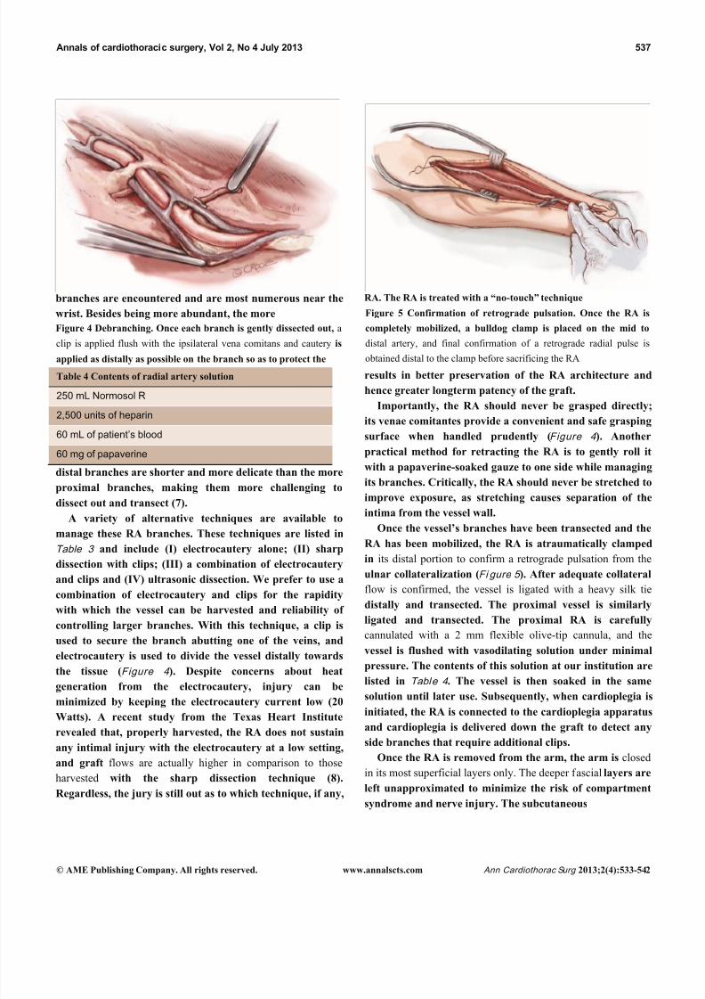
Figure 5 Confirmation of retrograde pulsation. Once the RA is
completely mobilized, a bulldog clamp is placed on the mid to
distal artery, and final confirmation of a retrograde radial pulse is
obtained distal to the clamp before sacrificing the RA
Table 4 Contents of radial artery solution
250 mL Normosol R
2,500 units of heparin
60 mL of patient’s blood
60 mg of papaverine
distal branches are shorter and more delicate than the more
proximal branches, making them more challenging to
dissect out and transect (7).
A variety of alternative techniques are available to
manage these RA branches. These techniques are listed in
Table 3 and include (I) electrocautery alone; (II) sharp
dissection with clips; (III) a combination of electrocautery
and clips and (IV) ultrasonic dissection. We prefer to use a
combination of electrocautery and clips for the rapidity
with which the vessel can be harvested and reliability of
controlling larger branches. With this technique, a clip is
used to secure the branch abutting one of the veins, and
electrocautery is used to divide the vessel distally towards
the tissue (Figure 4 ). Despite concerns about heat
generation from the electrocautery, injury can be
minimized by keeping the electrocautery current low (20Watts). A recent study from the Texas Heart Institute
revealed that, properly harvested, the RA does not sustain
any intimal injury with the electrocautery at a low setting,
and graft flows are actually higher in comparison to those
harvested with the sharp dissection technique (8).
Regardless, the jury is still out as to which technique, if any,
results in better preservation of the RA architecture and
hence greater longterm patency of the graft.
Importantly, the RA should never be grasped directly;
its venae comitantes provide a convenient and safe grasping
surface when handled prudently (Figure 4 ). Another
practical method for retracting the RA is to gently roll it
with a papaverine-soaked gauze to one side while managing
its branches. Critically, the RA should never be stretched to
improve exposure, as stretching causes separation of the
intima from the vessel wall.
Once the vessel’s branches have been transected and the
RA has been mobilized, the RA is atraumatically clamped
in its distal portion to confirm a retrograde pulsation from the
ulnar collateralization (Fi gure 5 ). After adequate collateral
flow is confirmed, the vessel is ligated with a heavy silk tie
distally and transected. The proximal vessel is similarly
ligated and transected. The proximal RA is carefully
cannulated with a 2 mm flexible olive-tip cannula, and the
vessel is flushed with vasodilating solution under minimal
pressure. The contents of this solution at our institution are
listed in Table 4 . The vessel is then soaked in the same
solution until later use. Subsequently, when cardioplegia isinitiated, the RA is connected to the cardioplegia apparatus
and cardioplegia is delivered down the graft to detect any
side branches that require additional clips.
Once the RA is removed from the arm, the arm is closed
in its most superficial layers only. The deeper fascial layers are
left unapproximated to minimize the risk of compartment
syndrome and nerve injury. The subcutaneous

8/11/2019 Harvesting the Radial Artery
http://slidepdf.com/reader/full/harvesting-the-radial-artery 6/11
538 Blitz et al. Harvesting the Radial Artery
© AME Publishing Company. All rights reserved. www.annalscts.com Ann Cardiothorac Surg 2013;2(4):533-542
to some degree) (Photo courtesy of Maquet)
layers are closed as per convention.
Endoscopic technique for radial artery harvest
Endoscopic Radial Artery Harvest (ERAH) has been rising
in popularity in recent years as a result of increasing
familiarity with endoscopic vein harvesting and expanding
use of the RA as a conduit. The specific ERAH technology
chosen depends on the experience of the harvester and the
individual institution. Prepping and positioning are the
same as for the open technique.
There are two categories of systems for ERAH: the open
system and the sealed system. The open system uses a
specialized retractor for endoscopic exposure, but CO2 is
not delivered in a pressurized fashion, as the system
remains open to the atmosphere. The closed system delivers
CO2 insufflation at a controlled pressure to aid visualization;
the wound is sealed at the scope entry site with a specialized
balloon. The authors are familiar with the latter technique
using the Vasoview Endoscopic Vessel Harvesting System
(Maquet) and this is the approach that will be described
below (F igures 6,7 ).
A 3 cm longitudinal incision is made over the RA, ending
1 cm proximal to the wrist flexion crease. The RA and its venae
comitantes are identified under direct vision (Fi gure 8 ). The
fascia overlying the pedicle is divided with scissors as far
proximally as possible under direct vision to create room
for scope entry.
A sterile tourniquet is then applied to the upper arm,and the entire arm is wrapped tightly with a sterile Esmark
bandage from its distal to proximal end (Fi gure 9 ). The
sterile tourniquet is inflated to 75 mmHg above the systolic
pressure (not to exceed 200 mmHg), and the Esmark
removed. This will create a bloodless field. It is important to
complete the open distal RA exposure prior to tourniquet
application to minimize ischemic time. The start and stop
times of tourniquet inflation should be noted and recorded, and
every effort expended to keep its duration under 60 minutes.
The components of the Vasoview System can be seen in
F igur es 6,7 . Fi gure 6 depicts the complete component setfor one of the more recent generations of the Vasoview
System. Fi gure 7 shows the Harvest cannula inserted into
the blunt-tipped trocar (BTT) port in situ in a human arm.
To commence the dissection with the Vasoview System, the
clear bullet tipped dissector is threaded on to the scope tip.
The blunt-tipped trocar (BTT) is then pre-loaded onto
Figure 6 Components of the Vasoview System. Components include the 7 mm scope with the dissection tip, the BTT port with an inflatable
balloon, the harvesting cannula and bipolar scissors, along with seals, adapters and connectors. In the first stage of the RA harvest, the 7 mm scope,
dissection tip and BTT port are used to dissect out the radial pedicle. In the second stage of the harvest, the 7 mm scope, the harvesting
cannula and bipolar scissors are used to manage the branches (Here, the Vasoview 7 System is shown; other generations vary

8/11/2019 Harvesting the Radial Artery
http://slidepdf.com/reader/full/harvesting-the-radial-artery 7/11

8/11/2019 Harvesting the Radial Artery
http://slidepdf.com/reader/full/harvesting-the-radial-artery 8/11
540 Blitz et al. Harvesting the Radial Artery
© AME Publishing Company. All rights reserved. www.annalscts.com Ann Cardiothorac Surg 2013;2(4):533-542
the scope, and the dissector is advanced anteriorly over the
RA. Once the dissector is advanced approximately 3 cm,
the BTT is slid down over the scope into the incision. The
BTT contains a balloon that is inflated with sequential 5 cc
aliquots of air (up to 25 cc) until a seal is created (Figure7 ). The gas line is connected to the insufflation port, and
CO2 is insufflated at a rate of 3-5 L/min under a pressure of
10-12 mmHg. The dissector is then used to bluntly
dissect the RA and its venae comitantes as a pedicle from
the surrounding tissue. The dissector is advanced
anteriorly (Figure 10 ), withdrawn, then advanced
osteriorly (Figure 11 ), and withdrawn once again.
Significantly, whenever the dissector is advanced, actual
contact with the RA itself should be avoided if possible;accordingly, the dissector is biased slightly to either side of
the RA during advancement, so that any contact made is
ith the venae comitantes
Figure 10 View from within the scope during anterior
advancement. The scope is being advanced anterior and to the left
over the RA, which can be seen in the 5 o’clock position, along
with its venae comitantes at the 4 and 6 o’clock positions. Note
that the scope is biased over one of the veins to minimize contact
with the RA itself (Photo courtesy of Maquet)
Figure 11 View from within the scope during posterior
advancement. The scope is being advanced posterior and to the
left under the RA, which can be seen in the 1 o’clock position,
along with its venae comitantes at the 11 and 2 o’clock positions.
Note again that the scope is biased over one of the veins to
minimize contact with the RA itself
(Photo courtesy of Maquet)

8/11/2019 Harvesting the Radial Artery
http://slidepdf.com/reader/full/harvesting-the-radial-artery 9/11
Annals of cardiothoracic surgery, Vol 2, No 4 July 2013 541
© AME Publishing Company. All rights reserved. www.annalscts.com Ann Cardiothorac Surg 2013;2(4):533-542
Figure 12 Fasciotomy of the BRM-FCRM fascia. A fasciotomy is
being performed via the harvesting cannula with a cauterizing tool
at the 2 o’clock position. The RA can be seen at the 5 o’clock
position
(Photo courtesy of Maquet)
instead. In addition, the scope should be slightly torqued
away from the pedicle, transmitting any forces to the
surrounding tissue. Finally, as the dissection proceeds and
branches are encountered, tissue should be judiciously
cleared around the branches and the branches themselves
should be minimally displaced. The dissector advancement
should be up to the level of the RRA or the venous plexus in
the antecubital fossa, depending on the scope’s relative
position with respect to the RA.
Once the dissection is complete, the scope is withdrawn,
the dissection tip is removed from the scope and the scope is
then inserted into the harvest cannula. The harvest cannula
contains several ports through which different tools can be
inserted and advanced (Figures 6,7 ). Via the harvest
cannula, the cautery instrument can be introduced to
perform a fasciotomy of the BRM-FCRM fascia (Fi gure 12 ).
This will create space to facilitate the harvest and to reduce
the risk of compartment syndrome developing in the
forearm postoperatively. A cautery instrument is then used
to divide the side branches of the RA, while a vessel cradle
keeps the RA displaced 2.5 cm away from the cautery
(Figure 13 ). The different generations of the Vasoview
System offer an assortment of dividing/ligating technologies,
including bipolar scissors, bipolar ligating bisector tool and
direct

8/11/2019 Harvesting the Radial Artery
http://slidepdf.com/reader/full/harvesting-the-radial-artery 10/11
542 Blitz et al. Harvesting the Radial Artery
© AME Publishing Company. All rights reserved. www.annalscts.com Ann Cardiothorac Surg 2013;2(4):533-542
Figure 14 Running the pedicle with the vessel cradle. Upon
reaching the distal end of the pedicle, the vessel cradle is used to
run the pedicle and ensure no branches have been missed (Photo
courtesy of Maquet)
current cut-and-seal ligating graspers. [One should refer to
the specific Instructions for Use (IFU) for each device to
learn the particulars of each]. Importantly, minimal
stretching or torqueing of the pedicle should be applied
when addressing the branches so as to not incur intimal
injury to the RA. Once all the side branches are divided
from the RA, the cradle is slid gently up and down the
pedicle to confirm completeness of debranching (Fi gure 14 ).
A 1 cm incision is made externally at the proximal end of
the harvest tunnel, 1 cm distal to the antecubital crease.
Prior to making the incision, its appropriate location is
verified by pushing down on the overlying skin while
visualizing within the tunnel with the scope. After the
incision is made, a hemostat is used to retrieve the RApedicle (Fi gure 15 ), bring it to skin level, ligate the proximal
stump, and divide the radial graft from the stump. The
endoscope is then use to withdraw the RA from the harvest
tunnel using the cradle, and the distal end is similarly
ligated and divided. The radial graft is then cannulated as
previously described, and flushed with solution. To ensure
hemostasis of the tunnel, the endoscope is reintroduced, the
tourniquet is released, and the tunnel is inspected for
potential bleeders that are then addressed. The incisions
are then closed, sterile dressings are applied, and an ace
bandage loosely wrapped around the forearm.
Comments
The RA has been assuming an increasingly prominent
Figure 15 Retrieval of RA. After the incision in the skin over the
antecubital space is complete, a mosquite clamp is placed via the
Figure 13 Debranching via the Harvesting Cannula. A branch of the RA is divided with the use of a cautery tool and the vessel cradle in
oth the illustration (A) and the endoscopic photo (B). In the latter, the cautery tool is seen at the 7 o’clock position. A vessel cradle is seen
from the 11 o’clock to the 2 o’clock position and is retracting the RA away from the cautery tool (Photo courtesy of Maquet)

8/11/2019 Harvesting the Radial Artery
http://slidepdf.com/reader/full/harvesting-the-radial-artery 11/11
Annals of cardiothoracic surgery, Vol 2, No 4 July 2013 543
© AME Publishing Company. All rights reserved. www.annalscts.com Ann Cardiothorac Surg 2013;2(4):533-542
incision and — under endoscopic vision — used to grasp the radial
pedicle 5-7 mm distal to the RRA. The pedicle is gingerly
withdrawn through the incision, the RA pedicle is divided just
distal to the clamp, and the vessel beneath the clamp is ligated
(Photo courtesy of Maquet)
role in arterial revascularization, often being used when
additional arterial conduits are desired in conjunction with
the internal mammary arteries. The surgical
armamentarium for harvesting is multifold, including (I)
either an open or endoscopic operative approach; (II)
alternative dissection planes and (III) alternative methods
of handling the RA branches. In this article we present both
our open and endoscopic approaches. Although much work
still needs to be done to fully elucidate which approaches or
techniques — if any — are superior, the most importantuniversal dictum is to pay great respect to the RA’s
propensity for vasospasm. A “no touch” technique will
Cite this article as: Blitz A, Osterday RM, Brodman RF.
Harvesting the radial artery. Ann Cardiothorac Surg
2013;2(4):533-542. doi: 10.3978/j.issn.2225-319X.2013.07.10
ensure the optimal quality and longevity of the RA conduit,
whatever harvesting methodology is chosen.
Acknowledgements
We would like to thank Maquet Getinge Group for
providing photographs and illustrations for the endoscopic
harvesting portion of the manuscript.
Disclosure: The authors declare no conflict of interest.
References
1. Deb S, Cohen EA, Singh SK, et al. Radial artery and
saphenous vein patency more than 5 years after coronary
artery bypass surgery: results from RAPS (Radial Artery
Patency Study). J Am Coll Cardiol 2012;60:28-35.
2. Reyes AT, Frame R, Brodman RF. Technique for
harvesting the radial artery as a coronary artery bypass
graft. Ann Thorac Surg 1995;59:118-26.
3. Conklin LD, Ferguson ER, Reardon MJ. The technical
aspects of radial artery harvesting. Tex Heart Inst J
2001;28:129-31.
4. Beldner S, Zlotolow DA, Melone CP Jr, et al. Anatomy of
the lateral antebrachial cutaneous and superficial radial
nerves in the forearm: a cadaveric and clinical study. J
Hand Surg Am 2005;30:1226-30.
5. Taggart DP, Mathur MN, Ahmad I. Skeletonization of the
radial artery: advantages over the pedicled technique.
Ann Thorac Surg 2001;72:298-9.
6. Sajja LR, Mannam G, Sompalli S. Extrafascially
harvested radial artery in CABG: technique of harvest,
complications, and mid-term angiographic patency. J
Card Surg 2005;20:440-8.
7. Strauch B. YH, Chen ZW, Liebling R. Forearm Region.
Atlas of Microvascular Surgery: Anatomy and Operative
Approaches. New York: Thieme Medical. 1993:44-83.
8. Marzban M, Arya R, Mandegar MH, et al. Sharp
dissection versus electrocautery for radial artery
harvesting. Tex Heart Inst J 2006;33:9-13.