purkinje cell maturation participates in the control of
TRANSCRIPT
Purkinje cell maturation participates in the control of
oligodendrocyte differentiation: role of Sonic Hedgehog
and Vitronectin
Lamia Bouslama-Oueghlani, Rosine Wehrle, Mohamed Doulazmi, Xiaoru
Chen, Yolande Lemaigre-Dubreuil, Isabelle Rivals, Constantino Sotelo,
Isabelle Dusart
To cite this version:
Lamia Bouslama-Oueghlani, Rosine Wehrle, Mohamed Doulazmi, Xiaoru Chen, YolandeLemaigre-Dubreuil, et al.. Purkinje cell maturation participates in the control of oligoden-drocyte differentiation: role of Sonic Hedgehog and Vitronectin. PLoS ONE, Public Library ofScience, 2012, 7 (11), pp.e49015. <10.1371/journal.pone.0049015>. <hal-00800364>
HAL Id: hal-00800364
https://hal-espci.archives-ouvertes.fr/hal-00800364
Submitted on 13 Mar 2013

HAL is a multi-disciplinary open accessarchive for the deposit and dissemination of sci-entific research documents, whether they are pub-lished or not. The documents may come fromteaching and research institutions in France orabroad, or from public or private research centers.
L’archive ouverte pluridisciplinaire HAL, estdestinee au depot et a la diffusion de documentsscientifiques de niveau recherche, publies ou non,emanant des etablissements d’enseignement et derecherche francais ou etrangers, des laboratoirespublics ou prives.
Purkinje Cell Maturation Participates in the Control ofOligodendrocyte Differentiation: Role of SonicHedgehog and VitronectinLamia Bouslama-Oueghlani1,2, Rosine Wehrle1,2, Mohamed Doulazmi1,2, Xiao Ru Chen1,2, Fanny Jaudon3,
Yolande Lemaigre-Dubreuil1,2, Isabelle Rivals4, Constantino Sotelo1,2,5, Isabelle Dusart1,2*
1 Neurobiologie des Processus Adaptatif, Universite Pierre et Marie Curie (UPMC Univ Paris 06), Paris, France, 2 Neurobiologie des Processus Adaptatif, CNRS (Centre
National de Recherche Scientifique), Paris, France, 3 Centre de Recherche de Biochimie Macromoleculaire, Universite Montpellier 1 et 2, CNRS UMR 5237, Montpellier,
France, 4 Equipe de statistique Appliquee, ESPCI ParisTech (Ecole Superieure de Physique et Chimie Industrielles de la Ville de Paris), Paris, France, 5 Instituto de
Neurociencias, Universidad Miguel Hernandez–CSIC, San Juan de Alicante, Spain
Abstract
Oligodendrocyte differentiation is temporally regulated during development by multiple factors. Here, we investigatedwhether the timing of oligodendrocyte differentiation might be controlled by neuronal differentiation in cerebellarorganotypic cultures. In these cultures, the slices taken from newborn mice show very few oligodendrocytes during the firstweek of culture (immature slices) whereas their number increases importantly during the second week (mature slices). First,we showed that mature cerebellar slices or their conditioned media stimulated oligodendrocyte differentiation in immatureslices thus demonstrating the existence of diffusible factors controlling oligodendrocyte differentiation. Using conditionedmedia from different models of slice culture in which the number of Purkinje cells varies drastically, we showed that theeffects of these differentiating factors were proportional to the number of Purkinje cells. To identify these diffusible factors,we first performed a transcriptome analysis with an Affymetrix array for cerebellar cortex and then real-time quantitativePCR on mRNAs extracted from fluorescent flow cytometry sorted (FACS) Purkinje cells of L7-GFP transgenic mice at differentages. These analyses revealed that during postnatal maturation, Purkinje cells down-regulate Sonic Hedgehog and up-regulate vitronectin. Then, we showed that Sonic Hedgehog stimulates the proliferation of oligodendrocyte precursor cellsand inhibits their differentiation. In contrast, vitronectin stimulates oligodendrocyte differentiation, whereas its inhibitionwith blocking antibodies abolishes the conditioned media effects. Altogether, these results suggest that Purkinje cellsparticipate in controlling the timing of oligodendrocyte differentiation in the cerebellum through the developmentallyregulated expression of diffusible molecules such as Sonic Hedgehog and vitronectin.
Citation: Bouslama-Oueghlani L, Wehrle R, Doulazmi M, Chen XR, Jaudon F, et al. (2012) Purkinje Cell Maturation Participates in the Control of OligodendrocyteDifferentiation: Role of Sonic Hedgehog and Vitronectin. PLoS ONE 7(11): e49015. doi:10.1371/journal.pone.0049015
Editor: Ya-Ping Tang, Louisiana State University Health Sciences Center, United States of America
Received November 24, 2011; Accepted October 8, 2012; Published November 14, 2012
Copyright: � 2012 Bouslama-Oueghlani et al. This is an open-access article distributed under the terms of the Creative Commons Attribution License, whichpermits unrestricted use, distribution, and reproduction in any medium, provided the original author and source are credited.
Funding: This work was supported by the Centre National de la Recherche Scientifique (CNRS): ATIP, University Pierre et Marie Curie (UPMC), the Institut NationalScientifique pour la Recherche Medicale (INSERM), Association pour la Recherche sur le Cancer (ARC, contract 3532), RGN (Reseau Genopole National, microarraysubvention) and the Agence National pour le Recherche (ANR, ANR-07-NEURO-043-01). The funders had no role in study design, data collection and analysis,decision to publish, or preparation of the manuscript.
Competing Interests: The authors have declared that no competing interests exist.
* E-mail: [email protected]
Introduction
Oligodendrocytes are central nervous system macroglial cells
that synthesize myelin, a multilayered membrane ensheathing
axons which facilitates rapid nerve conduction [1]. During
development, oligodendrocyte precursor cells (OPCs) divide and
migrate over long distances to reach their final destination where
they differentiate into mature oligodendrocytes and produce
myelin.
Neuron maturation affects oligodendrocyte survival and the
timing of myelin formation, OPCs nonetheless differentiate into
mature oligodendrocytes and generate a myelin sheath in the
absence of axons in vitro [2,3]. In the optic nerve, only the
oligodendrocytes ensheathing axons survive [4,5]. Oligodendro-
cytes are more abundant in transgenic mice with larger numbers
of axons [6]. Myelin formation is correlated with certain
parameters of axonal maturation, such as axon caliber and
neurofilament content [7–9]. Axonal factors which are directly
involved in controlling myelin formation include neuronal
electrical activity [10,11] and the downregulation of various
molecules in axonal membranes, including Jagged1, PSA-NCAM
(polysialic acid-neural cell adhesion molecule) and N-cadherin
[12–14]. Myelin membrane formation is coordinated by a large
number of proteins, through contact mechanisms and integrin
receptors [15]. Furthermore, Rosenberg and colleagues demon-
strated that myelin formation required an axonal microenviron-
ment and a critical density of OPCs [16].
The role of neurons in the switch between OPC proliferation
and differentiation into oligodendrocytes remains unclear. The
timing of this switch depends on both the intracellular timer and
extrinsic factors [17]. For several years, thyroid hormone (T3),
retinoic acid (RA), glucocorticoids and transforming growth factor
(TGFß) were the only molecules known to trigger the initial stages
of OPC differentiation [18,19]. More recently, neuronal activity
PLOS ONE | www.plosone.org 1 November 2012 | Volume 7 | Issue 11 | e49015
has also been shown to participate in OPC differentiation.
Purinergic receptor activation by non-synaptically released aden-
osine [20] stimulates the differentiation of OPCs into oligoden-
drocytes. Thus, reciprocal neuron-glial interactions are also
required for the complete conversion of OPCs into differentiated
oligodendrocytes. These neuron-glial interactions do not always
have positive effects; connective tissue growth factor (CFTG) has
been reported to inhibit the differentiation of OPCs into
oligodendrocytes through interactions with serum response factor
(SRF), a neuronal transcription factor [21].
In this study, we investigated the existence of neuronal soluble
factors controlling oligodendrocyte differentiation in an integrated
system. For that purpose, we used cerebellar organotypic cultures,
in which neuron-glial interactions mimic those occurring in vivo
and in which only one type of neuron, the Purkinje cell, is
myelinated [22]. We demonstrated that the maturation of Purkinje
cells is one of the key factors controlling the timing of
oligodendrocyte differentiation. Indeed, Purkinje cells timely
release two factors, Sonic Hedgehog (Shh) and vitronectin (VN),
which respectively stimulate OPC proliferation and oligodendro-
cyte differentiation.
Results
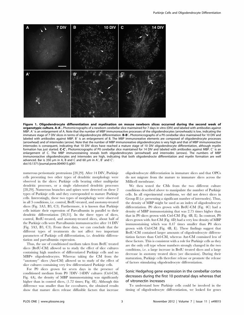
Newborn cerebellar slice cultures are immature foroligodendrocyte differentiation during the first week invitro and become mature during the second week
To analyze the timing of the oligodendrocyte differentiation
process in cerebellar slice cultures, we focused on the expression of
MBP because this protein is expressed in mature oligodendrocytes
(both pre- and myelinating oligodendrocytes [1,23]). MBP
immunostaining was detected both on the processes of mature
oligodendrocytes (Fig. 1A9, B9, C9) and the myelin segments
(internodes, Fig. 1B9, C9). The appearance of this protein therefore
reflects the last stages of oligodendrocyte differentiation.
P0 cerebellar slices grown for 7 DIV had very few MBP+oligodendrocytes (Fig. 1A, 1A9), and very few if any MBP+internodes were observed at this stage. Indeed, most of the slices
(over 75%) did not present any internodes (Fig. 1A), and we did
not detect more than 25 MBP+ internodes on the other slices. We
defined Group I as the slices containing less than 25 internodes
and Group II as the slices containing more than 26 internodes.
Thus, on P0–7 DIV slices, the density of MBP staining can be used
as an index of oligodendrocyte differentiation, since very few
internodes were present.
At 10 DIV, MBP+ oligodendrocytes were present throughout
the slices (Fig. 1B) and MBP+ internodes can be detected (Fig. 1B9).
At 10 DIV, over 50% of the slices exhibited more than 26
internodes per slice and were therefore in Group II. The process of
oligodendrocyte differentiation is well engaged, whereas myelina-
tion had only just started.
After 14 DIV, the number of MBP+ internodes increased
(Fig. 1C). Almost all of the slices contained more than 26
internodes and were therefore in Group II. Furthermore, myelin
tracts can often be observed (Fig. 1C9). At 14 DIV, the process of
myelination is already well engaged.
Our results therefore showed that most OPCs differentiated into
MBP+ oligodendrocytes between 7 and 10 DIV and most of the
myelination process occurred between 10 and 14 DIV. P0 slices
were thus considered to be immature for oligodendrocyte
differentiation during the first 7 DIV and to be mature during
the following week, in which rapid oligodendrocyte differentiation
occurred.
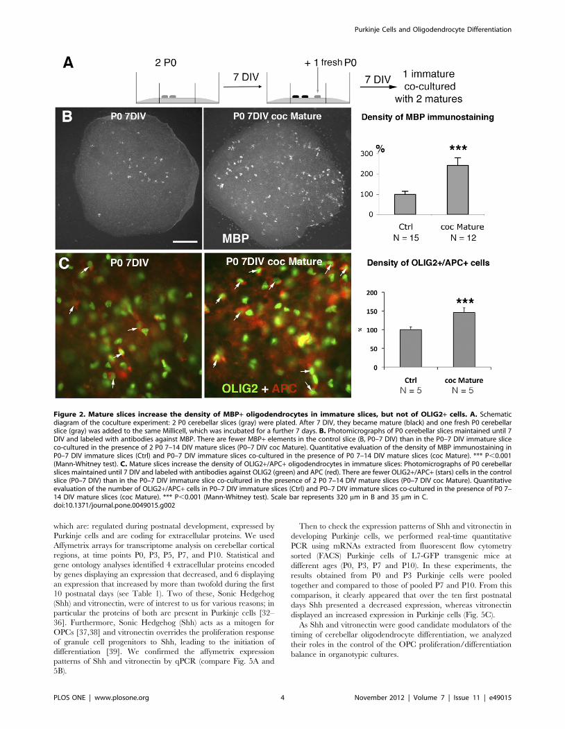
Mature cerebellar slices release factors that increaseoligodendrocyte differentiation in immature slices
We investigated whether, during the phase of oligodendrocyte
differentiation, cells in the slice culture (between 7 and 14 DIV)
were able to increase oligodendrocyte differentiation on immature
slices (7 DIV). To this aim, P0 cerebellar slices were grown in
culture until 7 DIV, when the differentiation of oligodendrocytes
was beginning and could be called ‘‘mature’’. At this time point,
fresh P0 slices (immature) were added to the Millicell (Fig. 2A)
such that the ratio of immature/mature slices was 1:2. After 7
more DIV, we analyzed the MBP immunostaining on the
immature slices (P0–7DIV control (ctrl) or grown in presence of
mature slices (coc Mature)). We did not detect any increase in the
presence of internodes; all of the P0–7 DIV slices (control slices or
slices grown in the presence of mature slices) were in Group I (i.e.
presenting less than 25 internodes). The MBP+ oligodendrocytes
were evenly distributed in the slices (Fig. 2B). The density of MBP
immunostaining was 2.44 times higher in immature slices grown in
the presence of mature slices (P0–7 DIV Coc-Mature) compared
with control immature slices (P,0.001, Fig. 2B).
Then to confirm that this increase of MBP immunostaining was
indeed due to an increase of the number of oligodendrocytes, we
evaluated the density of APC+/OLIG2+ cells on the slice cultures.
APC is expressed by oligodendrocytes and some astrocytes
[24,25], whereas OLIG2+ is expressed by both OPCs and
oligodendrocytes [26]. Thus, APC+/OLIG2+ cells are oligoden-
drocytes. We found a significant increase in the number of
OLIG2+/APC+ cells in 7 DIV slices grown in presence of mature
slices compared to 7 DIV control slices (Fig. 2C).
These results showed that mature slices release factors that
increase oligodendrocyte differentiation in immature ones. They
also suggest that the switch from OPC proliferation to oligoden-
drocyte differentiation in cerebellar organotypic cultures is
controlled by factors released from cerebellar cells at critical
stages of maturity. Then we investigated whether Purkinje cells
might influence the timing of oligodendrocyte differentiation in
cerebellar slices. Indeed the number of Purkinje cells can be
manipulated during the first week in vitro, i.e. before the addition
of immature slices.
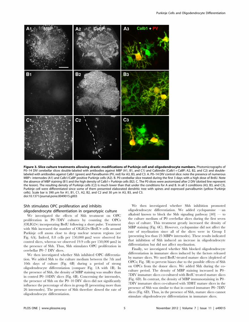
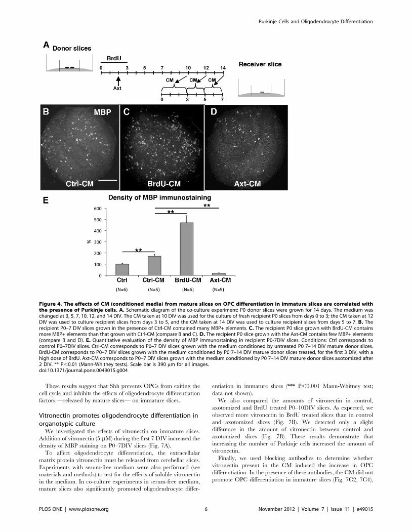
The oligodendrocyte differentiation effect of matureslices is proportional to the number of Purkinje cells
To investigate whether Purkinje cells might control the secretion
of these differentiation factors, we used two different slice-culture
models in which the numbers of Purkinje cells were markedly
different. To increase the number of Purkinje cells, we applied a
BrdU treatment that kills dividing cells during the first 3 DIV of
the culture [27]. In this model, after 14 DIV, the BrdU-treated
slices contained no MBP+ elements (Fig. 3B1) and much more
Purkinje cells than the P0–14DIV control slices (compare Figs. 3B2
with 3A2).
To decrease the number of Purkinje cells, we took advantage of
the fact that newborn Purkinje cells axotomized after 2 DIV, die in
large numbers in organotypic culture. Indeed, axotomized slices
contained small numbers of surviving Purkinje cells in the second
week of culture (Fig. 3C2). These axotomized slices contained
some MBP+ oligodendrocytes (Fig. 3C1).
We then evaluated whether the Purkinje cells have reached a
differentiated stage at 14 DIV in all 3 different conditions (Ctrl,
BrdU-treated slices or axotomy-treated slices).
We previously showed that at the time of the culture, newborn
Purkinje cells were bipolar, whereas after 7 DIV, most Purkinje
cells have retracted their primitive processes and developed
Purkinje Cells and Oligodendrocyte Differentiation
PLOS ONE | www.plosone.org 2 November 2012 | Volume 7 | Issue 11 | e49015
numerous perisomatic protrusions [28,29]. After 14 DIV, Purkinje
cells presenting two other types of dendritic morphology were
observed in the slices: Purkinje cells bearing either multipolar
dendritic processes, or a single elaborated dendritic processes
[28,29]. Numerous branches and spines were detected on these 2
types of Purkinje cells and they corresponded to mature Purkinje
cells. Interestingly, these two types of morphology were observed
in all 3 conditions, i.e. control, BrdU-treated, and axotomy-treated
slices (Fig. 3A3, B3, C3). Furthermore, it is known that Purkinje
cells initiate their expression of Parvalbumin in parallel to their
dendritic differentiation [30,31]. In the three types of slices,
control, BrdU-treated, and axotomy-treated slices, about half of
the Purkinje cells were Parvalbumin immunoreactive after 14 DIV
(Fig. 3A3, B3, C3). From these data, we can conclude that the
different types of treatments do not affect two important
parameters of Purkinje cell differentiation, i.e. dendritic differen-
tiation and parvalbumin expression.
Thus, the use of conditioned medium taken from BrdU treated
slices (BrdU-CM) allowed us to study the effect of slice cultures
containing high numbers of differentiated Purkinje cells and no
MBP+ oligodendrocytes. Whereas taking the CM from the
‘‘axotomy’’ slices (Axt-CM) allowed us to study of the effect of
slice cultures containing very few differentiated Purkinje cells.
For P0 slices grown for seven days in the presence of
conditioned medium from P0 7DIV–14DIV cultures (Ctrl-CM,
Fig. 4A), the density of MBP immunostaining was significantly
higher than in control slices, (1.72 times; Fig. 4E). Although this
difference was smaller than for co-cultures, the obtained results
show that mature slices release diffusible factors that increase
oligodendrocyte differentiation in immature slices and that OPCs
do not migrate from the mature to immature slices across the
Millicell membrane.
We then tested the CMs from the two different culture
conditions described above to manipulate the number of Purkinje
cells. In all experimental conditions, we did not detect slices in
Group II (i.e. presenting a significant number of internodes). Thus,
the density of MBP might be used as an index of oligodendrocyte
differentiation. P0 slices grown with BrdU-CM (Fig. 4C) had a
density of MBP immunostaining that was 2.75 times higher than
that in P0 slices grown with Ctrl-CM (Fig. 4B, E). In contrast, P0
slices grown with Axt-CM (Fig. 4D) had a very low density of MBP
immunostaining which was 8.47 times smaller than P0 slices
grown with Ctrl-CM (Fig. 4B, E). These findings suggest that
BrdU-CM contained larger amounts of oligodendrocyte differen-
tiation factors than Ctrl-CM, whereas Axt-CM contained less of
these factors. This is consistent with a role for Purkinje cells as they
are the only cell type whose numbers strongly changed in the two
conditions, i.e. a large increase in BrdU treated slices and a large
decrease in axotomy treated slices (see discussion). During their
maturation, Purkinje cells therefore release or promote the release
of factors stimulating oligodendrocyte differentiation.
Sonic Hedgehog gene expression in the cerebellar cortexdecreases during the first 10 postnatal days whereas thatof vitronectin increases
To understand how Purkinje cells could be involved in the
timing of oligodendrocyte differentiation, we looked for genes
Figure 1. Oligodendrocyte differentiation and myelination on mouse newborn slices occurred during the second week oforganotypic culture. A-A9. Photomicrographs of a newborn cerebellar slice maintained for 7 days in vitro (DIV) and labeled with antibodies againstMBP. A9 is an enlargement of A. Note that the number of MBP immunoreactive processes of the oligodendrocytes (arrowheads) is low, indicating theimmature stage of 7 DIV slices in terms of oligodendrocyte differentiation. B-B9. Photomicrographs of a P0 cerebellar slice maintained for 10 DIV andlabeled with antibodies against MBP. B9 is an enlargement of B. The MBP immunoreative elements are composed of oligodendrocyte processes(arrowhead) and of internodes (arrow). Note that the number of MBP immunoreative oligodendrocytes is very high and that of MBP immunoreactiveinternodes is consequent, indicating that 10 DIV slices have reached a mature stage of 10 DIV oligodendrocyte differentiation, although myelinformation has just started. C-C9. Photomicrographs of P0 cerebellar slice maintained for 14 DIV and labeled with antibodies against MBP. C9 is anenlargement of C. The MBP immunostaining reveals both oligodendrocytes (arrowhead) and internodes (arrows). The numbers of MBPimmunoreactive oligodendrocytes and internodes are high, indicating that both oligodendrocte differentiation and myelin formation are welladvanced. Bar is 350 mm in A, B and C and 60 mm in A9, B9 and C9.doi:10.1371/journal.pone.0049015.g001
Purkinje Cells and Oligodendrocyte Differentiation
PLOS ONE | www.plosone.org 3 November 2012 | Volume 7 | Issue 11 | e49015
which are: regulated during postnatal development, expressed by
Purkinje cells and are coding for extracellular proteins. We used
Affymetrix arrays for transcriptome analysis on cerebellar cortical
regions, at time points P0, P3, P5, P7, and P10. Statistical and
gene ontology analyses identified 4 extracellular proteins encoded
by genes displaying an expression that decreased, and 6 displaying
an expression that increased by more than twofold during the first
10 postnatal days (see Table 1). Two of these, Sonic Hedgehog
(Shh) and vitronectin, were of interest to us for various reasons; in
particular the proteins of both are present in Purkinje cells [32–
36]. Furthermore, Sonic Hedgehog (Shh) acts as a mitogen for
OPCs [37,38] and vitronectin overrides the proliferation response
of granule cell progenitors to Shh, leading to the initiation of
differentiation [39]. We confirmed the affymetrix expression
patterns of Shh and vitronectin by qPCR (compare Fig. 5A and
5B).
Then to check the expression patterns of Shh and vitronectin in
developing Purkinje cells, we performed real-time quantitative
PCR using mRNAs extracted from fluorescent flow cytometry
sorted (FACS) Purkinje cells of L7-GFP transgenic mice at
different ages (P0, P3, P7 and P10). In these experiments, the
results obtained from P0 and P3 Purkinje cells were pooled
together and compared to those of pooled P7 and P10. From this
comparison, it clearly appeared that over the ten first postnatal
days Shh presented a decreased expression, whereas vitronectin
displayed an increased expression in Purkinje cells (Fig. 5C).
As Shh and vitronectin were good candidate modulators of the
timing of cerebellar oligodendrocyte differentiation, we analyzed
their roles in the control of the OPC proliferation/differentiation
balance in organotypic cultures.
Figure 2. Mature slices increase the density of MBP+ oligodendrocytes in immature slices, but not of OLIG2+ cells. A. Schematicdiagram of the coculture experiment: 2 P0 cerebellar slices (gray) were plated. After 7 DIV, they became mature (black) and one fresh P0 cerebellarslice (gray) was added to the same Millicell, which was incubated for a further 7 days. B. Photomicrographs of P0 cerebellar slices maintained until 7DIV and labeled with antibodies against MBP. There are fewer MBP+ elements in the control slice (B, P0–7 DIV) than in the P0–7 DIV immature sliceco-cultured in the presence of 2 P0 7–14 DIV mature slices (P0–7 DIV coc Mature). Quantitative evaluation of the density of MBP immunostaining inP0–7 DIV immature slices (Ctrl) and P0–7 DIV immature slices co-cultured in the presence of P0 7–14 DIV mature slices (coc Mature). *** P,0.001(Mann-Whitney test). C. Mature slices increase the density of OLIG2+/APC+ oligodendrocytes in immature slices: Photomicrographs of P0 cerebellarslices maintained until 7 DIV and labeled with antibodies against OLIG2 (green) and APC (red). There are fewer OLIG2+/APC+ (stars) cells in the controlslice (P0–7 DIV) than in the P0–7 DIV immature slice co-cultured in the presence of 2 P0 7–14 DIV mature slices (P0–7 DIV coc Mature). Quantitativeevaluation of the number of OLIG2+/APC+ cells in P0–7 DIV immature slices (Ctrl) and P0–7 DIV immature slices co-cultured in the presence of P0 7–14 DIV mature slices (coc Mature). *** P,0.001 (Mann-Whitney test). Scale bar represents 320 mm in B and 35 mm in C.doi:10.1371/journal.pone.0049015.g002
Purkinje Cells and Oligodendrocyte Differentiation
PLOS ONE | www.plosone.org 4 November 2012 | Volume 7 | Issue 11 | e49015
Shh stimulates OPC proliferation and inhibitsoligodendrocyte differentiation in organotypic culture
We investigated the effects of Shh treatment on OPC
proliferation in P0–7DIV cultures by counting the OPCs
(OLIG2+) incorporating BrdU following a short pulse. Treatment
with Shh increased the number of OLIG2+/BrdU+ cells around
Purkinje cell axons close to deep nuclear neuron regions (see
Fig. 6A). Indeed, 8.8 cells per 150,000 mm2 were observed for
control slices, whereas we observed 19.9 cells per 150,000 mm2 in
the presence of Shh. Thus, Shh stimulates OPC proliferation in
cerebellar P0–7 DIV slices.
We then investigated whether Shh inhibited OPC differentia-
tion. We added Shh to the culture medium between the 7th and
10th days of culture (Fig. 6B) during a period of strong
oligodendrocyte differentiation (compare Fig. 1A with 1B). In
the presence of Shh, the density of MBP staining was smaller than
in control P0–10DIV slices (Fig. 6B). Concerning the internodes,
the presence of Shh on the P0–10 DIV slices did not significantly
influence the percentage of slices in group II (presenting more than
26 internodes). The presence of Shh therefore slowed the rate of
oligodendrocyte differentiation.
We then investigated whether Shh inhibition promoted
oligodendrocyte differentiation. We added cyclopamine —an
alkaloid known to block the Shh signaling pathway [40] — to
the culture medium of P0 cerebellar slices during the first seven
days of culture. This treatment greatly increased the density of
MBP staining (Fig. 6C). However, cyclopamine did not affect the
rate of myelination since all of the slices were in Group I
(presenting less than 25 MBP+ internodes). These results indicated
that inhibition of Shh induced an increase in oligodendrocyte
differentiation but did not affect myelination.
Finally, we investigated whether Shh blocked oligodendrocyte
differentiation in immature slices induced by the factors released
by mature slices. We used BrdU-treated mature slices (depleted of
OPCs; Fig. 3B) to prevent biases due to the possible effects of Shh
on OPCs from the donor slices. We added Shh during the co-
culture period. The density of MBP staining increased in P0–
7DIV immature slices co-cultured with BrdU treated mature slices
(Fig. 6D). In contrast, the density of MBP immunostaining in P0–
7DIV immature slices co-cultured with 3DBT mature slices in the
presence of Shh was similar to that in control immature P0–7DIV
slices (Fig. 6D). Thus, in the presence of Shh, mature slices cannot
stimulate oligodendrocyte differentiation in immature slices.
Figure 3. Slice culture treatments allowing drastic modifications of Purkinje cell and oligodendrocyte numbers. Photomicrographs ofP0–14 DIV cerebellar slices double-labeled with antibodies against MBP (A1, B1, and C1) and Calbindin (Calb1 = CaBP; A2, B2, and C2) and double-labeled with antibodies against Calb1 (green) and Parvalbumin (PV, red) for A3, B3, and C3. A. P0–14 DIV control slice: note the presence of numerousMBP+ internodes (A1) and Calb1/CaBP positive Purkinje cells (A2). B. P0 cerebellar slice treated during the first 3 days with a high dose of BrdU. Notethe absence of MBP staining (B1) and the high density of Calb1+ Purkinje cells (B2). C. The P0 slices were axotomized after 2 DIV (dotted line representthe lesion). The resulting density of Purkinje cells (C2) is much lower than that under the conditions for A and B. In all 3 conditions (A3, B3, and C3),Purkinje cell were differentiated since some of them presented elaborated dendritic tree with spines and expressed parvalbumin (yellow Purkinjecells). Scale bar is 390 mm for A1, B1, C1, A2, B2, and C2 and 30 mm in A3, B3, and C3.doi:10.1371/journal.pone.0049015.g003
Purkinje Cells and Oligodendrocyte Differentiation
PLOS ONE | www.plosone.org 5 November 2012 | Volume 7 | Issue 11 | e49015
These results suggest that Shh prevents OPCs from exiting the
cell cycle and inhibits the effects of oligodendrocyte differentiation
factors —released by mature slices— on immature slices.
Vitronectin promotes oligodendrocyte differentiation inorganotypic culture
We investigated the effects of vitronectin on immature slices.
Addition of vitronectin (5 mM) during the first 7 DIV increased the
density of MBP staining on P0–7DIV slices (Fig. 7A).
To affect oligodendrocyte differentiation, the extracellular
matrix protein vitronectin must be released from cerebellar slices.
Experiments with serum-free medium were also performed (see
materials and methods) to test for the effects of soluble vitronectin
in the medium. In co-culture experiments in serum-free medium,
mature slices also significantly promoted oligodendrocyte differ-
entiation in immature slices (*** P,0.001 Mann-Whitney test;
data not shown).
We also compared the amounts of vitronectin in control,
axotomized and BrdU treated P0–10DIV slices. As expected, we
observed more vitronectin in BrdU treated slices than in control
and axotomized slices (Fig. 7B). We detected only a slight
difference in the amount of vitronectin between control and
axotomized slices (Fig. 7B). These results demonstrate that
increasing the number of Purkinje cells increased the amount of
vitronectin.
Finally, we used blocking antibodies to determine whether
vitronectin present in the CM induced the increase in OPC
differentiation. In the presence of these antibodies, the CM did not
promote OPC differentiation in immature slices (Fig. 7C2, 7C4),
Figure 4. The effects of CM (conditioned media) from mature slices on OPC differentiation in immature slices are correlated withthe presence of Purkinje cells. A. Schematic diagram of the co-culture experiment: P0 donor slices were grown for 14 days. The medium waschanged at 3, 5, 7, 10, 12, and 14 DIV. The CM taken at 10 DIV was used for the culture of fresh recipient P0 slices from days 0 to 3; the CM taken at 12DIV was used to culture recipient slices from days 3 to 5, and the CM taken at 14 DIV was used to culture recipient slices from days 5 to 7. B. Therecipient P0–7 DIV slices grown in the presence of Ctrl-CM contained many MBP+ elements. C. The recipient P0 slice grown with BrdU-CM containsmore MBP+ elements than that grown with Ctrl-CM (compare B and C). D. The recipient P0 slice grown with the Axt-CM contains few MBP+ elements(compare B and D). E. Quantitative evaluation of the density of MBP immunostaining in recipient P0-7DIV slices. Conditions: Ctrl corresponds tocontrol P0–7DIV slices. Ctrl-CM corresponds to P0–7 DIV slices grown with the medium conditioned by untreated P0 7–14 DIV mature donor slices.BrdU-CM corresponds to P0–7 DIV slices grown with the medium conditioned by P0 7–14 DIV mature donor slices treated, for the first 3 DIV, with ahigh dose of BrdU. Axt-CM corresponds to P0–7 DIV slices grown with the medium conditioned by P0 7–14 DIV mature donor slices axotomized after2 DIV. ** P,0.01 (Mann-Whitney tests). Scale bar is 390 mm for all images.doi:10.1371/journal.pone.0049015.g004
Purkinje Cells and Oligodendrocyte Differentiation
PLOS ONE | www.plosone.org 6 November 2012 | Volume 7 | Issue 11 | e49015

whereas the CM alone (Fig. 7C1, 7C4), or in the presence of a
control antibody, promoted OPC differentiation (Fig. 7C3, 7C4).
Our findings suggest that Sonic Hedgehog and vitronectin play
important antagonistic roles in controlling the timing of OPC
differentiation during cerebellar development.
Discussion
Different cell types differentiate in parallel during development.
The synchronization of cell development is particularly important
for cell types that have strong interactions, such as oligodendro-
cytes and neurons. OPCs start to differentiate if a mitogenic
stimulus is removed or a differentiation stimulus is added. This cell
differentiation is inhibited in the presence of a mitogenic stimulus
and the absence of a differentiation stimulus [17]. After exiting the
cell cycle, OPCs become immature oligodendrocytes which
undergo different phases of differentiation [1,23]. The last two
phases of oligodendrocyte differentiation are the production of
myelin proteins and myelination. In the present study, we found
that Purkinje cells control the timing of oligodendrocyte differen-
tiation in cerebellar organotypic culture by releasing diffusible
factors. Indeed Shh and vitronectin, two proteins present in the
extracellular space and expressed by Purkinje cells, had opposite
patterns of expression during development and showed opposing
effects on oligodendrocyte differentiation.
Role of mature Purkinje cells in oligodendrocytedifferentiation in organotypic cultures
Mature slices promoted oligodendrocyte differentiation in
immature slices. We modified the number of Purkinje cells present
in the slices to investigate whether this cell type is involved in the
stimulation of oligodendrocyte differentiation during the second
week of culture. We have previously reported, that high doses of
BrdU treatment during the first three days of culture generates
cerebellar slices with large numbers of Purkinje cells and reactive
astrocytes, but with markedly decreased numbers of oligodendro-
cytes and microglial cells [27]. Furthermore, we have not detected
any changes in other neuronal population, such as granule cells
and molecular interneurons [27]. The increase in Purkinje-cell
number in BrdU-treated slices may be due to the depletion of
microglial cells, which enhances Purkinje cell survival in
organotypic culture [41].
In the present study, CM from mature slices treated with BrdU
had a stronger effect on oligodendrocyte differentiation than CM
from control mature slices. The almost complete absence of
oligodendrocytes and microglial cells from mature slices treated
with BrdU rules out the self-stimulation of oligodendrocyte
differentiation [18,42] and the release of factors from microglial
cells. The present study therefore implies a role for Purkinje cells
or astrocytes. The use of CM from axotomized mature slices favors
a role for Purkinje cells and not for reactive astrocytes. Indeed, no
oligodendrocyte differentiation was observed in slices treated with
CM from axotomy treated slices, despite the persistence of reactive
astrocytes in the manipulated slices (data not shown). However, we
cannot exclude that the reactivity of astrocytes is different between
BrdU and axotomy experiments and therefore that they release
different molecules under these two conditions, as recently
reported in a different context [43].
Altogether our findings indicate that among the cells in mature
slices, Purkinje cells and astrocytes might be responsible for
directly or indirectly releasing factors promoting oligodendrocyte
differentiation in immature cultures. We verified through the
analysis of two parameters (dendritic morphology and expression
of parvalbumin) that Purkinje cell differentiation occurred after the
two types of treatment (BrdU and axotomy).
During maturation, Purkinje cells down regulate theexpression of Shh and up regulate the expression ofvitronectin
We then hypothesized that during their maturation Purkinje
cells might express differentially genes to synchronize OPC
differentiation with their own differentiation. Transcriptome
analysis performed on cerebellar cortex during the first postnatal
week showed a decreasing expression profile of 4 genes and an
increasing expression profile of 6 genes. Interestingly, among
them, Shh and vitronectin were of special interest. Shh is well
known to be produced by Purkinje cells [32–35,39] and is a
mitogen for OPCs [37,38]. Interestingly, Gupta and colleagues
showed that vitronectin is expressed in Purkinje cells at P0 and this
expression is upregulated at P8 [36]. Last, vitronectin overrides the
proliferation response of granule cell progenitors to Shh, leading to
the initiation of their differentiation [39].
The transcriptome analysis has been performed on cerebellar
cortex. Nevertheless, the same profiles were observed for Shh and
Table 1. Selected gene candidates.
Affymetrix ID Gene Symbol P0/P0 P3/P0 P5/P0 P7/P0 P10/P0 Expression Pattern
1450716_at Adamts1 1.000 0.825 0.679 0.599 0.364 decreased
1426670_at Agrn 1.000 0.783 0.772 0.616 0.409 decreased
1451119_a_at Fbln1 1.000 1.172 0.806 0.720 0.412 decreased
1436869_at Shh 1.000 1.158 0.843 0.673 0.421 decreased
1424186_at Ccdc80 1.000 1.644 3.097 4.046 4.829 increased
1449581_at Emid1 1.000 1.816 1.848 1.993 2.525 increased
1424010_at Mfap4 1.000 1.344 1.982 2.699 2.063 increased
1417678_at Mmp24 1.000 0.980 1.367 1.374 2.041 increased
1451342_at Spon1 1.000 2.148 2.245 2.652 3.499 increased
1420484_a_at Vtn 1.000 1.640 2.284 2.717 3.334 increased
Expressions of the 10 transcription factors belonging to GO Cellular Component term ‘extracellular region/space’ (GO:0005576) and presenting a significant increase ordecrease of expression measured by Affymetrix from cerebellar cortices during the first 10 postnatal days. Values for P0, P3, P5, P7, and P10 are expressed as a ratio of P0values.doi:10.1371/journal.pone.0049015.t001
Purkinje Cells and Oligodendrocyte Differentiation
PLOS ONE | www.plosone.org 7 November 2012 | Volume 7 | Issue 11 | e49015
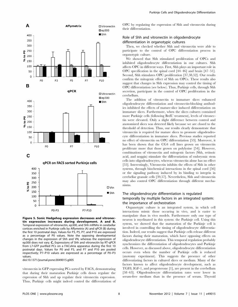
vitronectin in GFP expressing PCs sorted by FACS, demonstrating
that during their maturation Purkinje cells down regulate the
expression of Shh and up regulate their vitronectin expression.
Thus, Purkinje cells might indeed control the differentiation of
OPC by regulating the expression of Shh and vitronectin during
their differentiation.
Role of Shh and vitronectin in oligodendrocytedifferentiation in organotypic cultures
Then, we checked whether Shh and vitronectin were able to
participate to the control of OPC differentiation process in
organotypic culture.
We showed that Shh stimulated proliferation of OPCs and
inhibited oligodendrocyte differentiation in our cultures. Shh
affects OPC in different ways. First, Shh plays an important role in
OPC specification in the spinal cord [44–46] and brain [47–51].
Second, Shh stimulates OPC proliferation [37,38,52]. Our results
confirm the mitogenic effect of Shh on OPCs. These results also
suggest that changes in Shh expression may control the timing of
OPC differentiation (see below). Thus, Purkinje cells, through Shh
secretion, participate in the control of OPC proliferation in the
cerebellum.
The addition of vitronectin to immature slices enhanced
oligodendrocyte differentiation and vitronectin-blocking antibod-
ies inhibited the effects of mature-slice induced differentiation on
immature slices. Furthermore, when the slices cultures contained
more Purkinje cells (following BrdU treatment), levels of vitronec-
tin were elevated. Only a slight difference between control and
axotomized slices was detected likely because we are closed to the
threshold of detection. Thus, our results clearly demonstrate that
vitronectin is required for mature slices to promote oligodendro-
cyte differentiation in immature slices. Previous studies reported
no effect of vitronectin on OPC differentiation [53]. Moreover, it
has been shown that the CG4 cell lines grown on vitronectin
proliferate more that those grown on polylysine [54]. However,
combinations of vitronectin and mitogenic factors (Shh, retinoic
acid, and noggin) stimulate the differentiation of embryonic stem
cells into oligodendrocytes, whereas vitronectin alone has no effect
[55]. Interestingly, Vitronectin inhibits the effects of Shh in other
systems, through biochemical interactions in the spinal cord [56]
or the signaling pathway induced by its binding to integrin in
cerebellar granule cells [39,57]. Nevertheless, Shh and vitronectin
may also control OPC differentiation through different mecha-
nisms.
The oligodendrocyte differentiation is regulatedtemporally by multiple factors in an integrated system:the importance of orchestration
Organotypic culture is an integrated system, in which cell
interactions mimic those occuring in vivo, and is easier to
manipulate than in vivo models. Furthermore only one type of
neuron is myelinated in this system: the Purkinje cell. Using this
system, we showed that the maturation of the Purkinje cell is
involved in controlling the timing of oligodendrocyte differentia-
tion. Indeed, our results suggest that Purkinje cells release different
factors during their maturation, which have opposing effects on
oligodendrocyte differentiation. This temporal regulation probably
synchronizes the differentiation of oligodendrocytes and Purkinje
cells. However, as discussed above, oligodendrocyte differentiation
occurs even when the number of Purkinje cells is reduced
(axotomy experiment). This suggests the presence of other
differentiating factors in cultured slices or medium. Many of the
factors known to affect oligodendrocyte development, such as
TGFß, IGF-1, and progesterone [1], are present in the cerebellum
[58–63]. Oligodendrocyte differentiation rates were lower in
serum-free medium than in the presence of serum. Thyroid
Figure 5. Sonic Hedgehog expression decreases and vitronec-tin expression increases during development. A and B.Temporal expression of vitronectin, ep300, and Shh mRNAs in cerebellarcortices enriched in Purkinje cells by Affymetrix (A) and qPCR (B) duringthe first 10 postnatal days. Values for P3, P5, P7, and P10 are expressedas a percentage of P0 values. Note the opposing developmentalchanges in the expression of Shh and VN, whereas the expression ofep300 does not vary. C. Expressions of Shh and vitronectin by RT-qPCRfrom L7-GFP purified PCs on a FACsAria apparatus during the first 10postnatal days. Values for P0 and P3, and P7 and P10 are pooledrespectively; P7–P10 values are expressed as a percentage of P0–P3values.doi:10.1371/journal.pone.0049015.g005
Purkinje Cells and Oligodendrocyte Differentiation
PLOS ONE | www.plosone.org 8 November 2012 | Volume 7 | Issue 11 | e49015
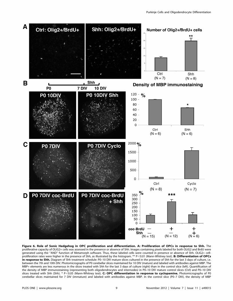
Figure 6. Role of Sonic Hedgehog in OPC proliferation and differentiation. A: Proliferation of OPCs in response to Shh. Theproliferative capacity of OLIG2+ cells was assessed in the presence or absence of Shh. Images containing pixels labeled for both OLIG2 and BrdU weregenerated using the ‘‘AND’’ function of Metamorph software. Thus, these labeled cells were counted in presence or absence of Shh. OLIG2+ cell-proliferation rates were higher in the presence of Shh, as illustrated by the histogram. ** P,0.01 (Mann-Whitney test). B: Differentiation of OPCsin response to Shh. Diagram of Shh treatment schedule: P0–10 DIV mature slices cultured in the presence of Shh for the last 3 days of culture, i.e.between the 7th and 10th DIV. Photomicrographs of P0 cerebellar slices maintained for 10 DIV (mature) and labeled with antibodies against MBP. TheMBP+ elements are less numerous in the slices treated with Shh for the last 3 days of culture (right) than in the control slice (left). Quantification ofthe density of MBP immunostaining (representing both oligodendrocytes and internodes) in P0–10 DIV mature control slices (Ctrl) and P0–10 DIVslices treated with Shh (Shh). * P,0.05 (Mann-Whitney test). C: OPC differentiation in response to cyclopamine. Photomicrographs of P0cerebellar slices maintained for 7 DIV (immature) and labeled with antibodies against MBP. In the control slice (P0–7 DIV), the density of MBP
Purkinje Cells and Oligodendrocyte Differentiation
PLOS ONE | www.plosone.org 9 November 2012 | Volume 7 | Issue 11 | e49015
hormones (TH) are crucial for both oligodendrocyte and Purkinje
cell differentiation [1,64]. Among the factors involved in
controlling OPC proliferation and differentiation, our study
identifies two molecules with developmentally regulated expression
patterns which are involved in the switch between OPC
proliferation and oligodendrocyte differentiation. Vitronectin has
been detected in active multiple sclerosis plaques [65] and a
decrease in Shh levels has been observed in the white matter of
patients with multiple sclerosis [66]. These actors are thus present,
but it is unclear as to whether they are correctly synchronized.
Determining how the timing of the various steps leading to
myelination or remyelination is controlled is therefore still a major
challenge.
The findings of the present study strongly imply that Purkinje
cells initially inhibit OPC differentiation during their maturation
by releasing Shh and then subsequently promote OPC differen-
tiation by producing vitronectin. Purkinje cells thus appear to
orchestrate OPC differentiation in cerebellar organotypic cultures.
Materials and Methods
Slice cultureAll procedures were submitted and approved by the Regional
Ethics Committee in Animal Experiment Nu3 of Ile-de-France
region (p3/2009/020). Cerebellar organotypic cultures were
established from newborn (P0) Swiss mice (Mus musculus, Janvier,
Le Genset St Isle, France), as previously described [67]. Mice were
decapitated and their brains were dissected out in cold Gey’s
balanced salt solution (Invitrogen, Cergy Pontoise, France)
supplemented with 5 mg/ml glucose. Cerebellar parasagittal slices
(350 mm thick) were cut on a McIlwain tissue chopper and
transferred onto 30 mm diameter Millipore culture inserts with
0.4 mm pores (Millicell, Millipore, Molsheim, France). Slices were
maintained in culture in six-well plates containing 1 ml of nutrient
medium per well, at 35uC, under a humidified atmosphere
containing 5% CO2. The nutrient medium consisted of 50% basal
medium with Earle’s salts (BME, Invitrogen), 25% Hanks’
balanced salt solution (Invitrogen), 25% horse serum (Invitrogen),
L-glutamine (1 mM), and 5 mg/ml glucose [68].
For each result, At least 5 animals and 25 slices were studied in
3 independent experiments. In the figures, ‘‘N’’ is the number of
animals. The experimental plan was designed in accordance with
the European Union Guidelines for the care and use of
experimental animals.
In experiments evaluating the effect of a decreased number of
Purkinje cells, tissue slices were dissected along the midline
between the dorsal and ventral halves, with two needles, under a
dissection microscope, after 2 days in vitro (DIV). The two parts
were gently separated to ensure complete axotomy and were then
re-apposed.
Exposure of cerebellar slices to Sonic Hedgehog,cyclopamine, vitronectin, anti-vitronectin antibody, or 5-bromo-2-deoxyuridine
We added Shh (3 mg/ml, R&D Systems, Lille, France),
cyclopamine (5 mM, Toronto Research Chemicals, North York,
Ontario, Canada), vitronectin (VN; 5 mM, Oxford Chemical
Research, Euromedex, Souffelweyersheim, France), goat anti-VN
antibody (0.2 mg/ml, Abcam, Cambridge, UK), or a control goat
antibody (goat anti-Brn3b, 0.2 mg/ml, Santa Cruz Biotechnology,
Santa Cruz, CA) to the medium. The medium and the added drug
were replaced every two to three days. Untreated slices were used
as controls.
Cerebellar slices were depleted of mature oligodendrocytes by
killing OPCs through the addition of 5-bromo-2-deoxyuridine
(BrdU; 150 mM; Sigma, Saint Louis, MO) in NaCl solution (9 g/l)
to the nutrient medium for the first 3 DIV (P0 slices with a 3-day
BrdU treatment: P0–3DBT [27].
To study the effect of Shh on OPC proliferation, both control
and treated (5 mM Shh) slices were incubated with BrdU (20 mM)
for 3 h before fixation.
Co-culture and conditioned medium (CM) experimentsP0 slices maintained in culture until 7 DIV contained very few
MBP (myelin basic protein) immunoreactive oligodendrocytes
(Fig. 1A, A9), whereas culture of these slices for an additional 3
DIV (10 DIV in total) resulted in the accumulation of MBP
immunoreactive oligodendrocytes and few MBP immunoreactive
internodes (myelin segments, Fig. 1B, B9). Seven DIV was
therefore used as the cutoff to distinguish between ‘‘immature
slices’’ and ‘‘mature slices’’. P0 slices were placed in culture and,
when these slices became mature after 7 DIV, fresh P0 slices were
added (immature).
A slice ratio of 2 matures: 1 immature (Fig. 2A) was used to
assess whether mature slices promoted oligodendrocyte differen-
tiation in immature slices. Co-cultures were incubated for 7 DIV
before fixation.
In some experiments, immature slices were cultured on
conditioned medium (CM). CM was obtained from cultures of
P0 slices, between the 8th and the 14th days of culture (Fig. 4A).
Different types of CM were generated by treating P0 slices to
modify the number of Purkinje cells; control P0 slices provided
Ctrl-CM, BrdU treated slices provided BrdU-CM, and axoto-
mized slices provided Axt-CM.
Antibodies and staining procedureMouse monoclonal antibodies against MBP (diluted 1/500,
Chemicon, MAB381, Millipore) were used to visualize mature
oligodendrocytes and myelin [22,27,69]. Goat antibodies against
OLIG2 (1/500 R&D Systems Europe, Abingdon, UK) were used
to visualize OPCs and oligodendrocytes [26]. Mouse monoclonal
antibodies against APC (1/1000, Calbiochem, VWR, Fontenay
sous bois, France) were used in combination with anti-OLIG2 to
immunostaining is low. In contrast, the density of MBP immunostaining is much higher in P0–7 DIV slices cultured in the presence of cyclopamine(P0–7 DIV Cyclo), a Shh inhibitor. Quantification of MBP immunostaining density in control (Ctrl) and cyclopamine-treated P0–7 DIV (Cyclo) immatureslices. The density of MBP immunostaining was determined. ** P,0.01 (Mann-Whitney test). D: OPC differentiation in co-culture experimentsin the presence or absence of Shh. Photomicrographs of P0 cerebellar slices labeled with antibodies against MBP at 7 DIV. These slices were co-cultured in the presence of P0 7–14 DIV mature slices treated with a high dose of BrdU during the first 3 days of culture (coc-BrdU) in the presence orabsence of Shh. In the presence of Shh, the density of MBP immunostaining present in this immature slice was similar to that of control P0–7 DIVslices (compare C left and D right). In contrast, in the absence of Shh, there were more MBP+ elements in P0–7 DIV slices co-cultured with BrdUtreated mature slices (coc-BrdU, left). Quantification of the density of MBP immunostaining in P0–7 DIV immature slices co-cultured in the presence orabsence of BrdU treated mature slices and/or Shh. *** P,0.001 (Mann-Whitney tests). Scale bar in A is 70 mm for A and 425 mm for B, C, and D.doi:10.1371/journal.pone.0049015.g006
Purkinje Cells and Oligodendrocyte Differentiation
PLOS ONE | www.plosone.org 10 November 2012 | Volume 7 | Issue 11 | e49015
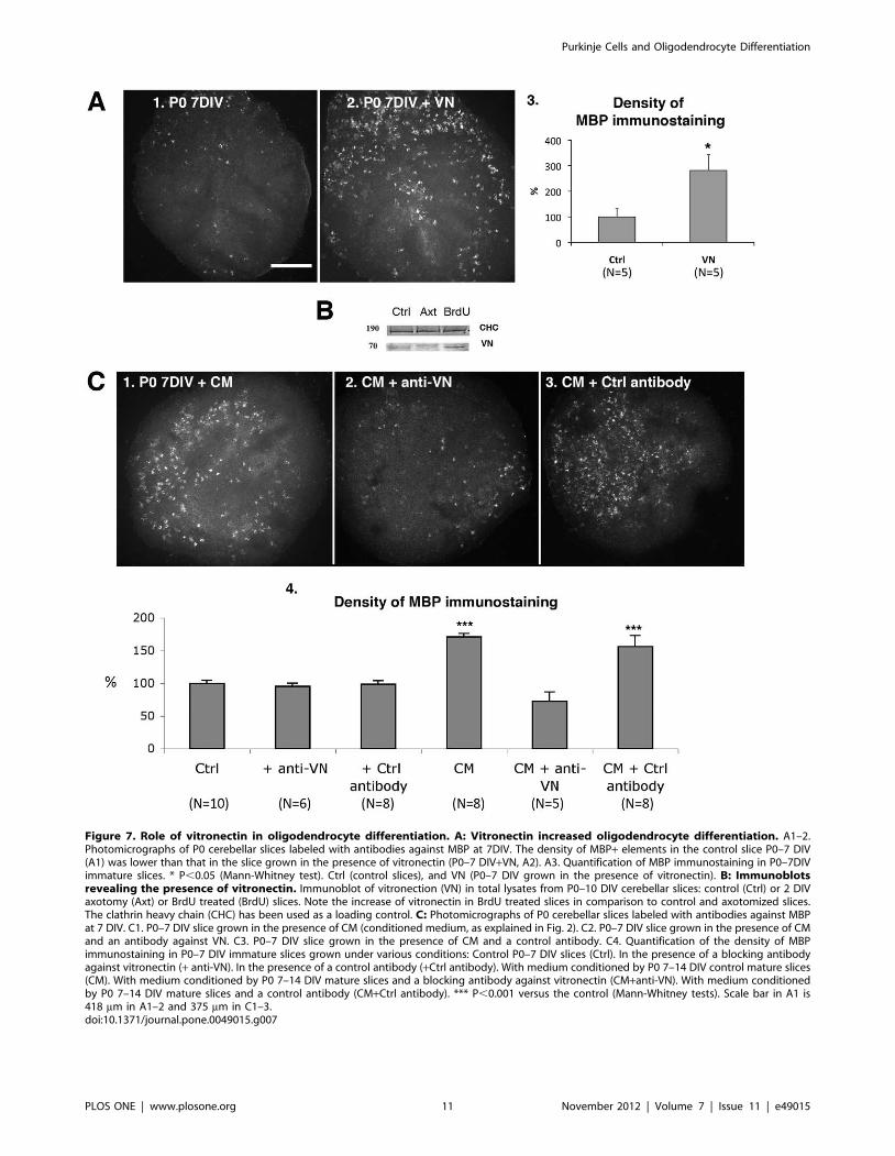
Figure 7. Role of vitronectin in oligodendrocyte differentiation. A: Vitronectin increased oligodendrocyte differentiation. A1–2.Photomicrographs of P0 cerebellar slices labeled with antibodies against MBP at 7DIV. The density of MBP+ elements in the control slice P0–7 DIV(A1) was lower than that in the slice grown in the presence of vitronectin (P0–7 DIV+VN, A2). A3. Quantification of MBP immunostaining in P0–7DIVimmature slices. * P,0.05 (Mann-Whitney test). Ctrl (control slices), and VN (P0–7 DIV grown in the presence of vitronectin). B: Immunoblotsrevealing the presence of vitronectin. Immunoblot of vitronection (VN) in total lysates from P0–10 DIV cerebellar slices: control (Ctrl) or 2 DIVaxotomy (Axt) or BrdU treated (BrdU) slices. Note the increase of vitronectin in BrdU treated slices in comparison to control and axotomized slices.The clathrin heavy chain (CHC) has been used as a loading control. C: Photomicrographs of P0 cerebellar slices labeled with antibodies against MBPat 7 DIV. C1. P0–7 DIV slice grown in the presence of CM (conditioned medium, as explained in Fig. 2). C2. P0–7 DIV slice grown in the presence of CMand an antibody against VN. C3. P0–7 DIV slice grown in the presence of CM and a control antibody. C4. Quantification of the density of MBPimmunostaining in P0–7 DIV immature slices grown under various conditions: Control P0–7 DIV slices (Ctrl). In the presence of a blocking antibodyagainst vitronectin (+ anti-VN). In the presence of a control antibody (+Ctrl antibody). With medium conditioned by P0 7–14 DIV control mature slices(CM). With medium conditioned by P0 7–14 DIV mature slices and a blocking antibody against vitronectin (CM+anti-VN). With medium conditionedby P0 7–14 DIV mature slices and a control antibody (CM+Ctrl antibody). *** P,0.001 versus the control (Mann-Whitney tests). Scale bar in A1 is418 mm in A1–2 and 375 mm in C1–3.doi:10.1371/journal.pone.0049015.g007
Purkinje Cells and Oligodendrocyte Differentiation
PLOS ONE | www.plosone.org 11 November 2012 | Volume 7 | Issue 11 | e49015
detect oligodendrocytes. Mouse monoclonal antibodies against
BrdU (1/400 Progen, Heidelberg, Germany) or were used to
identify proliferating cells. Rabbit polyclonal antibodies against
calbindin (Calb1; diluted 1/5,000; Swant, Bellinzona, Switzerland)
and goat antibodies against Parvalbumin (1/5000, Swant) were
used to visualize differentiated Purkinje cells [22].
After 7, 10, or 14 DIV, the cultures and co-cultures were fixed
by incubation in 4% paraformaldehyde in phosphate buffer
(0.1 M, pH 7.4) for 1 h at room temperature. Slices incubated
with anti-MBP antibody were washed in PBS (Invitrogen) and
immersed in Clark’s solution (95% ethanol/5% acetic acid) for
20 min at 4uC to extract some of the lipids —increasing the
accessibility of MBP antigens— and were then washed several
times with PBS. All slices were incubated for 1 h in PBS
containing 0.2% gelatin, 0.1% sodium azide (PBSGA) and
0.1 M lysine. This was followed by an overnight incubation at
room temperature with primary antibody diluted in PBSGA. The
primary antibodies were detected with the following secondary
antibodies: CY3-conjugated goat anti-mouse (1/200 dilution;
Jackson ImmunoResearch Laboratories Inc, West Baltimore Pike,
PA), FITC-conjugated sheep anti-rabbit (1/200 dilution, Chemi-
con), CY3-conjugated donkey anti goat (1/200 dilution, Jackson
Immunoresearch), Alexa Fluor 488-conjugated donkey anti mouse
(1/400 dilution, Invitrogen), AMCA-conjugated donkey anti
rabbit (1/50 dilution, Jackson Immunoresearch), CY3-conjugated
goat anti-rabbit (1/200 dilution; Jackson ImmunoResearch). Slices
incubated for 2 h with the secondary antibodies in PBSGA were
washed several times in PBS and mounted in Mowiol (Calbio-
chem). The sections were analyzed under a Leica DMR
microscope equipped with a Coolscan camera (Princeton Instru-
ments, Evry, France).
MBP immunostaining quantificationsMBP immunoreactive oligodendrocytes and internodes (myelin
segments) were quantified by acquiring an image of each slice
using the software, Metaview. The images were then analyzed
using the software, Metamorph (Universal Imaging Corporation).
Contours of the slices were drawn —the threshold was adjusted
based on the MBP immunostaining— and the density of MBP
immunostaining was determined for each slice. Means and
standard errors of the mean (SEM) were calculated for each type
of slice.
To distinguish between oligodendrocyte differentiation and
myelination, we performed a semi-quantification of MBP+internodes which did not consider the number of differentiated
oligodendrocytes. The65 pictures were enlarged on the computer
screen (zoom 4) to evaluate the number of internodes, and the first
26 internodes were pointed and counted using ImageJ software.
Two groups were specified: Group I included slices that either did
not contain MBP+ internodes (myelin segments) or contained less
than 25 internodes; group II included slices containing more than
26 internodes.
Quantification of OLIG2 positive and APC positive cellsOn slides with Calb1 labeling of Purkinje cells, pictures of areas
centered on white matter close to the deep nuclear neurons were
acquired for OLIG2 and APC using the Metaview software. Then,
number of cells positive for both OLIG2 and APC per
81,000 mm2 were determined manually using ImajeJ software.
Means and standard errors of the mean (SEM) were calculated for
each type of slice.
Quantification of OLIG2-positive and BrdU-positive cellsA proliferation index for OPCs was calculated by determining
the number of cells positive for both OLIG2 and BrdU. On slides
with Calb1 labeling of Purkinje cells, single areas of white matter
close to the deep nuclear neurons were chosen, 1 mm thick
confocal sections were acquired for OLIG2 and BrdU labeling
(Leica confocal microscope; Plateforme d’Imagerie, IFR83). Using
the software Metamorph, images containing pixels labeled for
both OLIG2 and BrdU were generated and the number of
double-stained cells per 150,000 mm2 was determined.
Statistic analysisMann-Whitney tests were used to compare two groups and
Kruskal-Wallis tests were used for multiple comparisons. P
values#0.05 were considered to significant. In the figures, the
values are represented as the percentage of the control.
Statistical analysis of Affymetrix data, microarrayannotation, and functional analysis
Affymetrix microarrays were used to assess patterns of gene
expression in murine cerebellar cortical areas. Total RNA was
extracted from dissected areas centered on the layer of Purkinje
cells from vermal lobules 5 and 6 of Swiss mice of various ages: P0,
P3, P5, P7, and P10 (4 replicates = 4 independent measurements
for each stage; different litters were used for each measurement).
RNA was extracted using RNAeasy Mini Kit (Qiagen, Courta-
boeuf, France). Following reverse transcription, cDNAs were
hybridized with Affymetrix microarrays (MOE430 GeneChip,
Affymetrix Platform, Institut Curie, Paris, France). All the
experimental procedures and results have been loaded on
ArrayExpress database (E-MEXP-3444, Experiment name: PC
transcriptome, http://www.ebi.ac.uk/arrayexpress/experiments/
E-MEXP-3444).
Matlab was used for mathematical manipulations and statistical
analysis. The aim was to identify transcripts displaying a significant
increase or decrease over time from P0 to P10.
The four replicates were used to estimate the intrinsic variability
of expression, or pure error, for each transcript. We used the pure
error in F-tests of lack of fit [70] for constant, affine, and quadratic
models. The Benjamini-Hochberg procedure [71] was used for
correction, with estimation of the number of true null hypotheses
[72] to guarantee an FDR of 1%.
A constant model was retained (i.e. no lack of fit) for 26,912 of
the 31,818 transcripts, an affine model was retained (lack of fit of
the constant model, but not of the affine model) for 4,474
transcripts, and a quadratic model was retained (lack of fit of the
constant and affine models, but not of the quadratic model) for the
remaining 432 transcripts. All ‘‘constant’’ transcripts were
discarded. We retained all of the ‘‘affine’’ transcripts as potential
candidates. We also searched for ‘‘quadratic’’ transcripts display-
ing continual increases or decreases. One quadratic transcript was
identified with a continually decreasing pattern over time. Two
lists were therefore generated: 2,262 affine transcripts showing
continuous increases in expression and 2,213 transcripts with
continuous decreases in expression (2,212 quadratic, 1 affine).
Among the latter, we selected transcripts with at least a two-fold
difference in expression (higher or lower) at P10 compared with
P0. This gave a list of 1190 transcripts, from which we further
discarded those with a very low level of expression (below 45), to
give a final list of 627 candidate transcripts.
Gene annotations were expanded and upgraded with NCBI
Entrez Gene ID, Unigene, and PubMed for the remaining 627
candidate probesets. Transcribed sequences and expression
Purkinje Cells and Oligodendrocyte Differentiation
PLOS ONE | www.plosone.org 12 November 2012 | Volume 7 | Issue 11 | e49015
sequence tags (ESTs) with no identified function were eliminated
from the reported lists. Gene annotations for the MOE430
GeneChip obtained from Webgestalt (web-based gene set analysis
toolkit [73]) and from the Affymetrix website were compared.
Gene ontology (GO) terms were applied and 10 genes were
identified based on the GO cellular component term ‘‘extracellular
region/space’’ (GO:0005576).
Quantitative RT-PCRRT-PCR quantified in real time was carried out on two
independent sets of mRNA for each age (P0, P7, P10), to assess
Shh and VN mRNA levels using a LightCycler 480 real-time PCR
system (Roche). First-strand cDNAs were synthesized from 500 ng
total RNA (Thermo Scientific, Surrey, UK), in accordance with
the manufacturer’s instructions. The reaction mixture contained
16 SYBR Green I Master (Roche Diagnostics, Mannheim,
Germany), 1.5 ng of the first-strand cDNA and 240 nM of each
forward and reverse primer, in a total volume of 13 ml. Thermal
cycle parameters were: 95uC for 5 min, followed by 40 cycles of
95uC for 10 s, 56uC for 20 s, and 72uC for 20 s. All primers were
synthesized by Eurofins MWG Operon (Ebersberg, Germany).
The following primer sequences were used: VN forward: 59-
GCCAGCAAGAAGTGTCAGTG-39, VN reverse: 59-
GCTTTAGAGTGCCGTCCGTC-39, Shh forward: 59-
TCACCCCCAATTACAACCCC-39, Shh reverse: 59-
ACTCCTCTGAATGATGGCCG-39, Ep300 forward: 59-
GAGGGTAAGAAATGACTCCAGTG-39, Ep300 reverse: 59-
CAGTTGAGGGTTTTTGCTTTC-39. Data were analyzed by
the LightCycler 480 software, release 1.4.9 (Roche), and target-
transcript expression was quantified using the second-derivative
maximum method. Control reactions without reverse transcriptase
were performed to check for genomic DNA contamination. All
samples were normalized with respect to the reference gene,
Ep300, which displays minimal variation during the first 10 days
of cerebellar development. Two independent experiments were
used for quantification. Data are presented as means 6 SEM.
Purkinje cell sortingPurkinje cells were isolated from BacL7-GFP mice as described
previously [74]. Briefly, cubes (0.5-mm3) of cerebella at indicated
ages were digested for 10 min at 37uC with 0.025% trypsin (type I;
Sigma-Aldrich, Saint Louis, MO) in dissociation solution consist-
ing of Ca2+-free Hanks’ balanced salt solution (HBSS) containing
3 mg/ml BSA, 15 mM Hepes, 1.5 mM MgSO4, and 3 mg/ml
glucose (pH 7.4). The enzymatic reaction was stopped by adding
of one volume of dissociation solution containing 0.25 mg/ml
soybean trypsin inhibitor, 40 mg/ml DNase I, 50 mM APV,
20 mM DNQX and 0.1 mM TTX (all from Sigma-Aldrich).
Tissues were triturated mildly by sequential passage through wide-
bore and fine-tipped pipettes. Cells were filtered through a 40 mm
nylon mesh, and were resuspended in Ca2+- and Mg2+-free
dissociation solution at a final concentration of 5.106 cells per ml.
To label the dead cells, PI (Propidium iodide; Sigma-Aldrich) was
added at a final concentration of 2 mg/ml. Cell sorting was
performed on a FACsAria machine. The sorting decision was
based on the measurements of FSC, PI fluorescence and GFP
fluorescence.
RNA preparation and real time quantitative RT-PCR forpurified Purkinje cells
Total RNA was extracted from approximately 3.104 purified
Purkinje cells at indicated stages with Trizol reagent (Invitrogen).
The cDNA was prepared by reverse transcription of 100 ng RNA
using SuperScript III First-Strand Synthesis System (Invitrogen)
with an oligo-dT primer according to the manufacturer’s
instructions. The resulting cDNA was used as a template for real
time PCR using a Light Cycler 480 thermocycler (384 plates,
Roche Diagnostics) with a home-made SYBR Green QPCR
master mix [75]. Thermal cycling parameters were 2 min at 95uC,
followed by 45 cycles of 95uC for 10 s, 64uC for 15 s and 72uC for
25 s. To normalize expression data, multiple internal control genes
(TATA box binding protein, Actin beta and Tubulin beta 2A)
were selected as described by Vandesompele et al. [76]. Sequences
of the primers used can be obtained upon request.
Western blotsCerbellar slices were lysed in RIPA buffer (50 mM Hepes,
150 MM NaCl, 5 mM EDTA, 1% NP-40, 0.5% SDS; pH 7.7).
Lysates were clarified by centrifugation. The protein pellet was
collected by centrifugation, washed with cold acetone and
recentrifuged. The pellet was then allowed to air dry and
resuspended in 10 mM Tris pH 8. The DCA protein assay
(Biorad, Hercules, California) was used to determine protein levels.
Samples (2 mg of CM proteins) were denatured by heating for
3 min at 95uC in Laemmli buffer, separated by 10% SDS-PAGE,
and transferred to a polyvinylidene difluoride membrane (PVDF,
Amersham Biosciences, GE Healthcare, UK). TBS supplemented
with 0.1% Tween-20 and 5% dry milk powder was used for
blocking and antibody incubations. Primary (rabbit polyclonal
anti-VN, 1/1000) and secondary Alkaline phosphatase conjugated
goat anti-rabbit polyclonal antibody (1/7500, Promega, Madison,
WI). Membranes were incubated overnight at 4uC with the
primary antibody and then for 1 h at room temperature with the
secondary antibody. They were then washed three times in TBS
supplemented with 0.1% Tween-20 and once in TBS. The
alkaline phosphatase-conjugated antibody was detected by NBT/
BCIP (Promega). A prestained protein ladder (Fermentas,
Euromedex, Souffelweyersheim, France) was used as the size
marker.
Acknowledgments
We thank Isabelle Caille, Beatrice Durand and Rachel Sherrard for helpful
discussion, Richard Schwarzman, Plateforme imagerie IFR83, for
assistance with confocal microscopy and Julien Dahan for his work on
microarray analysis.
Author Contributions
Conceived and designed the experiments: LB CS ID. Performed the
experiments: LB RW XC FJ YLD ID. Analyzed the data: LB RW MD
YLD IR CS ID. Wrote the paper: LB CS ID.
References
1. Baumann N, Pham-Dinh D (2001) Biology of oligodendrocyte and myelin in the
mammalian central nervous system. Physiol Rev 81:871–927.
2. Dubois-Dalcq M, Behar T, Hudson L, Lazzarini RA (1986) Emergence of three
myelin proteins in oligodendrocytes cultured without neurons. J Cell Biol
102:384–392.
3. Sarlieve LL, Rao GS, Campbell GL, Pieringer RA (1980) Investigations on
myelination in vitro: biochemical and morphological changes in cultures of
dissociated brain cells from embryonic mice. Brain Res 189:79–90.
4. Barres BA, Jacobson MD, Schmid R, Sendtner M, Raff MC (1993) Does
oligodendrocyte survival depend on axons? Cur Biol 3:489–497.
Purkinje Cells and Oligodendrocyte Differentiation
PLOS ONE | www.plosone.org 13 November 2012 | Volume 7 | Issue 11 | e49015
5. Trapp BD, Nishiyama A, Cheng D, Macklin W (1997) Differentiation and death
of premyelinating oligodendrocytes in developing rodent brain. J Cell Biol
137:459–468.
6. Burne JF, Staple JK, Raff MC (1996) Glial cells are increased proportionally in
transgenic optic nerves with increased numbers of axons. J Neurosci 16:2064–
2073.
7. Friede RL, Bischhausen R (1982) How are sheath dimensions affected by axon
caliber and internode length? Brain Res 235:335–350.
8. Cole JS, Messing A, Trojanowski JQ, Lee VM (1994) Modulation of axon
diameter and neurofilaments by hypomyelinating Schwann cells in transgenic
mice. J Neurosci 14:6956–6966.
9. Hsieh ST, Kidd GJ, Crawford TO, Xu Z, Lin WM, et al. (1994) Regional
modulation of neurofilament organization by myelination in normal axons.
J Neurosci 14:6392–6401.
10. Demerens C, Stankoff B, Logak M, Anglade P, Allinquant B, et al. (1996)
Induction of myelination in the central nervous system by electrical activity. Proc
Natl Acad Sci U S A 93:9887–9892.
11. Wake H, Lee PR, Fields RD (2011) Control of local protein synthesis and initial
events in myelination by action potentials. Science 333:1647–1651.
12. Wang S, Sdrulla AD, diSibio G, Bush G, Nofziger D, et al. (1998) Notch
receptor activation inhibits oligodendrocyte differentiation. Neuron 21:63–75.
13. Charles P, Hernandez MP, Stankoff B, Aigrot MS, Colin C, et al. (2000)
Negative regulation of central nervous system myelination by polysialylated-
neural cell adhesion molecule. Proc Natl Acad Sci U S A 97:7585–7590.
14. Schnadelbach O, Ozen I, Blaschuk OW, Meyer RL, Fawcett JW (2001) N-
cadherin is involved in axon-oligodendrocyte contact and myelination. Mol Cell
Neurosci 17:1084–1093.
15. Laursen LS, Ffrench-Constant C (2007) Adhesion molecules in the regulation of
CNS myelination. Neuron Glia Biol 3:367–375.
16. Rosenberg SS, Kelland EE, Tokar E, De la Torre AR, Chan JR (2008) The
geometric and spatial constraints of the microenvironment induce oligodendro-
cyte differentiation. Proc Natl Acad Sci U S A 105:14662–14667.
17. Durand B, Raff M (2000) A cell-intrinsic timer that operates during
oligodendrocyte development. Bioessays 22:64–71.
18. McKinnon RD, Piras G, Ida JA, Dubois-Dalcq M (1993) A role for TGF-beta in
oligodendrocyte differentiation. J Cell Biol 121:1397–1407.
19. Barres BA, Lazar MA, Raff MC (1994) A novel role for thyroid hormone,
glucocorticoids and retinoic acid in timing oligodendrocyte development.
Development 120:1097–1108.
20. Stevens B, Porta S, Haak LL, Gallo V, Fields RD (2002) Adenosine. A neuron-
glial transmitter promoting myelination in the CNS in response to action
potentials. Neuron 36:855–868.
21. Stritt C, Stern S, Harting K, Manke T, Sinske D, et al. (2009) Paracrine control
of oligodendrocyte differentiation by SRF-directed neuronal gene expression.
Nature Neurosci 12:418–427.
22. Dusart I, Airaksinen MS, Sotelo C (1997) Purkinje cell survival and axonal
regeneration are age dependent: an in vitro study. J Neurosci 17:3710–3726.
23. Emery B (2010) Regulation of oligodendrocyte differentiation and myelination.
Science 330:779–782.
24. Bhat RV, Axt KJ, Fosnaugh JS, Smith KJ, Johnson KA, et al. (1996) Expression
of the APC tumor suppressor protein in oligodendroglia. Glia 17:169–174.
25. Casella GT, Bunge MB, Wood PM (2004) Improved immunocytochemical
identification of neural, endothelial, and inflammatory cell types in paraffin-
embedded injured adult rat spinal cord. J Neurosci Meth 139:1–11.
26. Ligon KL, Alberta JA, Kho AT, Weiss J, Kwaan MR, et al. (2004) The
oligodendroglial lineage marker OLIG2 is universally expressed in diffuse
gliomas. J Neuropathol Exp Neurol 63:499–509.
27. Bouslama-Oueghlani L, Wehrle R, Sotelo C, Dusart I (2003) The developmental
loss of the ability of Purkinje cells to regenerate their axons occurs in the absence
of myelin: an in vitro model to prevent myelination. J Neurosci 23:8318–8329.
28. Boukhtouche F, Janmaat S, Vodjdani G, Gautheron V, Mallet J, et al. (2006)
Retinoid-related orphan receptor alpha controls the early steps of Purkinje cell
dendritic differentiation. J Neurosci 26:1531–1538.
29. Poulain FE, Chauvin S, Wehrle R, Desclaux M, Mallet J, et al. (2008) SCLIP is
crucial for the formation and development of the Purkinje cell dendritic arbor.
J Neurosci 28:7387–7398.
30. Solbach S, Celio MR (1991) Ontogeny of the calcium binding protein
parvalbumin in the rat nervous system. Anat Embryol 184:103–124.
31. Sotelo C (2008) Development of ‘‘Pinceaux’’ formations and dendritic
translocation of climbing fibers during the acquisition of the balance between
glutamatergic and gamma-aminobutyric acidergic inputs in developing Purkinje
cells. J Comp Neurol 506:240–262.
32. Dahmane N, Ruiz i Altaba A (1999) Sonic hedgehog regulates the growth and
patterning of the cerebellum. Development 126:3089–3100.
33. Traiffort E, Charytoniuk D, Watroba L, Faure H, Sales N, et al. (1999) Discrete
localizations of hedgehog signalling components in the developing and adult rat
nervous system. Eur J Neurosci 11:3199–3214.
34. Wallace VA (1999) Purkinje-cell-derived Sonic hedgehog regulates granule
neuron precursor cell proliferation in the developing mouse cerebellum. Curr
Biol 9:445–448.
35. Wechsler-Reya RJ, Scott MP (1999) Control of neuronal precursor proliferation
in the cerebellum by Sonic Hedgehog. Neuron 22:103–114.
36. Gupta SK, Meiri KF, Mahfooz K, Bharti U, Mani S (2010) Coordination
between extrinsic extracellular matrix cues and intrinsic responses to orient the
centrosome in polarizing cerebellar granule neurons. J Neurosci 30:2755–2766.
37. Loulier K, Ruat M, Traiffort E (2006) Increase of proliferating oligodendroglial
progenitors in the adult mouse brain upon Sonic hedgehog delivery in the lateral
ventricle. J Neurochem 98:530–542.
38. Lelievre V, Ghiani CA, Seksenyan A, Gressens P, de Vellis J, et al. (2006)
Growth factor-dependent actions of PACAP on oligodendrocyte progenitor
proliferation. Regulatory Peptides 137:58–66.
39. Pons S, Trejo JL, Martinez-Morales JR, Marti E (2001) Vitronectin regulates
Sonic hedgehog activity during cerebellum development through CREB
phosphorylation. Development 128:1481–1492.
40. Cooper MK, Porter JA, Young KE, Beachy PA (1998) Teratogen-mediated
inhibition of target tissue response to Shh signaling. Science 280:1603–1607.
41. Marin-Teva JL, Dusart I, Colin C, Gervais A, van Rooijen N, et al. (2004)
Microglia promote the death of developing Purkinje cells. Neuron 41:535–547.
42. Louis JC, Muir D, Varon S (1992) Autocrine inhibition of mitotic activity in
cultured oligodendrocyte-type-2 astrocyte (O-2A) precursor cells. Glia 6:30–38.
43. Nash B, Thomson CE, Linington C, Arthur AT, McClure JD, et al. (2011)
Functional duality of astrocytes in myelination. J Neurosci 31:13028–13038.
44. Poncet C, Soula C, Trousse F, Kan P, Hirsinger E, et al. (1996) Induction of
oligodendrocyte progenitors in the trunk neural tube by ventralizing signals:
effects of notochord and floor plate grafts, and of sonic hedgehog. Mech Dev
60:13–32.
45. Soula C, Danesin C, Kan P, Grob M, Poncet C, et al. (2001) Distinct sites of
origin of oligodendrocytes and somatic motoneurons in the chick spinal cord:
oligodendrocytes arise from Nkx2.2-expressing progenitors by a Shh-dependent
mechanism. Development 128:1369–1379.
46. Danesin C, Agius E, Escalas N, Ai X, Emerson C, et al. (2006) Ventral neural
progenitors switch toward an oligodendroglial fate in response to increased Sonic
hedgehog (Shh) activity: involvement of Sulfatase 1 in modulating Shh signaling
in the ventral spinal cord. J Neurosci 26:5037–5048.
47. Alberta JA, Park SK, Mora J, Yuk D, Pawlitzky I (2001) Sonic hedgehog is
required during an early phase of oligodendrocyte development in mammalian
brain. Mol Cell Neurosci 18:434–441.
48. Spassky N, Heydon K, Mangatal A, Jankovski A, Olivier C, et al. (2001) Sonic
hedgehog-dependent emergence of oligodendrocytes in the telencephalon:
evidence for a source of oligodendrocytes in the olfactory bulb that is
independent of PDGFRalpha signaling. Development 128:4993–5004.
49. Tekki-Kessaris N, Woodruff R, Hall AC, Gaffield W, Kimura S, et al. (2001)
Hedgehog-dependent oligodendrocyte lineage specification in the telencephalon.
Development 128:2545–2554.
50. Yung SY, Gokhan S, Jurcsak J, Molero AE, Abrajano JJ, et al. (2002)
Differential modulation of BMP signaling promotes the elaboration of cerebral
cortical GABAergic neurons or oligodendrocytes from a common sonic
hedgehog-responsive ventral forebrain progenitor species. Proc Natl Acad
Sci U S A 99:16273–16278.
51. Chojnacki A, Weiss S (2004) Isolation of a novel platelet-derived growth factor-
responsive precursor from the embryonic ventral forebrain. J Neurosci
24:10888–10899.
52. Merchan P, Bribian A, Sanchez-Camacho C, Lezameta M, Bovolenta P, et al.
(2007) Sonic hedgehog promotes the migration and proliferation of optic nerve
oligodendrocyte precursors. Mol Cell Neurosci 36:355–368.
53. Buttery PC, ffrench-Constant C (1999) Laminin-2/integrin interactions enhance
myelin membrane formation by oligodendrocytes. Mol Cell Neurosci 14:199–
212.
54. Blaschuk KL, Frost EE, ffrench-Constant C (2000) The regulation of
proliferation and differentiation in oligodendrocyte progenitor cells by alphaV
integrins. Development 127(9):1961–1969.
55. Gil JE, Woo DH, Shim JH, Kim SE, You HJ, et al. (2009) Vitronectin promotes
oligodendrocyte differentiation during neurogenesis of human embryonic stem
cells. FEBS letters 583:561–567.
56. Pons S, Marti E (2000) Sonic hedgehog synergizes with the extracellular matrix
protein vitronectin to induce spinal motor neuron differentiation. Development
127:333–342.
57. Wechsler-Reya RJ (2001) Caught in the matrix: how vitronectin controls
neuronal differentiation. Trends Neurosci 24:680–682.
58. Constam DB, Schmid P, Aguzzi A, Schachner M, Fontana A (1994) Transient
production of TGF-beta 2 by postnatal cerebellar neurons and its effect on
neuroblast proliferation. Eur J Neurosci 6:766–778.
59. Tsutsui K (2006) Biosynthesis and organizing action of neurosteroids in the
developing Purkinje cell. Cerebellum 5:89–96.
60. Ghoumari AM, Dusart I, El-Etr M, Tronche F, Sotelo C, et al. (2003)
Mifepristone (RU486) protects Purkinje cells from cell death in organotypic slice
cultures of postnatal rat and mouse cerebellum. Proc Natl Acad Sci U S A
100:7953–7958.
61. Ghoumari AM, Baulieu EE, Schumacher M (2005) Progesterone increases
oligodendroglial cell proliferation in rat cerebellar slice cultures. Neurosci
135:47–58.
62. Bartlett WP, Li XS, Williams M, Benkovic S (1991) Localization of insulin-like
growth factor-1 mRNA in murine central nervous system during postnatal
development. Dev Biol 147:239–250.
Purkinje Cells and Oligodendrocyte Differentiation
PLOS ONE | www.plosone.org 14 November 2012 | Volume 7 | Issue 11 | e49015
63. Sherrard RM, Richardson NA, Sara VR (1997) Localisation of insulin-like
growth factor-I (IGF-I) immunoreactivity in the olivocerebellar system ofdeveloping and adult rats. Dev Brain Res 98:102–113.
64. Kapfhammer JP (2004) Cellular and molecular control of dendritic growth and
development of cerebellar Purkinje cells. Prog Histochem Cytochem 39:131–182.
65. Sobel RA, Chen M, Maeda A, Hinojoza JR (1995) Vitronectin and integrinvitronectin receptor localization in multiple sclerosis lesions. J Neuropathol Exp
Neurol 54:202–213.
66. Mastronardi FG, daCruz LA, Wang H, Boggs J, Moscarello MA (2003) Theamount of sonic hedgehog in multiple sclerosis white matter is decreased and
cleavage to the signaling peptide is deficient. Mult Scler 9:362–371.67. Ghoumari AM, Wehrle R, De Zeeuw CI, Sotelo C, Dusart I (2002) Inhibition of
protein kinase C prevents Purkinje cell death but does not affect axonalregeneration. J Neurosci 22:3531–3542.
68. Stoppini L, Buchs PA, Muller D (1991) A simple method for organotypic
cultures of nervous tissue. J Neurosci Methods 37:173–182.69. Bouslama-Oueghlani L, Wehrle R, Sotelo C, Dusart I (2005) Heterogeneity of
NG2-expressing cells in the newborn mouse cerebellum. Dev Biol 285:409–21
70. Draper, Smith (1981) Applied Regression Analysis (John Wiley and Sons
editors).
71. Benjamini Y, Hochberg Y (1995) Controlling the false discovery rate: A practical
and powerful approach to multiple testing. J Roy Statist Soc Ser B 57:289–300.
72. Storey JD, Tibshirani R (2003) Statistical significance for genomewide studies.
Proc Natl Acad Sci U S A 100:9440–9445.
73. Zhang B, Kirov S, Snoddy J (2005) WebGestalt: an integrated system for
exploring gene sets in various biological contexts. Nucleic Acids Res 33(Web
Server issue):W741–748.
74. Tomomura M, Rice DS, Morgan JI, Yuzaki M (2001) Purification of Purkinje
cells by fluorescence-activated cell sorting from transgenic mice that express
green fluorescent protein. Eur J Neurosci 14:57–63.
75. Lutfalla G, Uze G (2006) Performing quantitative reverse-transcribed polymer-
ase chain reaction experiments. Methods Enzymol 410:386–400.
76. Vandesompele J, De Preter K, Pattyn F, Poppe B, Van Roy N, et al. (2002)
Accurate normalization of real-time quantitative RT-PCR data by geometric
averaging of multiple internal control genes. Genome Biology 3:research0034.1–
0034.11.
Purkinje Cells and Oligodendrocyte Differentiation
PLOS ONE | www.plosone.org 15 November 2012 | Volume 7 | Issue 11 | e49015