chemical heterogeneity in purkinje and
TRANSCRIPT
Proc. Natl Acad. Sci. USAVol. 78, No. 12, pp. 7787-7791, December 1981Neurobiology
Chemical heterogeneity in cerebellar Purkinje cells: Existence andcoexistence of glutamic acid decarboxylase-like and motilin-likeimmunoreactivities
(morphology/sagittal microzones/motilin/glutamate dehydrogenase)
VICTORIA CHAN-PALAY*, GAJANAN NILAVERt, SANFORD L. PALAY*, MARGERY C. BEINFELD§,EARL A. ZIMMERMANt, JANG-YEN WU$, AND THOMAS L. O'DONOHUEIIDepartments of *Neurobiology and *Anatomy, Harvard Medical School, Boston, Massachusetts 02115; tDepartment of Neurology, Columbia University College ofPhysicians and Surgeons, New York, New York 10032; IDepartment of Pharmacology, St. Louis University Medical School, St. Louis, Missouri 63104; ¶Departmentof Cell Biology, Baylor University College of Medicine, Houston, Texas 77030; and INational Institute of Mental Health, Bethesda, Maryland 20205
Contributed by Sanford L. Palay, July 24, 1981
ABSTRACT Purkinje neurons of the cerebellar cortex forma chemically and morphologically heterogeneous population con-taining some members that have y-aminobutyric acid (GABA),others that have immunoreactivity for motilin, and a small numberthat have both. The remaining 30-40% of all Purkinje cells haveneither of these two neuroactive substances, leaving possibilitiesfor other transmitter candidates. The evidence was compiled fromdouble-staining immunocytochemical procedures performed onsingle sections ofthe cerebellum and brain stem in rat, mouse, andmonkey. Two polyclonal antibodies were applied in succession,one directed against the midregion andCOOH terminus ofthe 22-amino acid polypeptide motilin and the other against glutamic aciddecarboxylase (glutamate decarboxylase; L-glutamate 1-carboxy-lyase, EC 4.1.1.15), the rate-limiting enzyme in the synthesis ofthe neurotransmitter GABA. The staining combinations employedthe immunoperoxidase method, with different chromogens fordistinguishing the motilin-like immunoreactivity from glutamicacid decarboxylase immunoreactivity by different colors, or theimmunoperoxidase method for one antiserum and immunofluo-rescence for the other. The locations of both motilin and GABAcell types were mapped. The recognition of motilin in Purkinjecells calls for experimental definition of the role of this substancein the cerebellum and for reevaluation ofthe roles ofPurkinje cellsand of GABA in cerebellar function. The significant motilin rep-resentation in the flocculus, paraflocculus, and vermis suggeststhat it may be the Purkinje cell mediative chemical in the vesti-bular parts of the cerebellum. However, the presence of GABAas well in the same regions indicates that the chemical preferencemay be at least bimodal.
A considerable literature supports the identification of y-ami-nobutyric acid (GABA) as a major inhibitory transmitter in thecerebellum. GABA has been localized by autoradiography withuptake of tritiated GABA (1) or binding of tritiated muscimolfor muscimol-GABA receptor sites (2, 3), and by immunocy-tochemistry with antibodies directed against glutamic acid de-carboxylase (GAD; glutamate decarboxylase; L-glutamate 1-car-boxy-lyase, EC 4.1.1.15), the rate-limiting enzyme for thesynthesis ofGABA from glutamic acid (4-8), and with antibodiesagainst y-aminobutyric acid transaminase, the enzyme that de-grades GABA (4, 9). Ofthe cerebellar neurons, a majority ofthesmaller interneurons e.g., stellate, basket, and Golgi cells-arelabeled consistently by these cytochemical methods for locali-zation of GABA. The Purkinje cells, however, have remained
enigmatic. Although a considerable number in any species canbe labeled by the autoradiographic and immunocytochemicalmethods for GABA, the rest of the Purkinje cells remain un-labeled (8-11). In recent studies with in vivo injections of an-tibodies against GAD combined with anterograde and retro-grade transport as a means for tracing chemically specificpathways (8), the significance ofthis patchy labeling of Purkinjecells was questioned. Does it mean that not all Purkinje cellscontain GABA and GAD immunoreactivity, or that the GABAcontent in some Purkinje cells may be cyclic, so that GAD levelsand therefore GAD immunoreactivity may fluctuate in time?If not all Purkinje cells contain GABA, then what are the otherpossible Purkinje cell neurotransmitters? A search among otherneuroactive substances revealed that although monoamines andamino acids may characterize some fiber systems and may becandidates for transmitters ofsome cell types in the cerebellum,none of them applied to the Purkinje cells.
Recent studies indicate that motilin, a 22-amino acid poly-peptide isolated from porcine gut 10 years ago (12), is presentin the cerebellum (13). Motilin stimulates enteric smooth mus-cle (12, 14), has endocrine effects when administered system-ically (15, 16), and has an excitatory effect on neurons of thecerebral cortex and spinal cord (17). Motilin-like immunoreac-tivity (motilin-i) is detectable in the brain ofa number ofspecies,particularly in the hypothalamus and pituitary, and less con-sistently in the cerebellum (18-21). The present investigation,in which double labeling techniques and polyclonal antibodiesdirected against the enzyme GAD and synthetic porcine motilinare used, demonstrates that these two substances can exist inPurkinje cells and even together in the same cells.
MATERIALS AND METHODS
The cerebellar cortex and deep cerebellar nuclei from adultmice, rats, and a monkey (Macaca fascicularis) were used inthese experiments. The brains from these animals were per-fused with 4% (wt/vol) formaldehyde in 0.12 M sodium phos-phate buffer (pH 7.4) or with Bouin's picric acid/glacial aceticacid fixative, or they were immersed in the latter. The tissueswere fixed overnight and prepared as follows: 50-,um-thick vi-bratome sections offormaldehyde-fixed tissue embedded in 7%agar, 6-,um-thick serial paraffin sections of Bouin's solution-
Abbreviations: GABA, y-aminobutyric acid; GAD, glutamic acid de-carboxylase; GAD-i; GAD immunoreactivity; motilin-i, motilin-likeimmunoreactivity.
The publication costs ofthis article were defrayed in part by page chargepayment. This article must therefore be hereby marked "advertise-ment" in accordance with 18 U. S. C. §1734 solely to indicate this fact.
7787
Dow
nloa
ded
by g
uest
on
Feb
ruar
y 15
, 202
2
7788 Neurobiology: Chan-Palay etaP
fixed material, 10-,um-thick cryostat sections of formaldehyde-fixed material. In several animals, colchicine, a drug that in-hibits fast axonal transport (22), was given as a single intracis-ternal injection (30 jig/100 g of body weight) 24 hr prior to theperfusion.GAD Antibody. GAD was isolated and purified from mouse
brain, and the purity of the enzyme was established from theresults of polyacrylamide gel electrophoresis, high-speed sedi-mentation equilibrium, and sodium dodecyl sulfate/polyacryl-amide gel electrophoresis. Antiserum to purified GAD was pro-duced in rabbits by biweekly injections of 50 ,ug of thedecarboxylase in complete Freund's adjuvant into the infras-capular muscles. The rabbits were bled 1 week after the fourthinjection and afterwards. The GAD antiserum was character-ized by enzyme inhibition, immunodiffusion, microcomple-ment fixation, and immunoelectrophoresis (23). The antiseraused in these studies have been tested extensively in the cer-ebellar system (4-6, 6, 8).
Motilin Antibody. Synthetic porcine motilin (Peninsula Lab-oratories, San Carlos, CA) was conjugated to bovine thyroglob-ulin by the glutaraldehyde method (24), and the antiserumraised in rabbits by G.N. was characterized by radioimmu-noassay (25) and found to be directed towards the mid-portionand COOH-terminal regions of the peptide. Preliminary im-munostaining studies revealed positive labeling with this anti-body in hypothalamus, pituitary, and cerebellum in rat, mouse,and man (13). Preincubation of the antiserum at a dilution of1:1000 with 1 ttg of synthetic porcine motilin abolished stainingin hypothalamus and pituitary in rat. Preincubation of the anti-serum at 1:1000 dilution with 100 ug of motilin resulted in re-duced cerebellar staining. Reactivity was abolished in all pro-cesses, but 50% of reactive Purkinje cell somata remained.Preabsorption of this antiserum with rat cerebellar extractblocked staining in hypothalamus and pituitary as well as incerebellum. The antigenic material in the cerebellum is distinctfrom but closely related to synthetic porcine motilin (13). It ispossible that an antibody in the antiserum reacts with a partic-
ular steric conformation of the motilin molecule or even a largepre-motilin molecular form present in the sites of synthesiswithin Purkinje cell somata.
Histological Procedures. The process ofdouble staining withthe two antibodies, one against motilin and the other againstGAD in sequential order, was applied to single sections so thateach section was stained twice. The paraffin was removed fromparaffin sections prior to hydration. All sections were rinsed inTris-HCl buffer (0.01 M, pH 7.6) and treated with 0.5% H202in Tris buffer for 30 min to abolish endogenous peroxidase ac-tivity before incubation with antibody. The most successful se-quence was obtained with the initial use of motilin antibodyfollowed by the GAD antibody. Motilin antiserum (dilution1:400 to 1:600) in Tris buffer was used for primary incubation(overnight at 4°C or 2 hr at 37°C) in a humid atmosphere. Thesections were then stained by the indirect peroxidase-antiper-oxidase method (26) followed by diaminobenzidine visualiza-tion. Subsequently, the same sections were reincubated in theGAD antiserum (1:50 dilution, overnight at 4°C or 2 hr at 37°C),and stained by the indirect immunofluorescence method ofCoons (27). The fluorophore used was either fluorescein iso-thiocyanate or rhodamine conjugated to-goat anti-rabbit serum(1:50 dilution or 1:250 dilution). Glycerin was put on the sec-tions, then coverslips; the sections were examined with fluo-rescence optics and with Nomarski interference optics. Anotherdouble-staining combination employed the immunoperoxidasemethod with different chromagens for distinguishing motilin-ifrom GAD immunoreactivity (GAD-i) by separate colors. Inthese sequences, motilin was revealed first by reaction withdiaminobenzidine for a brown reaction product, and the samesections were then treated with GAD antibody bound to thesame bridging secondary reagents but substituting 4-chloro-1-naphthol (28) to obtain a blue reaction product. Single repre-sentative sections of cerebellum in rat, mouse, and monkey, aswell as sets of serial sections through rat and mouse brains eachspaced 60 ,um apart, were stained for mapping. For the presentpurposes, Purkinje cells stainingwith the anti-GAD serum were
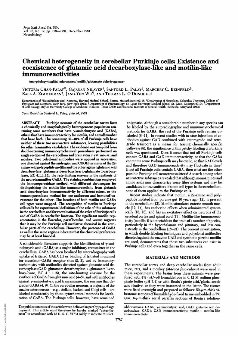
FIG. 1. Schematic drawing of the distribution of motilin-i (M-i) Purkirne cells (A) and GAD-i Purkinje cells (e) ine coronal section of the cer-ebellum. Motilin-i cells and GAD-i cells are both more-concentrated in the flocculus (fl) and dorsal and ventral paraflocculus (pfl) than elsewhere.Both cell types occur in the vermis and participate in the formation of the sagittal microzones (arrows). Motilin-i terminal axon projections in thedeep cerebellar nuclei-dentate (D), interpositus (I), fastigial (F), and lateral vestibular nucleus (LV)-are represented on the left and a comparablerepresentation for GAD-i terminal axon projections is shown on the right. Motilin-i is heaviest in the dentate and GAD-i is heaviest in the lateralvestibular nucleus.
Proc. Nad Acad. Scii. USA 78 (1981)
Dow
nloa
ded
by g
uest
on
Feb
ruar
y 15
, 202
2
Neurobiology: Chan-Palay et aL
considered to be GABA-containing neurons, and those withmotilin-i were considered to contain motilin or motilin precursor.
OBSERVATIONS AND DISCUSSIONM-i was observed in Purkinje cell somata, dendrites, and axonalplexuses in the cerebellar cortices of all three species exam-ined-rat, mouse, and monkey-and in the terminal projec-tions of Purkinje cells in the deep cerebellar nuclei and vesti-bular nuclei. The motilin Purkinje cells were most numerousin the flocculus and paraflocculus, where 60-70% ofall Purkinjecells were immunoreactive. The lateral hemispheres had con-siderable numbers of these neurons arranged in large groups,punctuated by 1-12 or more Purkinje cells that were not mo-tilin-immunoreactive. The paravermis and vermis of the cere-bellum consistently contained fewer Purkinje cells with distinctmotilin-i than did the lateral cerebellum. The motilin-Purkinjecells in the vermis were scattered singly or contributed to theformation of three narrow sagittal microbands in lobules I-IV.These three bands were precisely oriented with respect to themidline of the vermis (see Fig. 1)-a midsagittal band, two tofour Purkinje cells wide in each folium and composed of thesomata and dendritic trees lying precisely in the midline of the
4'~~~~~~~~~~~~~~~~4
JAl
A~~~~~~~~~~
JD A h a" +S''J-~~~~~~~~~~-
.2~~ ~ ~ ~ .
WA ~ 0Zr,D,.,; ',^ ;
Proc. Natd Acad. Sci. USA 78 (1981) 7789
cerebellum, and two parasagittal bands, each approximately 100Am from either side of midline and made up of the somata anddendrites of six to eight Purkinje cells both clustered and sep-arate from one another. In lobules V and VI of the vermis, thethree microzones were supplemented by a greater number(10-16) of narrower microzones each 50 Am wide, made up ofone to three Purkinje cells. Regardless of their distribution,Purkinje cells with motilin-i were large cells, 20-35 Am in di-ameter, round or almost round, containing prominent nucleithat occupied much of the somatic volume, thus resulting in ahigh nucleocytoplasmic ratio (Figs. 2 and 3). The emergenceofthe primary dendrite was also prominent. Some Purkinje cellswere intensely reactive, whereas others were less. Intracister-nally administered colchicine did not increase the numbers ofstained Purkinje cells but did increase the intensity of stainingwithin somata and processes. Under closer examination, themotilin-i within somata occurred not only in the cytoplasmicmatrix but also in the large Nissl bodies that these cells possessand in the protrusions ofNissl substance into the pale unstainednucleus (10). Varicose axons with motilin-i bordering the somataof Purkinje cells were best visualized on these somata withouteither motilin-i or GAD-i. These axons are likely to be the re-
I
it<-'~~~~:.AA.* ,--.~~~~~ R* ~ ~ o
4k,~.
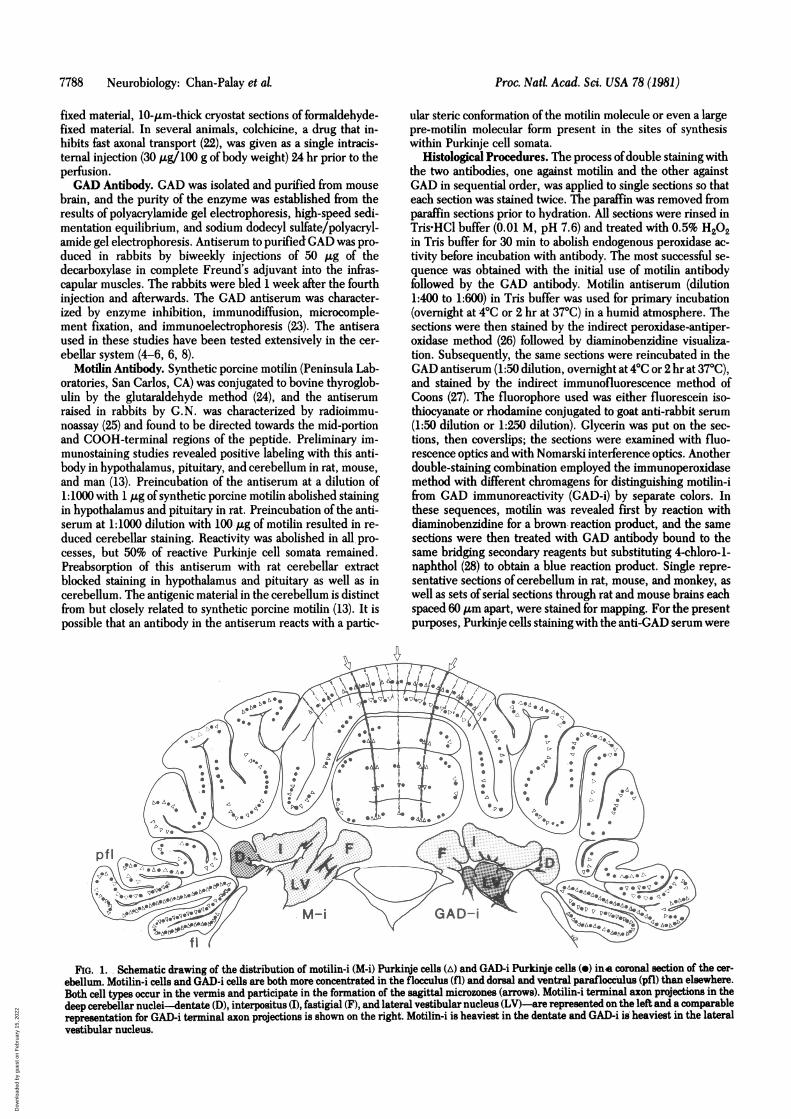
FIG. 2. Montage of photomicrographs showing the sagittal microzones in the midline (broken line) and on either side in the vermis of the mousecerebellum. Motilin-i in Purkinje cells detected by immunoperoxidase techniques with diaminobenzidine (arrows) appears darker than GAD-i de-tectedby chloronaphthol (crossed arrows). A number of cells have reaction products to both motilin-i and GAD-i and therefore appear black or darkestin this figure (large arrows). The microzones are formed by foci of motilin-i and GAD4i Purkinje cells together with their dendritic trees (bracket).(x 100.)
Dow
nloa
ded
by g
uest
on
Feb
ruar
y 15
, 202
2
7790 Neurobiology: Chan-Palay etalP
AL
t'
I X,
'8 ';": s>1a% , Xis Asd '.F v
t,0A, '{, e 0 St.''~ML_
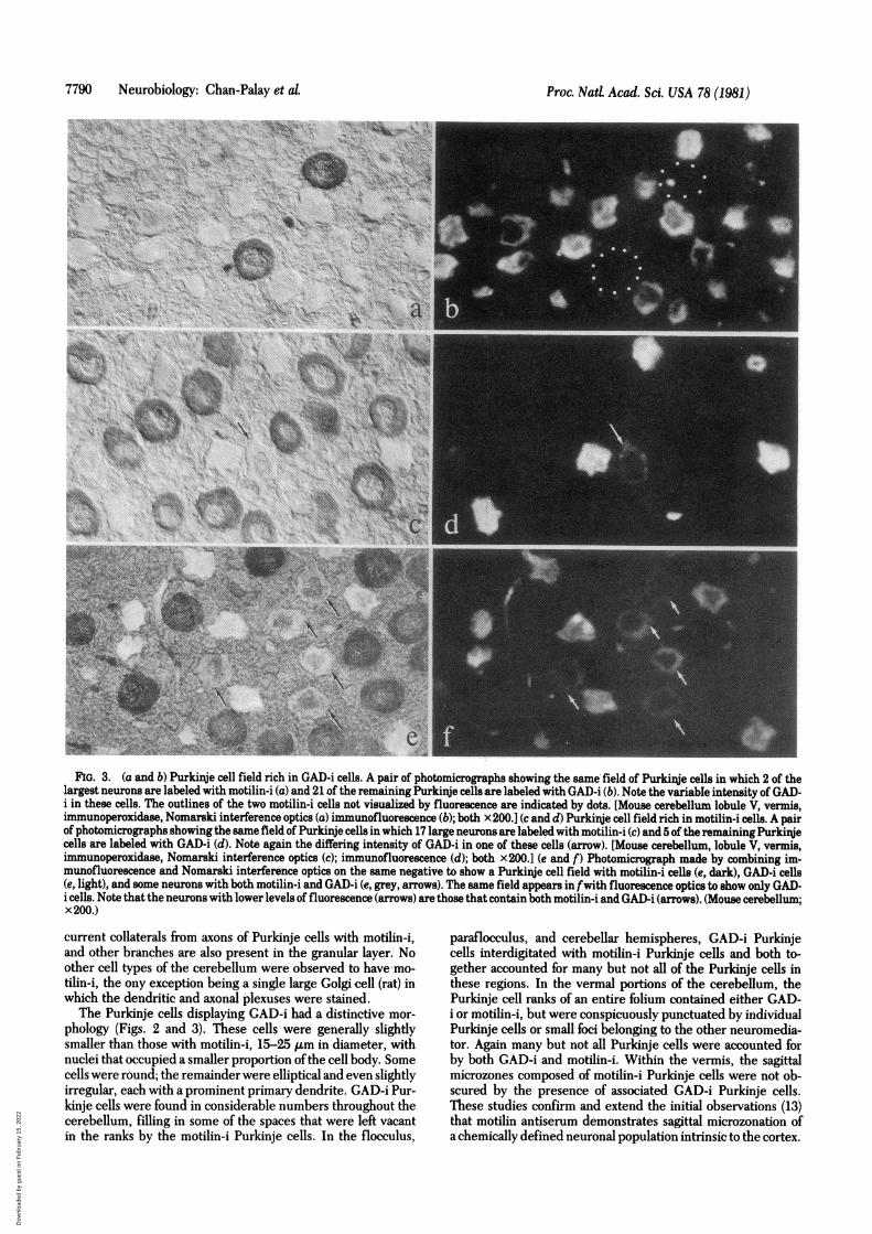
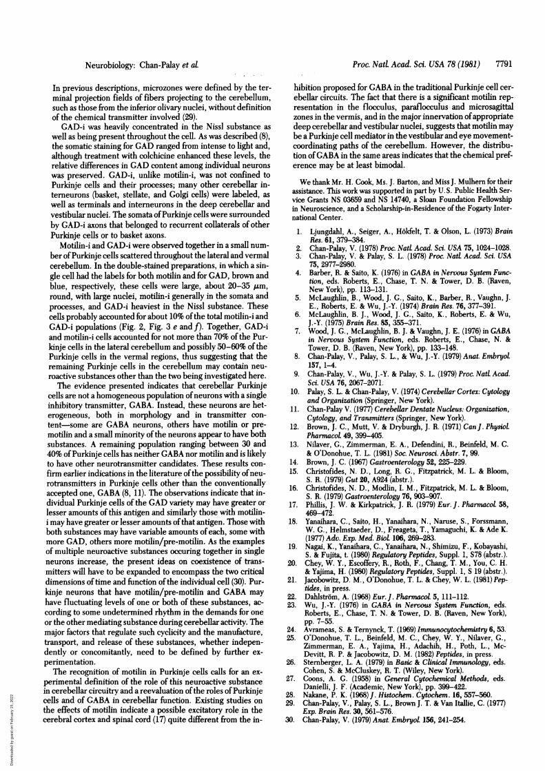
FIG. 3. (a and b) Purkinje cell field rich in GAD-i cells. A pair of photomicrographs showing the same field of Purkinje cells in which 2 of thelargest neurons are labeled with motilin-i (a) and 21 of the remaining Purkinje cellsare labeled with GAD-i (b). Note the variable intensity of GAD-i in these cells. The outlines of the two motilin-i cells not visualized by fluorescence are indicated by dots. [Mouse cerebellum lobule V, vermis,immunoperoxidase, Nomarski interference optics (a) immunofluorescence (b); both x200.] (c and d) Purkinje cell field rich in motilin-i cells. A pairof photomicrographs showing the same field of Purkinje cells in which 17 large neurons are labeled with motilin-i (c) and 5 of the remainingPurkinjecells are labeled with GAD-i (d). Note again the differing intensity of GAD-i in one of these cells (arrow). [Mouse cerebellum, lobule V, vermis,immunoperoxidase, Nomarski interference optics (c); immunofluorescence (d); both x200.] (e and f) Photomicrograph made by combining im-munofluorescence and Nomarski interference optics on the same negative to show a Purkinje cell field with motilin-i cells (e, dark), GAD-i cells(e, light), and some neurons with both motilin-i and GAD-i (e, grey, arrows). The same field appears in fwith fluorescence optics to show only GAD-i cells. Note that the neurons with lower levels of fluorescence (arrows) are those that contain both motilin-i and GAD-i (arrows). (Mouse cerebellum;x 200.)
current collaterals from axons of Purkinje cells with motilin-i,and other branches are also present in the granular layer. Noother cell types of the cerebellum were observed to have mo-tilin-i, the ony exception being a single large Golgi cell (rat) inwhich the dendritic and axonal plexuses were stained.
The Purkinje cells displaying GAD-i had a distinctive mor-phology (Figs. 2 and 3). These cells were generally slightlysmaller than those with motilin-i, 15-25 ,um in diameter, withnuclei that occupied a smaller proportion ofthe cell body. Somecells were round; the remainder were elliptical and even slightlyirregular, each with a prominent primary dendrite. GAD-i Pur-kinje cells were found in considerable numbers throughout thecerebellum, filling in some of the spaces that were left vacantin the ranks by the motilin-i Purkinje cells. In the flocculus,
paraflocculus, and cerebellar hemispheres, GAD-i Purkinjecells interdigitated with motilin-i Purkinje cells and both to-gether accounted for many but not all of the Purkinje cells inthese regions. In the vermal portions of the cerebellum, thePurkinje cell ranks of an entire folium contained either GAD-i or motilin-i, but were conspicuously punctuated by individualPurkinje cells or small foci belonging to the other neuromedia-tor. Again many but not all Purkinje cells were accounted forby both GAD-i and motilin-i. Within the vermis, the sagittalmicrozones composed of motilin-i Purkinje cells were not ob-scured by the presence of associated GAD-i Purkinje cells.These studies confirm and extend the initial observations (13)that motilin antiserum demonstrates sagittal microzonation ofa chemically defined neuronal population intrinsic to the cortex.
Proc. Nad Acad. Sci. USA 78 (1981)
Dow
nloa
ded
by g
uest
on
Feb
ruar
y 15
, 202
2
Proc. NatL Acad. Sci. USA 78 (1981) 7791
In previous descriptions, microzones were defined by the ter-minal projection fields of fibers projecting to the cerebellum,such as those from the inferior olivary nuclei, without definitionof the chemical transmitter involved (29).
GAD-i was heavily concentrated in the Nissl substance as
well as being present throughout the cell. As was described (8),the somatic staining for GAD ranged from intense to light and,although treatment with colchicine enhanced these levels, therelative differences in GAD content among individual neurons
was preserved. GAD-i, unlike motilin-i, was not confined toPurkinje cells and their processes; many other cerebellar in-terneurons (basket, stellate, and Golgi cells) were labeled, as
well as terminals and interneurons in the deep cerebellar andvestibular nuclei. The somata ofPurkinje cells were surroundedby GAD-i axons that belonged to recurrent collaterals of otherPurkinje cells or to basket axons.
Motilin-i and GAD-i were observed together in a small num-ber ofPurkinje cells scattered throughout the lateral and vermalcerebellum. In the double-stained preparations, in which a sin-gle cell had the labels for both motilin and for GAD, brown andblue, respectively, these cells were large, about 20-35 Am,round, with large nuclei, motilin-i generally in the somata andprocesses, and GAD-i heaviest in the Nissl substance. Thesecells probably accounted for about 10% of the total motilin-i andGAD-i populations (Fig. 2, Fig. 3 e and f). Together, GAD-iand motilin-i cells accounted for not more than 70% of the Pur-kinje cells in the lateral cerebellum and possibly 50-60% of thePurkinje cells in the vermal regions, thus suggesting that theremaining Purkinje cells in the cerebellum may contain neu-
roactive substances other than the two being investigated here.The evidence presented indicates that cerebellar Purkinje
cells are not a homogeneous population ofneurons with a singleinhibitory transmitter, GABA. Instead, these neurons are het-erogeneous, both in morphology and in transmitter con-
tent-some are GABA neurons, others have motilin or pre-
motilin and a small minority ofthe neurons appear to have bothsubstances. A remaining population ranging between 30 and40% of Purkinje cells has neither GABA nor motilin and is likelyto have other neurotransmitter candidates. These results con-
firm earlier indications in the literature ofthe possibility ofneu-rotransmitters in Purkinje cells other than the conventionallyaccepted one, GABA (8, 11). The observations indicate that in-dividual Purkinje cells of the GAD variety may have greater or
lesser amounts of this antigen and similarly those with motilin-i may have greater or lesser amounts ofthat antigen. Those withboth substances may have variable amounts of each, some withmore GAD, others more motilin/pre-motilin. As the examplesof multiple neuroactive substances occuring together in singleneurons increase, the present ideas on coexistence of trans-mitters will have to be expanded to encompass the two criticaldimensions oftime and function of the individual cell (30). Pur-kinje neurons that have motilin/pre-motilin and GABA may
have fluctuating levels of one or both of these substances, ac-
cording to some undetermined rhythm in the demands for oneor the other mediating substance during cerebellar activity. Themajor factors that regulate such cyclicity and the manufacture,transport, and release of these substances, whether indepen-dently or concomitantly, need to be defined by further ex-
perimentation.The recognition of motilin in Purkinje cells calls for an ex-
perimental definition of the role of this neuroactive substancein cerebellar circuitry and a reevaluation ofthe roles ofPurkinjecells and of GABA in cerebellar function. Existing studies on
the effects of motilin indicate a possible excitatory role in thecerebral cortex and spinal cord (17) quite different from the in-
hibition proposed for GABA in the traditional Purkinje cell cer-ebellar circuits. The fact that there is a significant motilin rep-resentation in the flocculus, paraflocculus and microsagittalzones in the vermis, and in the major innervation of appropriatedeep cerebellar and vestibular nuclei, suggests that motilin maybe a Purkinje cell mediator in the vestibular and eye movement-coordinating paths of the cerebellum. However, the distribu-tion of GABA in the same areas indicates that the chemical pref-erence may be at least bimodal.
We thank Mr. H. Cook, Ms. J. Barton, and Miss J. Mulhern for theirassistance. This work was supported in part by U.S. Public Health Ser-vice Grants NS 03659 and NS 14740, a Sloan Foundation Fellowshipin Neuroscience, and a Scholarship-in-Residence of the Fogarty Inter-national Center.
1. Ljungdahl, A., Seiger, A., Hbkfelt, T. & Olson, L. (1973) BrainRes. 61, 379-384.
2. Chan-Palay, V. (1978) Proc. Natl Acad. Sci. USA 75, 1024-1028.3. Chan-Palay, V. & Palay, S. L. (1978) Proc. Natl Acad. Sci. USA
75, 2977-2980.4. Barber, R. & Saito, K. (1976) in GABA in Nervous System Func-
tion, eds. Roberts, E., Chase, T. N. & Tower, D. B. (Raven,New York), pp. 113-131.
5. McLaughlin, B., Wood, J. G., Saito, K., Barber, R., Vaughn, J.E., Roberts, E. & Wu, J.-Y. (1974) Brain Res. 76, 377-391.
6. McLaughlin, B. J., Wood, J. G., Saito, K., Roberts, E. & Wu,J.-Y. (1975) Brain Res. 85, 355-371.
7. Wood, J. G., McLaughlin, B. J. & Vaughn, J. E. (1976) in GABAin Nervous System Function, eds. Roberts, E., Chase, N. &Tower, D. B. (Raven, New York), pp. 133-148.
8. Chan-Palay, V., Palay, S. L., & Wu, J.-Y. (1979) Anat. Embryol157, 1-4.
9. Chan-Palay, V., Wu, J.-Y. & Palay, S. L. (1979) Proc. NatL Acad.Sci. USA 76, 2067-2071.
10. Palay, S. L. & Chan-Palay, V. (1974) Cerebellar Cortex: Cytologyand Organization (Springer, New York).
11. Chan-Palay V. (1977) Cerebellar Dentate Nucleus: Organization,Cytology, and Transmitters (Springer, New York).
12. Brown, J. C., Mutt, V. & Dryburgh, J. R. (1971) Can J. PhysiolPharmacol 49, 399-405.
13. Nilaver, G., Zimmerman, E. A., Defendini, R., Beinfeld, M. C.& O'Donohue, T. L. (1981) Soc. Neurosci. Abstr. 7, 99.
14. Brown, J. C. (1967) Gastroenterology 52, 225-229.15. Christofides, N. D., Long, R. G., Fitzpatrick, M. L. & Bloom,
S. R. (1979) Gut 20, A924 (abstr.).16. Christofides, N. D., Modlin, I. M., Fitzpatrick, M. L. & Bloom,
S. R. (1979) Gastroenterology 76, 903-907.17. Phillis, J. W. & Kirkpatrick, J. R. (1979) Eur. J. Pharmacol 58,
469-472.18. Yanaihara, C., Saito, H., Yanaihara, N., Naruse, S., Forssmann,
W. G., Helmstaeder, D., Freageta, T., Yamaguchi, K. & Ade K.(1977) Adv. Exp. Med. Biol 106, 269-283.
19. Nagai, K., Yanaihara, C., Yanaihara, N., Shimizu, F., Kobayashi,S. & Fujita, t. (1980) Regulatory Peptides, Suppl. 1, S78 (abstr.).
20. Chey, W. Y., Escoffery, R., Roth, F., Chang, T. M., You, C. H.& Yajima, H. (1980) Regulatory Peptides, Suppl. 1, S 19 (abstr.).
21. Jacobowitz, D. M., O'Donohue, T. L. & Chey, W. L. (1981) Pep-tides, in press.
22. Dahlstrom, A. (1968) Eur. J. Pharmacoi 5, 111-112.23. Wu, J.-Y. (1976) in GABA in Nervous System Function, eds.
Roberts, E., Chase, T. N. & Tower, D. B. (Raven, New York),pp. 7-55.
24. Avrameas, S. & Ternynck, T. (1969) Immunocytochemistry 6, 53.25. O'Donohue, T. L., Beinfeld, M. C., Chey, W. Y., Nilaver, G.,
Zimmerman, E. A., Yajima, H., Adachih, H., Poth, L., Mc-Devitt, R. P. & Jacobowitz, D. M. (1982) Peptides, in press.
26. Sternberger, L. A. (1979) in Basic & Clinical Immunology, eds.Cohen, S. & McCluskey, R. T. (Wiley, New York).
27. Coons, A. G. (1958) in General Cytochemical Methods, eds.Danielli, J. F. (Academic, New York), pp. 399-422.
28. Nakane, P. K. (1968) J. Histochem. Cytochem. 16, 557-560.29. Chan-Palay, V., Palay, S. L., Brown J. T. & Van Itallie, C. (1977)
Exp. Brain Res. 30, 561-576.30. Chan-Palay, V. (1979) Anat. Embryot 156, 241-254.
Neurobiology: Chan-Palay et aL
Dow
nloa
ded
by g
uest
on
Feb
ruar
y 15
, 202
2