propionobacter acnes infection as an occult cause of ... · (orif) for a proximal humerus fracture...
TRANSCRIPT
CLINICAL RESEARCH
Propionobacter acnes Infection as an Occult Causeof Postoperative Shoulder Pain
A Case Series
Peter J. Millett MD, MSc, Yi-Meng Yen MD, PhD,
Connie S. Price MD, Marilee P. Horan MPH,
Olivier A. van der Meijden MD, Florian Elser MD
Received: 6 August 2010 / Accepted: 3 January 2011 / Published online: 15 January 2011
� The Association of Bone and Joint Surgeons1 2011
Abstract
Background Infections after shoulder surgery are poten-
tially devastating complications. Propionibacterium acnes
is recognized as a causal agent in shoulder infections. The
clinical presentation is usually insidious and nonspecific,
but a P. acnes infection could be an occult cause of post-
operative shoulder pain.
Questions/purposes What are the clinical and microbio-
logic characteristics of a postsurgical P. acnes shoulder
infection and how should it be addressed?
Patients and Methods Ten patients with an average age
of 57 years presented with P. acnes postsurgical shoulder
infection. Clinical infection signs and surgical history were
assessed and joint aspirates and tissue biopsy specimens
were obtained. Diagnosis was confirmed by microbiologic
cultures.
Results At the time of confirmation of the diagnosis,
clinical signs of infection were absent. C-reactive protein
and erythrocyte sedimentation rates were inconsistently
elevated. Cultures took a mean 7 days to confirm organism
growth. The average time from surgery to diagnosis of
infection was 1.8 years (range, 0.07–8.0 years). All patients
underwent irrigation and debridement and were treated
with antibiotics for 6 weeks.
Conclusions P. acnes shoulder infections should be con-
sidered as a cause for persistent, unexplained shoulder
pain. Shoulder aspirations and tissue samples should be
obtained. Surgical debridement and intravenous antibiotics
are necessary treatment modalities.
Level of Evidence Level IV, Prognostic study. See the
Guidelines for Authors for a complete description of levels
of evidence.
Investigation performed at the Steadman Philippon Research Institute,
Vail, Colorado.
One or more of the authors, (PJM, FE, OAvdM) have received
funding from a commercial entity (Arthrex, Inc, Naples, FL)
something of value (exceeding the equivalent of $500 - US) not
related to this manuscript or research. PJM is a consultant and
receives payments from Arthrex and has stock options in Game
Ready. FE and OAvdM’s positions were supported by Arthrex. This
research was supported by The Steadman Philippon Research
Institute, which is a 501(c)3 nonprofit institution supported financially
by private donations and corporate support from the following
entities: Smith & Nephew Endoscopy (Memphis, TN), Arthrex,
ArthroCare Corp (Austin, TX), Siemens USA (Washington, DC),
OrthoRehab, Inc (Tempe, AZ), and Ossur Americas (Foothill
Ranch, CA).
Each author certifies that the institution approved the human protocol
for this investigation, that all investigations were conducted in
conformity with ethical principles of research, and that informed
consent for participation in the study was obtained.
This work was performed at the Steadman Phillipon
Research Institute.
P. J. Millett (&), M. P. Horan, O. A. van der Meijden
Steadman Phillipon Research Institute, 181 West Meadow Drive,
Suite 1000, Vail, CO 81657, USA
e-mail: [email protected]
M. P. Horan
e-mail: [email protected]
O. A. van der Meijden
e-mail: [email protected]
Y.-M. Yen
Department of Pediatric Orthopaedics, Division of Sports
Medicine Children’s Hospital Boston, Harvard Medical School,
Boston, MA, USA
e-mail: [email protected]
123
Clin Orthop Relat Res (2011) 469:2824–2830
DOI 10.1007/s11999-011-1767-4
Introduction
Infection after shoulder surgery is a potentially devastat-
ing complication. The incidence of infection after rotator
cuff repair ranges between 0.27% to 1.9% [3, 18, 27, 28,
37], whereas the incidence after shoulder arthroplasty
ranges from 0% and 15.4% [9, 11, 38, 41, 44]. Infections
after proximal humerus fractures are less common [4, 29,
32, 40, 46].
P. acnes is being recognized as a causal agent in shoulder
infections with increasing frequency [3, 7, 14, 18, 33, 35, 37,
39, 45]. This nonspore-forming microaerophilic bacillus has
a low level of virulence but has been established as a path-
ogen in endocarditis [30, 42], meningitis [5, 43], arthritis
[23], osteomyelitis [1, 31] and spondylitis [10, 13]. It has
been described as the infectious etiology in 16% of pros-
thetic shoulder infections [38] and in 21% of revision
shoulder arthroplasties [22]. In another series, P. acnes was
the major etiology of postoperative septic arthritis, com-
prising 56% of shoulder infections [26]. Herrera et al. [18] in
2002 reported the incidence of deep infection after open
rotator cuff repair to be 0.27% to 1.7%. Athwal et al. [3]
found an incidence of 0.43% after different techniques of
rotator cuff reconstructions. P. acnes was reported as the
most common infecting organism in both studies.
In contrast, reports of P. acnes in lower-extremity
infections, including prosthetic joint infections or after
arthroscopy, are rare [8, 25, 31, 38]. This is likely
because P. acnes is predominant in the pilosebaceous
follicles [34] that are most prevalent in the head, neck,
and thorax. P. acnes is a dominant anaerobic bacteria
isolated from the healthy skin in moist areas such as the
axilla. This is another reason why the shoulder is thought
to have a propensity for infections with this type of
microorganism [14].
Because it is difficult to detect, the actual number of
P. acnes shoulder infections probably has been underesti-
mated. P. acnes is a fastidious organism, requiring
prolonged growth duration and anaerobic conditions to
grow in culture. When isolated, it often is disregarded as a
contaminant owing to its presence as a commensal of
upper-body skin sites. Furthermore, the clinical presenta-
tion of P. acnes is usually insidious and nonspecific.
Patients present without the typical signs of infection such
as fever or local inflammatory reaction and invariably have
normal leukocyte counts (WBCs) [22].
To increase our awareness of the potential of P. acnes
infection as an occult cause of postoperative shoulder pain,
we investigated a series of patients diagnosed with post-
surgical P. acnes shoulder infections to answer the
following questions: (1) What characterizes a P. acnes
infection of the shoulder, in clinical presentation and
quantification of laboratory markers? (2) How are the
microbiologic diagnosis and analysis best obtained?
(3) How should this infection be treated and what clinical
results of therapy can be expected?
Patients and Methods
We retrospectively reviewed all patients diagnosed with
postsurgical deep P. acnes shoulder infections at our clinic
between June 2006 and November 2007. Inclusion criteria
required culture-positive specimens obtained deep to the
deltoid in patients with symptoms of pain, prosthetic dys-
function, or joint sepsis. Patients with superficial wound
infections involving the skin or subcutaneous tissue were
excluded. We identified 10 patients, including two women
and eight men, with P. acnes shoulder infections. The mean
age of the patients was 57.8 years (range, 24–81 years)
(Table 1). Their average followup was 1.7 years (range,
3 months to 2.5 years). The research protocol was reviewed
and approved by the local institutional review board.
For the purpose of this study, a deep shoulder infection
was defined as a positive bacterial culture from specimens
obtained deep to the deltoid muscle. A symptomatic
infection was defined as any one of the following symp-
toms was present: pain, prosthetic dysfunction (decreased
ROM or joint instability), swelling, drainage, fever, leu-
kocytosis, elevated inflammatory markers, or purulence in
the joint. The index operation was defined as the last
operation that preceded the diagnosis of infection.
Table 1. Demographics of included patients
Characteristic Value
Number of patients 10
Male/female 8/2
Mean age (years) 57.8
Index operation performed at other facility 6/10
Index operation
RTC repair 4/10
ORIF humerus fracture 1/10
TSA 5/10
RTC = rotator cuff; ORIF = open reduction and internal fixation; TSA
= total shoulder arthroplasty.
C. S. Price
Infection Prevention/Hospital Epidemiology and Division of
Infectious Diseases, Denver Health and Hospital, University of
Colorado School of Medicine, East Aurora, CO, USA
e-mail: [email protected]
F. Elser
Department of Traumatology, Technical University Munich,
Klinikumrechts der Isar, Technical University Munich, Munich,
Germany
e-mail: [email protected]
Volume 469, Number 10, October 2011 P. acnes Infection after Shoulder Surgery 2825
123
A successful outcome was defined as eradication of
infection and elimination of pain and joint dysfunction.
The index operation was arthroscopic rotator cuff repair
in four patients, open reduction and internal fixation
(ORIF) for a proximal humerus fracture in one patient, and
shoulder arthroplasty in five patients. Four patients had
infections developed from after surgery at our facility and
six sought treatment from us for ongoing shoulder prob-
lems after having the index procedure at an outside facility.
Medical comorbidities that may have been contributing
factors included Type II diabetes in two individuals and
hepatitis C in one.
Comprehensive histories and physical examinations
were obtained from all patients. Pain (location, duration,
and character), swelling, appearance (erythema or lack
thereof), and ROM were assessed for each patient. All
underwent screening laboratory studies including WBC,
C-reactive protein (CRP), and erythrocyte sedimentation
rate (ESR). From these clinical data, the following data
elements were abstracted: demographics; description of
index operation, facility, and timing of index procedure
in relation to definitive diagnosis of infection; medical
comorbidities; duration of infection symptoms; chief
complaint and description of symptoms at time of presen-
tation in our clinic; physical examination findings;
operative findings at time of initial debridement; laboratory
findings; treatment regimen; and timing and clinical out-
come at last followup.
All culture specimens were obtained before adminis-
tration of any prophylactic or therapeutic antibiotic
therapy. Before surgery, all the shoulders were aspirated.
Nine of the 10 shoulders were aspirated under flouroscopic
guidance to ensure accurate needle placement. One patient
underwent aspiration in the outpatient office but this patient
had a massive rotator cuff tear with a contiguous suba-
cromial and intraarticular space. Most of the time, no fluid
was obtained. In all suspected cases that had a negative
aspiration, the joint was lavaged with saline before making
an incision, and the lavage fluid then was sent for culture.
Joint aspirates or lavage samples and a minimum of two
deep tissue samples (preferably three to five) were col-
lected during the operation, including joint capsule,
synovial lining, bone, or granulation tissue. An average of
three biopsy specimens (range, 1–6 specimens) were taken
from each shoulder to address the possible problem of
false-negative specimens as much as possible. After tissue
biopsies, antibiotic therapy was instituted intraoperatively
with empiric prophylactic antibiotics (typically a first-
generation cephalosporin) and then was modified under the
guidance of an infectious diseases expert to a therapeutic
regimen when the cultures turned positive. Patient speci-
mens were processed immediately after their arrival at the
laboratory, under sterile conditions in a laminar flow hood
for aerobic and anaerobic culture. For aerobic culture,
specimens were cultured on trypticase-soy agar containing
5% sheep blood (Remel, Lenexa, KS, USA), chocolate agar
(Remel), and MacConkey agar (Remel). Cultures were
incubated at 36�C under standard aerobic conditions. For
anaerobic culture, Brucella media (Anaerobe Systems,
Morgan Hill, CA, USA) were inoculated and cultured at
36�C in an anaerobic chamber. Media were checked daily
(aerobes) or every other day (anaerobes) for bacterial
growth. For tissue samples, thioglycolate broth (Remel)
also was inoculated and incubated in aerobic and anaerobic
conditions. Clouded broth was Gram stained and subcul-
tured onto appropriate agar plates. Cultures were
discontinued and declared negative if no growth was visi-
ble at 3 days for aerobes. As there was a high suspicion of
the possibility of P. acnes, prolonged testing was requested
for anaerobes. Orthopaedic specimens always were
requested to be held for 14 days. Positive colony growth
was identified by standard microbiologic procedures [19].
Pinpoint yellow- or buff-colored colony growth in anaer-
obic conditions was subjected to aerotolerance testing. A
positive aerotolerance test combined with a Gram stain
morphology showing pleomorphic gram-positive rods was
subjected to anaerobic catalase and indole testing, and
identification of P. acnes was confirmed using the RapIDTM
ANA II Identification System (Remel). Isolates confirmed
as P. acnes were subjected to b-lactamase testing.
The diagnosis of P. acnes infection was made with a
mean of three tissue biopsy specimens (capsule tissue,
bone, subacromial tissue) and a minimum of one biopsy
specimen on which the P. acnes organism grew. Pain and
functional outcomes were measured using the American
Shoulder and Elbow Surgeon (ASES) score (scale 0–100),
which has been validated for common shoulder disorders
such as rotator cuff disorders, instability, and osteoarthritis.
In addition, patients rated their daily pain on a 1 to 10 scale
with 1 being no pain and 10 being worst pain. Patients were
asked to rate how satisfied they were with their treatment
outcome on a 10-point scale, with 1 being very unsatisfied
and 10 being very satisfied.
All patients underwent a minimum of one surgical
debridement. Four of the five patients with shoulder pros-
theses underwent removal of their implants with insertion
of a antibiotic coated cement spacer. This subset had an
average of three surgical debridements. All but one patient
had reimplantation of a new prosthesis. Patients who had
an infection after rotator cuff arthroscopy underwent a
mean of one surgical debridement and the suture anchors
were removed, whereas the patient with the ORIF of a
proximal humerus fracture required only one debridement
and hardware removal in the same setting; the fracture was
not yet completely healed. In the subset of patients who
had undergone prior rotator cuff surgery, there were no
2826 Millett et al. Clinical Orthopaedics and Related Research1
123
obvious recurrent tears. In all 10 of these patients, the
definitive diagnosis of P. acnes infection was not known at
the time of surgery. At the time of the initial debridement
(seven were performed open, three performed arthroscop-
ically), all tissue specimens sent for immediate
intraoperative Gram stain were negative. Penicillin VK was
prescribed for three patients and intravenous ampicillin-
sulbactam was prescribed for nine patients. All patients
received antibiotics for a minimum of 6 weeks. None of the
10 patients had a b-lactamase-producing strain of P. acnes.
Patient treatment outcomes with a minimum 1 year fol-
lowup are reported for seven of the 10 patients. Subjective
followup was obtained through mail.
Results
The most consistent complaint of all patients was persistent
shoulder pain (Table 2). Only one patient had symptoms of
fever, swelling, and active wound drainage (this was a
subacute case diagnosed 4 weeks after arthroscopic rotator
cuff repair). For the nine patients who had pain as their
primary complaint, swelling around the shoulder was found
in one additional patient; three patients also had decreased
ROM or stiffness; and one patient had erythema. Despite
prolonged symptoms of pain, with a mean duration of
symptoms of 3 months (range, 1–9 months), the mean time
from the index surgery to diagnosis of infection was
1.8 years (range, 26 days to 8 years).
The preoperative WBCs in these 10 patients were nor-
mal (normal range, 5 9 103/mm3 to 10 9 103/mm3), and
ESR and CRP (normal range, 1–25 mm/hour and\10 mg/
dL, respectively) were not consistently elevated. The WBC
averaged 6.8 9 103/mm3 (range, 4.8–10.4 9 103/mm3).
The average CRP was 9.1 (range, \ 3–31 mg/dL). The
CRP was elevated above normal limits in two of
10 patients (average, 26; range, 21–31 mg/dL). The ESR
averaged 6.9 mm/hour (range, 1–10 mm/hour) in eight
patients within normal limits; the average ESR for two
patients with elevated levels was 29 mm/hour (range,
19–39 mm/hour). P. acnes took an average of 7 days
(range, 5–13 days) to grow on the cultures of all 10
patients. P. acnes grew on only five of 10 lavage or aspi-
ration samples.
All but three patients remained pain-free at clinical
followup (Table 2). One patient initially did well and then
had increased pain development. This patient progressed to
another surgery and had positive cultures of P. acnes. This
could be considered a failure of our treatment as opposed to
a new infection. The two other patients reporting continued
pain had the lowest satisfaction with surgical outcomes
scores. The average postoperative ASES score for patients
with more than 1-year followup (eight patients) was
78 points. Overall, the average patient satisfaction score
with surgical outcomes in patients who did not progress to
another surgery was 7 of 10 points. Four of the five patients
who had arthroplasties maintained their ROM, with one
patient electing not to have prosthesis reimplantation.
Discussion
Infections after shoulder surgery are a potentially devas-
tating complication. P. acnes is recognized as a causal
agent in shoulder infections. The clinical presentation is
usually insidious and nonspecific, but a P. acnes infection
could be an occult cause of postoperative shoulder pain. To
increase the awareness of P. acnes infection as a cause of
postoperative shoulder pain, we attempted to answer the
following questions: What characterizes a P. acnes infec-
tion of the shoulder from clinical and microbiologic
perspectives? How should this infection be treated and
what outcome can be expected?
This study has several limitations inherent in retro-
spective reviews of charts and laboratory results. It is a
small case series, representing a conglomerate of shoulder
infections at a referral sports medicine clinic, which could
result in selection bias. Patient treatment outcomes are
encouraging, yet the study’s sample size and lack of spe-
cific followup makes it difficult to make specific
recommendations. This report nevertheless is intended to
alert orthopaedic surgeons that patients can present with
subclinical infections after shoulder surgery and it may
take prolonged anaerobic culture of up to 14 days to
identify this slow-growing fastidious bacterium. Although
there is a risk that we included patients who had P. acnes
contamination in our cohort, the clinical improvement after
surgical debridement and antibiotic therapy makes us
believe the possibility of false-positives was less likely.
Insidious protracted pain was the most frequent pre-
sentation of infection attributable to P. acnes. The
nonspecific clinical findings and P. acnes’ indolent nature
have posed challenges to others in diagnosing a P. acnes
infection after shoulder surgery [22]. Only one patient
(10%) in our patient population had wound drainage and
fever. Assessment of a comparable patient selection after
total shoulder arthroplasty found none presented with fever
[19]. However, Athwal et al. [3] noticed wound drainage in
72% and erythema in 64% of the patients. This discrepancy
might be attributable to different bacteria, as organisms
other than P. acnes grew on 19 of 39 specimens [3]. The
standard laboratory data often are negative or nonspecific.
As in previous studies [3, 14], ESR and CRP levels have
not been reliable indicators for infection.
Patients were diagnosed an average of 1.8 years after
their index operations, with symptomatic complaints
Volume 469, Number 10, October 2011 P. acnes Infection after Shoulder Surgery 2827
123
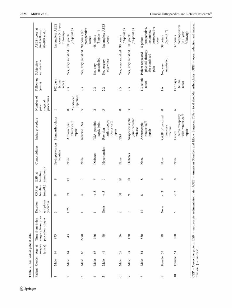
Ta
ble
2.
Ind
ivid
ual
pat
ien
td
ata
Pat
ien
tG
end
erA
ge
at
surg
ery
(yea
rs)
Tim
efr
om
ind
ex
surg
ery
fro
mp
rio
r
pro
ced
ure
(day
s)
Du
rati
on
of
sym
pto
ms
(mo
nth
s)
CR
Pat
bas
elin
e
(mg
/dL
)
ES
Rat
bas
elin
e
(mm
/ho
ur)
Co
mo
rbid
itie
sIn
dex
pro
ced
ure
Nu
mb
ero
f
pri
or
surg
ical
pro
ced
ure
s
Fo
llo
w-u
p
(yea
rs)
Su
bje
ctiv
e
sati
sfac
tio
n
AS
ES
sco
reat
fin
alfo
llo
wu
p
(0–
10
0sc
ale)
1M
ale
69
97
33
88
Pre
hy
per
ten
sio
n
hep
atit
is
Hem
iart
hro
pla
sty
11
02
day
s
(cli
nic
no
te)
Inco
mp
lete
AS
ES
sco
res
(\1
-yea
r
foll
ow
up
)
2M
ale
64
43
1.2
52
13
9N
on
eA
rth
rosc
op
ic
rota
tor
cuff
rep
air
0 2co
rtis
on
e
inje
ctio
ns
2.3
Yes
,v
ery
sati
sfied
10
0p
oin
ts
(27
-po
int:)
3M
ale
66
27
90
14
7N
on
eR
ever
seT
SA
52
.3Y
es,
ver
ysa
tisfi
ed9
0p
oin
ts(n
o
pre
op
erat
ive
sco
re)
4M
ale
63
96
61
\3
5D
iab
etes
TS
A,
po
ssib
le
sep
tic
join
t
22
.2N
o,
ver
y
un
sati
sfied
48
po
ints
(3-p
oin
t:)
5M
ale
46
90
No
ne
\3
1H
yp
erte
nsi
on
Rev
isio
n
arth
rosc
op
ic
rota
tor
cuff
rep
air
32
.2N
o,
surg
ery
else
wh
ere
Inco
mp
lete
AS
ES
sco
res
6M
ale
57
26
23
11
9N
on
eT
SA
02
.5Y
es,
ver
ysa
tisfi
ed9
0p
oin
ts
(53
-po
int:)
7M
ale
24
12
09
91
0D
iab
etes
Su
spec
ted
sep
tic
join
tca
psu
lar
rele
ase
32
.3Y
es,
ver
ysa
tisfi
ed1
00
po
ints
(85
-po
int:)
8M
ale
81
55
01
28
8N
on
eA
rth
rosc
op
ic
rota
tor
cuff
rep
air
11
.3(c
lin
ic
no
te)
Pat
ien
tre
qu
este
d
ph
ysi
cal
ther
apy
for
con
tin
ued
pai
n
8p
oin
ts
pre
op
erat
ive,
inco
mp
lete
po
sto
per
ativ
e
sco
re
9F
emal
e5
39
8N
on
e\
38
No
ne
OR
IFo
fp
rox
imal
hu
mer
us
frac
ture
01
.6N
o,
ver
y
un
sati
sfied
38
po
ints
(6-p
oin
t:)
10
Fem
ale
51
90
05
\3
8N
on
eF
aile
d
hem
iart
hro
pla
sty
wit
hro
tato
rcu
ff
rep
air
61
57
day
s
(cli
nic
no
te)
32
po
ints
pre
op
op
erat
ive
(\1
-yea
r
foll
ow
up
)
CR
P=
C-r
eact
ive
pro
tein
;E
SR
=er
yth
rocy
tese
dim
enta
tio
nra
te;
AS
ES
=A
mer
ican
Sh
ou
lder
and
Elb
ow
Su
rgeo
n;
TS
A=
tota
lsh
ou
lder
arth
rop
last
y;
OR
IF=
op
enre
du
ctio
nan
din
tern
al
fix
atio
n;:
=in
crea
se.
2828 Millett et al. Clinical Orthopaedics and Related Research1
123
averaging 3 months. This is similar to the report by Zeller
et al. [45], who classified the majority of infections in their
series as late chronic infections as opposed to acute post-
operative infections. One patient in our study presented
8 years after the index operation. It was unclear whether
the infection was indolent for those years or acquired
secondarily.
Previously reported risk factors associated with P. acnes
infections were male gender, first surgery of the day, and
increased duration of the surgical procedure [11]. Acute
outbreak of P. acnes was decreased by improved ventila-
tion systems and cleaning of the operating room [11].
Although the majority of our patients were men, it is
unlikely the ventilation systems or cleaning played a role as
most infections in our patients developed at outside insti-
tutions and procedures are performed in high-efficiency
particulate air-filtered rooms.
P. acnes required prolonged incubation in the laboratory
to detect growth, 5 to 13 days; similar ranges are supported
by other studies [21, 26, 36]. In most clinical laboratories
in the United States, conventional cultures are held only for
5 days [19]. Based on our findings and those of others, we
recommend cultures be held for at least 14 days [18].
Furthermore, similar to others [15, 21], we found intraop-
erative Gram stains unhelpful in diagnosing P. acnes
infection. At the time of the initial debridement, all spec-
imens sent for intraoperative Gram stain in our series were
negative. There are recent reports on PCR in combination
with sequencing of 16S rDNA [16] and multiplex PCR of
sonification fluid in detecting periprosthetic P. acnes joint
infections [2] in patients who had false-negative P. acnes
cultures. A limitation of this testing technique is the time it
takes to confirm a diagnosis; the results come in too late to
help in the clinical diagnosis. Poor bacterial DNA extrac-
tion properties also may limit sensitivity of the test, along
with possible cross-contamination from the patient’s or
staff’s skin. Molecular testing may be useful in the future
to confirm the presence of P. acnes, but it was not available
to us at the time of this study.
P. acnes often is susceptible to penicillin, tetracyclines,
chloramphenicol, erythromycin, and vancomycin, but
resistance is increasing. Sixty percent of isolates are
resistant to at least one antibiotic, likely owing to wide-
spread antibiotic use for acne vulgaris [12, 33]. Information
on resistance is gleaned mainly from cases of acne vulga-
ris, so correlations with the much less common systemic
infections are unclear. In our series, none of the isolates
possessed a b-lactamase, suggesting good susceptibilities
to penicillin and its derivatives. Therefore, penicillin G,
2 mU intravenously every 4 hours or via continuous infu-
sion, was the most focused therapy for serious deep
infections [23]. Other b-lactam antibiotics also have been
used successfully for serious infection [20]. Alternative
agents, if susceptible, may include clindamycin, vanco-
mycin, or a combination therapy with vancomycin and
doxycycline [17, 24]. Unlike other anaerobes, most P. ac-
nes are resistant to metronidazole [42]. Combination
therapy of rifampin and amoxicillin [10] and rifampin with
either penicillin or linezolid [6] also have been reported
successful for treatment of P. acnes shoulder prosthesis
infections. There are no clear data on prosthesis retention;
thus, removal of foreign bodies or a two-stage joint
arthroplasty for arthroplasty infections should be consid-
ered in difficult cases.
Infection by P. acnes is a potentially devastating com-
plication after shoulder surgery. Nonspecific complaints of
pain and stiffness were the most common findings, whereas
the typical clinical signs of infection usually are absent. As
this is an indolent type of infection, the normal laboratory
indicators of sepsis are unreliable. Coupled with difficulty
in growing P. acnes in culture, this makes the diagnosis of a
P. acnes infection easy to miss. Given the incidence, better
awareness of the problem and possibly a change in pro-
phylaxis might be considered in the future. Although it is
possible to eliminate infections of P. acnes with surgical
debridement and appropriate antibiotics, patients should be
counseled on potential permanent functional limitations of
their shoulder.
References
1. Abolnik IZ, Eaton JV, Sexton DJ. Propionibacterium acnes ver-
tebral osteomyelitis following lumbar puncture: case report and
review. Clin Infect Dis. 1995;21:694–695.
2. Achermann Y, Vogt M, Leunig M, Wust J, Trampuz A. Improved
diagnosis of periprosthetic joint infection by multiplex PCR of
sonification fluid from removed implants. J Clin Microbiol. 2010;
48:1208–1214.
3. Athwal GS, Sperling JW, Rispoli DM, Cofield RH. Deep infec-
tion after rotator cuff repair. J Shoulder Elbow Surg. 2007;16:
306–311.
4. Athwal GS, Sperling JW, Rispoli DM, Cofield RH. Acute deep
infection after surgical fixation of proximal humeral fractures.
J Shoulder Elbow Surg. 2007;16:408–412.
5. Barazi SA, Gnanalingham KK, Chopra I, van Dellen JR. Delayed
postoperative intracerebral abscess caused by Propionibacterium
acnes: case report and review of the literature. Br J Neurosurg.2003;17:336–339.
6. Bayston R, Nuradeen B, Ashraf W, Freeman BJ. Antibiotics for
the eradication of Propionibacterium acnes biofilms in surgical
infection. J Antimicrob Chemother. 2007;60:1298–1301.
7. Berthelot P, Carricajo A, Aubert G, Akhavan H, Gazielly D,
Lucht F. Outbreak of postoperative shoulder arthritis due to
Propionibacterium acnes infection in nondebilitated patients.
Infect Control Hosp Epidemiol. 2006;27:987–990.
8. Carlsson AS, Josefsson G, Lindberg L. Revision with gentamicin-
impregnated cement for deep infections in total hip arthroplasties.
J Bone Joint Surg Am. 1978;60:1059–1064.
9. Cheung EV, Sperling JW, Cofield RH. Infection associated with
hematoma formation after shoulder arthroplasty. Clin OrthopRelat Res. 2008;466:1363–1367.
Volume 469, Number 10, October 2011 P. acnes Infection after Shoulder Surgery 2829
123
10. Chia JK, Nakata MN. Intervertebral diskitis caused by Propion-
ibacterium acnes: a report of four cases. Clin Infect Dis. 1996;23:
643–644.
11. Cofield RH, Edgerton BC. Total shoulder arthroplasty:
complications and revision surgery. Instr Course Lect. 1990;39:
449–462.
12. Cooper AJ. Systematic review of Propionibacterium acnes
resistance to systemic antibiotics. Med J Aust. 1998;169:259–
261.
13. Crouzet J, Claudepierre P, Aribi EH, Malbrunot C, Beraneck L,
Juan LH, Larget-Piet B. Two cases of discitis due to Propioni-
bacterium acnes. Rev Rhum Engl Ed. 1998;65:68–71.
14. Dodson CC, Craig EV, Cordasco FA, Dines DM, Dines JS,
Dicarlo E, Brause BD, Warren RF. Propionibacterium acnes
infection after shoulder arthroplasty: a diagnostic challenge.
J Shoulder Elbow Surg. 2010:19:303–307.
15. Esteban J, Garcıa-Calvo G, Jimenez-Castillo P, Soriano F. Failure
of Gram stain to detect Propionibacterium acnes in specimens
from clinically significant infections. J Clin Microbiol. 1996;34:
2051.
16. Fenollar F, Roux V, Stein A, Drancourt M, Raoult D. Analysis of
525 samples to determine the usefulness of PCR amplification
and sequencing of the 16S rRNA gene for diagnosis of bone and
joint infections. J Clin Microbiol. 2006;44:1018–1028.
17. Fincher ME, Forsyth M, Rahimi SY. Successful management
of central nervous system infection due to Propionibacterium
acnes with vancomycin and doxycycline. South Med J. 2005;98:
118–121.
18. Herrera MF, Bauer G, Reynolds F, Wilk RM, Bigliani LU,
Levine WN. Infection after mini-open rotator cuff repair.
J Shoulder Elbow Surg. 2002;11:605–608.
19. Isenberg HD. Clinical Microbiology Procedures Handbook. 2nd
ed. Washington, DC: American Society for Microbiology; 1992.
20. James WD. Clinical practice: acne. N Engl J Med. 2005;352:
1463–1472.
21. Johnson MJ, Thatcher E, Cox ME. Techniques for control-
ling variability in Gram staining of obligate anaerobes. J ClinMicrobiol 1995;33:755–758.
22. Kelly JD. Positive culture rates in revision shoulder arthroplasty,
Clin Orthop Relat Res. 2009;467:2343–2348.
23. Kooijmans-Coutinho MF, Markusse HM, Dijkmans BA. Infec-
tious arthritis caused by Propionibacterium acnes: a report of two
cases. Ann Rheum Dis. 1989;48:851–852.
24. Lalani T, Person AK, Hedayati SS, Moore L, Murdoch DR,
Hoen B, Peterson G, Shahbaz H, Raoult D, Miro JM, Olaison L,
Snygg-Martino U, Suter F, Spelman D, Eykyn S, Strahilevitz J,
Van der Meer JT, Verhagen D, Baloch K, Abrutyn E, Cabell CH;
International Collaboration on Endocarditis Merged; Database
Study Group. Propionibacterium endocarditis: a case series
from the International Collaboration on Endocarditis Merged
Database and Prospective Cohort Study. Scand J Infect Dis. 2007;
39:840–848.
25. Launder WJ, Hungerford DS. Late infection of total hip
arthroplasty with Propionibacterium acnes: a case and review of
the literature. Clin Orthop Relat Res. 1981;157:170–177.
26. Levy PY, Fenollar F, Stein A, Borrione F, Cohen E, Lebail B,
Raoult D. Propionibacterium acnes postoperative shoulder
arthritis: an emerging clinical entity. Clin Infect Dis. 2008;46:
1884–1886.
27. Mansat P, Cofield RH, Kersten TE, Rowland CM. Complications
of rotator cuff repair. Orthop Clin North Am. 1997;28:205–213.
28. Mirzayan R, Itamura JM, Vangsness CT Jr, Holtom PD, Sherman
R, Patzakis MJ. Management of chronic deep infection following
rotator cuff repair. J Bone Joint Surg Am. 2000;82:1115–1121.
29. Moda SK, Chadha NS, Sangwan SS, Khurana DK, Dahiya AS,
Siwach RC. Open reduction and fixation of proximal humeral
fractures and fracture-dislocations. J Bone Joint Surg Br. 1990;
72:1050–1052.
30. Mohsen AH, Price A, Ridgway E, West JN, Green S,
McKendrick MW. Propionibacterium acnes endocarditis in a
native valve complicated by intraventricular abscess: a case
report and review. Scand J Infect Dis. 2001;33:379–380.
31. Morrey BF, Fitzgerald RH, Kelly PJ, Dobyns JH, Washington JA
2nd. Diphtheroid osteomyelitis. J Bone Joint Surg Am. 1977;59:
527–530.
32. Neer CS 2nd. Displaced proximal humeral fractures. II. Treat-
ment of three-part and four-part displacement. J Bone Joint SurgAm. 1970;52:1090–1103.
33. Nord CE, Oprica C. Antibiotic resistance in Propionibacterium
acnes: microbiological and clinical aspects. Anaerobe. 2006;12:
207–210.
34. Perry AL, Lambert PA. Propionibacterium acnes. Lett ApplMicrobiol. 2006;42:185–188.
35. Piper KE, Grigoris I, Butcher B, Rana J, Reilly H, Hamblen DL.
Poor predictive value of broad-range PCR for the detection
of arthroplasty infection in 92 cases. Acta Orthop. 2005;76:
341–346.
36. Schafer P, Fink B, Sandow D, Margull A, Berger I, Frommelt L.
Prolonged bacterial culture to identify late periprosthetic joint
infection: a promising strategy. Clin Infect Dis. 2008;47:1403–
1409.
37. Settecerri JJ, Pitner MA, Rock MG, Hanssen AD, Cofield RH.
Infection after rotator cuff repair. J Shoulder Elbow Surg. 1999;
8:1–5.
38. Sperling JW, Kozak TK, Hanssen AD, Cofield RH. Infection
after shoulder arthroplasty. Clin Orthop Relat Res. 2001;382:
206–216.
39. Sulkowski MS, Abolnik IZ, Morris EI, Granger DL. Infectious
arthritis due to Propionibacterium acnes in a prosthetic joint. ClinInfect Dis. 1994;19:224–225.
40. Szyszkowitz R, Seggl W, Schleifer P, Cundy PJ. Proximal
humeral fractures: management techniques and expected results.
Clin Orthop Relat Res. 1993;292:13–25.
41. Topolski MS, Chin PY, Sperling JW, Cofield RH. Revision
shoulder arthroplasty with positive intraoperative cultures: the
value of preoperative studies and intraoperative histology.
J Shoulder Elbow Surg. 2006;15:402–406.
42. Vanagt WY, Daenen WJ, Delhaas T. Propionibacterium acnes
endocarditis on an annuloplasty ring in an adolescent boy. Heart.2004;90:e56.
43. Viraraghavan R, Jantausch B, Campos J. Late-onset central
nervous system shunt infections with Propionibacterium acnes:
diagnosis and management. Clin Pediatr (Phila). 2004;43:
393–397.
44. Wirth MA, Rockwood CA Jr. Complications of shoulder
arthroplasty. Clin Orthop Relat Res. 1994;307:47–69.
45. Zeller V, Ghorbani A, Strady C, Leonard P, Mamoudy P,
Desplaces N. Propionibacterium acnes: an agent of prosthetic
joint infection and colonization. J Infect. 2007;55:119–124.
46. Zyto K, Ahrengart L, Sperber A, Tornkvist H. Treatment of
displaced proximal humeral fractures in elderly patients. J BoneJoint Surg Br. 1997;79:412–417.
2830 Millett et al. Clinical Orthopaedics and Related Research1
123