prolonged retention of glutaraldehyde-treated skin homografts in humans
TRANSCRIPT
British 3&omd of Plastic Surgery (1975), 28, 198-202
PROLONGED RETENTION OF GLUTARALDEHYDE-TREATED SKIN HOMOGRAFTS IN HUMANS
By I. SCHECHTER, M.D., Ph.D. Department of Chemical Immunology, The Weizmann Institute of Science, Rehovot, Israel
A. BELLDEGRIN, M.D., M. BEN-BASAT, M.D. and I. KAPLAN, M.D. Department of Plastic Surgery, Beilinson Hospital and Tel Aviv University Medical School,
Israel
ONE of us has previously reported that treatment of mouse skin homografts in vitro with glutaraldehyde (G.A.) prolonged their retention on the host from 12 to 39 days (Schechter, 1971). The G.A.-treated homografts were tightly bound to the recipient, they were free of inflammatory signs, and shrinkage of the graft was minimal. Further studies (Schechter, in preparation) have shown that G.A. treatment is effective in prolonging the retention of homografts in rats and in several heterogeneic combinations: mouse to rat, rat to mouse, guinea-pig to rat. The recipient animals did not exhibit sensitisation to subsequent challenge with G.A.-treated homografts or untreated homografts and did not develop cytotoxic antibodies. Histological examination showed that the G.A.-treated skin grafts are non-viable. Many of the findings were later confirmed by Im and Simmons (1972). The marked prolongation in the retention of G.A.-treated grafts in animals prompted us to evaluate the use of G.A.-treated homo- grafts clinically; we report here the preliminary results.
MATERIALS AND METHODS
Split thickness skin grafts were removed under aseptic conditions from cadavers within 12 hours of death. The grafts were soaked for 7 minutes in a saline solution containing 0*25 per cent G.A. and 0.1 per cent sodium bicarbonate. They were then soaked for IO minutes in a saline solution containing 1.5 per cent L-alanine and 0.1 per cent sodium bicarbonate and finally for 5 minutes in saline alone. In order to prevent folding and clumping of the grafts the solutions were shaken occasionally. The above manipulations were carried out at room temperature. The solutions were prepared before use and discarded thereafter.
The G.A.-treated grafts together with untreated grafts from the same donor were applied to patients immediately or kept in saline soaked gauze at +4”C up to 6 hours. In 6 cases the grafts were kept frozen at -20’ for 6 weeks. The grafts were placed on the wound without suturing and when possible without dressings. They were inspected daily for adherence to wound bed, consistency, colour and signs of inflamma- tion or maceration. Some biopsies were taken.
RESULTS
The study includes 21 patients, rg of whom suffered from deep burns ranging from 15 to 50 per cent of the body surface and 2 from extensive avulsion injuries.
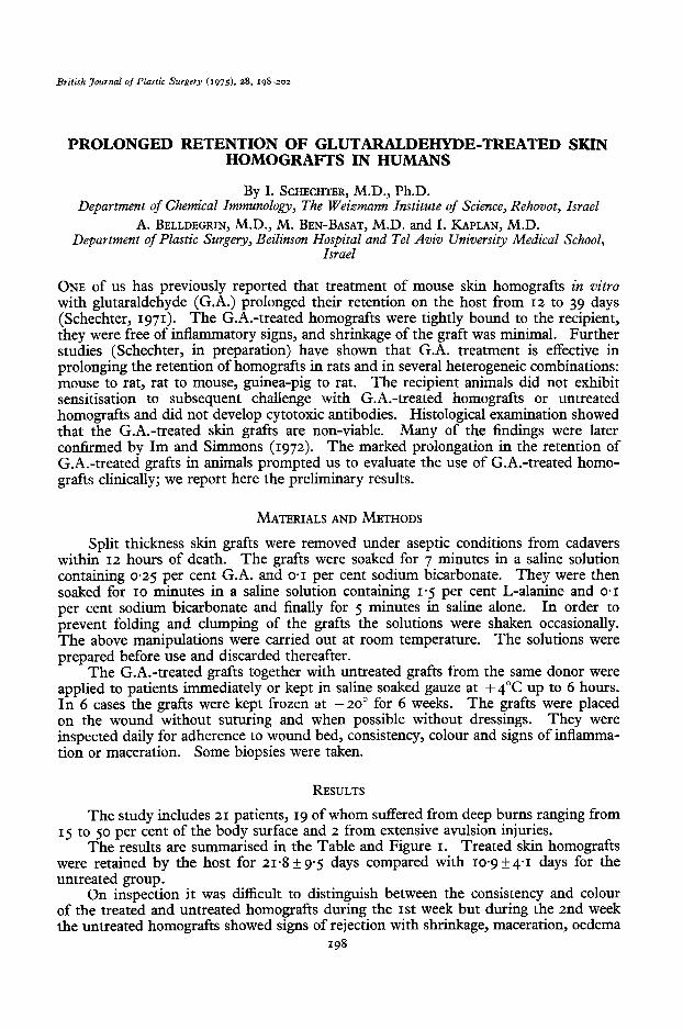
The results are summarised in the Table and Figure I. Treated skin homografts were retained by the host for 21.8 f 9.5 days compared with 10.9 + 4.1 days for the untreated group.
On inspection it was difficult to distinguish between the consistency and colour of the treated and untreated homografts during the 1st week but during the 2nd week the untreated homografts showed signs of rejection with shrinkage, maceration, oedema
198
RETENTION OF GLUTARALDEHYDE-TREATED SKIN HOMOGRAFTS 199
TABLE
Average Retention Time (A.R.T.) of Normal and Glutaraldehyde-Treated Skin Homografts
Total number A.R.T. & S.D. Group of grafts’ (days)
Untreated 24 1o.gt4.1
G.A.-treated 46 214tq
L Twenty-one patients were examined, some of whom received several grafts. Most patients (rS) received both treated and untreated grafts which were applied on wounds of similar quality.
and serous exudate (Fig. 2). By the 11th day most of the untreated grafts had been rejected.
In the G.A.-treated grafts the onset of rejection was delayed, it progressed slowly, and the process of rejection was not uniform. In some cases the treated grafts appeared to soften and then were detached from the recipient bed. In other cases, initially the grafts became hard in consistency and remained so for 2-3 weeks. Later they softened
50
40
30
20
IO
15-19
Days
20-24 25-29 >30
FIG. I. Effect of in vitro treatment with glutaraldehyde of human homografts on their retention time in the recipients. Columns represent percentage of grafts rejected at the indicated time interval. Empty
column, untreated grafts. Hatched column, G.A.-treated grafts.

200 BRITISH JOURNAL OF PLASTIC SURGERY
FIG. 2. Comparison between untreated and G.A.-treated homografts. Two untreated grafts (left and upper right) and one G.A.-treated graft (lower right) were applied to a wound in the region of the elbow. A, 7 days after application. All grafts are attached and look the same. B, 14 days after application. The untreated grafts are macerated and rejected. The G.A.-treated graft is preserved and attached to
the wound bed.
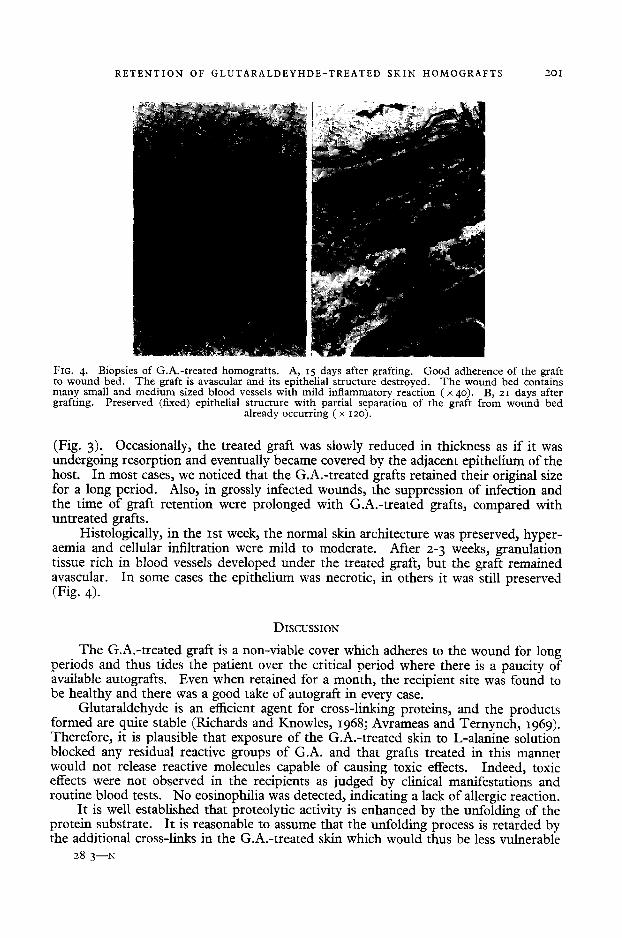
FIG. 3. Extensive avulsion injury with gross infection of right flank and thigh covered with several G.A.-treated homografts. For orientation, black dot indicates the same graft. A, 2 days after grafting. B, 23 days after grafting. The grafts are well attached to the site, they retain their original size and infection is markedly reduced. Note “tanned” appearance of the grafts. C, 35 days after grafting. Some of the grafts have become softened and are in the process of rejection (upper left); others exhibit
satisfactory retention (lower right).
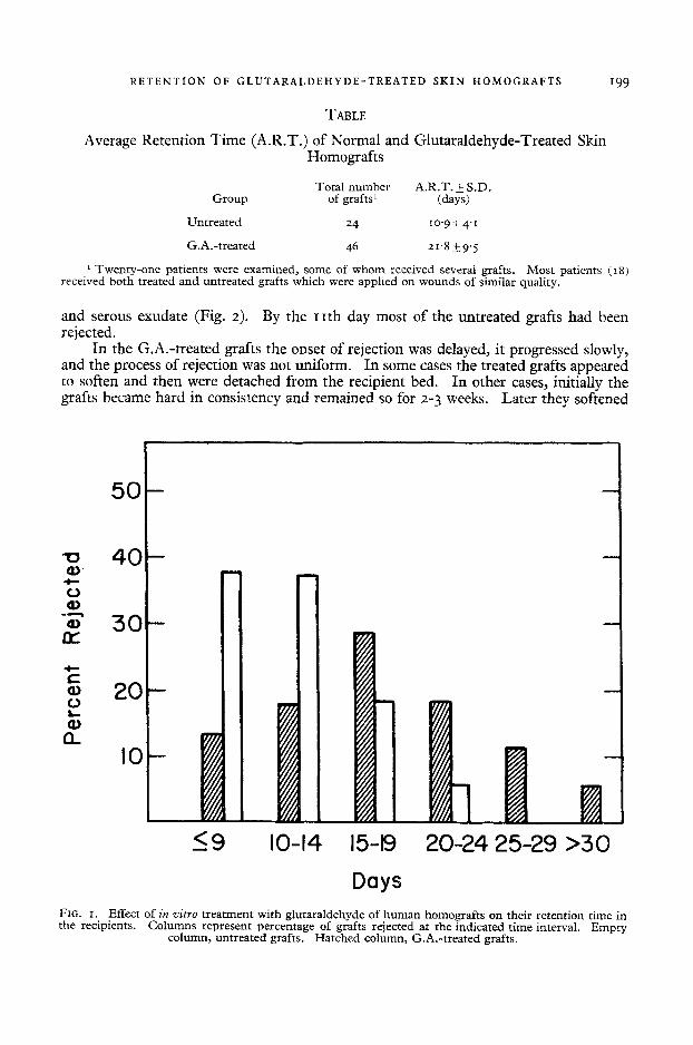
RETENTION OF GLUTARALDEYHDE-TREATED SKIN HOMOGRAFTS 201
FIG. 4. Biopsies of G.A.-treated homografts. A, 15 days after grafting. Good adherence of the graft to wound bed. The graft is avascular and its epnhelial structure destroyed. The wound bed contains many small and medium sized blood vessels with mild inflammatory reaction ( x 40). B, 21 days after grafting. Preserved (fixed) epithelial structure with partial separation of the graft from wound bed
already occurring ( x 120).
(Fig. 3). Occasionally, the treated graft was slowly reduced in thickness as if it was undergoing resorption and eventually became covered by the adjacent epithelium of the host. In most cases, we noticed that the G.A.-treated grafts retained their original size for a long period. Also, in grossly infected wounds, the suppression of infection and the time of graft retention were prolonged with G.A.-treated grafts, compared with untreated grafts.
Histologically, in the 1st week, the normal skin architecture was preserved, hyper- aemia and cellular infiltration were mild to moderate. After 2-3 weeks, granulation tissue rich in blood vessels developed under the treated graft, but the graft remained avascular. In some cases the epithelium was necrotic, in others it was still preserved (Fig. 4).
DISCUSSION
The G.A.-treated graft is a non-viable cover which adheres to the wound for long periods and thus tides the patient over the critical period where there is a paucity of available autografts. Even when retained for a month, the recipient site was found to be healthy and there was a good take of autograft in every case.
Glutaraldehyde is an efficient agent for cross-linking proteins, and the products formed are quite stable (Richards and Knowles, 1968; Avrameas and Ternynch, 1969). Therefore, it is plausible that exposure of the G.A.-treated skin to L-alanine solution blocked any residual reactive groups of G.A. and that grafts treated in this manner would not release reactive molecules capable of causing toxic effects. Indeed, toxic effects were not observed in the recipients as judged by clinical manifestations and routine blood tests. No eosinophilia was detected, indicating a lack of allergic reaction.
It is well established that proteolytic activity is enhanced by the unfolding of the protein substrate. It is reasonable to assume that the unfolding process is retarded by the additional cross-links in the G.A.-treated skin which would thus be less vulnerable
28..‘3-N

202 BRITISH JOURNAL OF PLASTIC SURGERY
to digestion and maceration by proteolytic enzymes in infected areas. This appeared to be the case in several grossly infected wounds where the treated grafts were retained (Fig. 3) while normal homografts were not.
Six treated grafts kept at -20°C for 6 weeks were used with complete success. This suggests that a bank of G.A.-treated skin may be practicable. It would be desirable to use heterograft skin because of the limited human supplies and since it has already been shown in experimental animals that G.A.-treated heterografts and homografts behave identically (Schechter, in preparation).
The course of rejection of G.A.-treated grafts was not uniform. This is probably due to variations in the state of the wound bed but it may also be due to the relatively low concentration of G.A. employed. It has been shown in animals that the prolonga- tion of graft retention increases with increasing G.A. concentration up to a certain level while at low G.A. concentrations of 0.1 per cent the grafts were soft and the process of rejection was similar to that of untreated grafts (Schechter, 1971; Im and Simmons, 1972).
The encouraging results obtained in this preliminary study justify further detailed investigations of the applicability and practicality of this method of graft preparation on a large scale.
SUMMARY
Treatment of cadaver skin homografts in vitro with glutaraldehyde significantly prolonged their average retention time from 10.9 to 214 days in 21 patients with burns and/or extensive soft tissue injuries.
The glutaraldehyde-treated homografts serve as a useful non-viable wound cover. They remain adherent to the wound bed for prolonged periods and support the formation of granulation tissue favourable for the subsequent acceptance of autografts. Toxic symptoms or allergic reactions were not noticed in any of the recipients.
Treatment of the skin with glutaraldehyde is simple to perform, requires minimal laboratory equipment, and is not time consuming.
REFERENCES
AVRAMEAS, S. and TERNYNCH, T. (1960). The cross-linking of proteins with glutaraldehyde and its use for the preparation of immunoadsorbents. Zvnmunochemistry, 6, 53.
1~~ H. Y. and SIMMONS, R. T. (1972). Mechanism for the prolonged survival of glutaral- dehyde-treated skin allografts. TravzspZuntation, 14, 527.
RICHARDS, F. M. and KNOWLES, J. R. (1968). Glutaraldehyde as a protein cross-linking reagent. Journal of Molecular Biology, 37, 23 I.
SCHECHTER, I. (1971). Prolonged survival of glutaraldehyde-treated skin homografts. Proceedings of the National Academy of Science, 68, 1590.