prof. andrás palkó md, phd - u-szeged.hu abd... · acute abdomen prof. andrás palkó md, phd...
TRANSCRIPT

Department of Radiology 1
Role of imaging in the evaluation of the acute abdomen
Prof. András Palkó MD, PhD
Department of Radiology
Faculty of General Medicine
University of Szeged
Hungary

Department of Radiology 2
Definition
• Sudden onset of severe symptoms requiring emergency
medical or surgical treatment,
– typically characterized by intensive abdominal pain
– may be associated with:
• muscular defense
• paralytic ileus or hyper- (retro-) –peristalsis
• nausea, vomiting, meteorism, hiccups
• fever, collapse, shock

Department of Radiology 3
Potential causes
• perforation
• obstruction
• ischemia
• bleeding
• infection
peritoneal irritation
GI paralysis & defanse musculaire

Department of Radiology 4
Acute abdomen – top 8
1. Acute appendicitis
2. Acute cholecystitis
3. Small bowel obstruction (SBO)
4. Acute gynecological disease
5. Acute pancreatitis
6. Renal colic
7. Perforated peptic ulcer
8. Acute diverticulitis
account for 90 % of patients referred to hospital

Department of Radiology 5
Role of imaging
• After stabilizing the patient’s condition rapid diagnosis
and therapeutic decision is essential to minimize
progression and reduce mortality.
• Clinical presentation, history, and lab test results are too
often non-specific.
• Imaging examinations are of utmost importance to
establish diagnosis.

Department of Radiology 6
What’s wrong? – clinical findings

Department of Radiology 7
• Intestinal – Acute appendicitis
– IBD
– Mesenteric adenitis
– Perforated carcinoma
– Epiploic appendagitis
– Omental infarction
– Cecal diverticulitis
– Infectious ileocolitis
– Sigmoid diverticulitis
– Obstructive sigmoid carcinoma
• Urological – Urinary tract obstruction
• Gynecological – PID
– Ovarian vein thrombosis
– Hemorrhagic ovarian cyst
– Rupture of ovarian dermoid
– Ovarian torsion
– Rupture of ectopic pregnancy
– Endometriosis
– Necrotic uterine leiomyoma
• Cholecystitis – Calculous
– Acalculous
– Emphysematous
• Hepatitis
• Liver abscess
• Spontaneous rupture
of a hepatic neoplasm
• Right-sided LB diverticulitis
• Retrocecal appendicitis
• Acute pancreatitis • Duodenal ulcer • Bowel obstruction • Abdominal aortic
aneurysm
• Bowel obstruction – SBO
• Adhesion
• Crohn’s
• Neoplasm
• Hernia
• Radiation
• Miscellaneous
– LBO
• Carcinoma
• Volvulus
• Diverticulitis
• Perforation • Peptic ulcer
• Diverticulitis
• Iatrogenic
• Bowel ischemia – Mesenteric artery
• Thromboembolism
• Atherosclerosis
• Dissection
– Mesenteric vein
• Hypercoagulable
• Neoplastic
• Strangulation, volvulus
• Infectious • Gastroenterocoliti
s
• Pseudomembranous colitis
• IBD • Ulcerative colitis
• Crohn’s disease

Department of Radiology 8
• (history, physical examination and lab tests)
• plain X-ray examination – abdomen
• erect or decubitus
• supine
– chest
• ultrasound
• computed tomography
Diagnostic algorithm

Department of Radiology 9
What’s wrong? – imaging findings
• Pathological gas accumulation
• Localized/diffuse fluid accumulation
• Diffuse/segmental gas/fluid levels
• Extravasation, leakage
• Pathological circulation
• Wall thickening (bull’s eye)
• Foreign body

Department of Radiology 10
Pathological gas accumulation
• Pneumoperitoneum – perforation
– iatrogenic
– per vaginam
• Pneumoretroperitoneum – perforation
– iatrogenic
• Bowel wall – pneumatosis
– infarction
– necrosis

Department of Radiology 11
Pathological gas accumulation
• Pneumoperitoneum – perforation
– iatrogenic
– per vaginam
• Pneumoretroperitoneum – perforation
– iatrogenic
• Bowel wall – pneumatosis
– infarction
– necrosis
Note: Ogilvie-sy!

Department of Radiology 12
Pathological gas accumulation

Department of Radiology 13
Pathological gas accumulation

Department of Radiology 14
Pathological gas accumulation
• Pneumoperitoneum – perforation
– iatrogenic
– per vaginam
• Pneumoretroperitoneum – perforation
– iatrogenic
• Bowel wall – pneumatosis
– infarction
– necrosis

Department of Radiology 15
Pathological gas accumulation
• Pneumoperitoneum – perforation
– iatrogenic
– per vaginam
• Pneumoretroperitoneum – perforation
– iatrogenic
• Bowel wall – pneumatosis
– infarction
– necrosis

Department of Radiology 16
Pathological gas accumulation
• Pneumoperitoneum – perforation
– iatrogenic
– per vaginam
• Pneumoretroperitoneum – perforation
– iatrogenic
• Bowel wall – pneumatosis
– infarction
– necrosis
Gellett LR et al: Emerg Med J 2002;19:480-481

Department of Radiology 17
Pathological gas accumulation
• Biliary system – sphincter Oddi incompetence
– postoperative
– spontaneous fistula
– emphysematous cholecystitis
• Portal vein – mesenteric infarction
– air embolus
– necrotizing enterocolitis
• Abscess

Department of Radiology 18
Pathological gas accumulation
• Biliary system – sphincter Oddi incompetence
– postoperative
– spontaneous fistula
– emphysematous cholecystitis
• Portal vein – mesenteric infarction
– air embolus
– necrotizing enterocolitis
• Abscess

Department of Radiology 19
Pathological gas accumulation
• Biliary system – sphincter Oddi incompetence
– postoperative
– spontaneous fistula
– emphysematous cholecystitis
• Portal vein – mesenteric infarction
– air embolus
– necrotizing enterocolitis
• Abscess

Department of Radiology 20
Pathological gas accumulation

Department of Radiology 21
Pathological gas accumulation

Department of Radiology 22
Pathological gas accumulation – gasless abdomen
• Normal abdomen
• Small bowel
obstruction/ischemia
• Ascites
• Surgery (e.g. total
colectomy)
• Gastroenteritis
• Large abdominal mass
Thompson WM: AJR, 191:1093–1099, 2008

Department of Radiology 23
Localized/diffuse fluid accumulation
• Ascites – cirrhosis
– tumor
– hypoalbuminaemia
– portal hypertension
– lymphatic obstruction
• Inflammation
• Perforation
• Abscess
Localized/diffuse fluid accumulation

Department of Radiology 24
Localized/diffuse fluid accumulation
• Ascites – cirrhosis
– tumor
– hypoalbuminaemia
– portal hypertension
– lymphatic obstruction
• Inflammation
• Perforation
• Abscess

Department of Radiology 25
Localized/diffuse fluid accumulation
• Ascites – cirrhosis
– tumor
– hypoalbuminaemia
– portal hypertension
– lymphatic obstruction
• Inflammation
• Perforation
• Abscess

Department of Radiology 26
Localized/diffuse fluid accumulation
• Ascites – cirrhosis
– tumor
– hypoalbuminaemia
– portal hypertension
– lymphatic obstruction
• Inflammation
• Perforation
• Abscess

Department of Radiology 27
Diffuse/segmental gas/fluid levels
• Paralytic ileus
– Diffuse
• peritonitis
• gastroenteritis
• abdominal pain
• other (postoperative, drug effect,
electrolyte imbalance, pneumonia,
retroperitoneal hemorrhage, etc.)
– Segmental (sentinel loop) • pancreatitis
• cholecystitis
• appendicitis,
• diverticulitis
• etc.

Department of Radiology 28
Diffuse/segmental gas/fluid levels
• Paralytic ileus
– Diffuse
• peritonitis
• gastroenteritis
• abdominal pain
• other (postoperative, drug effect,
electrolyte imbalance, pneumonia,
retroperitoneal hemorrhage, etc.)
– Segmental (sentinel loop) • pancreatitis
• cholecystitis
• appendicitis,
• diverticulitis
• etc.

Department of Radiology 29
Diffuse/segmental gas/fluid levels
Paulsen SR, RadioGraphics, 26:641, 2006
• Obstruction
– Small bowel
• Adhesion
• Crohn’s
• Neoplasm
• Hernia
• Radiation
• Miscellaneous
– Large bowel • Carcinoma
• Volvulus
• Diverticulitis
Department of Radiology 30
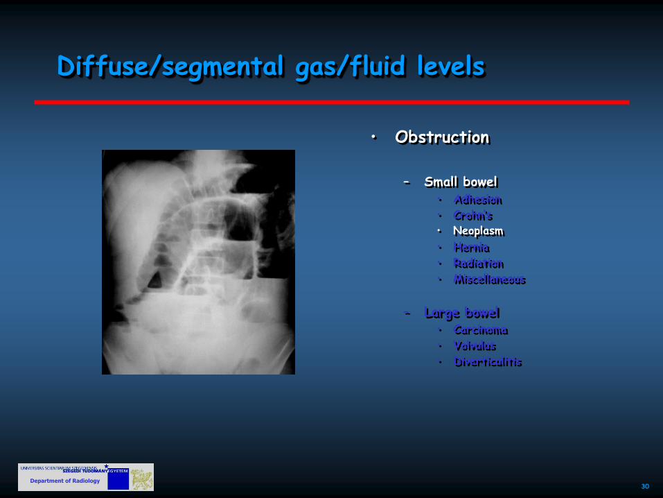
Diffuse/segmental gas/fluid levels
• Obstruction
– Small bowel
• Adhesion
• Crohn’s
• Neoplasm
• Hernia
• Radiation
• Miscellaneous
– Large bowel • Carcinoma
• Volvulus
• Diverticulitis

Department of Radiology 31
Diffuse/segmental gas/fluid levels
Takeyama,et al: RadioGraphics 2005; 25:997–1015
• Obstruction
– Small bowel
• Adhesion
• Crohn’s
• Neoplasm
• Hernia
• Radiation
• Miscellaneous
– Large bowel • Carcinoma
• Volvulus
• Diverticulitis

Department of Radiology 32
Diffuse/segmental gas/fluid levels
E M Anderson, Imaging 2006;18:198
• Obstruction
– Small bowel
• Adhesion
• Crohn’s
• Neoplasm
• Hernia
• Radiation
• Miscellaneous
– Large bowel • Carcinoma
• Volvulus
• Diverticulitis

Department of Radiology 33
Diffuse/segmental gas/fluid levels
www.nypemergency.org
• Obstruction
– Small bowel
• Adhesion
• Crohn’s
• Neoplasm
• Hernia
• Radiation
• Miscellaneous
– Large bowel • Carcinoma
• Volvulus
• Diverticulitis

Department of Radiology 34
Diffuse/segmental gas/fluid levels
• Obstruction
– Small bowel
• Adhesion
• Crohn’s
• Neoplasm
• Hernia
• Radiation
• Miscellaneous
– Large bowel • Carcinoma
• Volvulus
• Diverticulitis

Department of Radiology 35
Extravasation, leakage
• Perforation (see also pathologic gas/fluid accumulation)
• Bleeding
– Endoluminal
(ulcer, tumor, inflammation, iatrogenic, etc)
– Extraluminal
(aneurysm, tumor, iatrogenic, etc)
– Parenchymal organ
(tumor, iatrogenic, etc)

Department of Radiology 36
Extravasation, leakage
• Perforation (see also pathologic gas/fluid accumulation)
• Bleeding
– Endoluminal
(ulcer, tumor, inflammation, iatrogenic, etc)
– Extraluminal
(aneurysm, tumor, iatrogenic, etc)
– Parenchymal organ
(tumor, iatrogenic, etc)
Yoon W et al: Radiology 239:16-167, 2006
Detectable rate (arteriography): 0.5 – 1.5 mL/min
Department of Radiology 37
Extravasation, leakage
• Perforation (see also pathologic gas/fluid accumulation)
• Bleeding
– Endoluminal
(ulcer, tumor, inflammation, iatrogenic, etc)
– Extraluminal
(aneurysm, tumor, iatrogenic, etc)
– Parenchymal organ
(tumor, iatrogenic, etc)

Department of Radiology 38
Extravasation, leakage
• Perforation (see also pathologic gas/fluid accumulation)
• Bleeding
– Endoluminal
(ulcer, tumor, inflammation, iatrogenic, etc)
– Extraluminal
(aneurysm, tumor, iatrogenic, etc)
– Parenchymal organ
(tumor, iatrogenic, etc)

Department of Radiology 39
Pathological circulation
• Arterial – thromboembolism (MI, rheumatic
heart disease, atrial fibrillation,
etc.)
– arteriosclerosis
– dissection
– heart failure
– others (iatrogenic, idiopathic, strangulation/volvulus, septicemia,
extrinsic mass, etc.)
• Venous – thrombosis
– others (extrinsic mass, iatrogenic, – strangulation/volvulus, etc.)
Kirkpatrick IDC et al: Radiology, 229:91-98, 2003

Department of Radiology 40
Pathological circulation
• Arterial – thromboembolism (MI, rheumatic
heart disease, atrial fibrillation,
etc.)
– arteriosclerosis
– dissection
– heart failure
– others (iatrogenic, idiopathic, strangulation/volvulus, septicemia,
extrinsic mass, etc.)
• Venous – thrombosis
– others (extrinsic mass, iatrogenic, – strangulation/volvulus, etc.)
Shih MCP et al AJR 2007; 188:462-471

Department of Radiology 41
Pathological circulation
• Arterial – thromboembolism (MI, rheumatic
heart disease, atrial fibrillation,
etc.)
– arteriosclerosis
– dissection
– heart failure
– others (iatrogenic, idiopathic, strangulation/volvulus, septicemia,
extrinsic mass, etc.)
• Venous – thrombosis
– others (extrinsic mass, iatrogenic, – strangulation/volvulus, etc.)
McMahon MA et al: Radiographics, 30:445-460, 2010

Department of Radiology 42
Pathological circulation
• Arterial – thromboembolism (MI, rheumatic
heart disease, atrial fibrillation,
etc.)
– arteriosclerosis
– dissection
– heart failure
– others (iatrogenic, idiopathic, strangulation/volvulus, septicemia,
extrinsic mass, etc.)
• Venous – thrombosis
– others (extrinsic mass, iatrogenic, – strangulation/volvulus, etc.)
Department of Radiology 43
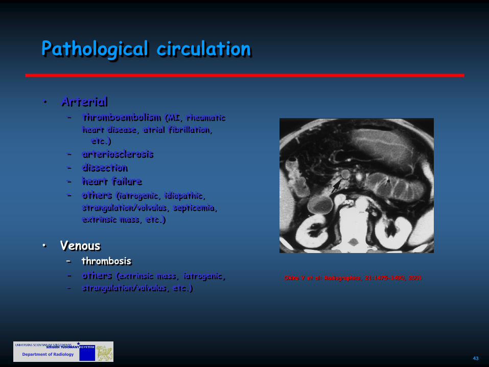
Pathological circulation
• Arterial – thromboembolism (MI, rheumatic
heart disease, atrial fibrillation,
etc.)
– arteriosclerosis
– dissection
– heart failure
– others (iatrogenic, idiopathic, strangulation/volvulus, septicemia,
extrinsic mass, etc.)
• Venous – thrombosis
– others (extrinsic mass, iatrogenic, – strangulation/volvulus, etc.)
Okino Y et al: Radiographics, 21:1475-1490, 2001

Department of Radiology 44
Wall thickening
• Infarction, ischemia
• Intramural hematoma
• Inflammation
– epiploic appendagitis
– Groove pancreatitis
• Infection
– appendicitis
– sigmoiditis
– typhlitis (neutropenic colitis)
– pseudomembranous colitis
(clostridium difficile)
– others (TBC, amebiasis, campylobacter,
shigella, etc.)
• Neoplasm
Rha SE et al: RadioGraphics, 20:29-42, 2000

Department of Radiology 45
Wall thickening
Dibbad R et al: The Internet Journal of Radiology, 12/1, 2010
• Infarction, ischemia
• Intramural hematoma
• Inflammation
– epiploic appendagitis
– Groove pancreatitis
• Infection
– appendicitis
– sigmoiditis
– typhlitis (neutropenic colitis)
– pseudomembranous colitis
(clostridium difficile)
– others (TBC, amebiasis, campylobacter,
shigella, etc.)
• Neoplasm

Department of Radiology 46
Wall thickening
Noah Gudel, DO; Lisa M. Rock, MD, Medscape 2008
• Infarction, ischemia
• Intramural hematoma
• Inflammation
– epiploic appendagitis
– Groove pancreatitis
• Infection
– appendicitis
– sigmoiditis
– typhlitis (neutropenic colitis)
– pseudomembranous colitis
(clostridium difficile)
– others (TBC, amebiasis, campylobacter,
shigella, etc.)
• Neoplasm

Department of Radiology 47
• Infarction, ischemia
• Intramural hematoma
• Inflammation
– epiploic appendagitis
– Groove pancreatitis
• Infection
– appendicitis
– sigmoiditis
– typhlitis (neutropenic colitis)
– pseudomembranous colitis
(clostridium difficile)
– others (TBC, amebiasis, campylobacter,
shigella, etc.)
• Neoplasm
Wall thickening

Department of Radiology 48
Wall thickening
• Infarction, ischemia
• Intramural hematoma
• Inflammation
– epiploic appendagitis
– Groove pancreatitis
• Infection
– appendicitis
– sigmoiditis
– typhlitis (neutropenic colitis)
– pseudomembranous colitis
(clostridium difficile)
– others (TBC, amebiasis, campylobacter,
shigella, etc.)
• Neoplasm

Department of Radiology 49
Wall thickening
Poletti PA et al, AJR 2004; 182:1159-1165
• Infarction, ischemia
• Intramural hematoma
• Inflammation
– epiploic appendagitis
– Groove pancreatitis
• Infection
– appendicitis
– sigmoiditis
– typhlitis (neutropenic colitis)
– pseudomembranous colitis
(clostridium difficile)
– others (TBC, amebiasis, campylobacter,
shigella, etc.)
• Neoplasm

Department of Radiology 50
Wall thickening
• Infarction, ischemia
• Intramural hematoma
• Inflammation
– epiploic appendagitis
– Groove pancreatitis
• Infection
– appendicitis
– sigmoiditis
– typhlitis (neutropenic colitis)
– pseudomembranous colitis
(clostridium difficile)
– others (TBC, amebiasis, campylobacter,
shigella, etc.)
• Neoplasm

Department of Radiology 51
Wall thickening
• Infarction, ischemia
• Intramural hematoma
• Inflammation
– epiploic appendagitis
– Groove pancreatitis
• Infection
– appendicitis
– sigmoiditis
– typhlitis (neutropenic colitis)
– pseudomembranous colitis
(clostridium difficile)
– others (TBC, amebiasis, campylobacter,
shigella, etc.)
• Neoplasm

Department of Radiology 52
Foreign body
• Accidental/intentional/iatrogenic
• Endoluminal/extraluminal
• W./w.o. perforation
Maglinte DDT: Radiology, 236:763-767, 2005

Department of Radiology 53
Acute abdomen - algorithm
• (history, physical examination and lab tests)
• plain X-ray examination – abdomen
• erect or decubitus
• supine
– chest
• ultrasound
• computed tomography

Department of Radiology 54
• (history, physical examination and lab tests)
• plain X-ray examination – abdomen
• erect or decubitus
• supine
– chest
• ultrasound
• computed tomography
?
Acute abdomen - algorithm

Department of Radiology 55