primary phagocytosis of viable neurons by microglia activated
TRANSCRIPT

RESEARCH Open Access
Primary phagocytosis of viable neurons bymicroglia activated with LPS or Aβ is dependenton calreticulin/LRP phagocytic signallingMichael Fricker1,2*, María José Oliva-Martín1 and Guy C Brown1*
Abstract
Background: Microglia are resident brain macrophages that can phagocytose dead, dying or viable neurons, whichmay be beneficial or detrimental in inflammatory, ischaemic and neurodegenerative brain pathologies. Cell deathcaused by phagocytosis of an otherwise viable cell is called ‘primary phagocytosis’ or ‘phagoptosis’. Calreticulin(CRT) exposure on the surface of cancer cells can promote their phagocytosis via LRP (low-density lipoproteinreceptor-related protein) on macrophages, but it is not known whether this occurs with neurons and microglia.
Methods: We used primary cultures of cerebellar neurons, astrocytes and microglia to investigate the potential roleof CRT/LRP phagocytic signalling in the phagocytosis of viable neurons by microglia stimulated withlipopolysaccharide (LPS) or nanomolar concentrations of amyloid-β peptide1-42 (Aβ). Exposure of CRT on theneuronal surface was investigated using surface biotinylation and western blotting. A phagocytosis assay was alsodeveloped using BV2 and PC12 cell lines to investigate CRT/LRP signalling in microglial phagocytosis of apoptoticcells.
Results: We found that BV2 microglia readily phagocytosed apoptotic PC12 cells, but this was inhibited by aCRT-blocking antibody or LRP-blocking protein (receptor-associated protein: RAP). Activation of primary ratmicroglia with LPS or Aβ resulted in loss of co-cultured cerebellar granule neurons, and this was blocked by RAP orantibodies against CRT or against LRP, preventing all neuronal loss and death. CRT was present on the surface ofviable neurons, and this exposure did not change in inflammatory conditions. CRT antibodies preventedmicroglia-induced neuronal loss when added to neurons, while LRP antibodies prevented neuronal loss whenadded to the microglia. Pre-binding of CRT to neurons promoted neuronal loss if activated microglia were added,but pre-binding of CRT to microglia or both cell types prevented microglia-induced neuronal loss.
Conclusions: CRT exposure on the surface of viable or apoptotic neurons appears to be required for theirphagocytosis via LRP receptors on activated microglia, but free CRT can block microglial phagocytosis of neuronsby acting on microglia. Phagocytosis of CRT-exposing neurons by microglia can be a direct cause of neuronal deathduring inflammation, and might therefore contribute to neurodegeneration and be prevented by blocking the CRT/LRP pathway.
Keywords: Phagocytosis, Neuron, Microglia, Calreticulin, LRP, Inflammation, Amyloid, Neurodegeneration, Celldeath, Phagoptosis
* Correspondence: [email protected]; [email protected] of Biochemistry, University of Cambridge, Tennis Court Road,Cambridge CB2 1QW, UK2Present address: HMRI, University of Newcastle, Newcastle upon Tyne, NSW,Australia
JOURNAL OF NEUROINFLAMMATION
© 2012 Fricker et al.; licensee BioMed Central Ltd. This is an Open Access article distributed under the terms of the CreativeCommons Attribution License (http://creativecommons.org/licenses/by/2.0), which permits unrestricted use, distribution, andreproduction in any medium, provided the original work is properly cited.
Fricker et al. Journal of Neuroinflammation 2012, 9:196http://www.jneuroinflammation.com/content/9/1/196
BackgroundMost neurological diseases involving neuronal loss areaccompanied by the appearance of activated microglia inthe affected tissues [1]. Microglia are resident brain ma-crophages that mediate the immune response againstCNS infections and clear cellular debris following injury.However, there is growing evidence that inflammatory-activated microglia actively participate in the death of neu-rons during neurodegenerative processes, for examplethrough release of reactive oxygen and nitrogen species(ROS/RNS) and release of pro-inflammatory neurotoxiccytokines and inflammatory mediators such as TNF-α [2].We have recently described a novel form of neuronaldeath mediated by inflammatory-activated microglia inwhich microglia phagocytose viable neurons, referred toas ‘primary phagocytosis’ or ‘phagoptosis’ [3,4]. Phagocy-tosis is normally thought to occur after the target cell hasundergone cell death, but we found that in inflammatoryconditions inhibition of phagocytic signalling rescues neu-rons both in vitro and in vivo, demonstrating that phago-cytosis can be a direct cause of neuronal death in modelsof inflammatory neurodegeneration [5-7].Phagocytosis is controlled by a complex array of sig-
nals. The interaction between a number of ‘eat-me’ and‘don’t-eat-me’ signals located on the target cell surfaceand their respective receptors on the phagocyte deter-mine whether or not phagocytosis takes place [8]. Thebest-characterised ‘eat-me’ signal is exposure of the phos-pholipid phosphatidylserine (PS) on the outer leaflet ofthe plasma membrane. In most viable cells that are notactivated, PS is almost exclusively localised on the innerleaflet of the plasma membrane due to an aminophospho-lipid translocase that pumps PS from the outer to theinner leaflet. Upon induction of cell death by apoptosis ornecrosis, PS becomes exposed on the cell surface due toinactivation of the translocase or activation of a scram-blase, which randomises phospholipid distribution bet-ween the inner and outer leaflets thus resulting in net PSexposure. However, PS exposure also occurs on the sur-face of viable cells when ‘activated’, usually as a result ofcalcium stimulation of the scramblase and inhibition of thetranslocase, for example during activation of all leucocytes[9-11], and on neurons exposed to oxidants from activatedmicroglia [5]. Exposed PS can be either bound directly bysome phagocyte receptors, such as Tim4, stabilin-1 and −2and BAI1, or bound by bridging proteins such as MFG-E8,which activates phagocytosis via the vitronectin receptor(αvβ3/5 integrin) [8]. Indeed we have shown that primaryphagocytosis of viable neurons by inflammatory-activatedmicroglia is mediated by microglia-induced PS expo-sure on viable neurons, evoking microglial phagocytosisvia MFG-E8 and the vitronectin receptor [5,7].Surface-exposed calreticulin (CRT) has been demon-
strated to act as an eat-me signal in a number of cell
types [12]. CRT, principally characterized as an endo-plasmic reticulum (ER)-resident chaperone, is constitu-tively expressed at the surface of numerous cancer celllines and its expression at the cell surface can be in-creased in the early stages of apoptosis induced by asubset of apoptotic stimuli including anthracyclins andUV irradiation [13,14]. CRT has been shown to act as anessential eat-me signal promoting phagocytosis of apop-totic cells and its activity can be modulated not only byincreasing exposure at the cell surface but also poten-tially by rearrangement of existing exposed CRT [15,16].Surface-exposed CRT is recognised by the phagocytic re-ceptor LRP (low-density lipoprotein receptor-relatedprotein) [15,17], although CRT is also found associatedwith LRP on the phagocyte membrane where it acts as aco-receptor for LRP ligands such as C1q and alpha-2-macroglobulin [18]. The constitutive expression of CRTon the surface of a number of cell types does not neces-sarily result in their phagocytosis as don’t-eat-me signalshave a dominant inhibitory effect on phagocytosis, forexample CD47 and its receptor SIRPα [13,15,19,20]. Therole and regulation of exposed CRT and LRP in media-ting phagocytosis of neurons by microglia is unknown.We therefore sought to test the requirement for CRT-and LRP-mediated signalling for phagocytosis of dyingneurons and primary phagocytosis of viable neurons inmodels of inflammatory neurodegeneration. Here wedemonstrate that neuronally exposed CRT is required asan eat-me signal for phagocytosis of both apoptotic andviable neurons by microglia, and that CRT is constitu-tively exposed on the surface of neurons but this onlypromotes phagocytosis in specific contexts, and indeedreleased CRT can inhibit phagocytosis at microglia.
MethodsAll experiments were performed in accordance with theUK Animals (Scientific Procedures) Act (1986) and ap-proved by the Cambridge University Local Research EthicsCommittee.
Cell culture and treatmentsMixed neuronal/glial cerebellar cultures were preparedfrom the cerebella of postnatal day 5 to 7 rats as pre-viously described [21] and were allowed to mature in vitrofor six to eight days prior to treatment. Pure microgliawere prepared from mixed cortical astroglial/microglialcultures as previously described [5]. BV2 microglial cellswere grown in Dulbecco’s modified Eagle’s medium(DMEM, Invitrogen, Carlsbad, CA, USA)) supplementedwith 10% fetal bovine serum (FBS, PAA Laboratories,Colbe, Germany). PC12 neuronal cells were grown inRoswell Park Memorial Institute medium (RPMI, Invitro-gen) supplemented with 10% FBS and 20% horse serum(Sigma-Aldrich, St Louis, MO, USA). PC12 were plated
Fricker et al. Journal of Neuroinflammation 2012, 9:196 Page 2 of 12http://www.jneuroinflammation.com/content/9/1/196
on collagen-coated tissue culture plates (0.5 mg/ml colla-gen, Sigma-Aldrich). All tissue culture medium was sup-plemented with 100 units/ml penicillin G, 100 μg/mlstreptomycin sulphate (Invitrogen). Reagents were pro-cured as follows: lipopolysaccharide (LPS), calreticulin(CRT), cytochalasin D (CytoD), 5-(and-6)-carboxytetra-methylrhodamine succinimidyl ester (TAMRA) were fromSigma-Aldrich. β 1–42 monomers (EZBiolab, Carmel,IN, USA) were prepared as previously described [6].Receptor-associated protein (RAP, R&D systems,Minneapolis, MN, USA), normal mouse IgG (mIgG,Santa Cruz Biotech, Santa Cruz, CA, USA), anti-CRTantibodies (Abcam, Cambridge, UK; Stressgen, Brussels,Belgium), anti-LRP (American Diagnostica Inc., Stamford,CT, USA), Alexa 488-labelled isolectin B4 (IB4, MolecularProbes, Eugene OR, USA). Neuronal and microglial cellsurvival was quantified three days after stimulation as pre-viously described [5]. Anti-CRT and anti-LRP blockingantibodies were Fc-blocked with an F(ab’)2 fragment anti-body (Jackson Immunoresearch, West Grove, PA, USA).Nitrite levels in culture supernatants were measured aspreviously described [5].
BV2 and PC12 phagocytosis assayBV2 were plated in 6-well plates in DMEM plus 0.5%FBS and were at approximately 60% confluency whentarget cells were added. PC12 in suspension were stainedfor 10 minutes with 50 μM TAMRA, washed in warmPBS and then plated in 10 cm collagen-coated dishes athigh density. UV-treated PC12 received 200 mJ/cm2 ir-radiation. Untreated and UV-treated PC12 were harvested16 hours after UV treatment by trypsinisation. PC12 targetcells were counted and resuspended in DMEM plus 0.5%FBS. Some 200,000 PC12 target cells were added to eachwell of BV2 (approximate four-fold excess of target PC12cells compared to BV2) followed by a two-hour incubationat 37°C. For FACS analysis, BV2 were stained with IB4(1 μg/ml) for 15 minutes prior to washing in PBS and brieftrypsinisation to detach cells. BV2 were then resuspendedin 200 μl PBS and FACS analysis performed using anAccuri C6 Flow Cytometer (BD Services, San Jose, CA,USA). Alexa 488 IB4 fluorescence was detected in FL1channel whilst TAMRA fluorescence was detected in FL2.For fluorescence microscopy, BV2 were labeled with IB4as above and washed briefly in PBS prior to labelling ofnuclear DNA with Hoechst 33342 [5]. Cells were imagedon an Olympus Fluoview 300 microscope (Olympus,Tokyo, Japan).
Transwell and microglial reconstitution experimentsFollowing six to seven days in vitro microglia were se-lectively eliminated from cerebellar cultures by adding50 mM L-leucine methyl ester (LME, Sigma-Aldrich).After three hours LME-containing medium was aspirated,
neurons washed once in warm HBSS (Invitrogen) andthen medium was replaced with conditioned mediumfrom sister cultures. Twenty-four hours later, 6.5 mm0.4 μm pore size polycarbonate transwell inserts (Corning,Sigma-Aldrich) that had been poly-L-lysine coated wereinserted and 25,000 microglia were plated onto the insert.After 24 hours, LPS was added at 100 ng/ml as indicatedin figure legends. Forty-eight hours later, transwell insertscontaining microglia were removed. During this timemicroglia were purified and plated in 6-well plates, left for24 hours and then incubated for a further 24 hours with100 ng/ml LPS. LPS-activated pure microglia were gentlyblown from wells after a brief incubation in Versene solu-tion (Invitrogen) at 37°C. At this point, blocking antibodywas added to the neurons or to purified LPS-activatedmicroglia in suspension for one hour at 37°C. Neuronswere washed three times in warm HBSS before condi-tioned medium from untreated sister cultures was addedback. LPS-activated microglia were washed three times inwarm HBSS, collected by centrifugation and counted. Atotal of 25,000 LPS-activated microglia was then addeddirectly back to neuronal cultures as indicated and plateswere spun briefly to allow microglia to settle. Cerebellarcultures were then incubated for six hours at 37°C beforeneuronal survival and numbers were assessed as describedabove. For addition of exogenous CRT, microglia wereeliminated from cerebellar granule cells (CGC) that hadbeen in vitro for seven days using LME as before. After24 hours, 1 μg/ml CRT (Sigma-Aldrich) was added dir-ectly to neurons and left to incubate for two hours, fol-lowed by three washes in warm HBSS. Pure microglia thathad either been left untreated or LPS activated for24 hours as described above were then added back to neu-rons as indicated at a density of 25,000 cells per well(24-well plate), plates incubated for six hours at 37°Cprior to quantification of neuronal number and survival.
Externalised protein biotinylation and pull-downSurface biotinylation was performed using the Pierce CellSurface Protein Isolation Kit including Sulfo-NHS-SS-Biotin as the labelling reagent (Thermo Fisher Scientific,Waltham, MA, USA). Cerebellar cultures were seeded in6-well plates and after six days in culture microglia wereeliminated with LME as described above. After 24 hours,a poly-L-lysine-coated transwell was inserted and 200,000purified microglia were plated on the transwell. Twenty-four hours later, 100 ng/ml LPS was added where indi-cated. After 48 hours, further incubation transwells wereremoved and neuron plates were transferred to ice andwashed several times in HBSS prior to addition of Sulfo-NHS-SS-Biotin. All subsequent steps including streptavi-din pull-down of biotinylated proteins followed the manu-facturer’s instructions. Neurons were lysed in a volume of500 μl and prior to pull-down a load sample of 40 μl was
Fricker et al. Journal of Neuroinflammation 2012, 9:196 Page 3 of 12http://www.jneuroinflammation.com/content/9/1/196
collected. Biotinylated proteins captured on streptavidinbeads were eluted by boiling in SDS gel loading buffer.Proteins were separated by SDS-PAGE and transferred tonitrocellulose membranes for western blot detection aspreviously described [22].
Statistical analysisStatistical analysis was performed using SPSS software.All results represent the mean value from at least threeseparate experiments (see figure legends) with each indi-vidual experiment having two replicates per conditionwith four fields counted per replicate. In figure legendsn = x refers to the number of separate experiments per-formed. Error bars represent the standard error of themean of experiments (SEM). Normality of data was veri-fied using the Shapiro-Wilk test. Data was analysedusing one-way ANOVA and post hoc Bonferroni test. Infigures * = P < 0.05, ** = P < 0.01, *** = P < 0.001.
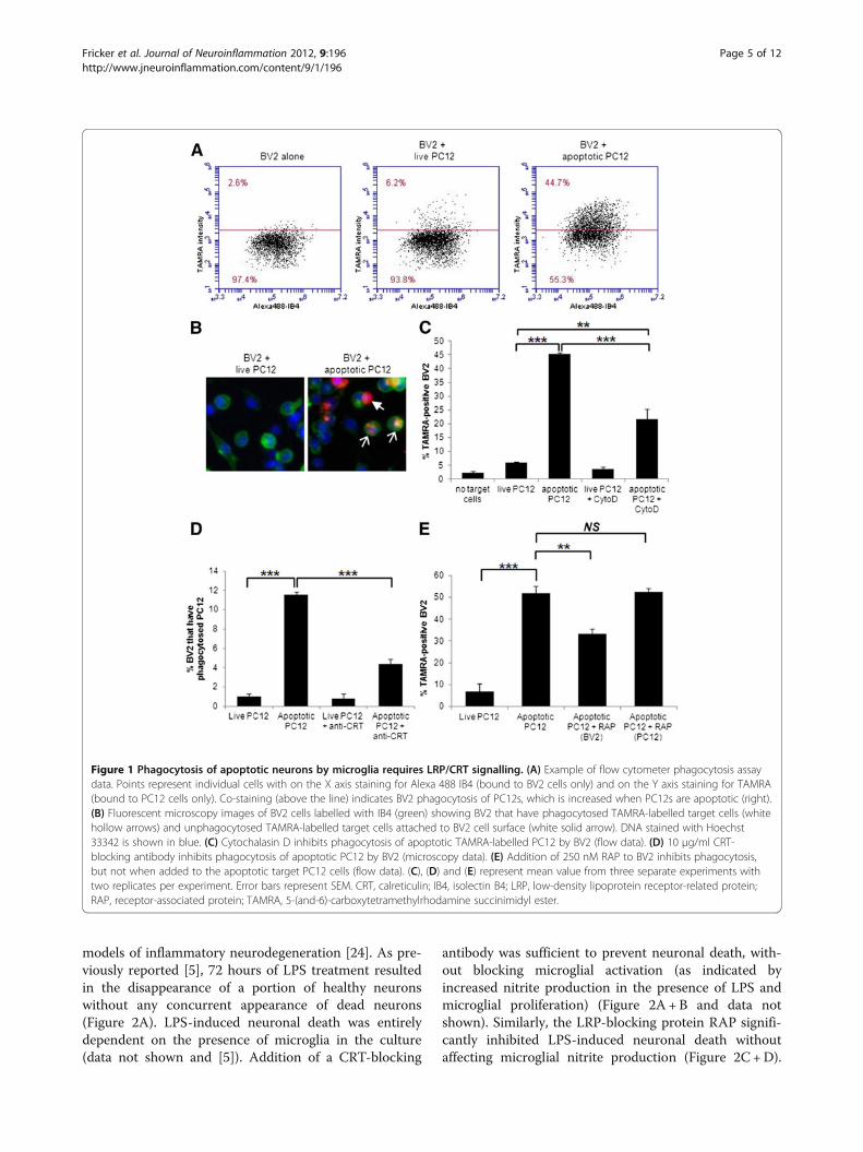
ResultsCRT/LRP signalling in phagocytosis of apoptotic PC12cells by BV2 microglial cellsCell-surface-exposed CRT has been demonstrated toplay an important role in mediating phagocytosis in vari-ous cancer cell lines, either as an eat-me signal on thetarget cell or as a co-receptor on the phagocytic cell sur-face. Whether exposed CRT plays a role in phagocytosisof neurons by microglia is unknown. Given the potentialimportance of phagocytosis of neurons by microglia inmodulating neurodegenerative processes, we sought totest whether phagocytosis of neurons by microgliainvolved CRT-mediated signalling. In order to test therole of CRT in phagocytosis of apoptotic neurons bymicroglia, we established a phagocytosis assay using thetransformed microglial cell line, BV2, and the cell linePC12 as target cells. BV2 cells were visualised withAlexa 488-labelled isolectin B4 (IB4) whilst PC12 cellswere separately labelled with red fluorescent 5-(and-6)-carboxytetramethylrhodamine succinimidyl ester (5(6)-TAMRA) prior to treatment and addition to BV2. Theextent of phagocytosis was evaluated by determining theproportion of BV2 cells that contained red fluorescence,indicative of phagocytosis of the TAMRA-labelled targetcells. Initially, red fluorescence of BV2 was evaluated byfluorescence microscopy and in later experiments FACSanalysis was employed (see figure legend for details)(Figure 1A+B). Some experiments were performedusing both fluorescence microscopy and flow cytometryfor data collection, and flow cytometry was more sensi-tive to low levels of TAMRA staining within the BV2cells, so that a higher level of labelled BV2 cells was seenby flow cytometry. However, the trends seen whenphagocytic inhibitors were added were similar and statis-tically significant with both methods (data not shown).
As expected very little phagocytosis occurred when livePC12 were added to BV2 (5.9% ± 0.5%, Figure 1C),but this greatly increased when apoptotic PC12 wereadded (45.3% ± 0.5%, Figure 1C). Cytochalasin D, awell-characterised inhibitor of phagocytosis, significantlyinhibited the phagocytosis of apoptotic cells by BV2, de-monstrating that phagocytosis of target cells wasrequired for TAMRA uptake by BV2 (Figure 1C). Wetested the requirement for cell surface CRT and LRPfor phagocytosis of apoptotic cells by microglia using aCRT-blocking antibody [19] and recombinant RAP, aLRP-binding protein, which inhibits ligand binding byLRP [23]. Addition of a CRT-blocking antibody to PC12cells prior to washing and then addition to BV2 was suffi-cient to significantly reduce phagocytosis of apoptoticPC12 cells by BV2 (live PC12, 1% ± 0.3%; apoptoticPC12, 11.5% ± 0.3%; apoptotic PC12 +CRT blocking,4.3% ± 0.5%; Figure 1D). RAP significantly inhibitedphagocytosis of apoptotic PC12 when incubated withBV2 prior to addition of target cells but not when incu-bated with apoptotic PC12 cells prior to addition to BV2(live PC12, 6.7%± 3.7%; apoptotic PC12, 52%±3.1%; apo-ptotic PC12+RAP microglia, 33.2%± 2.4%; apoptoticPC12+RAP neuron, 52.7%±1.4%; Figure 1E). Thusphagocytosis of apoptotic PC12 by the microglial cell lineBV2 requires recognition of CRT on the target cell mem-brane and ligand binding by LRP on the microglia.
CRT/LRP signalling in primary phagocytosis of viablecerebellar granule neurons by microglia activated by LPSor AβTumour cell lines and transformed cells frequently exposethe eat-me signal CRT at the cell surface and consequentlyCRT plays a pivotal role in determining whether or notthese cells are phagocytosed [13]. Less is known about therole of CRT in phagocytosis of primary cells, includingneurons. We recently demonstrated that in addition toclearance of apoptotic and necrotic debris, in certain cir-cumstances inflammatory-activated microglia may activelyparticipate in neurodegenerative processes by phagocytos-ing viable neurons both in vitro and in vivo [5,7].Inflammatory-activated microglia produce ROS/RNS thatinduce reversible exposure of PS on the neuronal sur-face, at which point microglia phagocytose the viable PS-exposing neurons resulting in neuronal death by ‘primaryphagocytosis’ [5]. A defining feature of primary phagocyt-osis as a form of cell death is that inhibition of any of thecritical components of the phagocytic signalling machin-ery is sufficient to rescue neurons, both in vitro andin vivo.We tested the role of CRT and LRP in primary phago-
cytosis using the TLR4 ligand LPS as an inflammatorystimulus. LPS has been well characterised by us andothers as a means of inducing microglial activation in
Fricker et al. Journal of Neuroinflammation 2012, 9:196 Page 4 of 12http://www.jneuroinflammation.com/content/9/1/196
models of inflammatory neurodegeneration [24]. As pre-viously reported [5], 72 hours of LPS treatment resultedin the disappearance of a portion of healthy neuronswithout any concurrent appearance of dead neurons(Figure 2A). LPS-induced neuronal death was entirelydependent on the presence of microglia in the culture(data not shown and [5]). Addition of a CRT-blocking
antibody was sufficient to prevent neuronal death, with-out blocking microglial activation (as indicated byincreased nitrite production in the presence of LPS andmicroglial proliferation) (Figure 2A+B and data notshown). Similarly, the LRP-blocking protein RAP signifi-cantly inhibited LPS-induced neuronal death withoutaffecting microglial nitrite production (Figure 2C+D).
Figure 1 Phagocytosis of apoptotic neurons by microglia requires LRP/CRT signalling. (A) Example of flow cytometer phagocytosis assaydata. Points represent individual cells with on the X axis staining for Alexa 488 IB4 (bound to BV2 cells only) and on the Y axis staining for TAMRA(bound to PC12 cells only). Co-staining (above the line) indicates BV2 phagocytosis of PC12s, which is increased when PC12s are apoptotic (right).(B) Fluorescent microscopy images of BV2 cells labelled with IB4 (green) showing BV2 that have phagocytosed TAMRA-labelled target cells (whitehollow arrows) and unphagocytosed TAMRA-labelled target cells attached to BV2 cell surface (white solid arrow). DNA stained with Hoechst33342 is shown in blue. (C) Cytochalasin D inhibits phagocytosis of apoptotic TAMRA-labelled PC12 by BV2 (flow data). (D) 10 μg/ml CRT-blocking antibody inhibits phagocytosis of apoptotic PC12 by BV2 (microscopy data). (E) Addition of 250 nM RAP to BV2 inhibits phagocytosis,but not when added to the apoptotic target PC12 cells (flow data). (C), (D) and (E) represent mean value from three separate experiments withtwo replicates per experiment. Error bars represent SEM. CRT, calreticulin; IB4, isolectin B4; LRP, low-density lipoprotein receptor-related protein;RAP, receptor-associated protein; TAMRA, 5-(and-6)-carboxytetramethylrhodamine succinimidyl ester.
Fricker et al. Journal of Neuroinflammation 2012, 9:196 Page 5 of 12http://www.jneuroinflammation.com/content/9/1/196

We also tested a LRP-blocking antibody [19] that providedalmost complete rescue from LPS-induced neuronal deathand had no effect on microglial activation as measured bynitrite production (Figure 2E+ F). Isotype-matched IgGcontrols had no effect on LPS-induced neuronal loss andmicroglial activation (2A, B, E + F).We recently reported that nanomolar concentrations
of amyloid-beta (Aβ) peptide that were not directlyneurotoxic were able to induce primary phagocytosis ofneurons by microglia in a PS-dependent manner [6]. Ofnote, Aβ-induced primary phagocytosis is not accompan-ied by the usual markers of microglial activation such asnitrite production, TNF-α release and microglial prolifera-tion, potentially reflective of a M2-type microglial phe-notype [25]. We tested whether CRT/LRP phagocyticsignalling may also be required for Aβ-induced primaryphagocytosis using the CRT-blocking antibody and RAP.As expected, Aβ induced a significant loss of neuronsafter 72 hours treatment, without increasing the numberof apoptotic or necrotic neurons (Figure 2G-J). However,addition of either CRT-blocking antibodies or LRP-blocking RAP was able to prevent Aβ-induced primaryphagocytosis and rescue viable neurons (Figure 2G-J). Insum, inhibition of the CRT/LRP phagocytic machinerywas sufficient to prevent primary phagocytosis of neuronsby microglia induced by either LPS or Aβ.
CRT acts as an ‘eat-me-if’ signal on neuronsThe above data indicated that CRT/LRP were necessaryfor primary phagocytosis of cerebellar granule neuronsby microglia to occur. As discussed above, two roles forsurface-exposed CRT have been described, one as an
eat-me signal on the target cell surface, acting as a bin-ding ligand for LRP on the phagocytic cell (described astrans) and a second role with CRT acting as a co-receptor with LRP for other ligands including comple-ment component C1q (cis) on the phagocyte membrane[15,18]. We sought to test the site of actions of bothCRT and LRP blocking agents and thus establishwhether CRT/LRP were operating in trans or cis duringprimary phagocytosis. We previously demonstrated thatmicroglia placed in a transwell were able to releaseROS/RNS upon activation and induce reversible PS flipof viable neurons, although physical contact was requiredfor primary phagocytosis to proceed [5]. These ‘primed’PS-exposing neurons were then rapidly phagocytosedwhen activated microglia were added back directly to theneurons. We used this model to test whether blockingagents would prevent primary phagocytosis if added toprimed neurons or if added to activated microglia prior tomixing of the two cell types (Figure 3A for schematic).Following addition of blocking agents, both neurons andmicroglia were washed to remove excess unbound block-ing agent before microglia were added back to the primedneurons. The LRP-blocking antibody prevented primaryphagocytosis when added to microglia but not whenadded to primed neurons, consistent with LRP being re-quired on the microglial/phagocyte surface (Figure 3B).Microglial counts showed that similar amounts of acti-vated microglia were added back to primed neuronal cul-tures in all conditions (data not shown). In contrast to theLRP-blocking antibody, the CRT-blocking antibody dis-played a significant neuroprotective effect when added di-rectly to primed neurons and no protective effect when
Figure 2 Disruption of CRT/LRP phagocytic signalling inhibits primary phagocytosis induced by LPS or Aβ. (A, C + E) Co-cultures ofcerebellar neurons and glia were treated with 100 ng/ml LPS for 72 hours in the presence of 1 μg/ml CRT-blocking antibody (A), 250 nM RAP (C)or 1 μg/ml LRP-blocking antibody (E). In (A) and (E) normal mouse IgG (mIgG) was added to control for non-specific effects of CRT- and LRP-blocking antibodies. In (C) and (I) ‘control’ refers to addition of PBS alone as RAP was dissolved in PBS prior to addition. Neuronal survival wasquantified using Hoechst/PI staining after 72 hours. (B, D+ F) Production of nitrite as a measure of microglial activation was measured in cellculture supernatants from experiments shown in (A) (B), (C) (D) and (E) (F). (G-J) Cerebellar co-cultures were treated with 250 nM Aβ peptide for72 hours in the presence of CRT-blocking antibody (G+H) or RAP (I + J) prior to quantification of neuronal survival (G+ I) and microglial density(H+ J). All data represent the mean value of three separate experiments (two replicates per experiment). Error bars represent SEM. Aβ, amyloid-βpeptide1-42; CRT, calreticulin; LPS, lipopolysaccharide; LRP, low-density lipoprotein receptor-related protein; RAP, receptor-associated protein.
Fricker et al. Journal of Neuroinflammation 2012, 9:196 Page 6 of 12http://www.jneuroinflammation.com/content/9/1/196

added to microglia (Figure 3C). Thus the data indicatethat CRT/LRP are required and operate in trans for pri-mary phagocytosis to proceed.Our data indicated that CRT exposure on the neuronal
cell surface was required for primary phagocytosis toproceed. We next tested whether exposed CRT alonewas sufficient to allow primary phagocytosis to occur.Several other groups have shown that addition of ex-ogenous CRT to cells is sufficient to allow or enhancephagocytosis of target cells. Intriguingly addition of ex-ogenous CRT to LPS-treated CGC cultures resulted in asignificant inhibition of primary phagocytosis, ratherthan an enhancement as hypothesised (Figure 4A).
Inflammatory activation of microglia as measured by ni-trite production was not affected by exogenous CRT(Figure 4B). Addition of exogenous CRT also inhibitedAβ-induced primary phagocytosis (Figure 4C+D). Wetested whether the inhibitory effect of exogenous CRTwas mediated by an action on the neuronal target cells oron the microglia using the method described in Figure 3A.When exogenous CRT was added to primed neuronsprior to readdition of activated microglia there was a slightincrease in primary phagocytosis, although this did notreach statistical significance (Figure 4E). However, incuba-tion of exogenous CRT with the activated microglia priorto adding them back to the primed neurons significantly
Figure 3 LRP blocking agents act on microglia and CRT blocking inhibits neuronal sites to prevent primary phagocytosis. (A) Schematicshowing experimental design for microglial elimination/reconstitution/inhibition studies. (B) 1 μg/ml LRP-blocking antibody inhibits LPS-inducedprimary phagocytosis of PS-exposing ‘primed’ neurons when antibody added to microglia but not when incubated with neuronal targets prior toreaddition of microglia (excess antibody was washed off cells prior to addition of microglia to neurons). Microglia were added to primedneuronal cultures for six hours followed by quantification of neuronal survival. (C) 1 μg/ml CRT-blocking antibody inhibits LPS-induced primaryphagocytosis when pre-incubated with primed neurons but not when pre-incubated with microglia. (B) and (C), data represent three separateexperiments with two replicates per experiment. mIgG=mouse IgG control. Error bars represent SEM. CRT, calreticulin; LPS, lipopolysaccharide;LRP, low-density lipoprotein receptor-related protein; PS, phosphatidylserine.
Fricker et al. Journal of Neuroinflammation 2012, 9:196 Page 7 of 12http://www.jneuroinflammation.com/content/9/1/196
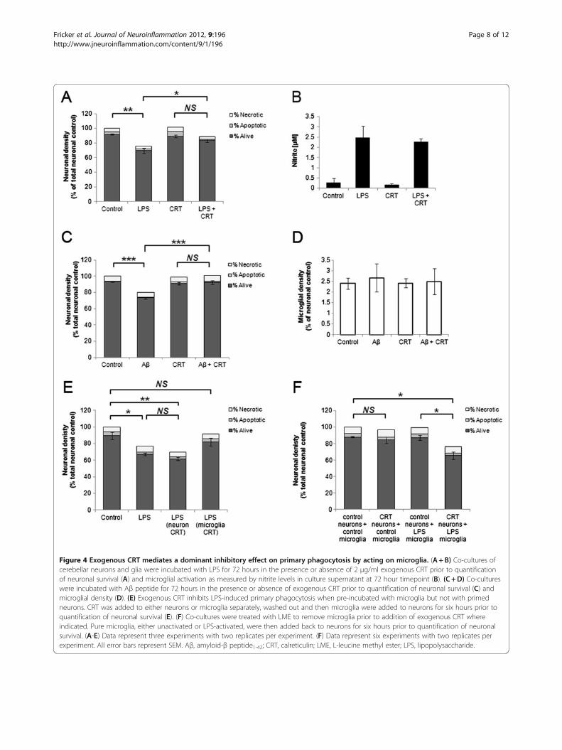
Figure 4 Exogenous CRT mediates a dominant inhibitory effect on primary phagocytosis by acting on microglia. (A+B) Co-cultures ofcerebellar neurons and glia were incubated with LPS for 72 hours in the presence or absence of 2 μg/ml exogenous CRT prior to quantificationof neuronal survival (A) and microglial activation as measured by nitrite levels in culture supernatant at 72 hour timepoint (B). (C +D) Co-cultureswere incubated with Aβ peptide for 72 hours in the presence or absence of exogenous CRT prior to quantification of neuronal survival (C) andmicroglial density (D). (E) Exogenous CRT inhibits LPS-induced primary phagocytosis when pre-incubated with microglia but not with primedneurons. CRT was added to either neurons or microglia separately, washed out and then microglia were added to neurons for six hours prior toquantification of neuronal survival (E). (F) Co-cultures were treated with LME to remove microglia prior to addition of exogenous CRT whereindicated. Pure microglia, either unactivated or LPS-activated, were then added back to neurons for six hours prior to quantification of neuronalsurvival. (A-E) Data represent three experiments with two replicates per experiment. (F) Data represent six experiments with two replicates perexperiment. All error bars represent SEM. Aβ, amyloid-β peptide1-42; CRT, calreticulin; LME, L-leucine methyl ester; LPS, lipopolysaccharide.
Fricker et al. Journal of Neuroinflammation 2012, 9:196 Page 8 of 12http://www.jneuroinflammation.com/content/9/1/196
inhibited primary phagocytosis (Figure 4E). Thus, exoge-nous CRT exerted a dominant inhibitory effect on primaryphagocytosis when added to microglia.To test whether surface CRT was sufficient to induce
phagocytosis of unprimed neurons we eliminated micro-glia from CGC with LME, incubated neurons with ex-ogenous CRT, washed off any excess unbound CRT andthen added back microglia that were either unactivatedor LPS-activated. Addition of exogenous CRT to neu-rons followed by unactivated microglia did not resultin any significant change in numbers of healthy ordead neurons. In contrast, when LPS-activated micro-glia were added back to neurons that had been incubatedwith exogenous CRT a significant loss of neurons was ob-served (unprimed neurons +LPS microglia, 87.4%±2.9%;unprimed neurons+ exogenous CRT+LPS microglia,65.9%± 4.5%; Figure 4F). Thus addition of exogenous CRTto unprimed neurons alone was not sufficient to allow pri-mary phagocytosis, except when microglia had beeninflammatory-activated with LPS.We previously demonstrated that inflammatory-activa-
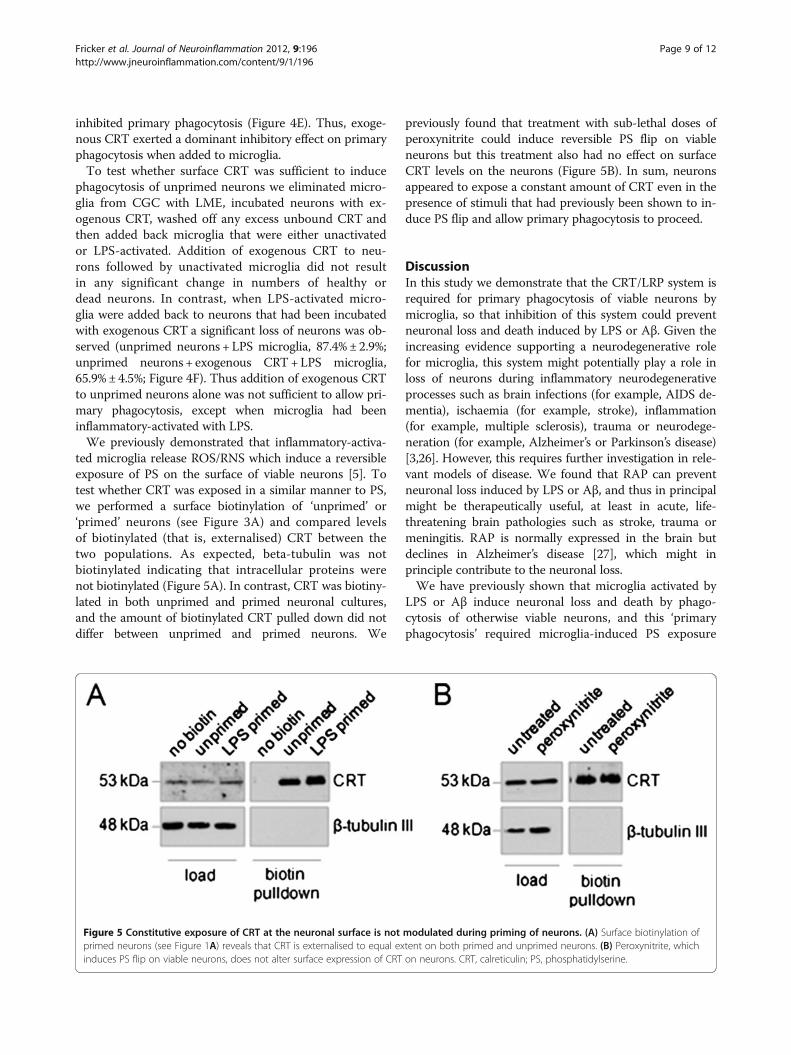
ted microglia release ROS/RNS which induce a reversibleexposure of PS on the surface of viable neurons [5]. Totest whether CRT was exposed in a similar manner to PS,we performed a surface biotinylation of ‘unprimed’ or‘primed’ neurons (see Figure 3A) and compared levelsof biotinylated (that is, externalised) CRT between thetwo populations. As expected, beta-tubulin was notbiotinylated indicating that intracellular proteins werenot biotinylated (Figure 5A). In contrast, CRT was biotiny-lated in both unprimed and primed neuronal cultures,and the amount of biotinylated CRT pulled down did notdiffer between unprimed and primed neurons. We
previously found that treatment with sub-lethal doses ofperoxynitrite could induce reversible PS flip on viableneurons but this treatment also had no effect on surfaceCRT levels on the neurons (Figure 5B). In sum, neuronsappeared to expose a constant amount of CRT even in thepresence of stimuli that had previously been shown to in-duce PS flip and allow primary phagocytosis to proceed.
DiscussionIn this study we demonstrate that the CRT/LRP system isrequired for primary phagocytosis of viable neurons bymicroglia, so that inhibition of this system could preventneuronal loss and death induced by LPS or Aβ. Given theincreasing evidence supporting a neurodegenerative rolefor microglia, this system might potentially play a role inloss of neurons during inflammatory neurodegenerativeprocesses such as brain infections (for example, AIDS de-mentia), ischaemia (for example, stroke), inflammation(for example, multiple sclerosis), trauma or neurodege-neration (for example, Alzheimer’s or Parkinson’s disease)[3,26]. However, this requires further investigation in rele-vant models of disease. We found that RAP can preventneuronal loss induced by LPS or Aβ, and thus in principalmight be therapeutically useful, at least in acute, life-threatening brain pathologies such as stroke, trauma ormeningitis. RAP is normally expressed in the brain butdeclines in Alzheimer’s disease [27], which might inprinciple contribute to the neuronal loss.We have previously shown that microglia activated by
LPS or Aβ induce neuronal loss and death by phago-cytosis of otherwise viable neurons, and this ‘primaryphagocytosis’ required microglia-induced PS exposure
Figure 5 Constitutive exposure of CRT at the neuronal surface is not modulated during priming of neurons. (A) Surface biotinylation ofprimed neurons (see Figure 1A) reveals that CRT is externalised to equal extent on both primed and unprimed neurons. (B) Peroxynitrite, whichinduces PS flip on viable neurons, does not alter surface expression of CRT on neurons. CRT, calreticulin; PS, phosphatidylserine.
Fricker et al. Journal of Neuroinflammation 2012, 9:196 Page 9 of 12http://www.jneuroinflammation.com/content/9/1/196
by neurons, bound by MFG-E8, which induced phago-cytosis of the neurons via microglial vitronectin recep-tors [5-7]. PS exposure on viable neurons was inducedby peroxynitrite production by microglia and phagocyt-osis of neurons occurred independently of apoptosis[5,7]. In the present work, we have found that the CRT/LRP pathway plays a permissive role for this inducedprimary phagocytosis, such that if the CRT/LRP pathwayis blocked primary phagocytosis can not proceed. Thefinding that blocking this phagocytic pathway with CRTantibodies, LRP antibodies, RAP or free CRT results inthe accumulation of live rather than dead neurons, againsupports the notion that the neurons are lost by primaryphagocytosis, rather than phagocytosis secondary to theneurons dying by some other means.We also found that CRT/LRP was important for BV2
microglial phagocytosis of apoptotic PC12 cells. If this istrue for primary neurons and microglia in vivo, thenblocking this system may be detrimental in a variety ofphysiological and pathological conditions by allowingdead neurons to accumulate and promote inflammation.However, for cancer cells, it has been shown that macro-phage phagocytosis of CRT-exposed cancer cells is im-munogenic as the macrophages present antigens fromthese cells [14]. This immunogenic phagocytosis of CRTexposing dead cells might be beneficial in the context ofbrain tumours or brain infections, but potentially detri-mental in other contexts such as MS or development. Itis possible that the CRT/LRP system contributes to thephagocytosis apoptotic and viable neurons arising duringdevelopment. In C.elegans mutation of a number of theCed genes (including Ced1, a proposed functional ho-mologue of LRP) involved in phagocytosis of dead cells incombination with a weak Ced3 (caspase) mutation resultsin survival of cells normally eliminated in the presence ofthe weak Ced3 mutation alone [28,29]. CRT knockout islethal in mice, and intriguingly 16% of CRT-null mice de-velop exencephaly of the brain characterised by failure toclose the neural tube, a process involving programmed celldeath [30,31].
Our data are consistent with a model in which CRTacts as an eat-me signal on the neuronal surface and isrecognised by LRP on the phagocytic membrane, as hasbeen described in other non-neuronal systems [15,17].LRP is known to be expressed and functional on microglia[32]. Through use of surface biotinylation, we demon-strated that CRT is constitutively expressed on the surfaceof cerebellar neurons. A previous report from Hossain andcolleagues demonstrated external localisation of CRT onrat hippocampal neurons where it co-localised withNMDA receptor and potentially played a role in modulat-ing Ca2+ influx into neurons [33]. The amount of CRT wasunchanged when neurons were primed for phagocytosis byinflammatory-activated microglia. Studies in non-neuronalcell types have shown that CRT-dependent phagocyt-osis does not necessarily require increased CRT exposure.In some cell types surface-exposed CRT accumulates inpatches on the cell surface, often in association withexposed PS [15,16,34]. CRT-dependent phagocytosis canalso be triggered by a reduction in don’t-eat-me signallingby CD47/SIRPα signalling [13,15,19]. However, we testeda CD47-blocking antibody and found that this had no ef-fect on phagocytosis of viable or apoptotic neuronal cellsin the presence or absence of inflammatory stimuli(data not shown). Gardai and colleagues demonstratedthat CRT knockout prevented phagocytosis of PS-exposing apoptotic cells, and that readdition of exogenousCRT restored phagocytosis although this phagocytosisremained PS-dependent [15]. Similarly, in our model wehave shown that LPS-activated microglia induce PS expos-ure on viable neurons and that PS recognition by MFG-E8is required for phagocytosis to proceed [5,7]. Thus whilstCRT is not increased on the surface of viable neurons dur-ing primary phagocytosis, the exposure of PS induced byLPS-activated microglia may be sufficient to allow phago-cytosis that occurs in a CRT-dependent manner. Wedemonstrated that incubation of viable neurons with ex-ogenous CRT allowed primary phagocytosis to proceed inthe presence of activated but not unactivated microglia. Itis possible that other types of neuropathologically relevant
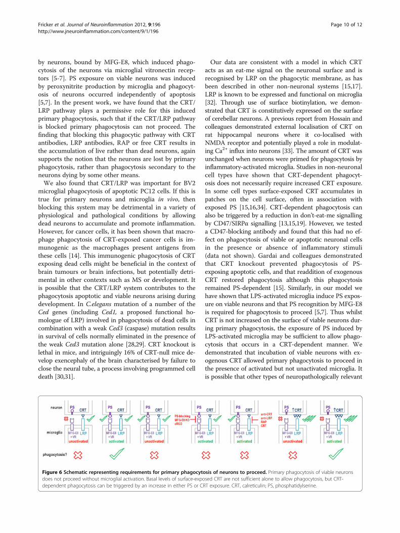
Figure 6 Schematic representing requirements for primary phagocytosis of neurons to proceed. Primary phagocytosis of viable neuronsdoes not proceed without microglial activation. Basal levels of surface-exposed CRT are not sufficient alone to allow phagocytosis, but CRT-dependent phagocytosis can be triggered by an increase in either PS or CRT exposure. CRT, calreticulin; PS, phosphatidylserine.
Fricker et al. Journal of Neuroinflammation 2012, 9:196 Page 10 of 12http://www.jneuroinflammation.com/content/9/1/196
stimuli may cause increased neuronal CRT exposure and,therefore, may cause neurodegeneration by primaryphagocytosis in this way.We found that addition of CRT to microglia or to both
microglia and neurons could block microglia-inducedneuronal loss. This may be because free CRT can acti-vate microglial phagocytosis/endocytosis via LRP in theabsence of bound neurons/target cells (as occurs inmacrophages [15]), resulting in downregulation of sur-face LRP and associated phagocytic machinery. This typeof mechanism might be involved in the neuroprotectiveeffect of peptide Y-P30. Y-P30 can bind CRT and wasreported to cause release of extracellular CRT in SH-SY5Y cells, as well as dissociation of CRT from mem-branes isolated from rat cortex. In conjunction with thisactivity, Y-P30 inhibited the appearance of microgliain vivo following lesioning of the cortex in rat [35].
ConclusionsCRT exposure on the surface of viable or apoptotic neu-rons is required for their phagocytosis via LRP receptorson microglia. CRT is exposed on neurons in the pre-sence or absence of inflammation, and appears to bepermissive for PS-induced phagocytosis. Based on ourpresent and previous data, we propose that CRT acts asan ‘eat-me-if ’ signal as illustrated in Figure 6. Phagocy-tosis of neurons by microglia normally requires: i) CRTexposure on neurons, AND ii) PS exposure on neurons,AND iii) activation of microglia. However, high levels ofCRT exposure on neurons can override the requirementfor PS exposure, and free CRT can block phagocytosis ofneurons by acting directly on microglia.
Competing interestsThe authors declare they have no competing interests.
Authors’ contributionsMF performed surface biotinylation, primary tissue culture and treatments,helped design the study and drafted the manuscript. MJOM performedapoptotic phagocytosis assays. GCB conceived and directed the study andhelped draft the manuscript. All authors read and approved the finalmanuscript.
AcknowledgementsThis work was funded by the Wellcome Trust (Grant RG50995).
Received: 27 March 2012 Accepted: 11 June 2012Published: 13 August 2012
References1. Kettenmann H, Hanisch UK, Noda M, Verkhratsky A: Physiology of
microglia. Physiol Rev 2011, 91:461–553.2. Brown GC, Neher JJ: Inflammatory neurodegeneration and mechanisms
of microglial killing of neurons. Mol Neurobiol 2010, 41:242–247.3. Neher JJ, Neniskyte U, Brown GC: Primary phagocytosis of neurons by
inflamed microglia: potential roles in neurodegeneration. FrontPharmacol 2012, 3:27.
4. Brown GC, Neher JJ: Eaten alive! Cell death by primary phagocytosis:‘phagoptosis’. Trends in Biochem Sci. in press.
5. Neher JJ, Neniskyte U, Zhao JW, Bal-Price A, Tolkovsky AM, Brown GC:Inhibition of microglial phagocytosis is sufficient to preventinflammatory neuronal death. J Immunol 2011, 186:4973–4983.
6. Neniskyte U, Neher JJ, Brown GC: Neuronal death induced by nanomolaramyloid β is mediated by primary phagocytosis of neurons by microglia.J Biol Chem 2011, 286:39904–39913.
7. Fricker M, Neher JJ, Zhao JW, Théry C, Tolkovsky AM, Brown GC: MFG-E8mediates primary phagocytosis of viable neurons duringneuroinflammation. J Neurosci 2012, 32:2657–2666.
8. Ravichandran KS: Beginnings of a good apoptotic meal: the find-me andeat-me signaling pathways. Immunity 2011, 35:445–455.
9. Elliott JI, Surprenant A, Marelli-Berg FM, Cooper JC, Cassady-Cain RL,Wooding C, Linton K, Alexander DR, Higgins CF: Membranephosphatidylserine distribution as a non-apoptotic signalling mechanismin lymphocytes. Nat Cell Biol 2005, 7:808–816.
10. Fischer K, Voelkl S, Berger J, Andreesen R, Pomorski T, Mackensen A:Antigen recognition induces phosphatidylserine exposure on the cellsurface of human CD8+ T cells. Blood 2006, 108:4094–4101.
11. Jitkaew S, Witasp E, Zhang S, Kagan VE, Fadeel B: Induction of caspase-and reactive oxygen species-independent phosphatidylserineexternalization in primary human neutrophils: role in macrophagerecognition and engulfment. J Leukoc Biol 2009, 85:427–437.
12. Martins I, Kepp O, Galluzzi L, Senovilla L, Schlemmer F, Adjemian S, MengerL, Michaud M, Zitvogel L, Kroemer G: Surface-exposed calreticulin in theinteraction between dying cells and phagocytes. Ann N Y Acad Sci 2010,1209:77–82.
13. Chao MP, Jaiswal S, Weissman-Tsukamoto R, Alizadeh AA, Gentles AJ,Volkmer J, Weiskopf K, Willingham SB, Raveh T, Park CY, Majeti R, WeissmanIL: Calreticulin is the dominant pro-phagocytic signal on multiple humancancers and is counterbalanced by CD47. Sci Transl Med 2010, 2:63ra94.
14. Obeid M, Tesniere A, Ghiringhelli F, Fimia GM, Apetoh L, Perfettini JL,Castedo M, Mignot G, Panaretakis T, Casares N, Métivier D, Larochette N,van Endert P, Ciccosanti F, Piacentini M, Zitvogel L, Kroemer G: Calreticulinexposure dictates the immunogenicity of cancer cell death. Nat Med2007, 13:54–61.
15. Gardai SJ, McPhillips KA, Frasch SC, Janssen WJ, Starefeldt A, Murphy-UllrichJE, Bratton DL, Oldenborg PA, Michalak M, Henson PM: Cell-surfacecalreticulin initiates clearance of viable or apoptotic cells through trans-activation of LRP on the phagocyte. Cell 2005, 123:321–334.
16. Kuraishi T, Manaka J, Kono M, Ishii H, Yamamoto N, Koizumi K, Shiratsuchi A,Lee BL, Higashida H, Nakanishi Y: Identification of calreticulin as a markerfor phagocytosis of apoptotic cells in Drosophila. Exp Cell Res 2007,313:500–510.
17. Garg AD, Krysko DV, Verfaillie T, Kaczmarek A, Ferreira GB, Marysael T, RubioN, Firczuk M, Mathieu C, Roebroek AJ, et al: A novel pathway combiningcalreticulin exposure and ATP secretion in immunogenic cancer celldeath. EMBO J 2012, 31:1062–1079.
18. Ogden CA, de Cathelineau A, Hoffmann PR, Bratton D, Ghebrehiwet B,Fadok VA, Henson PM: C1q and mannose binding lectin engagement ofcell surface calreticulin and CD91 initiates macropinocytosis and uptakeof apoptotic cells. J Exp Med 2001, 194:781–795.
19. Park YJ, Liu G, Lorne EF, Zhao X, Wang J, Tsuruta Y, Zmijewski J, Abraham E:PAI-1 inhibits neutrophil efferocytosis. Proc Natl Acad Sci USA 2008,105:11784–11789.
20. Jaiswal S, Jamieson CH, Pang WW, Park CY, Chao MP, Majeti R, Traver D, vanRooijen N, Weissman IL: CD47 is upregulated on circulatinghematopoietic stem cells and leukemia cells to avoid phagocytosis. Cell2009, 138:271–285.
21. Kinsner A, Pilotto V, Deininger S, Brown GC, Coecke S, Hartung T, Bal-PriceA: Inflammatory neurodegeneration induced by lipoteichoic acid fromStaphylococcus aureus is mediated by glia activation, nitrosative andoxidative stress, and caspase activation. J Neurochem 2005, 95:1132–1143.
22. Wong HK, Fricker M, Wyttenbach A, Villunger A, Michalak EM, Strasser A,Tolkovsky AM: Mutually exclusive subsets of BH3-only proteins areactivated by the p53 and c-Jun N-terminal kinase/c-Jun signalingpathways during cortical neuron apoptosis induced by arsenite. Mol CellBiol 2005, 25:8732–8747.
23. Herz J, Goldstein JL, Strickland DK, Ho YK, Brown MS: 39-kDa proteinmodulates binding of ligands to low density lipoprotein receptor-relatedprotein/alpha 2-macroglobulin receptor. J Biol Chem 1991,266:21232–21238.
Fricker et al. Journal of Neuroinflammation 2012, 9:196 Page 11 of 12http://www.jneuroinflammation.com/content/9/1/196
24. Burguillos MA, Deierborg T, Kavanagh E, Persson A, Hajji N, Garcia-Quintanilla A, Cano J, Brundin P, Englund E, Venero JL, Joseph B: Caspasesignalling controls microglia activation and neurotoxicity. Nature 2011,472:319–324.
25. Murray PJ, Wynn TA: Protective and pathogenic functions of macrophagesubsets. Nat Rev Immunol, 11:723–737.
26. Block ML, Zecca L, Hong JS: Microglia-mediated neurotoxicity: uncoveringthe molecular mechanisms. Nat Rev Neurosci 2007, 8:57–69.
27. Provias J, Jeynes B: Immunohistochemical detection of receptor-associated protein in normal human brain and Alzheimer’s disease.Patholog Res Int 2010, 2010:173496.
28. Reddien PW, Cameron S, Horvitz HR: Phagocytosis promotes programmedcell death in C. elegans. Nature 2001, 412:198–202.
29. Hoeppner DJ, Hengartner MO, Schnabel R: Engulfment genes cooperatewith ced-3 to promote cell death in Caenorhabditis elegans. Nature 2001,412:202–206.
30. Rauch F, Prud’homme J, Arabian A, Dedhar S, St-Arnaud R: Heart, brain,and body wall defects in mice lacking calreticulin. Exp Cell Res 2000,256:105–111.
31. Weil M, Jacobson MD, Raff MC: Is programmed cell death required forneural tube closure? Curr Biol 1997, 7:281–284.
32. Marzolo MP, von Bernhardi R, Bu G, Inestrosa NC: Expression of alpha(2)-macroglobulin receptor/low density lipoprotein receptor-related protein(LRP) in rat microglial cells. J Neurosci Res 2000, 60:401–411.
33. Hossain MA, Murayama N, Oka T, Nakajima T: Evidence of [Ca(2+)]ielevation by anti-calreticulin immunoreactive protein in neurons.Neurosci Res 2000, 36:285–290.
34. Tarr JM, Young PJ, Morse R, Shaw DJ, Haigh R, Petrov PG, Johnson SJ,Winyard PG, Eggleton P: A mechanism of release of calreticulin from cellsduring apoptosis. J Mol Biol 2010, 401:799–812.
35. Cunningham TJ, Jing H, Wang Y, Hodge L: Calreticulin binding and otherbiological activities of survival peptide Y-P30 including effects ofsystemic treatment of rats. Exp Neurol 2000, 163:457–468.
doi:10.1186/1742-2094-9-196Cite this article as: Fricker et al.: Primary phagocytosis of viable neuronsby microglia activated with LPS or Aβ is dependent on calreticulin/LRPphagocytic signalling. Journal of Neuroinflammation 2012 9:196.
Submit your next manuscript to BioMed Centraland take full advantage of:
• Convenient online submission
• Thorough peer review
• No space constraints or color figure charges
• Immediate publication on acceptance
• Inclusion in PubMed, CAS, Scopus and Google Scholar
• Research which is freely available for redistribution
Submit your manuscript at www.biomedcentral.com/submit
Fricker et al. Journal of Neuroinflammation 2012, 9:196 Page 12 of 12http://www.jneuroinflammation.com/content/9/1/196