phagocytosis assay of microglia for dead neurons … · microglia, we may control their...
TRANSCRIPT
http://www.bio-protocol.org/e1795 Vol 6, Iss 8, Apr 20, 2016
Copyright © 2016 The Authors; exclusive licensee Bio-protocol LLC. 1
Phagocytosis Assay of Microglia for Dead Neurons in Primary Rat Brain Cell Cultures
Xiurong Zhao*, Liyan Zhang, Shun-Ming Ting and Jaroslaw Aronowski
Stroke Program - Department of Neurology, The University of Texas Health Science Center at
Houston, McGovern Medical School, Texas, USA *For correspondence: [email protected]
[Abstract] Clearance of dead brain tissue including the dead neurons through phagocytosis is
an endogenous function of microglia in the brain, which is critical for inflammation resolution
after ischemic stroke or head trauma. By regulating the function or polarization status of
microglia, we may control their phagocytosis efficacy and therefore the cleanup process for the
dead brain tissue. We cultured rat cortical neurons and microglia from the same litter of
embryos. The cultured neurons are subjected to irradiation for inducing neuronal apoptosis.
After labeling with propidium iodide (PI), the dead neurons (DNs) are exposed to the cultured
microglia for phagocytosis assay. By counting the number of DNs in each microglia, we
calculate the phagocytosis index to quantify the phagocytosis efficacy of microglia toward DNs.
The protocol is divided into 4 sections: A) culturing rat cortical neurons from pre-natal rat
embryos, B) preparing dead neurons as phagocytosis target, C) culturing rat brain microglia, D)
quantifying phagocytosis index of microglia toward the dead neurons.
Materials and Reagents
1. Cell Strainer (100 μm and 40 μm) (BD, Falcon, catalog number: 431752 and 431750)
Note: Currently, it is “Corning, Falcon, catalog number: 431752 and 431750”.
2. Cell Lifter (VWR International, catalog number: 89030-910)
3. 100 mm Petri dish (Thermo Fisher Scientific, catalog number: 263991)
4. 60 mm TC dish (Corning, catalog number: CLS430166)
5. 75 cm2 TC flask (Corning, catalog number: 430825)
6. 24-well TC plate (BD, Falcon, catalog number: 353047)
Note: Currently, it is “Corning, Falcon, catalog number: 353047”.
7. 1 ml Syringe (BD, Falcon, catalog number: 305217)
8. Glass Pasteur pipet (VWR International, catalog number: 14673-043)
9. E-18 Pregnant Sprague Dawley (SD) rat (Charles River Laboratories International)
10. Pentobarbital (Sigma-Aldrich, catalog number: P-3761)
11. Neurobasal medium (Thermo Fisher Scientific, GibcoTM, catalog number: 21103)
12. B27 Supplement (Thermo Fisher Scientific, GibcoTM, catalog number: 17504)
13. Glutamine (Sigma-Aldrich, catalog number: G7513)
14. Penicillin/Streptomycin (100x) (GE Healthcare, HycloneTM, catalog number: SV30079)
15. DMEM (Corning, catalog number: 10-013-CM)
http://www.bio-protocol.org/e1795 Vol 6, Iss 8, Apr 20, 2016
Copyright © 2016 The Authors; exclusive licensee Bio-protocol LLC. 2
16. Propidium Iodide (PI) (Sigma-Aldrich, catalog number: P-4170)
17. DAPI (4’, 6-Diamidino-2-Phenylindole, Dilactate) (Invitrogen, catalog number: D3571)
Note: Currently, it is “Thermo Fisher Scientific, Molecular ProbesTM, catalog number:
D3571”.
18. 16% Paraformaldehyde (PFA) (Electron Microscopy Sciences, catalog number: 15170)
19. Fetal Bovine Serum (FBS) (GE Healthcare, HycloneTM, catalog number: SH30071)
20. Sodium pyruvate (Sigma-Aldrich, catalog number: P5280)
21. HEPES Buffer solution (Sigma-Aldrich, catalog number: 83264)
22. Sodium bicarbonate (Sigma-Aldrich, catalog number: S5761)
23. Poly-D-Lysine (Sigma-Aldrich, catalog number: P0889)
24. HBSS (Lonza, catalog number: 10-543F)
25. PBS (Corning, catalog number: 21-040-CV)
26. 0.4% Trypan blue (Sigma-Aldrich, catalog number: T8154)
27. Alexa Fluor 488-Phalloidin (Invitrogen, catalog number: A12379)
Note: Currently, it is “Thermo Fisher Scientific, Molecular ProbesTM, catalog number:
A12379”.
28. Mouse anti-MAP2 antibody (Sigma-Aldrich, catalog number: M4403)
29. Rabbit anti-mouse IgG-Alexa Fluor 488 (Thermo Fisher Scientific, InvitrogenTM, catalog
number: A11029)
30. Dissection buffer (see Recipes)
31. DMEM/FBS (see Recipes)
32. Neurobasal/B27 (see Recipes)
Notes:
a. All reagents and chemicals for cell cultures must be sterile. Without special indication,
all culture medium or buffer is used at room temperature (RT). And all operational
procedures for neuronal culture and microglia (MΦ) culture, including animal
dissecting, tissue triturating, cell plating or harvesting, and medium changing, have to
be performed in a dissecting hood or cell culture hood using sterile tools.
b. Poly-D-Lysine coated tissue culture (TC) plate. The TC dish and plate for the primary
neuronal cells or purified microglia must be pre-coated with Poly-D-lysine (0.1 mg/ml
in H2O) (2 ml for 60 mm2 TC dish, 0.5 ml per well for 24w-TC plate) for 30 min at 37 °C.
After washing with sterile water, the TC dishes/plates have to be air-dried in the cell
culture hood before use.
c. The age of the embryos for cell culture. Although E16 embryos are more suitable for
neuronal culture and post-natal 1-2-day-old pups are more suitable for glia culture, we
used the E-18 embryos to prepare the neuron culture and also the glial culture (to
isolate MΦ). Thus, the neurons and MΦ are prepared from the same litter of embryos.
The neuronal culture and microglia can be prepared from embryos or pups of different
litters.

http://www.bio-protocol.org/e1795 Vol 6, Iss 8, Apr 20, 2016
Copyright © 2016 The Authors; exclusive licensee Bio-protocol LLC. 3
d. The purity of neurons in the cortical neuronal culture. The Neurobasal/B27 medium is
serum-free medium, which is optional for neuronal culture. However, there are other
cell types in the cultures. In the 2-day-old neuronal cultures, the ratio of
MAP2+-neurons is 43.6 ± 23% (n = 10, Figure 1).
Figure 1. MAP2 immunofluorescence of 2-day-old rat cortical neurons in culture. The cortical neuron culture is fixed in 2% PFA and labeled by mouse anti-MAP2
antibody. The signals are visualized with Alexa Fluor 488 (Green). The nuclei of all cells
are labeled with DAPI (red). The arrows indicate the MAP2+-neurons. Scale bar = 50
µm.
e. All animal studies followed the guidelines outlined in Guide for the Care and Use of
Laboratory Animals from the National Institutes of Health and were approved by the
Animal Welfare Committee of The University of Texas Health Science Center at
Houston. f. The Irradiation operation followed the guidelines for Environment Health & Safety of
The University of Texas Health Science Center at Houston.
Equipment
1. Hemacytometer (VWR International, catalog number: 15170)
2. Cell culture CO2 incubator (LabX, Sanyo, model: MCO-19AIC)
3. Fluorescence microscopy with Imaging System, Olympus IX81 controlled by
MetaMorph 7.4
4. Dissecting microscope (Nikon Corporation, model: SMZ-27)
5. Digital controlled orbital shaker (Thermo Fisher Scientific, model: MaxQTM 2000)
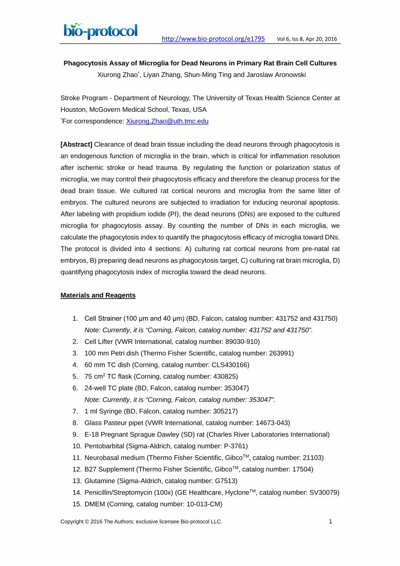
6. 137Cs Irradiation Source (J. L. SHEPHERD & ASSOCIATES, model: 143) (Figure 2)
http://www.bio-protocol.org/e1795 Vol 6, Iss 8, Apr 20, 2016
Copyright © 2016 The Authors; exclusive licensee Bio-protocol LLC. 4
Figure 2. Photos of the 137Cs irradiator. The cultured neurons grown in the 60 mm
TC dishes are loaded into the metal cylinder at top of the irradiator. Upon pressing the
red button on the right controller, the cylinder will rotate and go down until completely
being imbedded in the irradiator (the round bottom portion) for irradiation. By the
irradiation time (the left controller) is over, the irradiator will automatically rotate the
cylinder upward to its original position. By now, the irradiated cultures are ready to be
removed from the cylinder and returned to the CO2 incubator.
Software
1. MetaMorph software, version 7.4 Procedure
A. Culturing rat cortical neurons
1. Deeply anesthetize one E-18 pregnant rat by intraperitoneal injection of sodium
pentobarbital (50 mg/kg).
2. Prepare the surgical site with an appropriate skin disinfectant. We suggest 70%
alcohol.
3. Remove the embryos from uterus to one 100 mm Petri dish with 10 ml ice-cold
Dissection Buffer.
4. Remove the embryos’ brain using two surgical forceps and transfer the brains to
another Petri dish with 10 ml ice-cold Dissection Buffer and leave the cells in dish on
ice.
5. Under a dissecting microscope, after removing olfactory bulbs and meninges, the
cortices are separated from other brain regions, and rinsed in ice-cold Neurobasal/B27
medium once. The dissected brain cortices may be divided into two portions: One
portion will be used to culture neurons (see next step in this section) and another
portion will be used to culture microglia (see Section C).
http://www.bio-protocol.org/e1795 Vol 6, Iss 8, Apr 20, 2016
Copyright © 2016 The Authors; exclusive licensee Bio-protocol LLC. 5
6. Transfer the cortices in a 100 μm cell strainer placed in a 100 mm petri dish containing
10 ml ice-cold Neurobasal/B27 medium and mince the tissue in the same medium by
using a 1 ml-syringe plunger.
7. The dissociated cells passing through the 100 μm cell strainer (which is placed in a
100 mm petri dish) are applied onto another 40 μm cell strainer, which is placed on top
of a 50 ml centrifuge tube.
8. The collected cell suspension is subjected to a centrifugation step at 400 x g, 5 min.
The pelleted cells are suspended in Neurobasal/B27 medium (from 5 rat embryos) are
counted and seeded in Poly-D-lysine coated 60 mm TC dish (5 ml/dish) at a cell
density of 2.5 x 10⁷ cells/ml in Neurobasal/B27 medium (Kim et al., 2004). The yield of
the cells is about 2.5 x 10⁸ per brain, which can be seeded in 2 of 60 mm TC dishes.
9. Culture the cells in a CO2 incubator (5% CO2, 21% O2) at 37.0 ± 0.5 °C for 2 d.
10. After two days, the culture medium is completely replaced with fresh one and it is
ready for irradiation treatment.
B. Preparing dead neurons (DNs) as phagocytosis target
1. The 2-day-old neuronal cells grown in 60 mm TC dishes from the above (step A10) are
subjected to 137Cs irradiation, for 15 min (32Gy) (Kim et al., 2004). The culture dishes
are automatically rotated during radiation to ensure uniform exposure. The irradiation
operation is performed with supervision of the staff in the department of the
Environment Health and Safety.
2. After irradiation, the cells are returned to the original CO2 incubator and cultured in the
same culture medium for another 48 h. This incubation time after irradiation injury
allows the injured cells progressively undergo apoptosis. Using Trypan Blue, MAP2
immunofluorescence and DAPI staining, we confirmed that ≥95% neurons at this
stage are shrunken and dead (Figure 3). By comparing the cell number in the control
dish (Figure 3A) and in the irradiation-treated dish (Figure 3B). There is no significant
neuronal loss. The immunofluorescence procedures for MAP2 followed the same
method as we reported previously (Zhao et al., 2006).
Figure 3. MAP2 immunofluorescence of 4-day-old rat cortical neurons in culture. A. Control; B. 2 d after irradiation injury. The cortical neuron cultures are fixed in 2%
http://www.bio-protocol.org/e1795 Vol 6, Iss 8, Apr 20, 2016
Copyright © 2016 The Authors; exclusive licensee Bio-protocol LLC. 6
PFA and labeled by mouse anti-MAP2 antibody. The signals are visualized with Alexa
Fluor 488 (green). The nuclei are highlighted with DAPI (red). Scale bar = 50 μm
3. Label the dead neurons (DNs) in the culture dishes by adding 1 μg/ml of Propidium
Iodide (PI) directly into the culture medium and incubating for 10 min in the CO2
incubator.
4. Wash the cells with 10 ml PBS (37 °C), 3 times.
5. After removing the PBS, add 3 ml Neurobasal/B27 medium (37 °C) into each dish and
harvest the cells by using a Cell Lifter and then collect the cells in a 10 ml centrifuge
tube.
6. Triturate the cells using a Pasteur Pipet 5-10 strokes to ensure all cells to be
dissociated.
7. Apply the cell suspension through a 100 μm Cell Strainer and wash the strainer with 2
ml Neurobasal/B27 medium (37 °C).
8. Collect all passed cells in one 15 ml centrifuge tube and centrifuge at 400 x g for 10
min.
9. Re-suspend the cell pellet in 300 μl of DMEM/FBS per 60 mm TC dish and count the
cell number using one hemocytometer. Then, adjust the cell density to 5 x 10⁸ cells/ml
in the same culture media. Now the PI-DNs are ready to be used as phagocytosis
target at the next step for phagocytosis assay.
10. Keep the PI-DNs at 4 °C fridge and wrap the tube with foil to protect the cells from light.
The PI-DNs can be safely stored for one month.
C. Culturing rat brain microglia (MΦ)
Since we use embryos from the same litter to prepare the cortical neurons (Section A)
and also the microglia (Section C), we usually distribute equal number of embryos to
each culture. The beginning procedures for preparing microglia cultures are the same as
the steps A1-7 described in Section A.
1. This is next to the step A7 in Section A. The collected cell suspension is subjected to a
centrifugation step at 400 x g, 5 min. The pelleted cells are suspended in DMEM/FBS
(37 °C) at a ratio of 50 ml per brain and seeded in two 75 cm2 TC flasks (25 ml/flask).
2. The glia cells are cultured in CO2 incubator (5% CO2, 21% O2) at 37.0 ± 0.5 °C.
3. The culture media are completely changed to fresh one on d 4, d 8 and d 12.
4. On day 14, the astrocytes have grown into completely confluent. The MΦ become
loosely attached or freely floating in the culture medium. We harvest MΦ by subjecting
the flasks to a shaking procedure at 220 rpm for 10 min to detach MΦ from other glia
cells.
5. Collect the culture medium containing MΦ into 50 ml centrifuge tubes.
6. Centrifuge at 400 x g, 10 min.
http://www.bio-protocol.org/e1795 Vol 6, Iss 8, Apr 20, 2016
Copyright © 2016 The Authors; exclusive licensee Bio-protocol LLC. 7
7. After removing the supernatant, re-suspend the pelleted MΦ in 5 ml (per flask) fresh
DMEM/FBS (37 °C).
8. After cell counting, dilute the cells to 1 x 10⁵ cells/ml with DMEM/FBS. The yield of the
MΦ is about 1-2 x 10⁶ cells per flask.
9. Re-plate the MΦ in Poly-D-lysine coated 24w-TC plate at 5 x 10⁴ cells in 500 μl/well.
10. Culture the purified MΦ in CO2 incubator (5% CO2, 21% O2) at 37.0 ± 0.5 °C for 24 h.
And by now it is ready to be used at the next step for phagocytosis assay. By
immunofluorescence, we confirmed that more than 95% of the cells are CD68+-MΦ
and CD11b+- MΦ (Zhao et al., 2007).
D. Quantifying phagocytosis efficacy
1. Adding 10 μl of the PI-DNs (prepared at step B10) into the re-plated MΦ cultures
(prepared at step C10) at a target/effector ratio of 100:1 (DNs: MΦ).
2. Incubate the cells in CO2 incubator (5% CO2, 21% O2) at 37.0 ± 0.5 °C for 2 h.
3. At end of incubation, remove the free-floating PI-DNs by aspirating the culture medium
and fix the cells with 4% room temperature paraformaldehyde (PFA) for 10 min. It is
not necessary to have a washing step prior to fixture.
4. Rinse the cells by adding 1 ml PBS in each well, 3 times.
5. Incubate the cells in 1 ml of 0.1% Triton X-100 in PBS for 1 min.
6. Rinse the cells with 1 ml PBS per well, 3 times.
7. Label the cells with 300 μl of Alexa Fluor 488-Phalloidin at 1:100 dilution and 1 μg/ml
DAPI in PBS for 30 min.
8. Rinse the cells with 1 ml PBS, 3 times.
9. Observe the cells under a fluorescence microscopy and acquire the images using the
filter sets at Ex/Em of 490/520 nm for Phalloidin-labeled MΦ, Ex/Em of 550/575 nm for
PI-DNs and Ex/Em of 365/480 nm for DAPI-labeled nuclei.
10. By combining the images acquired with the above 3 sets of filters at the same position,
we may clearly observe the PI-DNs within each MΦ. The phagocytized DNs are
fluorescently red within Phalloidin-labeled green MΦ. The nuclei of the MΦ are blue
(Figure 4). Five images under 40x objective are recorded. The number of the
PI-positive neuronal nuclei in each MΦ is counted manually, on still images. At least
fifty MΦ cells from each condition should be analyzed. The average number of
phagocytized dead neurons (PI-DNs) within each MΦ is calculated and used as
Phagocytosis Index (Zhao et al., 2015).

http://www.bio-protocol.org/e1795 Vol 6, Iss 8, Apr 20, 2016
Copyright © 2016 The Authors; exclusive licensee Bio-protocol LLC. 8
Figure 4. Phalloidin-labeled microglia (green) after phagocytizing the PI-DNs (red). The nucleus is labeled with DAPI (blue). Scale bar = 20 μm. The arrows indicate
the phagocytized PI-DNs within the MΦ.
Notes
1. The incubation time of MΦ with dead neurons. We subject MΦ to PI-DNs for 2 h to
measure the Phagocytosis Index. To precisely quantify the phagocytosis efficacy, it is
necessary to perform a time course (1-4 h) study.
2. The number of cell counting. The size of DNs is not unique and the number within
each MΦ varies. Therefore, counting the number of DNs in each MΦ should measure
all MΦ cells within the image field. We count 50 of MΦ for each experimental condition.
However, more number counted, more precise data.
3. Bias of image acquisition. To determine the phagocytosis capacity of the MΦ toward
the PI-DNs, we suggest using a microscope with a motorized stage and CCD camera,
which is controlled by a computer software (e.g., MetaMorph software) that may allow
image stitching. This computer-controlled image acquisition system may permit
automatic and unbiased cell counting. However, the automatically counted data needs
to be confirmed manually. And when multiple batches of cell counting are repeated, it
is better done by the same person.
Recipes
1. Dissection buffer
HBSS
4.2 mM bicarbonate
1 mM pyruvate
20 mM HEPES
1 mg/ml BSA
1x Penicillin/Streptomycin (pH 7.25)
2. DMEM/FBS
http://www.bio-protocol.org/e1795 Vol 6, Iss 8, Apr 20, 2016
Copyright © 2016 The Authors; exclusive licensee Bio-protocol LLC. 9
DMEM
10% FBS
1x Penicillin/Streptomycin
3. Neurobasal/B27
Neurobasal culture medium
1x B27 supplement
0.5 mM L-Glutamine
1x Penicillin/Streptomycin
Acknowledgments
This protocol was supported in part by grants NS060768, NS064109 and NS084292 of
National Institute of Health, National Institute of Neurological Disorders and Stroke. For
citation, please refer to Zhao et al. (2015).
References
1. Kim, D. H., Zhao, X., Tu, C. H., Casaccia-Bonnefil, P. and Chao, M. V. (2004).
Prevention of apoptotic but not necrotic cell death following neuronal injury by
neurotrophins signaling through the tyrosine kinase receptor. J Neurosurg 100(1):
79-87.
2. Zhao, X., Ou, Z., Grotta, J. C., Waxham, N. and Aronowski, J. (2006).
Peroxisome-proliferator-activated receptor-gamma (PPARgamma) activation protects
neurons from NMDA excitotoxicity. Brain Res 1073-1074: 460-469.
3. Zhao, X., Sun, G., Zhang, J., Strong, R., Song, W., Gonzales, N., Grotta, J. C. and
Aronowski, J. (2007). Hematoma resolution as a target for intracerebral hemorrhage
treatment: role for peroxisome proliferator-activated receptor gamma in
microglia/macrophages. Ann Neurol 61(4): 352-362.
4. Zhao, X., Wang, H., Sun, G., Zhang, J., Edwards, N. J. and Aronowski, J. (2015).
Neuronal interleukin-4 as a modulator of microglial pathways and ischemic brain
damage. J Neurosci 35(32): 11281-11291.