prevention of behavioral deficits in rats exposed to...
TRANSCRIPT

ORIGINAL ARTICLE
Prevention of behavioral deficits in rats exposed to folatereceptor antibodies: implication in autismA Desai1, JM Sequeira2 and EV Quadros2
Folate receptor alpha (FRα) autoantibodies have been associated with fetal abnormalities and cerebral folate deficiency-relateddevelopmental disorders. Over 70% of the children with autism spectrum disorders (ASD) are positive for these autoantibodies andhigh-dose folinic acid is beneficial in treating these children. Here we show that antibodies (Abs) to the rat FRα administered duringgestation produce communication, learning and cognitive deficits in a rat model that can be prevented by folinic acid anddexamethasone. FRα Ab can trigger inflammation as well as block folate transport to the fetus and to the developing brain toproduce the functional deficits. In humans, exposure to FRα autoantibodies during fetal development and infancy could contributeto brain dysfunction such as that seen in ASD and other developmental disorders. Identifying women positive for the autoantibodyand treating them with high-dose folinic acid along with other interventions to lower the autoantibody titer are effective strategiesthat may be considered to reduce the risk of having a child with developmental deficits.
Molecular Psychiatry advance online publication, 20 September 2016; doi:10.1038/mp.2016.153
INTRODUCTIONFolate is an essential B-complex vitamin required for the transferof carbon units in the intermediary metabolism of amino acids,purines, pyrimidines and in the production of S-adenosylmethio-nine for methylation reactions.1 Folate is particularly essentialduring pregnancy as fetal development is a time of continuousDNA synthesis and cell division. Deficiency during pregnancycan produce fetal malformations and miscarriage.2,3 Folatesupplementation can prevent neural tube defect pregnancy4
and decrease the risk of autism spectrum disorders (ASD).5,6
Schmidt et al.7 reported that the protection afforded by prenatalfolic acid supplementation was particularly beneficial in mothersand children with the MTHFR 677 C4T variant genotype.7
Dietary folate insufficiency, gene defects or compromisedtransport can lead to metabolic folate deficiency. The identifica-tion of folate receptor alpha (FRα)-specific autoantibodies that canblock folate transport provided a potential mechanism for folatedeficiency in the fetus.8 These FRα autoantibodies have beenidentified in a majority of women with a history of neural tubedefect pregnancy,8,9 as well as with subfertility and pretermbirth.10,11 Direct proof that FRα-specific antibodies (Abs) areteratogenic to the embryo came from observations in a pregnantrat model of exposure to FRα Ab that caused resorption ofembryos at higher doses and neural tube and cranio-facialmalformations at lower doses.12 Folinic acid and dexamethasoneprevented malformations, suggesting that blocking of folatetransport to the embryo and Ab-mediated inflammation mayhave a role in the pathology.12
FRα autoantibodies have also been found in infants withcerebral folate deficiency (CFD), substantiating the important roleof folate in brain development, whereby FRα-specific autoanti-bodies could block folate transport across the choroid plexus.13–15
CFD is a developmental disorder with decreased level of
5-methyltetrahydrofolate (MTHF) in the cerebrospinal fluid.16
Consequently, these patients suffer from severe neurologicalsymptoms, including marked irritability, cerebellar ataxia, slowhead growth, psychomotor retardation and pyramidal tract signs.One-third also suffer from dyskinesia (for example, choreoathe-tosis and ballismus) and seizures.13,14 These autoantibodies havealso been found in other developmental disorders, including lowfunctioning autism,15 Rett syndrome17 and in ASD.18,19 Owing tothe high prevalence of ASD in children with CFD, and favorableresponse to folinic acid, reports have hypothesized that ASD maybe a less severe manifestation of CFD.18,20
Considering the high prevalence of FRα autoantibodies inchildren with CFD (89%)13 and ASD (~70%),18 we sought toestablish proof of hypothesis that FRα Abs can produce thepathology of behavioral and cognitive deficits. Preliminary studiesindicated that exposure to FRα Abs during gestation andpreweaning in a rat model produced severe learning andbehavioral deficits.21 Therefore, we determined behavioral deficitsin pups born to dams exposed to FRα Ab during gestation andevaluated the effect of folinic acid, which would provide adequatefolate, and dexamethasone, which would suppress inflammation,during gestational Ab exposure.
MATERIALS AND METHODSRat model of FRα Ab exposureTimed-pregnant postnatal day (PND) 50 Long Evans hooded rats (CharlesRiver Laboratories, Wilmington, MA, USA) were anesthetized on gestationalday (GD) 8 and a laparotomy was performed to record the number ofimplanted embryos. FRα Ab at a dose of 4 or 12 μg per embryo in 1 mlnormal rat serum was administered by intraperitoneal (IP) injection, 1 hafter the laparotomy. These doses were chosen because they allowed theimplanted embryos to be carried to term and produce live pups. Treatmentgroups received 1 mg of folinic acid (GD7–GD12) IP and/or 0.5 mg
1School of Graduate Studies, State University New York-Downstate Medical Center, Brooklyn, NY, USA and 2Department of Medicine, State University New York-Downstate MedicalCenter, Brooklyn, NY, USA. Correspondence: Professor E Quadros, Department of Medicine, SUNY-Downstate Medical Center, 450 Clarkson Avenue, Brooklyn, NY 11203, USA.E-mail: [email protected] 6 April 2016; revised 13 July 2016; accepted 18 July 2016
Molecular Psychiatry (2016) 00, 1–7© 2016 Macmillan Publishers Limited, part of Springer Nature. All rights reserved 1359-4184/16
www.nature.com/mp

dexamethasone (GD7–GD10) intramuscularly. As the 12 μg per embryopups showed more severe behavioral and cognitive deficits, this dose waschosen subsequently to evaluate response to folinic acid and dexametha-sone treatments.As shown in the flow chart (Supplementary Figure S1), pups born
underwent behavioral tests between PND4 and PND70. As sham controls,we used rats that were injected with an equivalent amount of pooledrabbit immunoglobulin G (IgG) from preimmune serum (NRIgG). Followingbirth, the number of pups born to each litter in all groups were countedand compared with the number of implanted embryos noted during thelaparotomy on GD8. Normal rats (simple controls) were also tested todetermine baseline measures and behavior. All rats were fed a normal dietcontaining 2 mg folic acid per kg chow as recommended by the AmericanInstitute of Nutrition (1977) and had free access to food and water. The ratswere maintained at 22 °C and on a 12 h light/dark cycle and theexperimental protocols were approved by the Animal Care and UseCommittee of the State University of New York, Downstate Medical Center,Brooklyn, NY, USA.
Testing for communication deficitsIsolated pups emit 40 kHz vocalizations, which normally elicits maternalsearch and retrieval of the pup, and thus has a crucial role in survival of thepup.22 On PND4, pups were individually separated from the mother in atemperature-controlled room (25 °C) and ultrasonic vocalizations emittedby the pup over a 3-min period were recorded and analyzed (Supplemen-tary Figure S2). Adult male ultrasonic vocalizations (on PND 50) uponinteraction with a female were also analyzed to determine whether adeficit in communication persisted into adulthood.22,23
Sociability studiesMale rats underwent a sociability paradigm on PND40–PND45 where eachrat’s inclination to approach and engage in social interaction with a samesex rat was assessed24 (Supplementary Figure S3).
Open field testWe used the open field test to assess locomotion as well as exploratory/anxiety-like behavior on PND40–PND4525 (Supplementary Figure S4).
Place avoidance tasksThe place avoidance tasks require recognition and segregation ofinformation obtained from both relevant and irrelevant stimuli andpermits the assessment of spatial memory formation26,27 (SupplementaryFigure S5). The tests, consisting of passive, active and conflict placeavoidance (PPA, APA, CPA), were conducted over 2 consecutive daysbetween PND60–PND70.21 Each place avoidance task consisted of 4–6trials of 10 min each with 10 min rest periods. A number of parameters arerecorded by an automated data acquisition system (Bio-Signal Group,Brooklyn, NY, USA).
Rabbit polyclonal antiserum to FRαRecombinant rat FRα was produced and purified and polyclonal antiserumwas generated in New Zealand white rabbits as previously described.21 TheIgG fraction was purified by affinity chromatography on a protein Acolumn and used in the study. The antiserum contained two types of Abs:binding and blocking Abs to rat FRα. Binding Ab is defined as an Abthat binds to an epitope distant from the folate-binding site and, therefore,does not interfere with folate binding to FRα28–30 (SupplementaryFigure S6A). Blocking Ab is defined as an Ab that prevents the bindingof folate when preincubated with apo FRα by virtue of the Ab directly orsterically interfering with folate binding30 (Supplementary Figure S6B).
ImmunohistochemistryTo examine inflammatory response induced by exposure to FRα Ab, GD14rats were injected with FRα Ab or NRIgG (12 μg per embryo, for ~ 16embryos). All animals received a second dose 16 h after the first dose. Sixhours later, animals were killed and tissues were collected. A similarprotocol was used to examine the effect of dexamethasone treatment inreducing the inflammation where animals were given 0.5 mg dexametha-sone IP, 3 h prior to Ab injection. Tissues were fixed in Carnoy’s solution,embedded in Paraplast (Fisher, Houston, TX, USA) and 5–7 μm sections
were cut for immunohistochemistry. Injected FRα Ab was localized byprobing sectioned tissue with peroxidase-conjugated goat anti-rabbit IgG(Pierce, Rockford, IL, USA) and then reacted with diaminobenzidine (DAB,Vector Laboratories, Burlingame, CA, USA) to develop the chromagen.Nuclear staining was carried out using hematoxylin (Vector Laboratories).For markers of inflammation, sections were incubated with mousemonoclonal anti-CD68 Ab (1:400 dilution, Abcam, Cambridge, MA, USA)or anti rat IL-1β Ab (5 μg ml− 1, R&D Systems, Minneapolis, MN, USA)overnight at 4 °C. Biotinylated anti-IgG was used as a secondary Ab,followed by incubation with ABC reagent and reaction with DAB todevelop the chromagen.
Folate uptake and FRα Ab localization studiesTo determine FRα Ab localization and its effect on folate uptake, GD14rats were administered IP with 12 μg of FRα Ab or NRIgG per embryo and5 μCi of 3HPGA in 1 ml saline on GD15. The rats were killed on GD16(48 h after the Ab dose). In another set of rats, a similar protocol wasfollowed except the 5μCi of 3HPGA was administered on GD16 andkilled on GD17 (72 h after Ab dose). Tissue was homogenized in 0.1 M
sodium phosphate buffer pH 7.4. Half of this homogenate was addedto scintillation fluid to determine folic acid (3HPGA) uptake. The other halfof the tissue was treated with glycine/HCl pH 2.5 to detach Ab fromreceptor, supernatant fraction neutralized with 1 M dibasic sodiumphosphate, followed by measuring immunoprecipitation of FRα labeledwith 3HPGA.In order to examine the effect of dexamethasone treatment, GD14
rats were given 0.5 mg dexamethasone intramuscularly. Three hours later,they received FRα Ab or NRIgG (12 μg per embryo). All animals received asecond dose of Ab 16 h later. Twenty hours after the first Ab or NRIgGinjection, all rats were injected with 5 μCi of 3HPGA. All animals were killed4 h later and tissues were collected (placenta, embryo, yolk sac, uterus).Radioactivity in the tissues was then determined as above.
Statistical analysisStatistical analysis of behavioral studies, which involved ⩾ 3 groups wascarried out using one-way analysis of variance (ANOVA). If the one-wayANOVA showed statistical significance (Po0.05), post-hoc analysis wascarried out using Tukey's Honest Significant Difference. For analysisbetween two groups, Student’s t-test was used to determine statisticalsignificance (GraphPad Software, La Jolla, CA, USA). Values plotted are themean and error bars represent s.e.m. Sample sizes (n) are indicated in thecorresponding figure or figure legend. Based on our published data (2% ofcontrols and 75% of Ab-exposed affected pups),21 we calculated thenecessary sample sizes. We needed pups from two dams in each group toyield a statistically significant result with 95% statistical power, accepting a95% confidence interval in a two-tailed post-hoc test following a significantmain effect ANOVA. This calculation was carried out using the StatisticalPower Calculator from DSS Research, Fort Worth, TX, USA.
RESULTSIn the various experimental and treatment groups, no decrease inthe number of live pups born was observed in any group. All pupswere examined for any gross abnormalities, and none were noted.There was also no difference in the ratio of males to females inlitters of each group. Pups were weighed on PND10 and PND25and showed no significant weight differences in the variousgroups (18.6 ± 0.5 and 70.8 ± 2.0 g, respectively).
Early communication deficitsFollowing administration of the rat FRα Ab at 4 or 12 μg perembryo to pregnant dams on GD8, a dose-dependent decrease inisolation-induced vocalizations was observed on PND4 comparedwith sham controls (Po0.001; Figure 1a). A significant improve-ment in the number of vocalizations was seen when folinic acid,dexamethasone or folinic acid and dexamethasone (Po0.001)were administered along with the Ab. Additionally, these pupsalso presented with a significant delay to first call; correctedin all treatment groups (Figure 1b, Po0.001) by folinic acid anddexamethasone.
Preventing folate receptor antibody-induced deficitsA Desai et al
2
Molecular Psychiatry (2016), 1 – 7 © 2016 Macmillan Publishers Limited, part of Springer Nature.
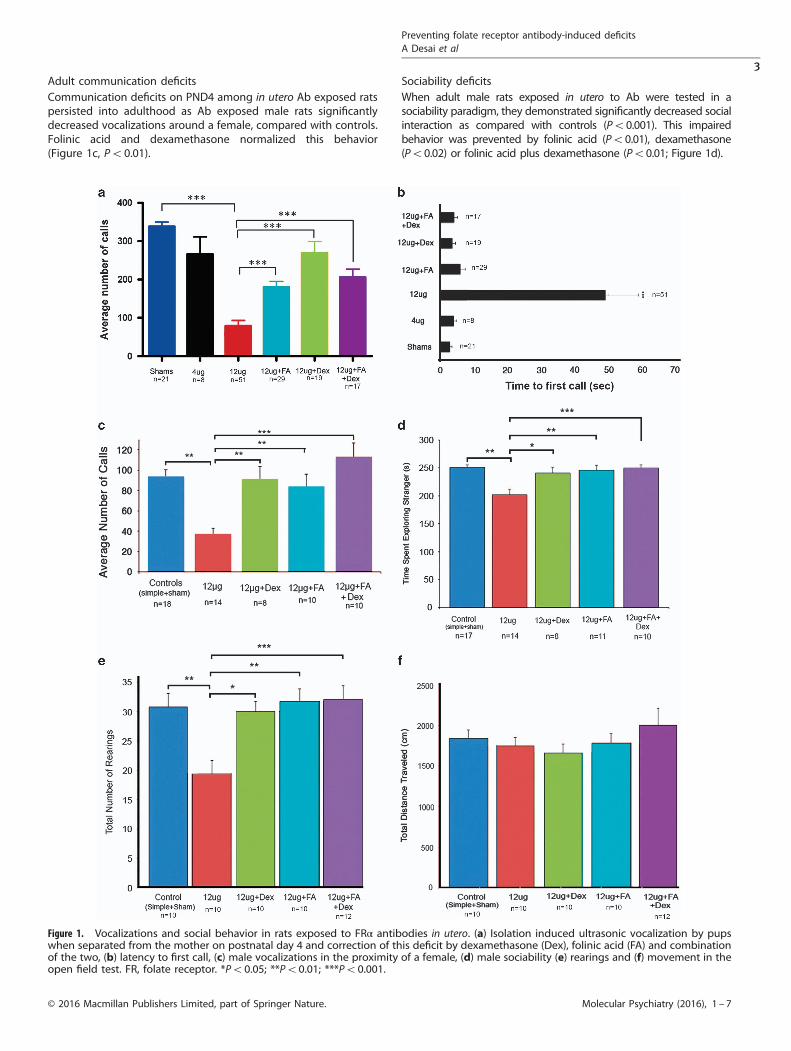
Adult communication deficitsCommunication deficits on PND4 among in utero Ab exposed ratspersisted into adulthood as Ab exposed male rats significantlydecreased vocalizations around a female, compared with controls.Folinic acid and dexamethasone normalized this behavior(Figure 1c, Po0.01).
Sociability deficitsWhen adult male rats exposed in utero to Ab were tested in asociability paradigm, they demonstrated significantly decreased socialinteraction as compared with controls (Po0.001). This impairedbehavior was prevented by folinic acid (Po0.01), dexamethasone(Po0.02) or folinic acid plus dexamethasone (Po0.01; Figure 1d).
Figure 1. Vocalizations and social behavior in rats exposed to FRα antibodies in utero. (a) Isolation induced ultrasonic vocalization by pupswhen separated from the mother on postnatal day 4 and correction of this deficit by dexamethasone (Dex), folinic acid (FA) and combinationof the two, (b) latency to first call, (c) male vocalizations in the proximity of a female, (d) male sociability (e) rearings and (f) movement in theopen field test. FR, folate receptor. *Po0.05; **Po0.01; ***Po0.001.
Preventing folate receptor antibody-induced deficitsA Desai et al
3
© 2016 Macmillan Publishers Limited, part of Springer Nature. Molecular Psychiatry (2016), 1 – 7

Open field testing for anxietyRats exposed to Ab in utero showed increased anxiety in the openfield test. These animals had significantly decreased numbers ofrearings compared with controls (Figure 1e, Po0.02). They didnot present with any significant decrease in total distancetraveled, indicating no motor abnormalities (Figure 1f).
Learning, memory and set-shifting deficitsThe rats were further tested for learning and memory in a seriesof tests of increasing complexity that evaluated their abilityto learn and remember a task over an extended period and thenbe able to switch to a new task. When animals exposed to FRαAb in utero were tested in a hierarchy of place avoidance tasksbetween PND60 and PND70, all animals could learn the PPAtask by successfully avoiding a stationary shock sector usingolfactory and visual cues. However, when they continued onto the APA task, 50% of the 4 μg, and 75% of 12 μg Ab-exposedanimals failed to learn the task (Figure 2a). Protection fromthese deficits was provided by administration of folinic acid,dexamethasone and folinic acid plus dexamethasone alongwith the FRα Ab (Figure 2a). A majority of the animals in alltreatment groups could successfully acquire the APA task, with thelearning curve for animals treated with folinic acid plusdexamethasone being almost identical to control animals(Figure 2b).
Not all of the 12 μg Ab/embryo-exposed rats that passed theactive avoidance task were able to pass the CPA task, (Figure 3a).Further, administration of folinic acid and dexamethasone rescuedthese rats from the CPA learning deficits (Figure 3a). Representa-tive tracings illustrating the movement of rats in the APA and CPAtasks are shown in Figure 3b, demonstrating the inability of theAb-exposed animals to learn the tasks as indicated by repeatedentry into the shock sector and the ability of the animals in thetreatment groups to learn the tasks. No significant differencebetween the treatment groups of folinic acid, dexamethasone orfolinic acid plus dexamethasone following Ab exposure was notedin any of the behavioral and cognitive studies, suggesting nodetrimental effect of the steroid use.
Figure 2. Learning deficits in rats exposed to FRα antibody in utero.(a) Active place avoidance test results and (b) learning curvesfor various treatment groups as indicated by decreased entrancesinto the shock zone in subsequent trials. ***Po0.001 comparedwith all the groups. APA, place avoidance; Dex, dexamethasone,FA, folinic acid.
Figure 3. Set-shifting deficits in rats exposed to FRα antibody inutero. (a) Conflict place avoidance test results for various treatmentgroups. Of the animals exposed to antibody during gestation, only25% could pass the active place avoidance task and 40% of thesecould not complete the conflict avoidance task. This is indicative ofimpairment in completing set-shifting tasks as 93% of sham animalsthat passed active place avoidance could pass conflict avoidance.This impairment was corrected by dexamethasone (Dex), folinic acid(FA) and folinic acid plus dexamethasone treatment. (b) Tracing themovement of the rat in the fourth trial of the active and conflictplace avoidance tasks.
Preventing folate receptor antibody-induced deficitsA Desai et al
4
Molecular Psychiatry (2016), 1 – 7 © 2016 Macmillan Publishers Limited, part of Springer Nature.

Folate uptake, localization of Ab and inflammationDuring fetal development, maternal folate is the only source offolate and this has to traverse the placental barrier in order toenter fetal circulation.31 One potential mechanism by which theAbs can affect brain development is by blocking folate transport
to the fetus. Ab localization was seen mostly in the placenta and inthe uterus and much less in the embryo (Figure 4a). Immuno-histochemical analysis showed extensive accumulation in theplacenta, the uterine wall and the yolk sac (Figure 4b-1) Within theembryo, Ab was mostly localized to epithelial cells in the head
Figure 4. Antibody (Ab) localization, effect on 3HPGA uptake and inflammation produced in gestational day 15 pregnant rats. (a) Antibodyaccumulation, (b) immunohistochemical localization, (c) blocking of 3HPGA uptake by antibody, (d) increase in inflammatory markers and (e)correction of this by dexamethasone (Dex), (f) correction of 3HPGA uptake by dexamethasone. IL, interleukin; NRIgG, immunoglobulin G frompreimmune serum; NS, not significant. *Po0.05; **Po0.01; ***Po0.001; ****Po0.0001.
Preventing folate receptor antibody-induced deficitsA Desai et al
5
© 2016 Macmillan Publishers Limited, part of Springer Nature. Molecular Psychiatry (2016), 1 – 7

region (Figure 4b-2) and to the epithelial cells of the choroidplexus (Figure 4b-3). During this period, folate transport to theembryo was significantly compromised as indicated by a 56% and41% decrease in 3HPGA accumulation in the placenta at 48 and72 h, respectively, and ~ 76% decrease in the embryo (Figure 4c).Ab localization within the placenta and yolk sac appeared to affectfolate transfer from the mother to the fetus and folate uptake inthe fetus. Localization of IgG was not seen in the NRIgG-injected dam.Rodent studies of maternal inflammation during pregnancy that
increase risk of neurodevelopmental deficits in the offspring haveshown increased CD68+ cells and interleukin (IL)-1β in theplacenta.32,33 This prompted us to examine whether the localiza-tion of this Ab in the placenta induced a similar inflammatoryresponse. Consistent with the presence of Ab on the placenta,increased expression of CD68+ cells and IL-1β was seen insimilar areas (Figure 4d). Dexamethasone treatment significantlydecreased the expression of both these markers for inflammation(Figures 4(d-1 and d-2)). Dexamethasone treatment also signifi-cantly increased folate transport to the placenta, yolk sac andamnion and the embryo (Figure 4e). Rats exposed to Ab in uterowere killed after PND 60 and examined for the presence of anystructural changes in the brain using coronal sections stained withhematoxylin–eosin and for markers of inflammation. No discern-able histological changes were observed compared with sham orsimple controls.
DISCUSSIONIn this study, we provide evidence that exposure to FRα-specificAb during gestation in a rat results in the birth of pups with severebehavioral and learning deficits. Many of these deficits mirror coredeficits of ASD, including deficits in communication, sociabilityand difficulty with set-shifting tasks. Lack of bonding, socialinteraction and verbal communication are some of the earlyindicators of autism development although these may not fullymanifest until later in life. In the rodent model, ultrasonicvocalizations are one of the earliest forms of communicationbetween the pups and their mother.22 Thus evaluating the ratpups on PND4 is ideal. As observed in this study, lack ofcommunication has been reported in many rodent models ofautism-like behavior.34–36 Additionally, we have shown thepresence of variable symptoms of ASD, including increasedanxiety in the open field test and learning deficits in the placeavoidance tasks. We have shown that both of these core deficitsand variable symptoms produced by Ab exposure can beprevented, or at the least attenuated, using gestational treatmentwith folinic acid and dexamethasone.Further, intellectual disability has been found to be highly
comorbid with ASD.37 We have previously shown that exposure toAb during gestation and the preweaning period in rats leads tolearning deficits.21 We have confirmed this effect of gestationalexposure in the current study and, more importantly, shown thatthese learning deficits can be prevented by treatment with folinicacid and dexamethasone.Overall, it appears that FRα Abs exert their effect on the fetus by
decreasing transplacental transport of folate, which results in fetalfolate deficiency. This explains why protection is afforded byfolinic acid treatment where folinic acid, a reduced form of folate,is taken up by the reduced folate carrier, rather than by FRα, whichis hindered by the Abs from transporting folate.Epidemiological studies suggest a clear association between
maternal exposure to infection and inflammation and increasedrisk of ASD in the offspring.38 Maternal cytokine expression,including expression in the placenta, can affect the developingembryo or fetus, resulting in neurological and behavioralabnormalities.39 These studies suggest that FRα Ab exposure inan animal model leads to a type II autoimmune reaction,40 where
there are increased Fc-receptor-bearing phagocytes, as demon-strated by the increased presence of CD68+ macrophages. Thesemacrophages appear to secrete increased amounts of IL-1βcytokine in the presence of FRα Ab, leading to increasedinflammatory response. When the inflammatory response issuppressed with dexamethasone, there is more folate uptakealong with rescue of behavioral deficits. Hence, the benefitafforded by dexamethasone treatment also appears to be theresult of increased folate uptake, where the dexamethasonesuppresses inflammatory reaction around FRα. The inflammatoryresponse owing to autologous Abs in the FRα autoimmunedisorder in humans may vary from that owing to the heterologousAbs we have used in our rat model. Regardless, the Abs appear tocause immune-mediated damage and block folate transport.These mechanisms of impaired folate transport can potentially
explain pregnancy-related disorders, including neural tube defectsin the presence of FRα autoimmune disorder. Daily folate intakeand folate supplementation during pregnancy may minimize theeffects of the autoantibody but may not be sufficient in those withhigher Ab titer. Therefore, identifying women positive for theautoantibody and treating them with high-dose folinic acid alongwith other interventions to lower the Ab titer are effectivestrategies for a favorable outcome. A case that utilized such astrategy showed positive results in preventing pregnancy-relatedcomplications owing to the presence of FRα autoimmunedisorder.41 Our rat study suggests that steroids and otherimmunosuppressant drugs could decrease placental inflammationinduced by the FRα autoantibody. Consequently, further investi-gation of similar treatment options to prevent pregnancy-relatedcomplications owing to FRα autoimmune disorder is warranted.It should be noted that the methods for the detection of FRα-
specific autoantibodies in the serum of patients provide a measureof autoantibody titer in serum.30 The FRα protein is a glycosyl-phosphatidylinositol-linked peripheral membrane protein andAbs directed against this protein would seek the target antigenon the membrane.42 Therefore, Ab in circulation is likely torepresent excess free Ab. As in many autoimmune disorders,fluctuations in Ab titer is a common occurrence,43,44 and thereforetesting patients multiple times during 2–6 months may benecessary to rule out the autoimmune disorder. The prevalenceof autoantibodies in 470% of children with autism18 andimprovements in the core symptoms of autism with folinic acidtreatment15,18 provides compelling evidence linking the auto-immune disorder with autism. Parental FRα autoantibodies witheither mother or father positive have also been linked to autism inchildren.19 Particularly, mothers of children with ASD were foundto have a significantly higher prevalence of FRα autoantibodiescompared with controls (26% vs 3.3%, Po0.01),19 and thereforeidentifying and treating these mothers could potentially reducethe risk of autism in the offspring.In conclusion, the findings of this study suggest severe
behavioral and cognitive changes mirroring ASD symptomsfollowing gestational Ab exposure in a rat model and protectionafforded by folinic acid and dexamethasone treatment. This hasmajor implications in the treatment of FRα autoimmune disorderin women during pregnancy and reducing the risk of autism in theoffspring.
CONFLICT OF INTERESTTwo of the authors (JMS and EVQ) are inventors on a US patent for the detection ofFRalpha autoantibodies issued to the Research Foundation of the State University ofNew York, USA. The other author declares no conflict of interest.
ACKNOWLEDGMENTSFunding for this work was provided by Autism Speaks grant no. 8202.
Preventing folate receptor antibody-induced deficitsA Desai et al
6
Molecular Psychiatry (2016), 1 – 7 © 2016 Macmillan Publishers Limited, part of Springer Nature.
AUTHOR CONTRIBUTIONSAD, JMS and EVQ designed the studies. AD and JMS conducted all experiments.AD, JMS and EVQ participated in the writing of the manuscript.
REFERENCES1 Wagner C. Biochemical role of folate in cellular metabolism. In: Bailey LB (ed).
Folate in Health and Disease. Marcel Dekker, Inc: New York, NY, USA, 1995,pp 23–42.
2 Tamura T, Picciano MF. Folate and human reproduction. Am J Clin Nutr 2006; 83:993–1016.
3 Geisel J. Folic acid and neural tube defects in pregnancy: a review. J PerinatNeonatal Nurs 2003; 17: 268–279.
4 MRC Vitamin Study Research Group. Prevention of neural tube defects: results ofthe Medical Research Council Vitamin Study. Lancet 1991; 338: 131–137.
5 Suren P, Roth C, Bresnahan M, Haugen M, Hornig M, Hirtz D et al. Associationbetween maternal use of folic acid supplements and risk of autism spectrumdisorders in children. JAMA 2013; 309: 570–577.
6 Steenweg-de Graaff J, Ghassabian A, Jaddoe VW, Tiemeier H, Roza SJ. Folateconcentrations during pregnancy and autistic traits in the offspring. The Gen-eration R Study. Eur J Public Health 2015; 25: 431–433.
7 Schmidt RJ, Tancredi DJ, Ozonoff S, Hansen RL, Hartiala J, Allayee H et al. Maternalpericonceptional folic acid intake and risk of autism spectrum disorders anddevelopmental delay in the CHARGE (CHildhood Autism Risks from Genetics andEnvironment) case-control study. Am J Clin Nutr 2012; 96: 80–89.
8 Rothenberg SP, da Costa MP, Sequeira JM, Cracco J, Roberts JL, Weedon J et al.Autoantibodies against folate receptors in women with a pregnancy complicatedby a neural-tube defect. N Engl J Med 2004; 350: 134–142.
9 Cabrera RM, Shaw GM, Ballard JL, Carmichael SL, Yang W, Lammer EJ et al.Autoantibodies to folate receptor during pregnancy and neural tube defect risk.J Reprod Immunol 2008; 79: 85–92.
10 Berrocal-Zaragoza MI, Fernandez-Ballart JD, Murphy MM, Cavalle-Busquets P,Sequeira JM, Quadros EV. Association between blocking folate receptor auto-antibodies and subfertility. Fertil Steril 2009; 91(4 Suppl): 1518–1521.
11 Vo HD, Sequeira JM, Quadros EV, Schwarz SM, Perenyi AR. The role of folatereceptor autoantibodies in preterm birth. Nutrition 2015; 31: 1224–1227.
12 da Costa M, Sequeira JM, Rothenberg SP, Weedon J. Antibodies to folate recep-tors impair embryogenesis and fetal development in the rat. Birth Defects Res AClin Mol Teratol 2003; 67: 837–847.
13 Ramaekers VT, Rothenberg SP, Sequeira JM, Opladen T, Blau N, Quadros EV et al.Autoantibodies to folate receptors in the cerebral folate deficiency syndrome. NEngl J Med 2005; 352: 1985–1991.
14 Ramaekers VT, Quadros E. Folate receptor autoimmunity in cerebral folate defi-ciency. In: Dale RC and Vincent A (eds). Inflammatory and Autoimmune Disorders ofthe Nervous System in Children, Mac Keith Press: London, UK, 2010, pp 302–316.
15 Ramaekers VT, Blau N, Sequeira JM, Nassogne MC, Quadros EV. Folate receptorautoimmunity and cerebral folate deficiency in low-functioning autism withneurological deficits. Neuropediatrics 2007; 38: 276–281.
16 Ramaekers VT, Blau N. Cerebral folate deficiency. Dev Med Child Neurol 2004; 46:843–851.
17 Ramaekers VT, Sequeira JM, Artuch R, Blau N, Temudo T, Ormazabal A et al. Folatereceptor autoantibodies and spinal fluid 5-methyltetrahydrofolate deficiency inRett syndrome. Neuropediatrics 2007; 38: 179–183.
18 Frye RE, Sequeira JM, Quadros EV, James SJ, Rossignol DA. Cerebral folate receptorautoantibodies in autism spectrum disorder. Mol Psychiatry 2013; 18: 369–381.
19 Ramaekers VT, Quadros EV, Sequeira JM. Role of folate receptor autoantibodies ininfantile autism. Mol Psychiatry 2013; 18: 270–271.
20 Moretti P, Peters SU, Del Gaudio D, Sahoo T, Hyland K, Bottiglieri T et al. Briefreport: autistic symptoms, developmental regression, mental retardation, epi-lepsy, and dyskinesias in CNS folate deficiency. J Autism Dev Disord 2008; 38:1170–1177.
21 Sequeira JM, Desai A, Berrocal-Zaragoza MI, Murphy MM, Fernandez-Ballart JD,Quadros EV. Exposure to folate receptor alpha antibodies during gestation and
weaning leads to severe behavioral deficits in rats: a pilot study. PLoS One 2016;11: e0152249.
22 Crawley JN. Designing mouse behavioral tasks relevant to autistic-like behaviors.Ment Retard Dev Disabil Res Rev 2004; 10: 248–258.
23 Portfors CV. Types and functions of ultrasonic vocalizations in laboratory ratsand mice. J Am Assoc Lab Anim Sci 2007; 46: 28–34.
24 Jamain S, Radyushkin K, Hammerschmidt K, Granon S, Boretius S, Varoqueaux Fet al. Reduced social interaction and ultrasonic communication in a mouse modelof monogenic heritable autism. Proc Natl Acad Sci USA 2008; 105: 1710–1715.
25 Walsh RN, Cummins RA. The Open-Field Test: a critical review. Psychol Bull 1976;83: 482–504.
26 Wesierska M, Adamska I, Malinowska M. Retrosplenial cortex lesion affectedsegregation of spatial information in place avoidance task in the rat. NeurobiolLearn Mem 2009; 91: 41–49.
27 Abdel Baki SG, Kao HY, Kelemen E, Fenton AA, Bergold PJ. A hierarchy of neu-robehavioral tasks discriminates between mild and moderate brain injury in rats.Brain Res 2009; 1280: 98–106.
28 Sadasivan E, Meng Y, Rothenberg SP. Coding sequence, genomic organizationand expression of a folate binding protein gene in the rat. Gene 2000; 254:219–228.
29 Sadasivan E, da Costa M, Rothenberg SP, Brink L. Purification, properties, andimmunological characterization of folate-binding proteins from humanleukemia cells. Biochim Biophys Acta 1987; 925: 36–47.
30 Sequeira JM, Ramaekers VT, Quadros EV. The diagnostic utility of folate receptorautoantibodies in blood. Clin Chem Lab Med 2013; 51: 545–554.
31 Yasuda S, Hasui S, Yamamoto C, Yoshioka C, Kobayashi M, Itagaki S et al. Placentalfolate transport during pregnancy. Biosci Biotechnol Biochem 2008; 72: 2277–2284.
32 Girard S, Tremblay L, Lepage M, Sebire G. IL-1 receptor antagonist protects againstplacental and neurodevelopmental defects induced by maternal inflammation.J Immunol 2010; 184: 3997–4005.
33 Ashdown H, Dumont Y, Ng M, Poole S, Boksa P, Luheshi GN. The role of cytokinesin mediating effects of prenatal infection on the fetus: implications for schizo-phrenia. Mol Psychiatry 2006; 11: 47–55.
34 Tyzio R, Nardou R, Ferrari DC, Tsintsadze T, Shahrokhi A, Eftekhari S et al. Oxytocin-mediated GABA inhibition during delivery attenuates autism pathogenesis inrodent offspring. Science 2014; 343: 675–679.
35 Hodgson RA, Guthrie DH, Varty GB. Duration of ultrasonic vocalizations in theisolated rat pup as a behavioral measure: sensitivity to anxiolytic andantidepressant drugs. Pharmacol Biochem Behav 2008; 88: 341–348.
36 Scattoni ML, Crawley J, Ricceri L. Ultrasonic vocalizations: a tool for behaviouralphenotyping of mouse models of neurodevelopmental disorders. Neurosci Bio-behav Rev 2009; 33: 508–515.
37 Newschaffer CJ, Croen LA, Daniels J, Giarelli E, Grether JK, Levy SE et al. Theepidemiology of autism spectrum disorders. Annu Rev Public Health 2007; 28:235–258.
38 Malkova NV, Yu CZ, Hsiao EY, Moore MJ, Patterson PH. Maternal immune acti-vation yields offspring displaying mouse versions of the three core symptomsof autism. Brain Behav Immun 2012; 26: 607–616.
39 Parker-Athill EC, Tan J. Maternal immune activation and autism spectrum disorder:interleukin-6 signaling as a key mechanistic pathway. Neurosignals 2010; 18:113–128.
40 Janeway CA Jr, Travers P, Walport M, Shlomchik MJ. Autoimmune responses aredirected against self antigens. Immunobiology: The Immune System in Health andDisease, 5th edn. Garland Science: New York, USA, 2001.
41 Shapira I, Sequeira JM, Quadros EV. Folate receptor autoantibodies in pregnancyrelated complications. Birth Defects Res A Clin Mol Teratol 2015; 103: 1028–1030.
42 Desai A, Sequeira JM, Quadros EV. The metabolic basis for developmentaldisorders due to defective folate transport. Biochimie 2016; 126: 31–42.
43 Out HJ, van Vliet M, de Groot PG, Derksen RH. Prospective study of fluctuations oflupus anticoagulant activity and anticardiolipin antibody titre in patients withsystemic lupus erythematosus. Ann Rheum Dis 1992; 51: 353–357.
44 Praprotnik S, Bozic B, Kveder T, Rozman B. Fluctuation of anti-Ro/SS-A antibodylevels in patients with systemic lupus erythematosus and Sjogren's syndrome: aprospective study. Clin Exp Rheumatol 1999; 17: 63–68.
Supplementary Information accompanies the paper on the Molecular Psychiatry website (http://www.nature.com/mp)
Preventing folate receptor antibody-induced deficitsA Desai et al
7
© 2016 Macmillan Publishers Limited, part of Springer Nature. Molecular Psychiatry (2016), 1 – 7