preparation of the root canal system
TRANSCRIPT
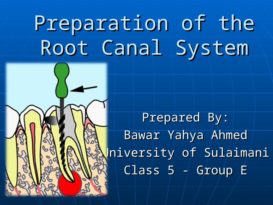
Preparation of the Root Preparation of the Root Canal SystemCanal System
Prepared By:Prepared By:
Bawar Yahya AhmedBawar Yahya Ahmed
University of SulaimaniUniversity of Sulaimani
Class 5 - Group EClass 5 - Group E
Root canal preparationRoot canal preparation
1. Cleaning 2. Shaping
Root canal preparationRoot canal preparation
Cleaning is the removal of all contents of the root canal system before and during shaping: infected materials, antigenic materials, organic substances, microflora, bacterial by products, food, caries, tissue remnants, denticles, pulp stones, collagen, inflammatory chemicals, contaminated canal filling materials, and dentinal debris created during canal shaping procedures. Cleaning entails accomplished by both mechanical instrumentation and chemically by irrigation.
Root canal preparationRoot canal preparation
Shaping is a mechanical process accomplished (with files, reamers, Gates-Glidden drills, slow speed burs, high speed diamonds-tipped drills, and sonic and ultrasonic, and nickel-titanium instruments of variable taper) to establishment of a specific cavity from that permits pluggers, spreaders, and other Obturation instruments to fit freely within the root canal system and to generate the pressures needed to transform and capture a maximal Obturation cushion, forcing gutta-percha and a microfilm of sealer into all foramina. Equally important, shaping facilitates three-dimensional cleaning by providing easy direct access for file, reamer, rotary instruments and irrigant during the treatment process.
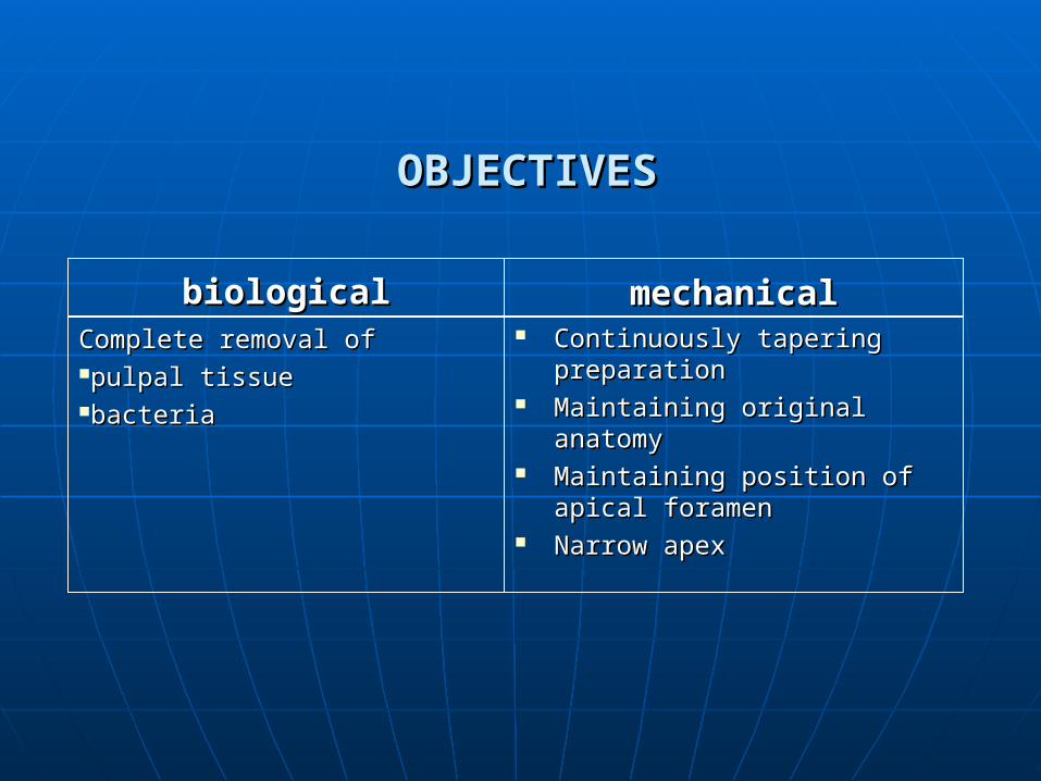
OBJECTIVESOBJECTIVES
biologicalbiologicalComplete removal ofComplete removal ofpulpal tissuepulpal tissuebacteriabacteria
mechanicalmechanical Continuously tapering Continuously tapering
preparationpreparation Maintaining original anatomyMaintaining original anatomy Maintaining position of apical Maintaining position of apical
foramenforamen Narrow apexNarrow apex
Objectives of root canal preparationObjectives of root canal preparation
1. Biological 2. Mechanical
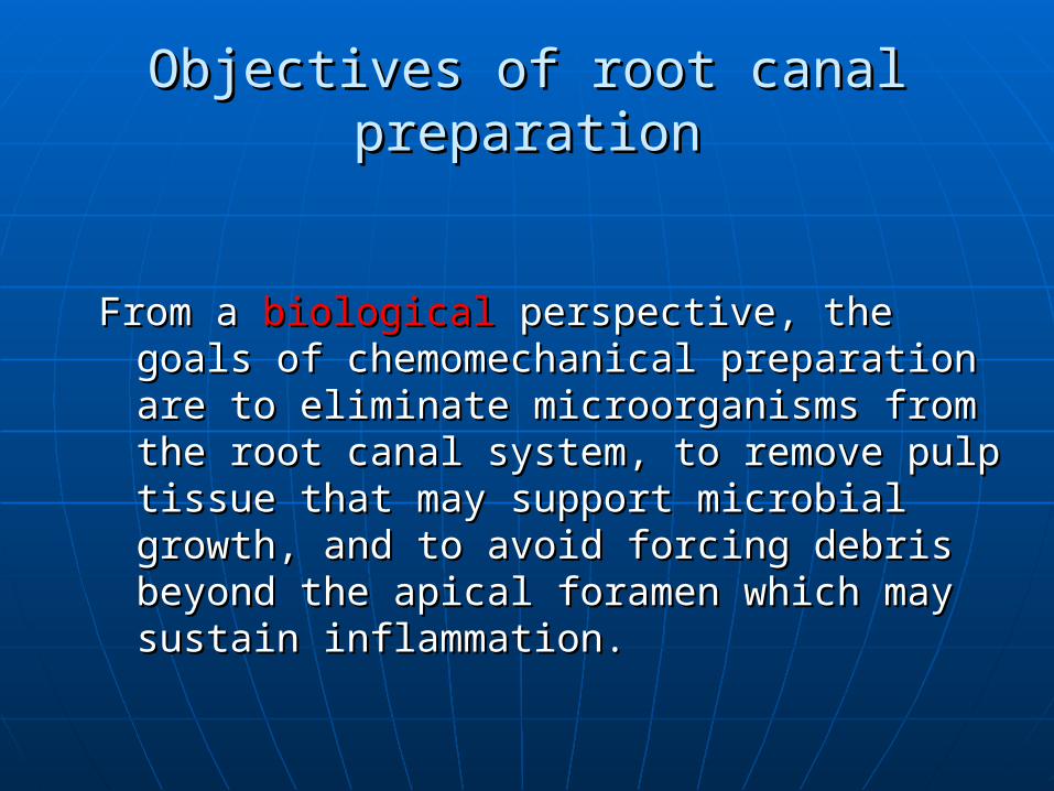
Objectives of root canal preparationObjectives of root canal preparation
From a From a biologicalbiological perspective, the goals of perspective, the goals of chemomechanical preparation are to chemomechanical preparation are to eliminate microorganisms from the root eliminate microorganisms from the root canal system, to remove pulp tissue that canal system, to remove pulp tissue that may support microbial growth, and to may support microbial growth, and to avoid forcing debris beyond the apical avoid forcing debris beyond the apical foramen which may sustain inflammation.foramen which may sustain inflammation.
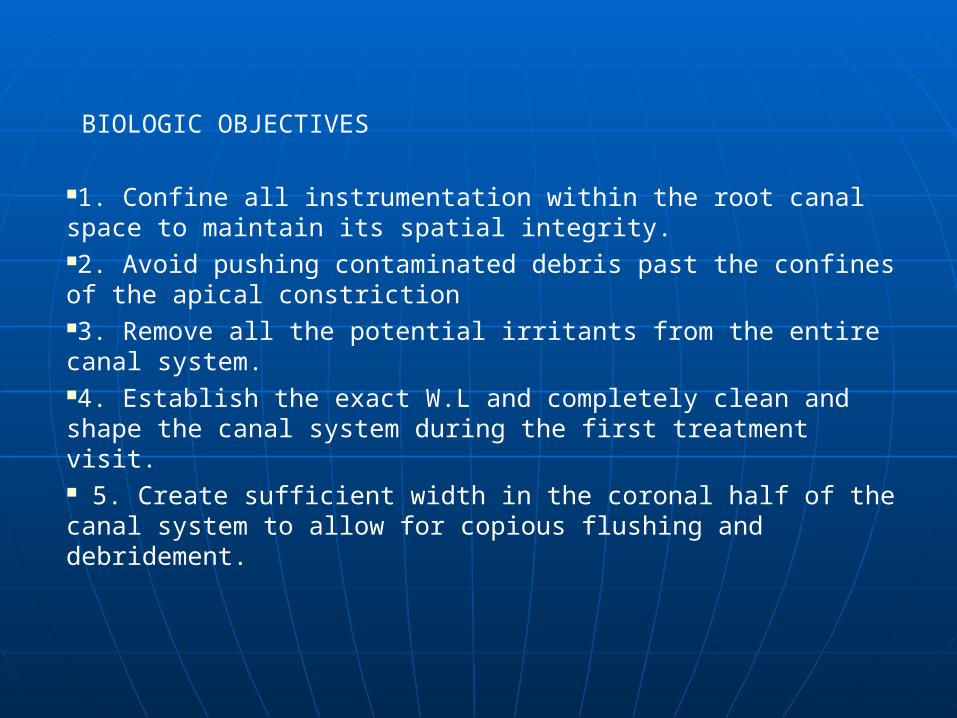
BIOLOGIC OBJECTIVES
1. Confine all instrumentation within the root canal space to maintain its spatial integrity. 2. Avoid pushing contaminated debris past the confines of the apical constriction3. Remove all the potential irritants from the entire canal system. 4. Establish the exact W.L and completely clean and shape the canal system during the first treatment visit. 5. Create sufficient width in the coronal half of the canal system to allow for copious flushing and debridement.
MECHANICAL OBJECTIVES
1. Develop a continuously tapering conical form in the root canal preparation. The final preparation of this system should be an exact replica of the original canal configuration in shape, taper.2. Prepare a sound apical dentine matrix at the DC junction. This provides the resistance form to the intraradicular cavity preparation. This also prevents the over- extension of instruments and controls the apical movement of gutta-percha sealer during obturation. 3. Prepare the canal to taper apically, with the narrowest cross- sectional diameter at the apical termination (apical dentin matrix). 4. Confine cleaning and shaping procedures to the canal system, thereby maintaining the spatial integrity of the apical foramen.
PrinciplesPrinciples
Note: the current concept of root canal preparation is not cleaning and shaping, but shaping and cleaning.
Principles of RCTPrinciples of RCT Principles of Endodontic cavity preparation Endodontic cavity
preparation may be separated into two anatomic divisions:
(a) Coronal preparation (b) Radicular preparation.
Principles of RCTPrinciples of RCT Black's principles of cavity preparation can be modified to include
the Root canal 'cavity' preparation
1. Coronal:
I. Outline FormII. Convenience FormIII. Removal of the remaining carious dentin (and defective
restorations) IV. Toilet of the cavity
Principles of RCTPrinciples of RCT Black's principles of cavity preparation can be modified to include
the Root canal 'cavity' preparation
2. Radicular:
I and II. Outline Form and Convenience Form IV. Toilet of the cavity V. Retention Form VI. Resistance Form
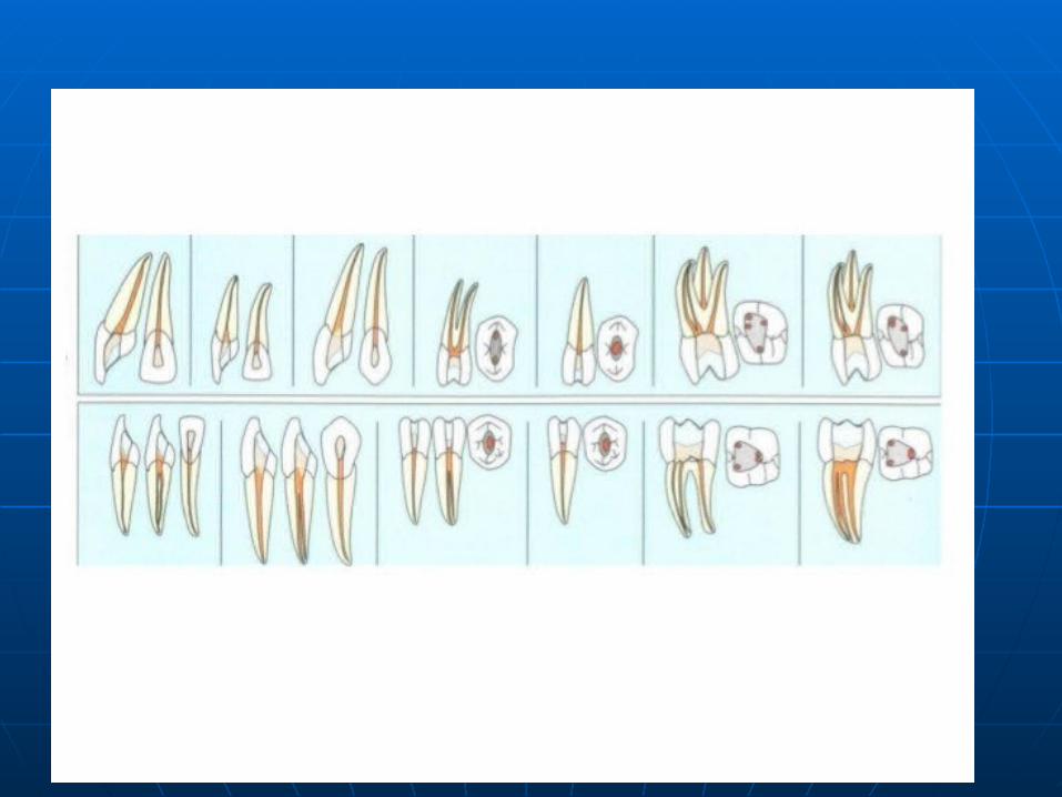
Outline FormOutline Form To achieve optimal preparation, three factors of anatomy must be considered:
(1)the size of the pulp chamber, (2)the shape of the pulp chamber, (3)the number of individual root canals,
their curvature, and their position internal
The finished outline form should accurately reflect the shape of the pulp chamber.
Convenience FormConvenience Form
Removing excess of coronal dentin, so as to allow passage of larger instruments, for better instrumentation, irrigation and obturation.
Four important benefits are gained through convenience form modifications: (1) unobstructed access to the canal orifice, (2) direct access to the apical foramen, (3) cavity expansion to accommodate filling techniques, and (4) complete authority over the enlarging instrument.
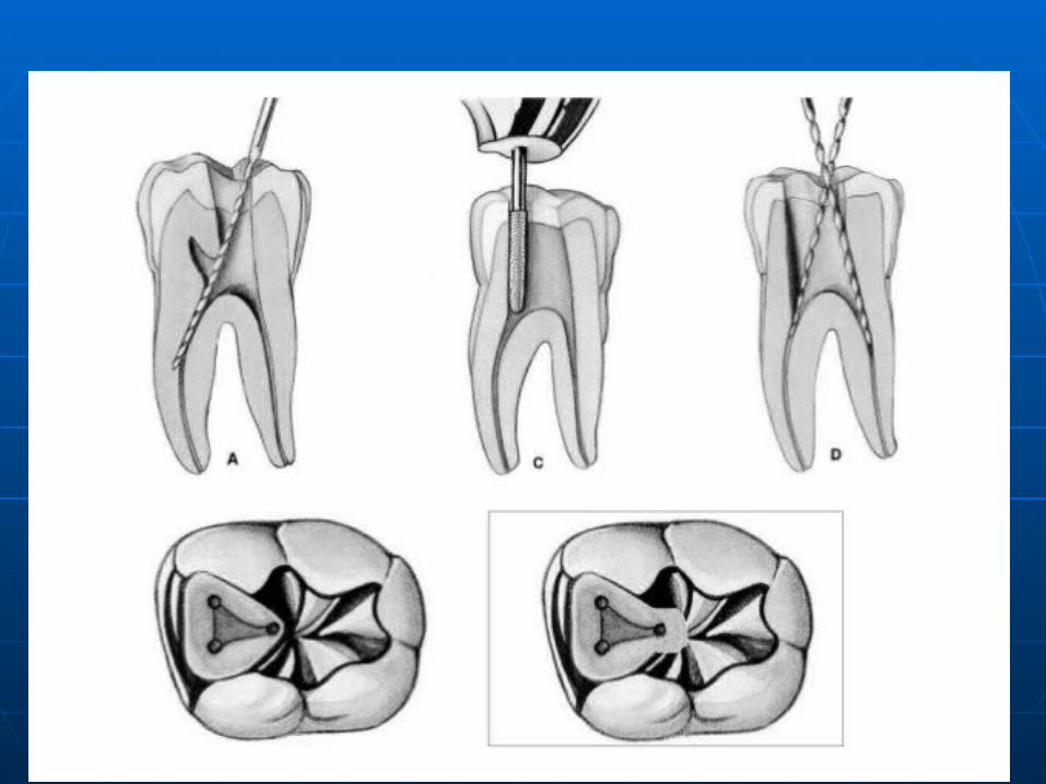
Removal of the remaining carious dentin (and defective restorations)
Removal of the Remaining Carious Dentin and Defective Restorations : Caries and defective restorations remaining in an endodontic cavity preparation must be removed for three reasons:
1. To eliminate mechanically as many bacteria as
possible from the interior of the tooth, 2. To eliminate the discolored tooth structure, that
may ultimately lead to staining of the crown, 3. To eliminate the possibility of any bacteria-laden
saliva leaking into the prepared cavity.
Toilet of the Cavity•All of the caries, debris, and necrotic material must be removed from the chamber before the radicular preparation is begun. •If the calcified or metallic debris is left in the chamber and carried into the canal, it may act as an obstruction during canal enlargement. • Soft debris carried from the chamber might increase the bacterial population in the canal
Retention form
Near parallel walls in the apical 2-3 mm ensure a snugly fitting G.P [ Apical TUG BACK ] .
Most crucial for preventing apical leakage.
Resistance form
Resistance to overfilling is provided by maintaining the integrity of the natural constriction of the apical preparation
Prevents over instrumentation Prevents forcing debris or obturating
material Provides a stop, against which G.P
can be compacted. .
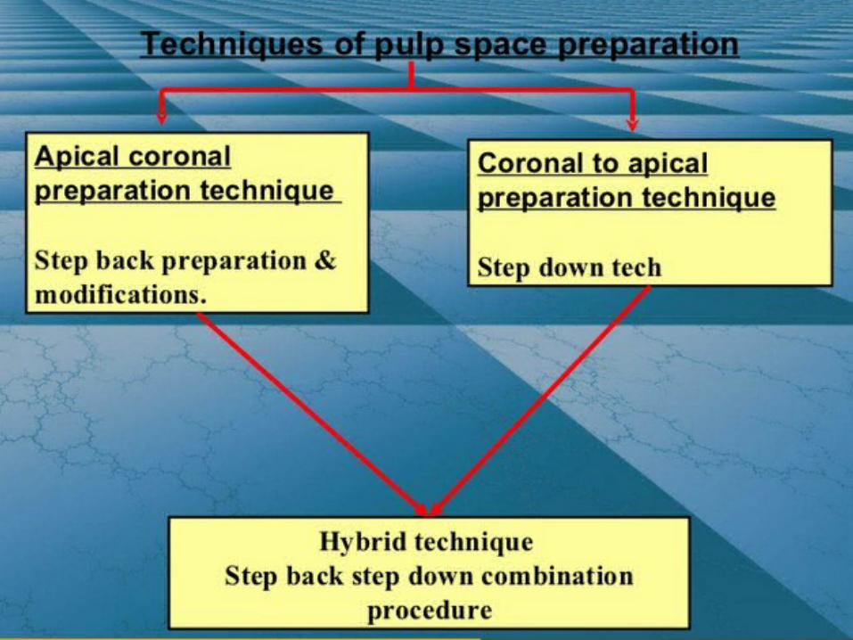
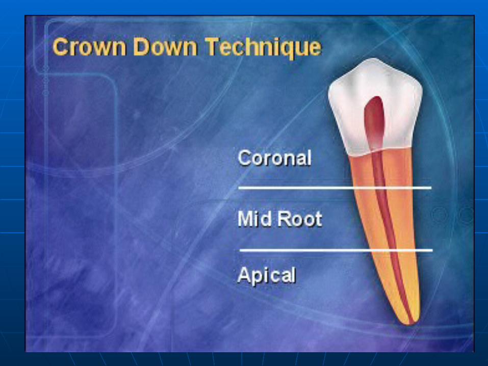
TYPES OF ROOT CANAL TYPES OF ROOT CANAL PREPARATIONSPREPARATIONS
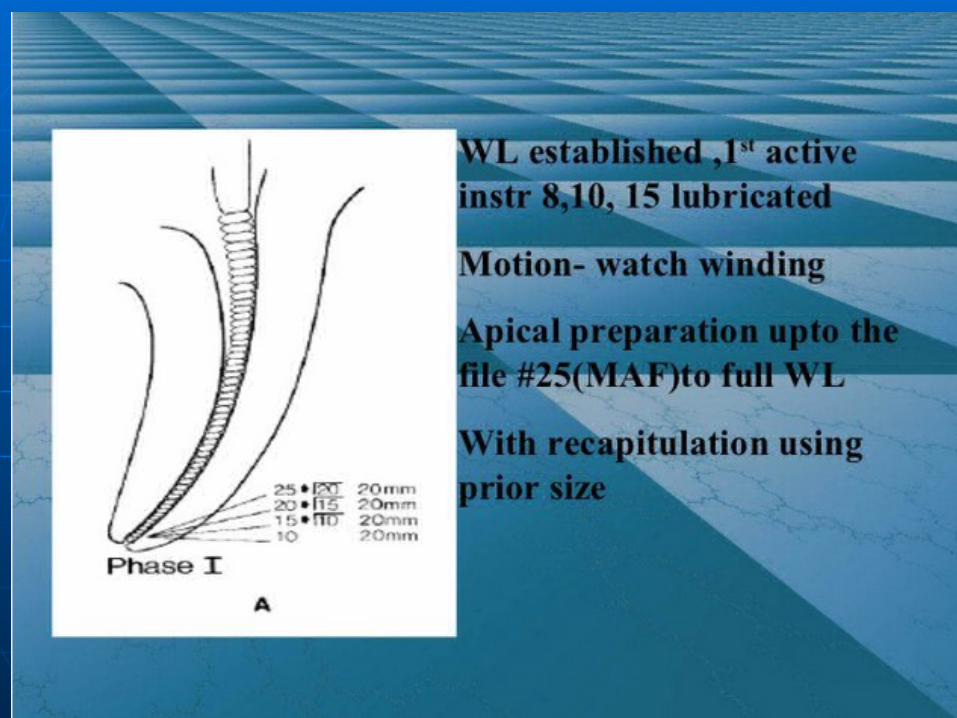
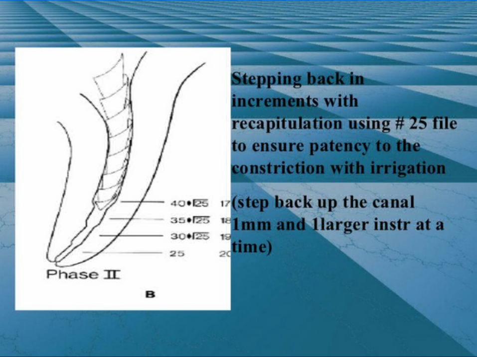
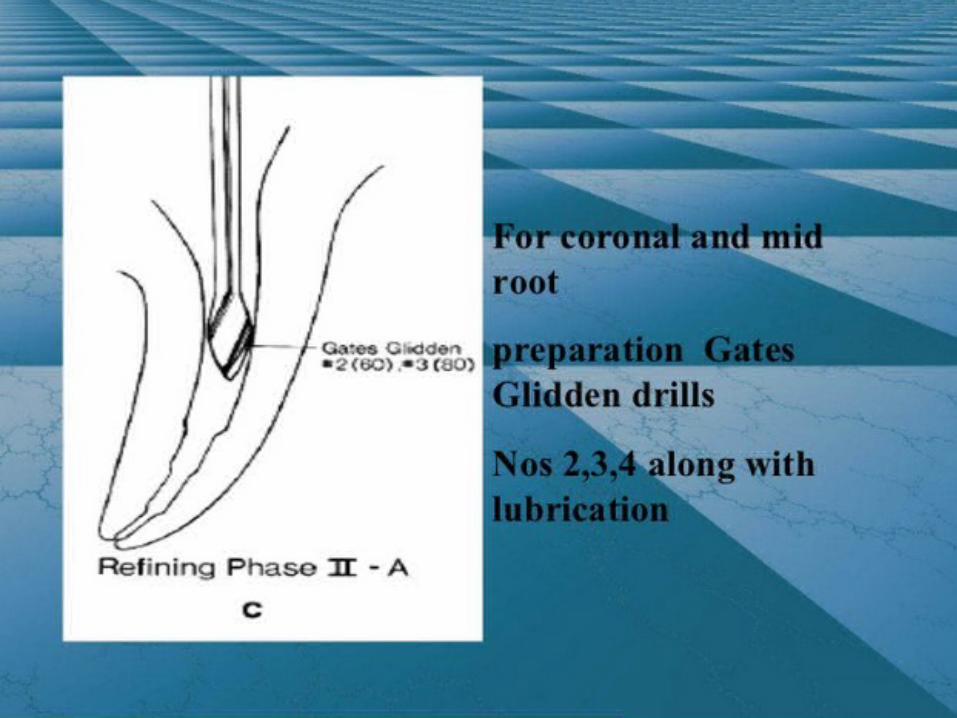
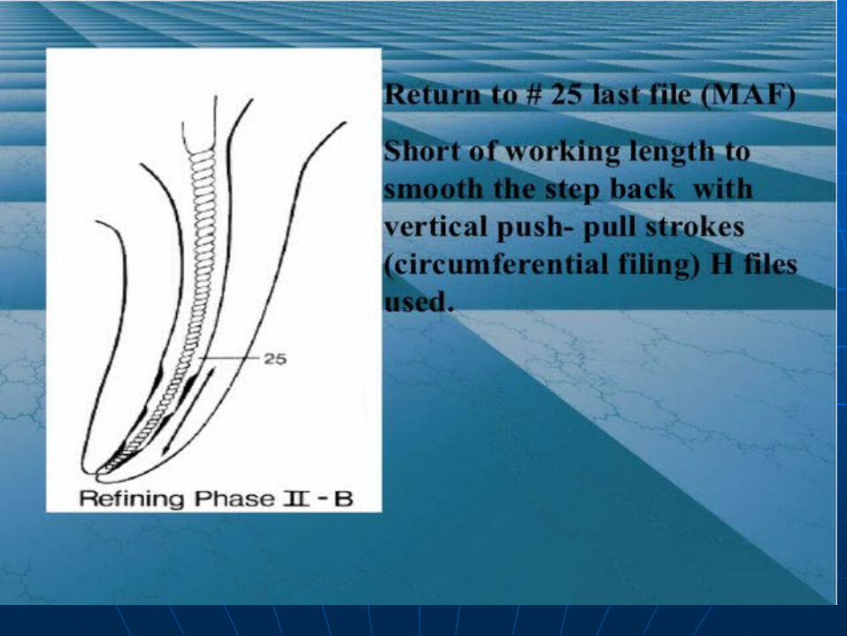
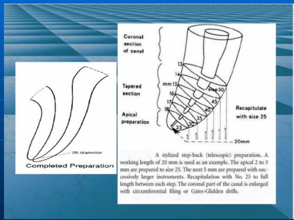
Crown downCrown down Step backStep back
hybridhybrid

Step backStep back

Filing techniquesFiling techniques
Watch windingWatch winding ReamingReaming FilingFiling Circumferential filingCircumferential filing AnticurvatureAnticurvature Balanced force techniqueBalanced force technique
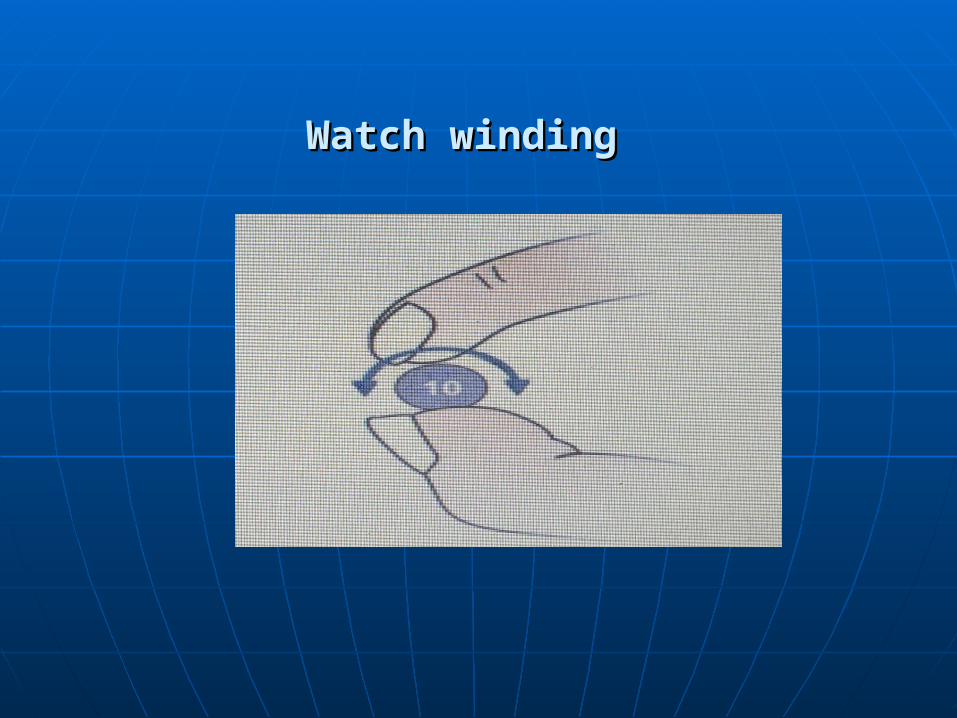
Watch windingWatch winding
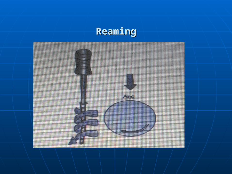
ReamingReaming
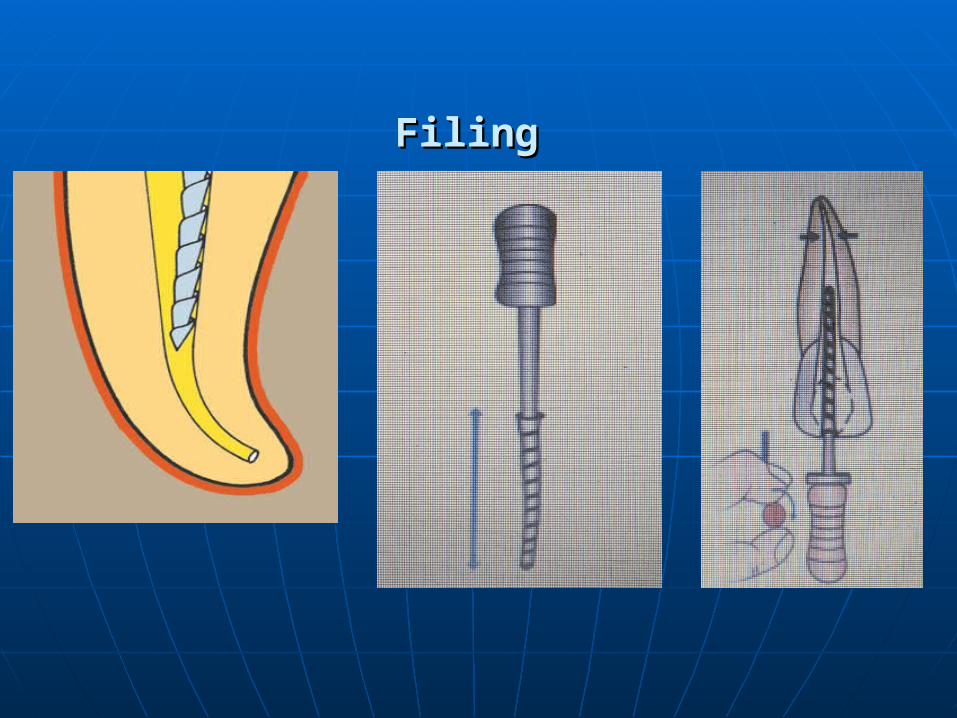
FilingFiling
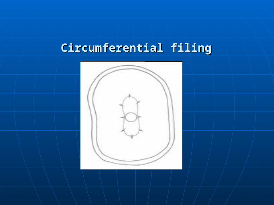
Circumferential filingCircumferential filing
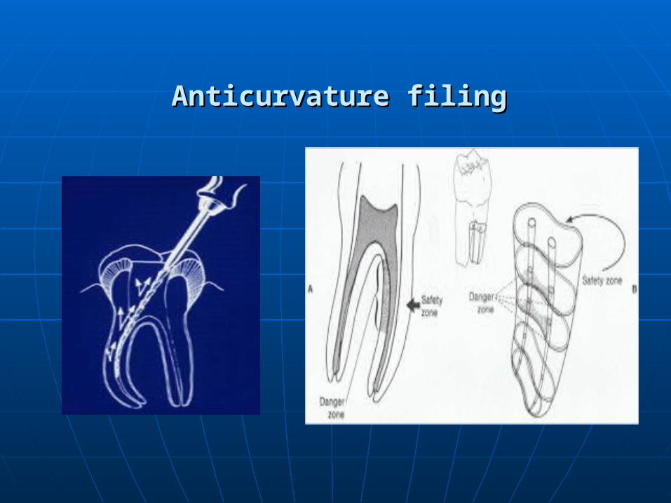
Anticurvature filingAnticurvature filing
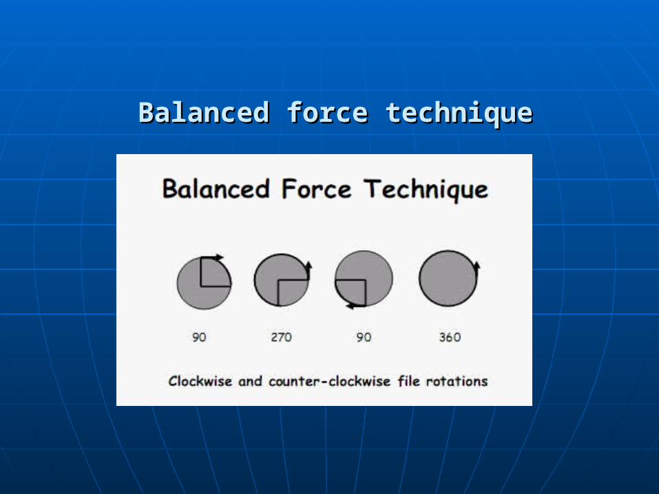
Balanced force techniqueBalanced force technique
IrrigationIrrigation
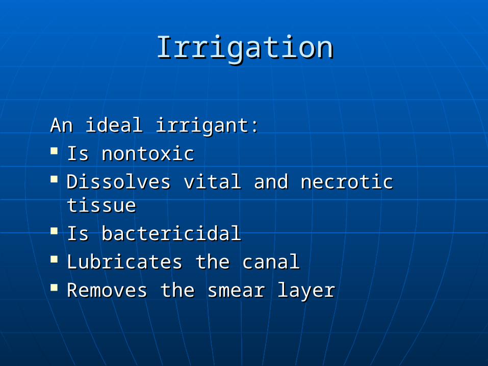
An ideal irrigant:An ideal irrigant: Is nontoxic Is nontoxic Dissolves vital and necrotic tissueDissolves vital and necrotic tissue Is bactericidalIs bactericidal Lubricates the canalLubricates the canal Removes the smear layerRemoves the smear layer
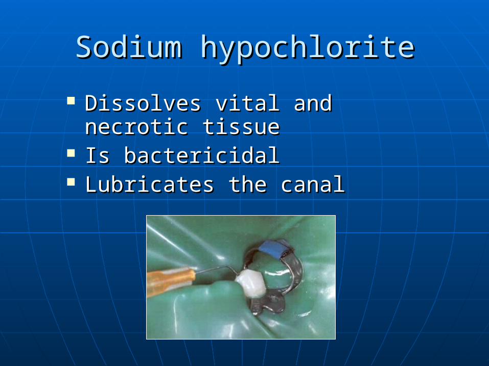
Sodium hypochloriteSodium hypochlorite
Dissolves vital and necrotic Dissolves vital and necrotic tissuetissue
Is bactericidalIs bactericidal Lubricates the canalLubricates the canal

Sodium HypochloriteSodium Hypochlorite
Cannot be considered non-
toxic!!!
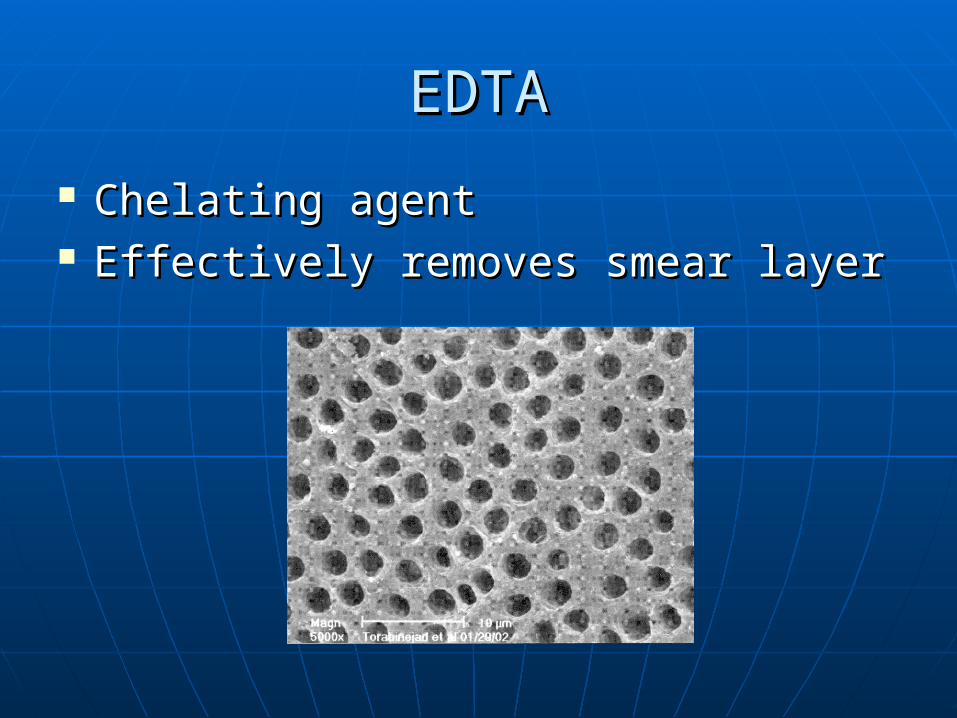
EDTAEDTA
Chelating agentChelating agent Effectively removes smear layerEffectively removes smear layer
InstrumentsInstruments
Instruments differ according to:Instruments differ according to: MetalMetal TaperTaper Tip designTip design Cross sectional geometryCross sectional geometry Length of cutting bladesLength of cutting blades Sizing Sizing
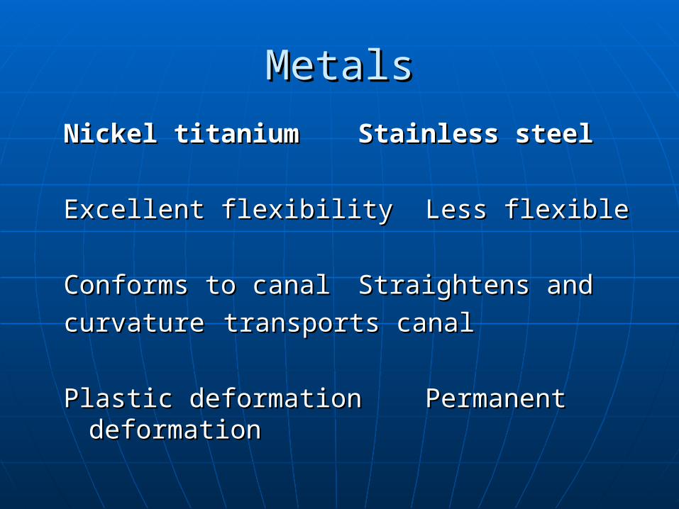
MetalsMetals
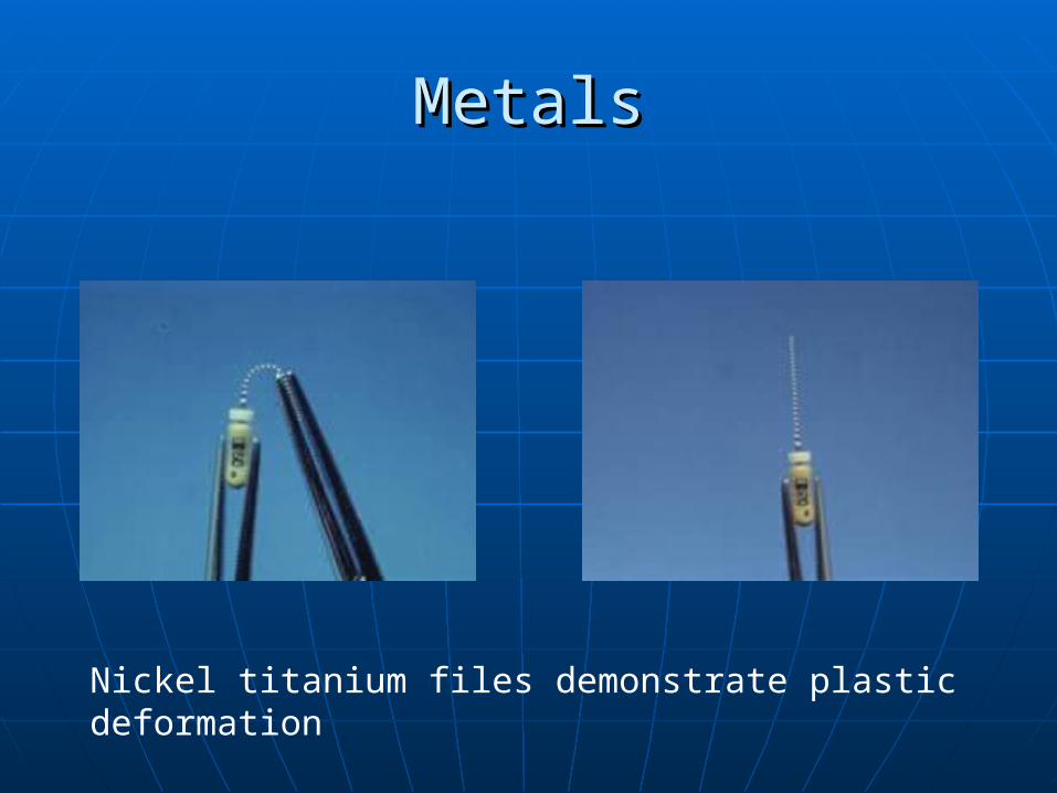
Nickel titaniumNickel titanium Stainless steelStainless steel
Excellent flexibilityExcellent flexibility Less flexibleLess flexible
Conforms to canal Conforms to canal Straightens and Straightens and
curvature curvature transports canaltransports canal
Plastic deformationPlastic deformation Permanent Permanent deformationdeformation

MetalsMetals
Stainless steel files demonstrate permanent deformation
MetalsMetals
Nickel titanium files demonstrate plastic deformation
TaperTaper
DefinitionDefinition
Increase in diameter per unit lengthIncrease in diameter per unit length
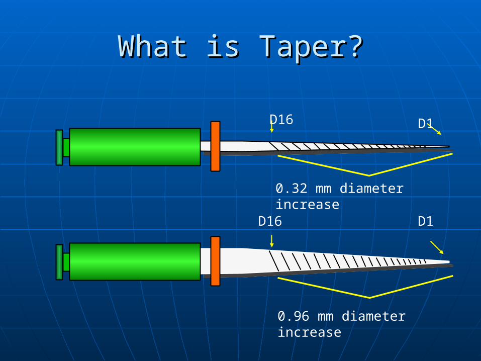
What is Taper?What is Taper?
0.32 mm diameter increase
D16 D1
0.96 mm diameter increase
D16 D1
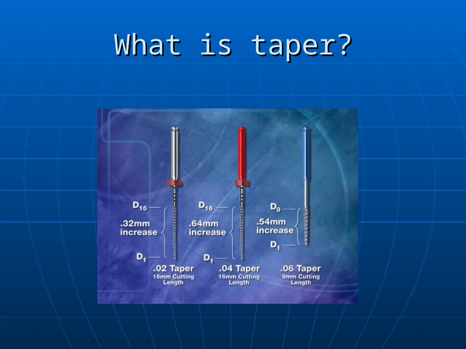
What is taper?What is taper?
TaperTaper
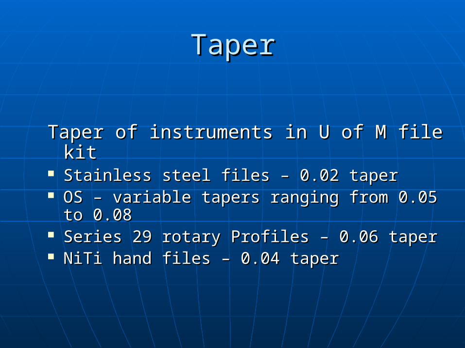
Taper of instruments in U of M file kitTaper of instruments in U of M file kit Stainless steel files – 0.02 taperStainless steel files – 0.02 taper OS – variable tapers ranging from 0.05 to OS – variable tapers ranging from 0.05 to
0.080.08 Series 29 rotary Profiles – 0.06 taperSeries 29 rotary Profiles – 0.06 taper NiTi hand files – 0.04 taperNiTi hand files – 0.04 taper
Tip DesignTip Design
Non-cutting tipNon-cutting tip Bullet nose (60 degree) tipBullet nose (60 degree) tip Smooth transition angle where tip Smooth transition angle where tip
meets flat radial landsmeets flat radial lands
Tip DesignTip Design
Designed to follow a pilot holeDesigned to follow a pilot hole Guides instrument through canal Guides instrument through canal
during preparationduring preparation
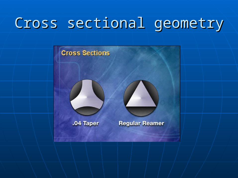
Cross sectional geometryCross sectional geometry

Cross sectional geometryCross sectional geometry
•• NotNotSelfSelf--ThreadingThreading
•• Gentle Planing Gentle Planing ActionAction
Cross sectional geometryCross sectional geometry
Radial lands separated by three u-Radial lands separated by three u-shaped flutesshaped flutes
Provide space for accumulation of Provide space for accumulation of debrisdebris
Moves debris out of canal Moves debris out of canal
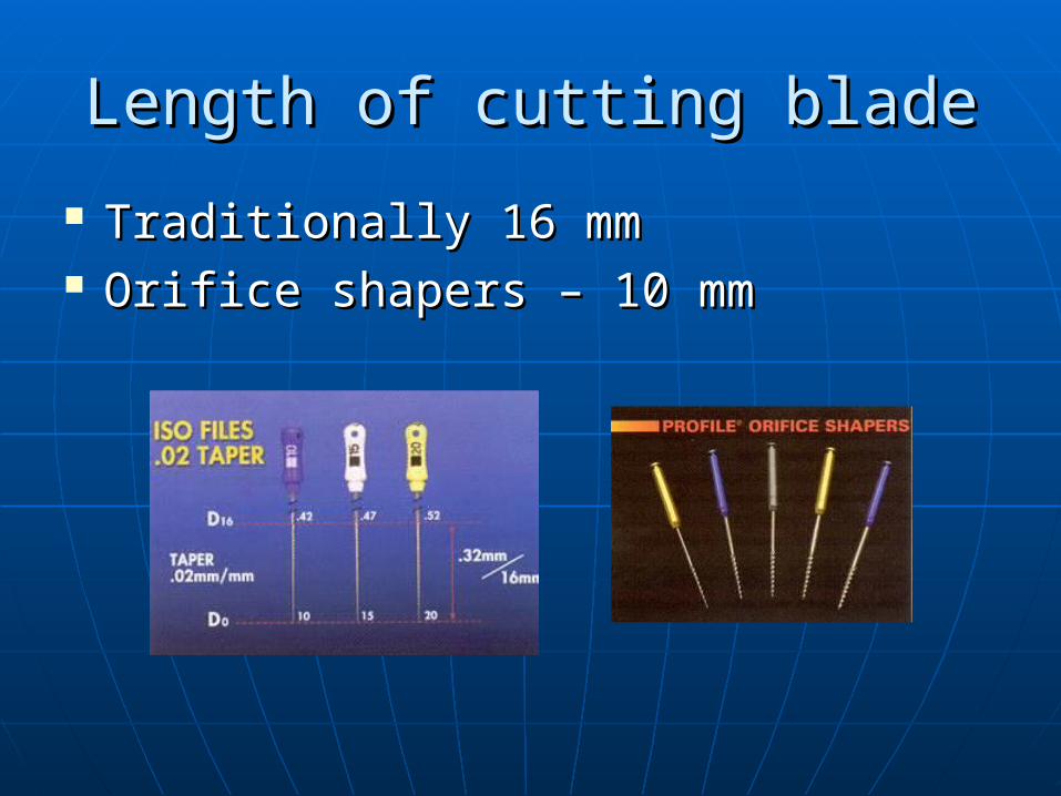
Length of cutting bladeLength of cutting blade
Traditionally 16 mmTraditionally 16 mm Orifice shapers – 10 mmOrifice shapers – 10 mm
Sizing of instrumentsSizing of instruments
ISO sizesISO sizes Number refers to tip diameter in Number refers to tip diameter in
tenths of mmtenths of mm The tip diameter increases by 0.05 The tip diameter increases by 0.05
mm from sizes 10 to 60, then by mm from sizes 10 to 60, then by 0.10 mm0.10 mm
Sizing of instrumentsSizing of instruments
% % increase in diameter from #10 increase in diameter from #10 to #15 file is 50%to #15 file is 50%
Difference between #55 and #60 Difference between #55 and #60 is only 9%is only 9%
Sizing of instrumentsSizing of instruments
Series 29Series 29 Progressive 29% increase in tip Progressive 29% increase in tip
diameter diameter Instruments are better spacedInstruments are better spaced More instruments in smaller sizes More instruments in smaller sizes
and fewer large instrumentsand fewer large instruments
REFERENCESREFERENCES
Endodontics, Volume 1 By John Ide Ingle, Leif K. Bakland COHEN’S PATHWAY OF PULPCOHEN’S PATHWAY OF PULP GOOGLEGOOGLE SlideshareSlideshare

TTHHAANNKK YYOOUU