evaluation of root canal preparation using rotary system ... · pdf fileevaluation of root...
TRANSCRIPT
Received: 2015.02.25Accepted: 2015.05.24
Published: 2015.06.20
Evaluation of Root Canal Preparation Using Rotary System and Hand Instruments Assessed by Micro-Computed Tomography
ABEF 1 Miranda Stavileci A 1 Veton Hoxha A 2 Ömer Görduysus D 3 Ilkan Tatar DE 4 Kjell Laperre D 4 Jeroen Hostens F 2 Selen Küçükkaya C 5 Edmond Muhaxheri
Corresponding Author: Miranda Stavileci, e-mail: [email protected] Source of support: Departmental sources
Background: Complete mechanical preparation of the root canal system is rarely achieved. Therefore, the purpose of this study was to evaluate and compare the root canal shaping efficacy of ProTaper rotary files and standard stain-less steel K-files using micro-computed tomography.
Material/Methods: Sixty extracted upper second premolars were selected and divided into 2 groups of 30 teeth each. Before preparation, all samples were scanned by micro-computed tomography. Thirty teeth were prepared with the ProTaper system and the other 30 with stainless steel files. After preparation, the untouched surface and root canal straightening were evaluated with micro-computed tomography. The percentage of untouched root ca-nal surface was calculated in the coronal, middle, and apical parts of the canal. We also calculated straighten-ing of the canal after root canal preparation. Results from the 2 groups were statistically compared using the Minitab statistical package.
Results: ProTaper rotary files left less untouched root canal surface compared with manual preparation in coronal, mid-dle, and apical sector (p<0.001). Similarly, there was a statistically significant difference in root canal straight-ening after preparation between the techniques (p<0.001).
Conclusions: Neither manual nor rotary techniques completely prepared the root canal, and both techniques caused slight straightening of the root canal.
MeSH Keywords: Root Canal Preparation • Stainless Steel • X-Ray Microtomography
Full-text PDF: http://www.basic.medscimonit.com/abstract/index/idArt/893950
Authors’ Contribution: Study Design A
Data Collection B Statistical Analysis CData Interpretation D
Manuscript Preparation E Literature Search FFunds Collection G
1 Department of Dental Pathology and Endodontics, University of Prishtina, Faculty of Medicine, Prishtina, Kosovo
2 Department of Endodontics, Hacettepe University, Faculty of Dentistry, Ankara, Turkey
3 Department of Anatomy, Hacettepe University, Faculty of Medicine, Ankara, Turkey
4 Bruker-microCT, Kontich, Belgium5 Lecturer of Statistics, American University in Kosovo (A.U.K.), Prishtina, Kosovo
2339 4 5 24
eISSN 2325-4416© Med Sci Monit Basic Res, 2015; 21: 123-130
DOI: 10.12659/MSMBR.893950
123
BIOTECHNOLOGY
This work is licensed under a Creative CommonsAttribution-NonCommercial-NoDerivs 3.0 Unported License
Indexed in: [Index Medicus/MEDLINE] [EMBASE/Excerpta Medica] [Chemical Abstracts/CAS] [Index Copernicus]

Background
Preparation of the root canal system includes both enlarge-ment and shaping of the complex endodontic space, together with its disinfection. However, complete mechanical prepara-tion of the root canal system is rarely achieved because of its variety and complexity [1,2]. Additionally, geometrical dissym-metry between the root canal and the preparation instrument may prevent the preparation instrument from acting efficiently on all canal walls. In simple, narrow, straight root canals with round cross-sections, most currently used rotary instruments will adequately clean and shape the canal, with favorable re-sults. However, in oval, flat, or curved root canals, rotary files often fail to adequately clean and shape the canal, leaving fins that may not have been touched [3,4].
Several types of endodontic instruments have been recommend-ed as being capable of achieving the primary objectives of root canal preparation [5,6]. The shaping of curved canals presents a problem for operators when stainless steel instruments are used, and all preparation techniques have a tendency to move the prepared canal from its original axis. Parameters affected by preparation, such as the angle of curvature determined by a line from the apical termination to the point of departure from a straight line drawn through the middle of the coronal part of the canal, can have a significant effect on endodon-tic treatment success. The use of rotary Ni-Ti files for root ca-nal preparation has significantly reduced the time required to prepare the root canal, with minimal deviations from the orig-inal canal path compared with manual instrumentation [5,7].
A variety of methodologies have been used to evaluate the shaping ability of endodontic instruments, including simulated root canal models, decalcification techniques, sectioning tech-niques, and radiographic comparison, but the limitations of these techniques have led researchers to look for new meth-ods that can produce more accurate results. Micro-computed tomography (micro CT) systems are now widely used in many academic fields. In recent years, micro CT has proved to be an efficient tool for evaluating the morphologic changes in the root canal shape before and after preparation [8]. Based on the fact that adequately cleaning and shaping of the root canal can optimize its disinfection and filling, the purpose of this study was to use micro CT to determine the percentage of untouched surface and the change in the angle of curva-ture after root canal preparation with stainless steel files and the ProTaper system in second upper premolars.
Material and Method
We used 60 intact maxillary second premolars that had been extracted for periodontal and orthodontic reasons. Prior to
extraction, patients were informed about and agreed to the scientific work to be performed. All teeth had fully developed roots and were stored in 10% formalin until use. Prior to the study, the teeth were washed with distilled water to remove residual formalin.
Each tooth was mounted in a sample holder before the micro CT scanning to allow reproducible orientation in the pre- and post-preparation scans. All teeth were scanned using either a SkyScan 1173 micro CT system (Bruker-microCT, Kontich, Belgium) with an isotropic voxel size of 22.86 µm at 70 kV/114 microA using a 1-mm aluminium filter or a SkyScan 1174 sys-tem (Bruker-microCT, Kontich, Belgium) with an isotropic vox-el size of 24 µm at 50 kV/800 microA. To allow scanning with-in the shortest possible period of time, we used 2 micro CT machines. Two-dimensional lateral projections of the samples were created over 360°, with a rotation step of 0.4°. A mod-ified Feldkamp algorithm (NRecon with a GPU recon server version 1.6.8.0, SkyScan, Kontich, Belgium) was used for re-construction of projection images, and 2-dimensional cross-sectional images were created. SkyScan Dataviewer software (Version 1.4.4, SkyScan, Kontich, Belgium) was used for dis-tance calculations.
The access cavities of all samples were prepared, and the root canals were localized and explored with size 15 K-files (Diadent, France) until their tips were visible to the apical fo-ramen. Working length was set at 1 mm from the apical fora-men. All samples were divided into 2 groups of 30 teeth each.
The root canals of the teeth in the first group were prepared with the ProTaper rotary system (Dentsply, Maillefer, Ballaigues, Switzerland) using a crown-down technique. In this technique, instrumentation was performed by using a Sx, S1, S2, F1, F2, and F3 sequence. Sx instrument was used to shape the coro-nal part of the canal, to continue with preparation of the mid-dle and apical sectors of the canal up to the working length with S1, S2 shaping files and F1, F2, and F3 finishing files. The X-Smart endodontic motor (Dentsply, Maillefer) was used at a rotation 250 rpm, introducing the instruments passively into the root canal. The root canals of teeth in the second group were prepared with stainless steel K-files (Diadent, France) us-ing a step-back technique. Apical enlargement was performed with an instrument size of up to No. 30 for both techniques. The root canals of both groups were irrigated with 2 ml of 3% sodium hypochlorite (Ultradent products, Inc., South Jordan, USA) after each file use. When the preparation was completed, each sample was inserted into the micro CT scanner and teeth were re-scanned (using the same parameters used for the ini-tial scan) for comparison with the pre-preparation images. All scans were recorded on a computer in bitmap image format.
124
Stavileci M. et al.: Effects on canal shaping
© Med Sci Monit Basic Res, 2015; 21: 123-130BIOTECHNOLOGY
This work is licensed under a Creative CommonsAttribution-NonCommercial-NoDerivs 3.0 Unported License
Indexed in: [Index Medicus/MEDLINE] [EMBASE/Excerpta Medica] [Chemical Abstracts/CAS] [Index Copernicus]
A B C
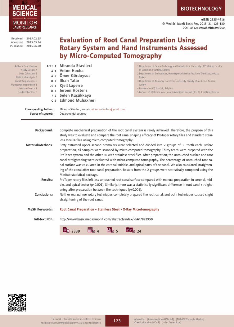
Figure 1. Three dimensional image of tooth with surface rendering (A) Before preparation; (B) After preparation; (C) Superimposition.
A
D
B
E
C
F
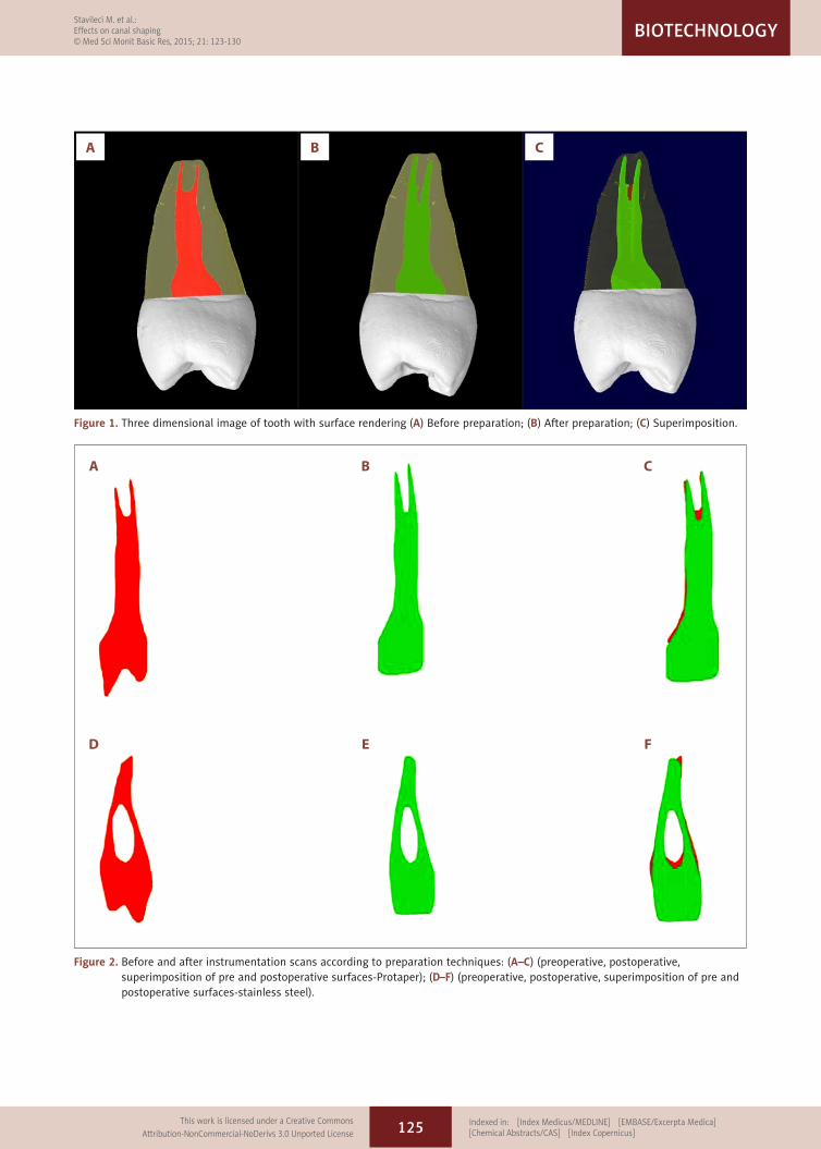
Figure 2. Before and after instrumentation scans according to preparation techniques: (A–C) (preoperative, postoperative, superimposition of pre and postoperative surfaces-Protaper); (D–F) (preoperative, postoperative, superimposition of pre and postoperative surfaces-stainless steel).
125
Stavileci M. et al.: Effects on canal shaping© Med Sci Monit Basic Res, 2015; 21: 123-130
BIOTECHNOLOGY
This work is licensed under a Creative CommonsAttribution-NonCommercial-NoDerivs 3.0 Unported License
Indexed in: [Index Medicus/MEDLINE] [EMBASE/Excerpta Medica] [Chemical Abstracts/CAS] [Index Copernicus]

CTVox software (version 2.4.0, Bruker-microCT, Kontich, Belgium) and CTVol software (version 2.2.3.0, Bruker-microCT, Kontich, Belgium) were used for 3-dimensional visualization and qualitative evaluation, respectively, of the pre- and post-instrumented canals (Figure 1A–1C). The color red was used to indicate the preoperative canal surface and the color green to indicate the postoperative canal surface (Figure 2A–2F).
Post-preparation changes in all canal parameters were calculat-ed by subtracting the scores for the treated canals from those recorded for the untreated canals. The AutoCAD 2012-CDW
program was used to analyze the 15 superimposed cross-sec-tional images for each sample (5 for each sector of the tooth: coronal, middle, and apical) and to evaluate the percentage of uninstrumented walls (Figures 3 and 4).
The percentage of uninstrumented walls was calculated by de-termining how much of the root canal perimeter was treated in cross-section. The same program was used to calculate dif-ferences in straightening of the canal between the 2 root canal preparation techniques (Figure 5). The angle of curvature was considered to be the angle formed by a line joining the apical
Figure 3. Untouched surface after preparation- root canal cross sectional images.
Figure 4. Before and after preparation scans – Auto Cad program evaluation of untouched surface.
126
Stavileci M. et al.: Effects on canal shaping
© Med Sci Monit Basic Res, 2015; 21: 123-130BIOTECHNOLOGY
This work is licensed under a Creative CommonsAttribution-NonCommercial-NoDerivs 3.0 Unported License
Indexed in: [Index Medicus/MEDLINE] [EMBASE/Excerpta Medica] [Chemical Abstracts/CAS] [Index Copernicus]
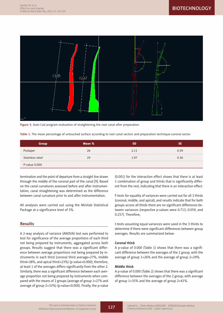
termination and the point of departure from a straight line drawn through the middle of the coronal part of the canal [9]. Based on the canal curvatures assessed before and after instrumen-tation, canal straightening was determined as the difference between canal curvature prior to and after instrumentation.
All analyses were carried out using the Minitab Statistical Package at a significance level of 5%.
Results
A 2-way analysis of variance (ANOVA) test was performed to test for significance of the average proportions of each third not being prepared by instruments, aggregated across both groups. Results suggest that there was a significant differ-ence between average proportions not being prepared by in-struments in each third (coronal third average=27%, middle third=38%, and apical third=23%) (p-value=0.000); therefore, at least 1 of the averages differs significantly from the other 2. Similarly, there was a significant difference between each aver-age proportion not being prepared by instruments when com-pared with the means of 2 groups (average of group 1=27% and average of group 2=32%) (p-value=0.000). Finally, the p-value
(0.001) for the interaction effect shows that there is at least 1 combination of group and thirds that is significantly differ-ent from the rest, indicating that there is an interaction effect.
F-tests for equality of variances were carried out for all 3 thirds (coronal, middle, and apical), and results indicate that for both groups across all thirds there are no significant differences be-tween variances (respective p-values were 0.713, 0.059, and 0.257). Therefore,
t-tests assuming equal variances were used in the 3 thirds to determine if there were significant differences between group averages. Results are summarized below:
Coronal third:A p-value of 0.000 (Table 1) shows that there was a signifi-cant difference between the averages of the 2 group, with the average of group 1=26% and the average of group 2=29%.
Middle third:A p-value of 0.000 (Table 2) shows that there was a significant difference between the averages of the 2 group, with average of group 1=35% and the average of group 2=41%.
Figure 5. Auto Cad program evaluation of straightening the root canal after preparation.
Group Mean % SD SE
Protaper 26 2.11 0.39
Stainless steel 29 1.97 0.36
P-value 0.000
Table 1. The mean percentage of untouched surface according to root canal sectors and preparation technique-coronal sector.
127
Stavileci M. et al.: Effects on canal shaping© Med Sci Monit Basic Res, 2015; 21: 123-130
BIOTECHNOLOGY
This work is licensed under a Creative CommonsAttribution-NonCommercial-NoDerivs 3.0 Unported License
Indexed in: [Index Medicus/MEDLINE] [EMBASE/Excerpta Medica] [Chemical Abstracts/CAS] [Index Copernicus]

Apical third:A p-value of 0.000 (Table 3) shows that there was a significant difference between the averages of the 2 groups, with average of group 1=20% and the average of group 2=25%.
This part of the analysis involves performing a 1-way ANOVA for equality of means (averages) between each third for individual groups. In the group in which the ProTaper system was used, the p-value of 0.000 shows that there was a significant difference between the averages of the 3 thirds, with coronal third=26%, middle third average=35%, and apical third average=20%.
In the group in which stainless steel files were used, the p-value of 0.000 shows that there was a significant difference between the thirds averages, with coronal third average 29%, middle third average 41%, and apical third average 25%.
The second evaluated parameter, the straightening of the ca-nal after root canal preparation, is presented in Table 4.
An F-test for equality of variances with a p-value of 0.656 indi-cates that these 2 samples had equal variances, thus a t-test assuming equal variances was performed. A p-value of 0.000 shows that there was a significant difference between the 2 group averages of angle of curving after the treatment, with the average of the 1st group=5o and the average of the 2nd group=11o.
Discussion
The simultaneous needs to enlarge the root canal and pre-serve its anatomy are a challenge for the practitioner. In this study, we analyzed the effect of canal instrumentation using the variables of untouched surface and the straightening of the curvature after root canal preparation. The method used in the current study – 3-dimensionally reconstructed images of the canal system – allowed us to evaluate morphological changes after root canal preparation.
The percentage of untouched canal surface is very important to characterize the completeness of root canal preparation; it should be as low as possible to enable good endodontic treat-ment. Superimposed micro CT reconstructions in 3 sectors of the root canal showed that the use of the ProTaper system resulted in less untouched surface than did the use of hand files. However, oval and long oval canals are common in most roots that contain 2 canals in the same root and represent a challenge in endodontics.
Numerous studies using extracted human teeth have discov-ered uninstrumented areas with remaining debris in all ar-eas of the canals, irrespective of the preparation technique. Cleanliness was found to decrease from the coronal to the api-cal part of the root canal [10,11]. Peters et al. [12] found that,
Group Mean % SD SE
Protaper 20 1.49 0.27
Stainless steel 25 1.84 0.34
P-value 0.000
Table 3. The mean percentage of untouched surface according to root canal sectors and preparation technique-apical sector.
Group Mean (°) Standard deviation (SD) Standard error (SE)
Protaper 5.00 1.34 0.24
Stainless steel 11.00 1.23 0.22
P-value 0.000
Table 4. The straightening of the canal after root canal preparation.
Group Mean % SD SE
Protaper 35 2.22 0.41
Stainless steel 41 1.55 0.28
P-value 0.000
Table 2. The mean percentage of untouched surface according to root canal sectors and preparation technique-middle sector.
128
Stavileci M. et al.: Effects on canal shaping
© Med Sci Monit Basic Res, 2015; 21: 123-130BIOTECHNOLOGY
This work is licensed under a Creative CommonsAttribution-NonCommercial-NoDerivs 3.0 Unported License
Indexed in: [Index Medicus/MEDLINE] [EMBASE/Excerpta Medica] [Chemical Abstracts/CAS] [Index Copernicus]

after preparation of the root canals of maxillary first molars with K-type hand files and 3 rotary Ni Ti files, 35% or more of the canals’ dentine surface was left untouched, with very little difference between all experimental groups. Paque et al. [13] reported that a variable portion of the surface area of oval-shaped root canals in mandibular molars was left unprepared regardless of the instrumentation technique used; they found less unprepared canal perimeter in oval canal shapes created with rotary instruments, ranging from 25% to 35%. Weiger et al. [14] showed that 44–68% of the canal surface was unpre-pared in long oval canals. Rotary instruments with a great-er than 4% taper have been shown to be more efficient than hand files in preparing root canals. However, even the Ni Ti instruments that were used in this study did not completely prepare oval root canal walls [15]. Tan and Messer [16] found that instrumentation with larger file sizes using rotary Ni Ti instruments resulted in significantly cleaner canals in the api-cal 3 mm than did hand instrumentation. Divergent results were obtained by Yin et al. [17], who reported that hand files removed more dentin, resulting in less unprepared surface compared with the ProTaper system, which produced 50.6% untouched surface. These contradictory results may be ex-plained by differences in methodology, preparation technique, and tooth selection.
The other parameter that was examined in this study was ca-nal straightening, which can be classified as a procedural er-ror. The present data suggest that the manual instrumen-tation technique resulted in more root canal straightening compared with the ProTaper system. The preoperative canal curvature affects the type and frequency of instrumentation errors [18,19]. Several studies have revealed that Ni Ti instru-ments maintain the original canal curvature better than stain-less steel files, especially in the apical region of the root ca-nal. Ni-Ti instruments produce significantly less straightening
and better-centered preparations, reducing the potential for iatrogenic errors [10,20,21]. Although Rahman et al. [22] and Capar et al. [23] concluded that instrumentation systems used in their studies tended to induce various degrees of dentinal damage during root canal preparation, Pagliosa et al. [24] re-ported that the ProTaper system can safely be used in instru-mentation of curved canals at full working length with satis-factory preservation of the original canal shape. This may be due to their flexibility and mechanical memory.
Micro CT provides images at a resolution of up to 22.86 µm, making it an excellent method for the evaluation of morpho-logical root canal changes after canal preparation. In our study, evaluation with this technique suggests that both manual and rotary endodontic instruments have limited ability to prepare and clean root canals, emphasizing the importance of anti-bacterial agent use for enhanced disinfection of the root ca-nal system.
Conclusions
On the basis of the results of this micro CT study it may be concluded that: (1) both manual preparation with stainless steel files and motorized preparation with ProTaper left un-prepared root canal surface, and (2) both techniques caused slight straightening of the root canal after preparation.
Acknowledgements
This research was completed in Hacettepe University, Faculty of Dentistry, Department of Endodontics and Hacettepe University, Faculty of Medicine, Department of Anatomy, Ankara, Turkey. The authors thank the Bruker-microCT Company for helping in data reconstruction.
References:
1. De-Deus G, Barino B, Zamolyi RQ et al: Suboptimal debridement quality produced by the single file F2 Protaper technique in oval shaped canals. J Endod, 2010; 36: 1897–900
2. Paque F, Barbakow F, Peters OA: Root canal preparation with Endo-Eze AET: changes in root canal shape assessed by micro CT. Int Endod J, 2005; 38: 456–64
3. Whitworth J: Methods of filling root canals: principles and practice. Endod Topics, 2005; 12: 2–24
4. De-Deus G, Gurgel-Filho ED, Magalhaes KM, Coutihno-Filho T: A laborato-ry analysis of gutta-percha filled area obtained using Thermafil, system B and lateral condensation. Int Endod J, 2006; 39: 378–83
5. Camara AC, Aguiar CM, Figueiredo JAP: Assessment of the deviation after biomechanical preparation of the coronal, middle and apical thirds of root canals instrumented with three Hero Rotary systems. J Endod, 2007; 33: 1460–63
6. Schafer E, Erler M, Dammaschke T: Comparative study on the shaping abil-ity and cleaning efficiency of rotary two instruments. Part 1. Shaping abil-ity in simulated curved canals. Int Endod J, 2006; 39: 196–202
7. Aguiar CM, Camara AC: Radiological evaluation of the morphological chang-es of root canals shaped with ProTaperTM and RaCeTMrotary instruments. Aust Endod J, 2008; 34: 115–19
8. Peters OA, Laib A, Ruegsegger P, Barbakow F: Three dimensional analysis of root canal geometry by high resolution computed tomography. J Dent Res, 2000; 79: 1405–9
9. Pruett JP, Clement DJ, Carnes DL: Cyclic fatigue testing of Nickel-Titanium endodontic instruments. J Endod, 1997; 23: 77–85
10. Schafer E, Lohmann D: Efficiency of rotary Ni-TiFlexMaster instruments compared with stainless steel hand K-flexofile-Part 2. Cleaning effective-ness and instrumentation results in severely curved root canals of extract-ed teeth. Int Endod J, 2002; 35: 514–21
11. Schafer E, Schlingemann R: Efficiency of rotary Ni-Ti K3 instruments com-pared with stainless steel hand K-Flexofile, Part 2. Cleaning effectiveness and shaping ability in severely curved root canals of extracted teeth. Int Endod J, 2003; 36: 208–17
12. Peters OA, Schonenberger K, Laib A: Effects of four Ni-Ti preparation tech-niques on root canal geometry assessed by micro CT. Int Endod J, 2001; 34: 221–30
129
Stavileci M. et al.: Effects on canal shaping© Med Sci Monit Basic Res, 2015; 21: 123-130
BIOTECHNOLOGY
This work is licensed under a Creative CommonsAttribution-NonCommercial-NoDerivs 3.0 Unported License
Indexed in: [Index Medicus/MEDLINE] [EMBASE/Excerpta Medica] [Chemical Abstracts/CAS] [Index Copernicus]

13. Paque F, Balmer M, AttinTh, Peters O: Preparation of oval shaped root ca-nals in mandibular molars using Ni-Ti rotary instruments: A micro CT study. J Endod, 2010; 36(4): 703–7
14. Weiger R, El Ayouti A, Lost C: Efficiency of hand and rotary instruments in shaping oval root canals. J Endod, 2002; 28: 580–83
15. El Ayouti A, Chu AL, Kimonis I et al: Efficassy of rotary instruments with greater taper in preparing oval root canals. Int Endod J, 2008; 41: 1088–92
16. Tan BT, Messer HH: The quality of apical canal preparation using hand and rotary instruments with specific criteria for enlargement based on initial apical file size. J Endod, 2002; 28: 658–64
17. Yin C, Cheung GS, Zhang C et al: Micro CT comparison of Ni-Ti rotary vs. traditional instrumentation in C shaped root canal system. J Endod, 2010; 36: 708–12
18. Nagy CD, Bartha K, Bernath M et al: The effect of root canal morphology on canal shape following instrumentation using different techniques. Int Endod J, 1997; 29: 113–17
19. Byrant ST, Dummer PM, Pitoni C et al: Shaping ability of.04 and.06 taper Profile rotary Ni-Ti instruments in simulated root canals. Int Endod J, 1999; 32: 155–64
20. Gluskin AH, Brown DC, Buchanan LS: A reconstructed computerized tomo-graphic comparison of Ni-Ti rotary GT files versus traditional instruments in canals shaped by novice operators. Int End J, 2001; 34: 476–84
21. Esposito PT, Cunningham CJ: A comparison of canal preparation with Ni-Ti and stainless steel instruments. J Endod, 1995; 21: 173–76
22. Rahman H, Chandra A, Singh S: In vitro evaluation of dentinal microcrack formation during root canal preparation by different Ni-Ti system. Indian Journal of Restorative Dentistry, 2014; 3: 43–47
23. Capar ID, Arslan H, Akcay M, Uysal B: Effects of ProTaper Universal, ProTaper Next and HyFlex instruments on crack formation in dentin. J Endod, 2014; 40: 1482–84
24. Pagliosa A, Sousa-Neto MD, Versiani MA et al: Computed tomography eval-uation of rotary systems on the root canal transportation and centering ability. Braz Oral Res, 2015; 29: 1–7
130
Stavileci M. et al.: Effects on canal shaping
© Med Sci Monit Basic Res, 2015; 21: 123-130BIOTECHNOLOGY
This work is licensed under a Creative CommonsAttribution-NonCommercial-NoDerivs 3.0 Unported License
Indexed in: [Index Medicus/MEDLINE] [EMBASE/Excerpta Medica] [Chemical Abstracts/CAS] [Index Copernicus]