preimplantation genetic screening (pgs) current ppt
TRANSCRIPT
8
Preimplantation Genetic Testing: Current Status and Future Prospects
Eduardo C. Lau1, Marleen M. Janson1, Carl B. Ball1,2, Mark R. Roesler3, Peter VanTuinen1,
David P. Bick1,4 and Estil Y. Strawn1,3
1 Medical College of Wisconsin, 2Alverno College,
3Froedtert Hospital, 4Children's Hospital of Wisconsin
USA
1. Introduction
While in vitro fertilization (IVF) is most often employed as a remedy for infertility, a discussion of the field would not be complete if it did not address the application of IVF to avoid genetic disorders. IVF makes it possible to assess the genetic status of the embryo before establishing a pregnancy when couples are at risk for an affected child. Physicians in the field will benefit from being informed about the diverse set of molecular and cytogenetic technologies employed in preimplantation genetic diagnosis (PGD) and screening (PGS), and from understanding their relative power and limitations as tools for genetic counseling.
PGD and pre-implantation genetic screening (PGS) refer to two distinct types of clinical procedure that help a couple to have a healthy child: PGD determines the embryo’s genotype, while PGS assesses an embryo’s karyotype and has been used in screening chromosomal aneuploidy.
PGD and PGS require the use of the IVF technique. These technologies, initiated in the late 1980s as an alternative to prenatal diagnosis (PND), allows a couple at risk of a genetic disorder to give birth to an unaffected child by avoiding selective termination of an affected pregnancy. Genetic disorders could be due to either a single gene disorder, or an abnormal number or structure of chromosomes. The current agreed upon indications (Cooper & Jungheim, 2010) for PGD and PGS include:
1. Screening for embryo chromosomal aneuploidy in cases of advanced maternal age or known parental translocation
2. Family history indicating risk for known autosomal Mendelian genetic disorders 3. Sex selection with family history indicating risk for X-linked disorders 4. Sex selection for family balancing, e.g. parental preference for a male or female 5. Human leukocyte antigen (HLA) matching to achieve a child to provide hematopoietic
progenitor cells from cord blood to an existing sibling who requires bone marrow transplantation
www.intechopen.com
In Vitro Fertilization – Innovative Clinical and Laboratory Aspects
138
The medical need for these services is significant. The U.S. Centers for Disease Control and Prevention estimates that more than 6,000 single-gene disorders affect approximately 1 in 300 live-births (Benson and Haith, 2009), while cytogenetic abnormalities appear at about twice this rate in live births and cause approximately ¼ of miscarriages and stillbirths (Thiesen & Shaffer, 2010). PGD makes it possible to assess the genetic status of at-risk embryos prior to implantation and initiation of a potentially affected pregnancy. There still remains considerable controversy regarding the need for and the ability of PGD to increase implantation rates for IVF.
1.1 The milestones of PGD and PGS development
PGD began in the late 1980's with the pioneering work by Handyside and colleagues in selecting embryo gender by polymerase chain reaction (PCR) amplification (Handyside et al., 1990), and diagnosis for a recessive autosomal disorder (Handyside et al., 1992).
PGS was subsequently developed by Munne’s team for gender determination (Munne et al., 1993), and by Handyside’s team for aneuploidy screening (Schrurs et al., 1993) using fluorescent in-situ hybridization (FISH). Other PGD milestones include its application for chromosomal translocations (Munne et al., 1998), and for HLA matching that led to the first HLA matched baby unaffected with Fanconi anemia (Verlinsky et al., 2001; Grewal et al., 2004).
Between the birth of the first PGD baby in 1989 and the year 2000, about 500 babies were born worldwide using PGD. Since then, the PGD has been on a steep rise: about 1000 PGD babies were born worldwide between 2000 and 2002, and 1500 more PGD babies were born between 2002 and 2004 (Verlinsky et al., 2004; ESHRE PGD Consortium, 2002; Harper et al., 2010). In the past 21 years, while thousands of babies have been born following PGD, there are no confirmed reports of increased fetal abnormalities following PGD.
1.2 Assisted reproductive technologies that support preimplanation genetic testing
Preimplementation genetic testing is supported by advances in assisted reproductive technology (ART). Methodologies of particular importance include intracytoplasmic sperm injection (ICSI), laser-assisted biopsies of embryos at the cleavage and blastocyst stages, sperm sorting, and cryopreservation of biopsied embryos.
1.2.1 Intracytoplasmic sperm injection
In conventional IVF each egg is combined with several thousand motile sperms on the day of egg retrieval to fertilize the egg. As a result, numerous sperms are present around the fertilized egg. At the next step, embryo biopsy, these excess sperms can contaminate the biopsy and contamination of the embryonic sample with sperm DNA can lead to incorrect PGD results. To guard against this risk a single sperm can be injected into each egg. This procedure is known as intracytoplasmic sperm injection (ICSI; Palermo et al., 1992; Harton et al., 2011a).
Through ICSI eggs are fertilized and embryos are formed. On day 3 of fertilization, a single blastomere is removed from each cleavage-stage embryo having 6-8 cells for PGD. When embryos develop into blastocysts on day 5 of fertilization, embryos selected by PGD are
www.intechopen.com
Preimplantation Genetic Testing: Current Status and Future Prospects
139
transferred to the mother. Alternatively, a few cells can be removed from the trophectoderm of a blastocyst for PGD on day 5, but the embryo will need to be transferred on day 6. Otherwise, the blastocysts need to be stored frozen and transferred at the next fertilization cycle.
1.2.2 Laser-assisted biopsy of embryos
PGD is accomplished by evaluating genetic material in polar bodies from unfertilized and fertilized oocytes (not performed here), blastomeres from cleavage-stage embryos, or trophectoderm cells from blastocysts. Depending on the developmental stage (oocyte, embryo or blastocyst), the zona pellucida can be breeched by one of three methods: mechanical zona drilling, acidified Tyrodes solution or laser.
At present, the cleavage stage is most widely used for embryo biopsy, and drilling a hole in the zona pellucida with a laser beam is the predominant method (Harton et al., 2011b). On day 3 of fertilization when normally developing embroys reach the 8-cell stage, one or two blastomeres are aspirated through the opening of a cleavage-stage embryo for PGD using a glass capillary pipette. This is also known as blastomere biopsy.
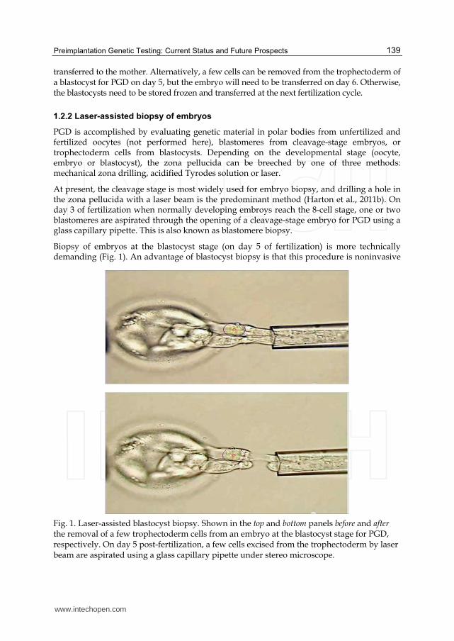
Biopsy of embryos at the blastocyst stage (on day 5 of fertilization) is more technically demanding (Fig. 1). An advantage of blastocyst biopsy is that this procedure is noninvasive
Fig. 1. Laser-assisted blastocyst biopsy. Shown in the top and bottom panels before and after the removal of a few trophectoderm cells from an embryo at the blastocyst stage for PGD, respectively. On day 5 post-fertilization, a few cells excised from the trophectoderm by laser beam are aspirated using a glass capillary pipette under stereo microscope.
www.intechopen.com
In Vitro Fertilization – Innovative Clinical and Laboratory Aspects
140
to the inner cell mass of the embryo because cells are removed only from the trophectoderm that will become the placenta. A further advantage is that the removal of multiple cells for PGD will significantly lower the allele drop-out (ADO) rate and increase the accuracy of testing. A drawback of blasocyst testing is that it allows less time for PGD, and thus the embryo may need to be frozen for transfer in the next fertilization cycle if test results are not obtained within a day.
1.2.3 Sperm sorting
After binding non-intercalating fluorescent dye to their DNA, X- and Y-bearing sperms are separated by flow-activated cell sorting based on the difference in total DNA content. The purity is greater than 70% for Y-bearing sperms, but greater than 90% for X-bearing sperms. Sperm sorting can be used for preconception sex selection used in family balancing, or used in combination with IVF-PGD to prevent the transmission of X-linked recessive diseases.
1.2.4 Cryopreservation of biopsied embryos at the blastocyst stage
Using the standard freeze-thaw method, the survival rate is low when cleavage-stage embryos (on day 3 of fertilization) are biopsied and cryopreserved. The survival rate can, however, be improved by vitrification after incubating the cleavage-stage embryos for 6-8 h following biopsy (Zheng et al., 2005), or by cryopreservation in CJ3 medium (Stachecki et al., 2005). Recently higher survival rates of biopsied cleavage-stage embryos have been obtained by vitrification at the blastocyst stage (Magli et al., 2006; Zhang et al., 2009).
2. Current technologies for preimplantation genetic testing
At the Medical College of Wisconsin and Froedtert Hospital (Milwaukee, Wisconsin, U.S.A.) we provide PGD and PDS services for medically indicated conditions (Bick & Lau, 2006; Swanson et al, 2007). PGS for chromosomal aneuploidies (monosomy or trisomy) and chromosomal translocations is performed by FISH (Swanson et al., 2007). We also provide PGD for HLA matching (Bick et al., 2008), diagnosis of specific single-gene disorders (Swanson et al., 2007; Lau et al., 2010), and gender selection in cases of X-linked diseases (E.C. Lau, K. Wang & M.M. Janson, unpublished).
For a family with a child who needs bone marrow transplantation, PGD for HLA matching (tissue typing) can facilitate birth of an HLA-matched donor infant (Bick et al., 2008; Verlinsky et al., 2001). Hematopoietic progenitor cells are then transplanted from cord blood or bone marrow of the PGD infant to save the life of the affected sibling. In addition, we have developed PGD assays for a few common and/or severe childhood genetic diseases, such as spinal muscular atrophy, sickle cell anemia, autosomal recessive polycystic kidney disease (ARPKD), and cystic fibrosis.
2.1 PGD by multiplex PCR
Single-cell multiplex PCR has been used in the PGD assays for HLA matching, spinal muscular atrophy (SMA) and sickle cell anemia. In PGD for these disorders we perform multiplex PCR of single blastomeres followed by haplotyping analysis of embryos with several linked short tandem repeat (STR) markers (Bick et al., 2008). Multiple markers are used due to the high incidence of allele-drop-out for single-cell PCR.
www.intechopen.com
Preimplantation Genetic Testing: Current Status and Future Prospects
141
In PGD for SMA, we perform multiplex nested PCR of single blastomeres followed by restriction cleavage analysis (Swanson et al., 2007). This technique detects the restriction fragment length polymorphisms (RFLPs) of the SMN1 and SMN2 genes at exons 7 and 8. We use Hinf I for cleaving exon 7 (Daniels et al., 2001), and Dde I for cleaving exon 8 of the SMN genes (Malcov et al., 2004). We use both fast PCR reaction mixture and rapid restriction enzymes to shorten the turn-around-time of PGD for SMA (E.C. Lau, unpublished). SMA, an autosomal recessive motor neuron disorder, is the most common fatal genetic disorder in childhood. It affects 1 in 6,000 to 1 in 10,000 live births, and has carrier frequency of 1 in 40 to 1 in 60 in the population. Deletion of both copies of SMN1 gene accounts for 95% of SMA cases. SMN2 gene is highly homologous to SMN1, but cannot substitute for SMN1 gene functions (Prior & Russman, 2011).
In PGD for sickle cell anemia, we perform multiplex PCR of single blastomeres followed by detection of β-globin (HBB) gene mutations by mini-sequencing, which is also known as single-base primer extension (Kobayashi et al., 1995; Heinrich et al., 2009). Sickle cell anemia is an autosomal recessive disorder caused by single-base mutation in codon 6 of HBB gene, that substitutes a thymine for adenine. A conformational change in the hemoglobin S (Hb S) molecule reduces its ability to carry oxygen. Other types of sickle cell diseases result from co-inheritance of HbS with abnormal globin β-chain variants, such as sickle hemoglobin C disease (Hb SC), and sickle β-thalassemia. In PGD for sickle cell diseases, linkage analysis of HBB haplotypes is performed with linked STRs located within the HBB gene and its flanking regions (E.C. Lau, A.F. Licht & M.M. Janson, unpublished).
The major drawback of PGD by multiplex PCR is the interaction of PCR primers, which results in a long and tedious optimization process to work out a robust mixture for each patient family (see Section 3.1).
2.2 PGD by whole-genome amplification
A major challenge of PGD is to amplify DNA from single blastomeres and perform genotyping analysis within the time constraints of the IVF cycle. In order to meet the turn-around-time of 30 hours or less for PGD from receiving the biopsied cells to clinical report, we have developed a fast and reliable protocol for whole-genome amplification (WGA) from single cells using multiple displacement amplification (MDA; Lau et al., 2010). Amplification is necessary since a single cell does not contain enough DNA to fulfill these assays.
2.2.1 Techniques for single-cell whole-genome amplification
The earliest method for whole-genome amplification (WGA) from single cells was PCR-based primer-extension preamplification (PEP) of single sperms (Zhang et al. 1992), that was adapted for the amplification of single blastomeres in PGD (Sermon et al., 1996). Like other PCR-based methods for WGA, the drawbacks of PEP method were incomplete genome coverage, and amplification bias (103 to 106 folds) between genomic loci in amplified products.
More recent technologies for WGA from single cells are multiple displacement amplification (MDA; Handyside et al., 2004; Lau et al., 2010; Dean et al., 2002) and PicoPlex® library methods. MDA is a non-PCR and isothermal method for DNA amplification. Handyside and colleagues used a commercial MDA kit to amplify single blastomeres for PGD (Handyside et
www.intechopen.com
In Vitro Fertilization – Innovative Clinical and Laboratory Aspects
142
al., 2004; Hellani et al., 2004), but the reaction time was 16 hours, and the ADO rates for genotyping MDA products were as high as 34%. While commercial kits for MDA are optimized for greater than 10 ng input genomic DNA, the use of MDA for WGA of single cells is not a standard application supported by the kit manufacturers (Coskun & Alsmadi, 2007). We modified the reaction of a commercial rapid-MDA kit for single cells, and obtained genotyping results of less than 10% ADO after 4-h MDA reaction (Lau et al., 2010).
An alternative method for WGA from single cells is the PicoPlex® kit, which is adapted from the OmniPlex® technique for genomic DNA (Langmore 2002). The PicoPlex® library kit for WGA is a PCR-based technique that requires the fragmentation of large genomic DNA prior to the construction of a PCR-amplifiable library. Both MDA and PicoPlex® methods for single-cell WGA have been used in array-based preimplantation testing (Hellani et al., 2008; Johnson et al., 2010; see Section 3.1).
2.2.2 Linkage approach for embryo analysis
Besides direct mutation detection, we use a linkage approach for embryo and familial analysis in PGD. For PGD of single-gene disorders, genotyping analysis is performed for embryos using STR markers linked with the disease genes, followed by haplotyping analysis.
“Preimplantation genetic haplotyping” (PGH) is a test procedure for which the first round is whole genome amplification (WGA) of single blastomeres, and the second round is genotyping using whole-genome amplified products as template. A comparison of sizes and genotypes of STRs among family members allows inferences of the haplotypes for parents and an affected child. If the STR loci are closely linked to and flank the disease locus, unaffected embryos can be identified by comparing their haplotypes to those of the parents and affected sibling. The advantage of using single-cell WGA in the first round of PGD is that a single common protocol is used to make many copies of the entire genome for subsequent analysis.
We have successfully applied PGH to PGD for single-gene recessive disorders, such as autosomal recessive polycystic kidney disease (ARPKD; Lau et al. 2010) and cystic fibrosis (E.C. Lau, M.M. Janson & T. Boyle, unpublished). ARPKD is caused by mutations in the PKHD1 gene, which is located at chromosome 6p12.2 and codes for fibrocystin protein. Although 2-5% of all cases of polycystic kidney diseases (PKD) are ARPKD, more than 75% of all PKD cases that present clinically in the first month of life are ARPKD, with a mortality rate of about 25% in the first month of life (Sweeney & Avner, 2006; Dell & Avner, 2008). ARPKD affects approximately 1 in 20,000 live-births, with a carrier frequency of 1 in 70 individuals. The first PGD case for a family at risk for autosomal recessive polycystic kidney disease (ARPKD) was successful using PGH (Lau et al., 2010). A PGD child, whose sibling died at birth from the disease and whose parents were both carriers of the gene, was born healthy with normal kidneys.
We have also applied the PGH approach to PGD for cystic fibrosis (CF). CF is the most common lethal autosomal recessive disorder among Caucasians of northern European ancestry, and is a common genetic cause of infant mortality. The carrier frequency among Caucasians in the United States is approximately 1 in 25, but is lower in other racial groups. Over 1,000 different mutations in the CFTR gene for CF have been described, but fewer than 25 occur with appreciable frequency (Amos et al. 2006).
www.intechopen.com

Preimplantation Genetic Testing: Current Status and Future Prospects
143
PGD for single-gene disorders by PGH is a general approach based on linkage analysis. It does not require prior knowledge of the exact nucleotide sites of the mutations within a gene. PGH saves the time required to develop custom PGH assays for individual alleles, and thus is particularly useful for PGD of single-gene disorders involving many rare or private mutations. However, the haplotyping approach for PGD also has limitations. The method would be unable to determine the parental haplotypes when the affected child is deceased and a DNA specimen is unavailable. In addition, PGH will not infer the correct parental haplotypes if the affected inherited a chromosome that had undergone recombination at the disease locus (Altarescu et al., 2008).
2.3 PGD for gender selection
The application of PGD for gender selection offers an alternative to single-gene assays for individual X-linked diseases. Instead of developing custom PGD assays for individual X-linked diseases, it is more practical to provide a single PGD assay of gender identification for families at risk of X-linked diseases. To prevent the transmission of X-linked Mendelian recessive diseases, PGD is used to select female embryos offspring. Because two copies of the mutant X allele are required for the diseases to occur in females, daughters of unaffected fathers will at worst be carriers for the trait. By contast, in males only one copy of the mutant X allele is required for the disease to occur and so the male child of a carrier mother has a 50% risk of having the disease.
2.3.1 Applications of PGD for gender selection
The main medical use of PGD for gender selection is to prevent the transmission of X-linked Mendelian recessive disorders by giving birth to female offspring. Many X-linked Mendelian recessive disorders, such as Duchenne muscular dystrophy (DMD), and hemophilia A and B, are rarely seen in females because the child is unlikely to inherit two copies of the recessive allele. This may be because the condition is rare, or because affected males are reproductively disadvantaged.
PGD for gender selection may also be used for non-Mendelian disorders that are significantly more prevalent in one sex. For the prevention of these inherited disorders, the gender of offspring is selected based on the seriousness of inherited condition, the risk ratio in either sex, and the options for disease treatment (Amor & Cameron, 2008).
“Non-medical” applications of PGD for family balancing (also known as “social sexing” or “social sex selection”) are more controversial. There is no broad cultural preference for male or female offspring in the U.S., but there is a preference of males in some countries, such as China, India and the Middle East. This non-medical use of PGD for gender selection and family balancing is prohibited in many countries such as China, because it could disrupt the sex ratio of the population.
2.3.2 Method of PGD for gender selection
We have performed PGD for gender selection by WGA of single blastomeres by MDA, followed by multiplex PCR for detecting X- and Y-linked genetic loci (E.C. Lau, M.M. Janson & K. Wang, unpublished). The Medical College of Wisconsin and Froedtert Hospital do not
www.intechopen.com
In Vitro Fertilization – Innovative Clinical and Laboratory Aspects
144
support non-medical use of PGD. We provide PGD for gender selection to help families with X-linked disorders give birth to unaffected children.
2.3.3 Sperm separation in combination with PGD for selecting female embryos
Sperm sorting techniques prior to IVF can be combined with post-fertilization molecular diagnosis of the resulting embryos. To prevent the birth of affected children, female embryos can be selected by PGD after forming embryos through ICSI with X-bearing sperm. For families at risk for rare X-linked recessive disorders, such as Wiskott-Aldrich syndrome (WAS) and monocarboxylate transporter 8-specific thyroid hormone cell transporter (THCT) deficiency, sperm sorting can be combined with PGD for selecting female embryos instead of developing custom PGD assays for these disorders.
PGD for gender selection can also be used to increase the odds of conceiving an HLA-matched, unaffected sibling donor. For a family with an X-linked recessive disorder that has an affected child in need of a hematopoietic progenitor cell transplant from cord blood or bone marrow, the combined use of sperm sorting and PGD for gender selection increases the chance of finding an HLA-matched and unaffected sibling donor to approximately 1 in 5. Otherwise, the chance of finding an HLA-matched female embryo by PGD is only approximately 1 in 10.
2.4 PGS/PGD for cytogenetic anomalies
Preimplantation screening for cytogenetic anomalies, in particular aneuploidies and translocations, has been performed using either FISH probes or array-comparative genomic hybridization. We shall discuss the challenges and limitations of both methods.
2.4.1 Aneuploidy for whole chromosomes
The adoption of FISH in a preimplantation context was a natural adaptation of FISH techniques as used in prenatal diagnosis (PND), where its value in assessing aneuploidy is unassailable. In contrast to prenatal specimens, only single, or at most two cells, are available for preimplantation genetic testing (PGT), and thus single FISH probe cocktails must employ at least 5 fluorochromes to cover the most prevalent liveborn aneuploidies, namely, 13, 18, 21, the X and Y. The availability of commercially produced cocktails has alleviated the need to “home brew” probes, but has necessitated the acquisition of more comprehensive fluorescent filter sets than those used for most routine two- or three-color FISH.
Following biopsy of an embryo the single blastomeres are placed in a hypotonic solution, and then treated with acetic acid methanol to fix and spread the cells. All these are performed by an embryologist. Ideally this will result in a uniformly flat nucleus with little or no overlying cytoplasm. Larger swelled nuclei which have not ruptured and have therefore preserved the nuclear contents are desired in order to separate the hybridization signals, especially for the large centromere targets, 18, X and Y. Subsequent steps are performed in the cytogenetics laboratory. From this point forward a major departure from PND, where typically 50 nuclei are assessed, is the reliance on a single nucleus for aneuploidy diagnosis. Thus, there is a need for consistency in the fixation and spreading,
www.intechopen.com
Preimplantation Genetic Testing: Current Status and Future Prospects
145
and freedom from artifacts such as spurious extra signals or weak signals, which are inconsequential in multi-cell analyses.
The partial coverage of a 5-probe set can be partly overcome by sequential hybridizations to the same nucleus. The quality and consistency of a second or subsequent hybridization are , however, generally progressively inferior due to the damaging hot treatments at 73 oC, which are necessary for denaturation and stringent washing. Preliminary indications of commercial development of a 6-step reduced-temperature sequential hybridization strategy to cover all 24 chromosomes have not to our knowledge been realized, and routine widespread use of such a strategy may not be technically realistic.
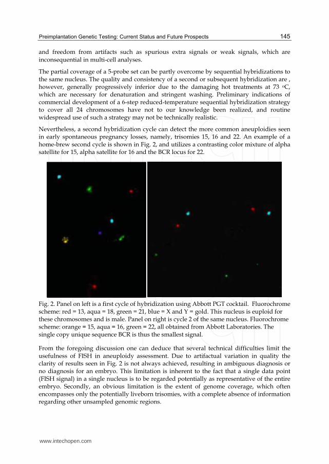
Nevertheless, a second hybridization cycle can detect the more common aneuploidies seen in early spontaneous pregnancy losses, namely, trisomies 15, 16 and 22. An example of a home-brew second cycle is shown in Fig. 2, and utilizes a contrasting color mixture of alpha satellite for 15, alpha satellite for 16 and the BCR locus for 22.
Fig. 2. Panel on left is a first cycle of hybridization using Abbott PGT cocktail. Fluorochrome scheme: red = 13, aqua = 18, green = 21, blue = X and Y = gold. This nucleus is euploid for these chromosomes and is male. Panel on right is cycle 2 of the same nucleus. Fluorochrome scheme: orange = 15, aqua = 16, green = 22, all obtained from Abbott Laboratories. The single copy unique sequence BCR is thus the smallest signal.
From the foregoing discussion one can deduce that several technical difficulties limit the usefulness of FISH in aneuploidy assessment. Due to artifactual variation in quality the clarity of results seen in Fig. 2 is not always achieved, resulting in ambiguous diagnosis or no diagnosis for an embryo. This limitation is inherent to the fact that a single data point (FISH signal) in a single nucleus is to be regarded potentially as representative of the entire embryo. Secondly, an obvious limitation is the extent of genome coverage, which often encompasses only the potentially liveborn trisomies, with a complete absence of information regarding other unsampled genomic regions.
www.intechopen.com
In Vitro Fertilization – Innovative Clinical and Laboratory Aspects
146
Both these limitations are overcome by the use of genomic copy number microarrays (see
Section 3 below). Firstly, each chromosome is represented by many data points by array
hybridization, making 1, 2 or 3-copy number calls more statistically reliable. Secondly, all
the chromosomes are represented on arrays, thus all aneupoloides leading to early
pregnancy loss can be detected.
2.4.2 Aneuploidy for sub-chromosomal regions
Arrays using single-cell amplified DNA may not yet be necessarily sensitive enough to
detect sub-chromosomal aneuploidy. Examples may include small extra marker
chromosomes or unbalanced segregants of reciprocal translocation, both of which can
lead to unfavorable pregnancy outcomes. FISH may continue to be of utility in assessing
aneuploidy from translocation of small but genetically significant regions that have been
associated with developmental delay or pregnancy loss. We have used a FISH strategy
when the microarray service cannot provide assurance of reliable coverage of small
regions.
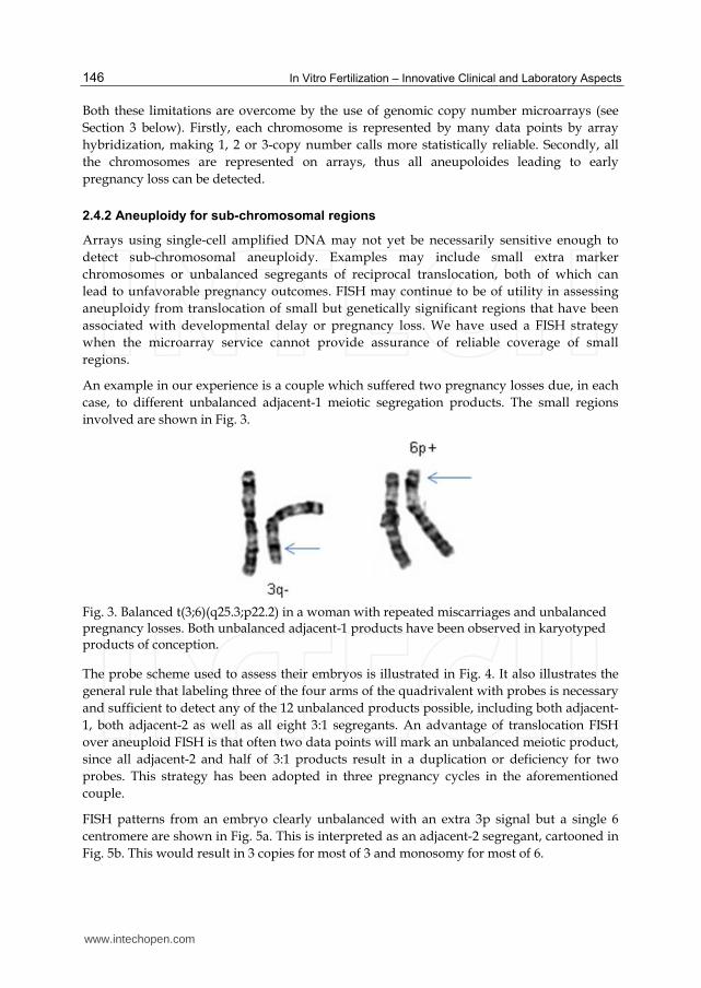
An example in our experience is a couple which suffered two pregnancy losses due, in each
case, to different unbalanced adjacent-1 meiotic segregation products. The small regions
involved are shown in Fig. 3.
Fig. 3. Balanced t(3;6)(q25.3;p22.2) in a woman with repeated miscarriages and unbalanced pregnancy losses. Both unbalanced adjacent-1 products have been observed in karyotyped products of conception.
The probe scheme used to assess their embryos is illustrated in Fig. 4. It also illustrates the
general rule that labeling three of the four arms of the quadrivalent with probes is necessary
and sufficient to detect any of the 12 unbalanced products possible, including both adjacent-
1, both adjacent-2 as well as all eight 3:1 segregants. An advantage of translocation FISH
over aneuploid FISH is that often two data points will mark an unbalanced meiotic product,
since all adjacent-2 and half of 3:1 products result in a duplication or deficiency for two
probes. This strategy has been adopted in three pregnancy cycles in the aforementioned
couple.
FISH patterns from an embryo clearly unbalanced with an extra 3p signal but a single 6
centromere are shown in Fig. 5a. This is interpreted as an adjacent-2 segregant, cartooned in
Fig. 5b. This would result in 3 copies for most of 3 and monosomy for most of 6.
www.intechopen.com
Preimplantation Genetic Testing: Current Status and Future Prospects
147
Fig. 4. Top, scheme to detect unbalanced t(3;6) meiotic products: subtelomere green (3p) and orange (3q) as well as 6 centromere (aqua). Three contrasting probes are sufficient to detect all unbalanced products. Bottom, this interphase pattern is balanced, any other pattern is unbalanced, following fertilization.
Fig. 5. Left, shows FISH patterns with an extra 3p signal and deficient for a 6 centromere signal. This is one of two cells from this embryo showing adjacent-2 segregation. Right, depicts this unbalanced complement.
To date adjacent-1 and adjacent-2 segregants have been detected in this couple, including those equivalent to both of the previous cytogenetically documented pregnancy losses.
It should be emphasized that both FISH and microarray approaches are limited in predictive
value by strictly biological considerations. It is apparent by numerous studies that many
early embryos are mosaic for aneuploidy. In addition, mosaic embryos often “correct”,
leading to normal outcomes following an aneuploid finding based on a single cell (Barbash-
Hazan et al, 2009). Thus, both false negative and false positive findings may be expected
3 6p q
www.intechopen.com

In Vitro Fertilization – Innovative Clinical and Laboratory Aspects
148
from these single cell approaches. These considerations must be kept in mind when
counseling couples to the value and accuracy of anueploid PGD.
3. Technological advances in preimplantation genetic testing
Robust WGA protocols (see section 2.2 above) and high-throughput DNA technologies such as microarrays and next generation sequencing (NGS) make it reasonable to envision PGD by interrogating the complete genome of a single blastomere. Completing a whole-genome analysis within the time frame of an IVF cycle via either technology remains a technical challenge. Genomic strategies, however, offer the promise of universal diagnosis and screening methods with consequent advantages in cost efficiency and diagnostic power. Emerging technologies for whole genome analysis via DNA microarrays and DNA sequencing are rapidly becoming more powerful and more cost effective, and it will soon be feasible to apply these genomic tools widely in reproductive medicine.
3.1 PGD/PGS by microarrays
DNA microarray technologies measure hybridization between the subject’s DNA (the “target”) and a matrix of known DNA sequences (the array “features”) immobilized on a solid state matrix. Depending upon the array platform and hybridization protocol, microarrays can reveal gains or losses of genome segments or determine the subject’s genotype for SNPs.
Array-comparative genomic hybridization (aCGH) has become an established alternative to FISH for PGS of aneuploidies, unbalanced translocations and complex karyotypes with multiple rearrangements (Hellani et al., 2008; Van den Veyver et al., 2009; Harper & Harton, 2010; Alfarawati et al., 2011; Fiorentino et al., 2011; Vanneste et al., 2011). In this procedure, target and control genomic DNAs are mixed and competitively hybridized to the same array. Changes in the hybridization ratio of target to control at a region indicate a gain or loss of material relative to the control genome. Since aCGH interrogates every chromosome and reveals events below the limits of microscopic detection, it is able to identify chromosome anomalies that a standard 8- or 12-chromosome FISH might fail to detect. However, aCGH does not detect balanced rearrangements or triploidy, where the target:control DNAs hybridized to the array features is constant along the genome (Thornhill et al., 2008; Harper & Harton, 2010; Fiorentino et al., 2011).
Single-gene disorders are amenable to whole-genome analysis using SNP microarrays. The SNP array features include alternative alleles for a large number of polymorphisms, and hybridization indicates which SNP allele(s) are present in the target genome. The SNP genotypes of two parents and a reference child define maternal and paternal haplotypes at a gene of interest, and linkage then establishes the genetic risk for a second child based on its combination of parental haplotypes. The analysis is similar to STR haplotyping discussed previously (see section 2.2.2). While custom PGD linkage assays have been developed for less than 10% of the known single gene disorders, a single microarray generates predictive SNP haplotypes for the entire genome.
Handyside et al. (2010) employed SNP genotype data to create “karyomaps” that represent the parental haplotypes and points of recombination along a child’s chromosomes. In this
www.intechopen.com
Preimplantation Genetic Testing: Current Status and Future Prospects
149
work, SNP haplotypes were shown to coincide with the inheritance of cystic fibrosis in families where both parents were carriers for CFTR mutations (Handyside et al., 2010).
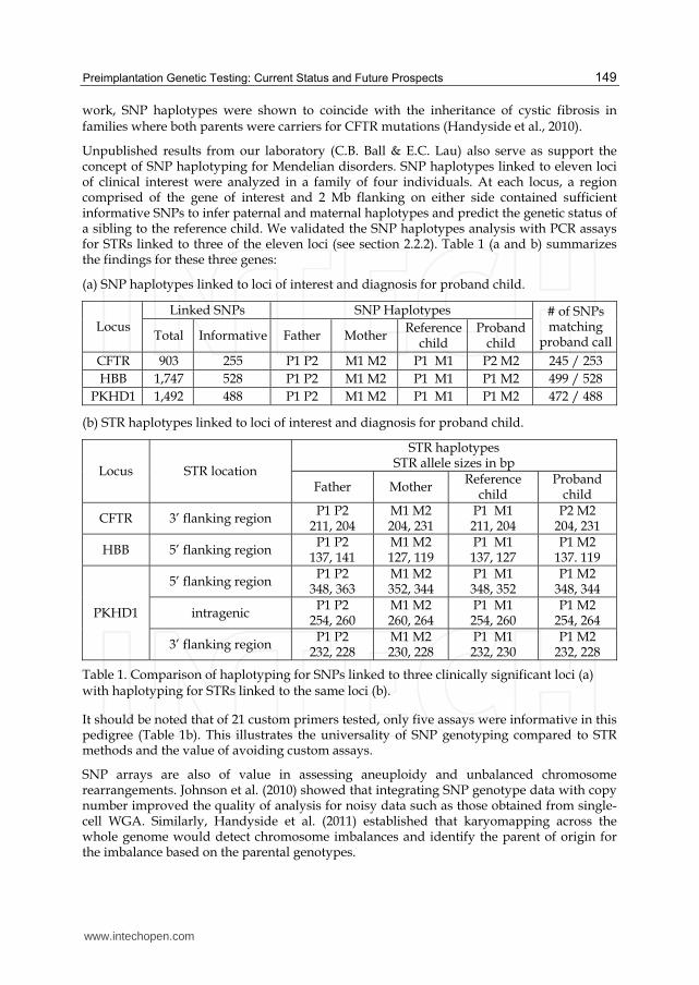
Unpublished results from our laboratory (C.B. Ball & E.C. Lau) also serve as support the concept of SNP haplotyping for Mendelian disorders. SNP haplotypes linked to eleven loci of clinical interest were analyzed in a family of four individuals. At each locus, a region comprised of the gene of interest and 2 Mb flanking on either side contained sufficient informative SNPs to infer paternal and maternal haplotypes and predict the genetic status of a sibling to the reference child. We validated the SNP haplotypes analysis with PCR assays for STRs linked to three of the eleven loci (see section 2.2.2). Table 1 (a and b) summarizes the findings for these three genes:
(a) SNP haplotypes linked to loci of interest and diagnosis for proband child.
Locus Linked SNPs SNP Haplotypes # of SNPs
matching proband call Total Informative Father Mother
Referencechild
Probandchild
CFTR 903 255 P1 P2 M1 M2 P1 M1 P2 M2 245 / 253
HBB 1,747 528 P1 P2 M1 M2 P1 M1 P1 M2 499 / 528
PKHD1 1,492 488 P1 P2 M1 M2 P1 M1 P1 M2 472 / 488
(b) STR haplotypes linked to loci of interest and diagnosis for proband child.
Locus STR location
STR haplotypesSTR allele sizes in bp
Father Mother Reference
childProband
child
CFTR 3’ flanking region P1 P2
211, 204 M1 M2204, 231
P1 M1211, 204
P2 M2 204, 231
HBB 5’ flanking region P1 P2
137, 141 M1 M2127, 119
P1 M1137, 127
P1 M2 137. 119
PKHD1
5’ flanking region P1 P2
348, 363 M1 M2352, 344
P1 M1348, 352
P1 M2 348, 344
intragenic P1 P2
254, 260 M1 M2260, 264
P1 M1254, 260
P1 M2 254, 264
3’ flanking region P1 P2
232, 228 M1 M2230, 228
P1 M1232, 230
P1 M2 232, 228
Table 1. Comparison of haplotyping for SNPs linked to three clinically significant loci (a) with haplotyping for STRs linked to the same loci (b).
It should be noted that of 21 custom primers tested, only five assays were informative in this pedigree (Table 1b). This illustrates the universality of SNP genotyping compared to STR methods and the value of avoiding custom assays.
SNP arrays are also of value in assessing aneuploidy and unbalanced chromosome rearrangements. Johnson et al. (2010) showed that integrating SNP genotype data with copy number improved the quality of analysis for noisy data such as those obtained from single-cell WGA. Similarly, Handyside et al. (2011) established that karyomapping across the whole genome would detect chromosome imbalances and identify the parent of origin for the imbalance based on the parental genotypes.
www.intechopen.com

In Vitro Fertilization – Innovative Clinical and Laboratory Aspects
150
3.2 Future PGD/PGS by DNA sequencing
Besides high-density microarrays, SNPs can be detected genome-wide by DNA sequencing,
which provides genotyping analysis at the highest resolution. For PGD of single-gene
disorders, DNA sequencing would be the future method of choice. The assay can interrogate
the entire genome, or target specific regions and genes of special interest. In principle,
sequencing methods can search directly for a targeted base change, or generate SNP
haplotypes for a linkage-based diagnosis. Second-generation sequencing systems, such as
Roche 454 system, Illumina sequencers and ABI SOLiD system, have not been used for PGD
due to the high cost and slow turn-around-time.
Even if the cost of whole genome DNA sequencing falls to $1,000, it is still too costly for
routine clinical PGD when the total cost of sequencing the parental samples, affected child
and several embryos is considered. In contrast to accurately determining the genotypes of
every SNP that requires sequencing at high genome coverage, it may lower the cost of PGD
using a haplotying approach that would require a lower genome coverage to identify the
SNP haplotypes of embryos for transfer.
The recently released third-generation DNA sequencers, such as the Single-Molecular Real-
Time System RS (Pacific Biosciences, CA), has enabled more rapid and cost effective
genomic sequencing. Future sequencing technologies, such as DNA transistor nanopore
sequencing system (Roche-IBM), and DNA tunneling silicon nanopore sequencer, will
benefit PGD when the sequencing cost falls below $250, and the turn-around-time to be less
than 24 hours.
4. Genetic counseling for PGD/PGS
Genetic counseling is an essential step for patients contemplating PGD (Swanson et al.,
2007). During the counseling session information is gathered including the patient’s medical
and reproductive history, the partner’s medical and reproductive history, and their family
histories. Both the patient and her partner should attend the counseling session. If an
indication of risk is not covered, it is essential that the counselor obtains the physician
and/or laboratory records supporting the information. Preimplantation genetic testing
should not be undertaken without a firm diagnosis. With this information in hand the
counselor can discuss disorder(s) in the family, the severity and variability of such
condition(s), limitations of genotype/phenotype and the patient’s probability of affected
children. All of this information will help a patient decide whether PGD would be a
reasonable option for her situation.
If PGD appears to be an option, then information is provided detailing the testing that can
be carried out on single embryonic cells. Limitations of such testing are discussed including
the patient’s anticipated success rate for IVF with PGD and the possibility of both false
positive and false negative PGD results. Prenatal testing through chorionic villus sampling
(CVS) or amniocentesis is recommended to confirm the results of PGD. Patients should be
reminded of alternative reproductive options including use of donor gametes, prenatal
diagnosis (PND), accepting genetic risk without further testing, adoption, and having no
children or no additional children (Harton et al., 2011c).
www.intechopen.com
Preimplantation Genetic Testing: Current Status and Future Prospects
151
By combining the information from the reproductive medicine specialist and the genetic counselor, the patient can decide whether to pursue PGD. It is important that the counseling provided should be non-directive, to enable the patient to reach her own conclusion about the suitability of PGD (Shenfield et al., 2003).
5. Future prospects for preimplantation genetic diagnosis
Severe genetic disorders are debilitating, expensive and incurable conditions. The patient, the family and society at large each have an interest in avoiding the birth of a child with such a disease. PGD provides that option in a cost-effective manner, without resort to termination of an affected pregnancy.
5.1 Current status of choice between PGD/PGS and PND in the U.S.
Prenatal diagnosis (PND) of fetuses is performed by either amniocentesis or CVS performed at different stages of fetal development, but the procedures are invasive and parents may ultimately decide to terminate an affected fetus. Even though PGD has been available for 21 years, PND following natural conception still prevails in the U.S. For couples at risk of severe inherited genetic disorders, abortion of affected pregnancy is still the most common option, especially in those States which do not have mandatory insurance coverage for IVF and PGD.
5.2 Medical and economic considerations of universal access to PGD/PGS
The medical community has recently begun to address the medical and economic implications of implementing a national PGD program to help couples who are carriers of severe single-gene disorders.
About 1000 children affected with cystic fibrosis (CF) are born annually in the U.S., in some part due to reluctance to terminate affected pregnancies. There is the potential to save 33 billion dollars in lifetime medical care for those affected with this disorder if carrier parents had the option of undergoing government-backed or insurance-mandated PGD and IVF (Tur-Kaspa et al., 2010). For couples who are carriers of severe inherited genetic disorders, prevention of affected pregnancy by PGD may be a preferred option to the termination of affected fetuses (Davis et al., 2009). Thus, economic and medical considerations favor a universal and affordable access to IVF, PGD or PGS services for carrier couples of severe single-gene disorders such as CF, or for individuals at risk for transmitting chromosomal translocations, but cannot afford it (Handyside, 2010).
6. Ethical concerns
The non-medical use of PGD and PGS presents some ethical concerns. While PGD has mainly been used to select embryos unaffected with severe genetic diseases to avoid the transmission of some medical conditions, PGD may also have the potential to select an embryo affected with the same disability or disease that affects the parents, such as deafness. A deaf child born to a deaf couple would be better suited to the parents' shared culture.
There is resistance regarding the practice of reproductive technologies including IVF, PND, abortion, sperm sorting, PGD/PGS, and embryonic stem cells. In fact, many countries have
www.intechopen.com
In Vitro Fertilization – Innovative Clinical and Laboratory Aspects
152
banned the use of PGD for gender selection, but it is permitted in the U.S. The opponents of “social sex selection” argue that the consequences of preimplantation procedures such as sperm sorting, PGD and PGS would artificially unequalize the ratio of females to males. The opponents of PGD and PGS fear that these technologies could be used for eugenic purposes, or increasing uses of PGD may open the door to other eugenic technologies.
PGD has successfully been used to prevent the transmission of single-gene disorders with Mendelian inheritance, but has not been used for complex genetic disorders because there is insufficient amount of amplified DNA from a single blastomere with high fidelity for testing a large number of genes and single-nucleotide polymorphisms (SNPs). Complex genetic disorders (e.g. diabetes mellitus), athletic ability and intelligence are controlled by many genes and environmental factors. Similarly, physical traits (e.g. body height), and cosmetic traits (e.g. hair color, eye color and skin pigmentation) are determined by multiple genes and SNPs (Sulem et al., 2007). With the recent advancement in blastocyst biopsy for removing multiple cells, cryopreservation of blastocysts, whole-genome amplification methods that generate amplified DNA with high fidelity, it has been technically feasible to provide preimplantation testing for complex genetic disorders and human traits. When PGD expands its scope to such a non-medical realm in potential attempts to make “designer babies”, it will spark more debates and controversy that might lead to legislation in this area.
7. Conclusion
Preimplantation genetic testing is an important application of IVF and has broad interest to reproductive medicine practitioners. Economic and medical considerations favor a universal and affordable access to IVF and PGD/PGS services for carrier couples of severe inherited genetic disorders. In addition to current methodologies, preimplantation testing is an expanding field poised to adopt cutting-edge genomic technologies for new advances in preventative medical care. Technologies for interrogating whole genomes via via SNP microarrays microarrays and DNA sequencing are rapidly becoming more powerful and more cost-effective. It will soon be feasible to apply these genomic tools widely in reproductive medicine.
8. Acknowledgments
The authors gratefully acknowledge the excellent support of our colleagues in the PGD team Amy Granlund, Bridget Lawler, Barbara Szlendakova and Julie McCarrier.
9. References
Alfarawati, S.; Fragouli, E.; Colls, P. & Wells, D. (2011) First births after preimplantation genetic diagnosis of structural chromosome abnormalities using comparative genomic hybridization and microarray analysis. Human Reproduction, Vol 26, No. 6, pp. 1560-1574.
Altarescu, G.; Geva, T.E.; Brooks, B.; Margalioth, E.; Levy-Lahad, E. & Renbaum, P. (2008). PGD on a recombinant allele: crossover between the TSC2 gene and ‘linked’ markers impairs accurate diagnosis. Prenatal Diagnosis, Vol.28, No.10, pp. 929-933.
www.intechopen.com
Preimplantation Genetic Testing: Current Status and Future Prospects
153
Amor, D.J. & Cameron, C. PGD gender selection for non-Mendelian disorders with unequal sex incidence. (2008). Human Reproduction, Vol.23, No.4, pp. 729-734.
Amos, J.; Feldman, G.L.; Grody, W.W.; Monaghan, K.; Palomaki, G.E.; Prior, T.W.; Richards, C.S & Watson, M.S. (2006). Technical standards and guidelines for CFTR mutation testing 2006 edition. Available from
http://www.acmg.net/Pages/ACMG_Activities/stds-2002/cf.htm Barbash-Hazan, S.; Frumkin, T.; Malcov, M.; Yaron, Y.; Cohen, T.; Azern, F.; Amit, A. & Ben-
Yosef, D. (2009). Preimplantation aneuploid embryos undergo self-correction in correlation with their developmental potential. Fertility & Sterility, Vol.92, No.3, pp. 890-896.
Bick, D.P. & Lau, E.C. (2006). Preimplantation genetic diagnosis. Pediatric Clinics of North America, Vol.53, No.4, pp. 559-577.
Bick, S.L.; Bick, B.P.; Wells, B.E.; Roesler, M.R.; Strawn, E.Y. & Lau, E.C. (2008). Preimplantation HLA haplotyping using tri-, tetra-, and pentanucleotide short tandem repeats for HLA matching. Journal of Assisted Reproduction and Genetics, Vol.25, pp. 323-331.
Benson, J.B. & Haith, M.M. (2009). Diseases and Disorders in Infancy and Early Childhood. Academic Press,. p. 210.
Coskun, S. & Alsmadi, O. (2007). Whole genome amplification from a single cell: a new era for preimplantation genetic diagnosis. Prenatal Diagnosis, Vol.27, pp. 297-302.
Cooper, A.R. & Jungheim E.S. (2010). Preimplantation genetic testing: indications and controversies. Clinical Laboratory Medicine, Vol.30, No.3, pp. 519-531.
Daniels G.; Pettigrew R.; Thornhill A.; Abbs S.; Lashwood A.; O'Mahony F. et al. (2001). Six unaffected livebirths following preimplantation diagnostic for spinal muscular atrophy. Moleclar Human Reproduction, Vol.7, No.10, pp. 995-1000.
Davis, L.B.; Champion, S.J.; Fair, S.Q.; Baker, V.L. & Garber, A.M. (2009). A cost-benefit analysis of preimplantation genetic diagnosis for carrier couples of cystic fibrosis. Fertility & Sterility, Vol.93, No.6, pp. 1793-1804.
Dean, F.B.; Hosono, S.; Fang, L.; Wu, X.; Faruqi, A.F.; Bray-Ward, P. et al. (2002). Comprehensive human genome amplification using multiple displacement amplification. Proceedings of the National Academy of Sciences USA, Vol.99, pp. 5261-5266.
Dell, K.M. & Avner, E.D. (July 2008). Autosomal recessive polycystic kidney disease. In: Gene Reviews, NIH, NCBI Bookshelf Available from:
http://www.ncbi.nlm.nih.gov/books/NBK1326/. ESHRE PGD Consortium Steering Committee. (2002). ESHRE Preimplantation Genetic
Diagnosis Consortium: data collection III (May 2001). Human Reproduction, Vol.17, No.1, pp.233-246.
Fiorentino, F.; Spizzichino, L.; Bono, S.; Biricik, A.; Kokkali, G.; Rienzi, L. et al. (2011). PGD for reciprocal and Robertsonian translocations using array comparative genomic hybridization. Human Reproduction, Vol.26, No.7, pp. 1925-1935.
Grewal, S.S.; Kahn, J.P.; MacMillan, M.L;, Ramsay, N.K & Wagner, J.E. (2004). Successful hematopoietic stem cell transplantation for Fanconi anemia from an unaffected HLA-genotype-identical sibling selected using preimplantation genetic diagnosis. Blood, Vol.103, pp. 1147-1151.
www.intechopen.com
In Vitro Fertilization – Innovative Clinical and Laboratory Aspects
154
Handyside, A.H. (2010). Preimplantation genetic diagnosis after 20 years. Reproductive BioMedicine Online, Vol.21, No.3, pp. 280-282.
Handyside, A.H.; Kontogianni, E.H.; Hardy, K. & Winston, R.M. (1990). Pregnancies from biopsied human preimplantation embryos sexed by Y-specific DNA amplification. Nature, Vol.344, pp. 768-770.
Handyside, A.H.; Lesko, J.G.; Tarin, J.J.; Winston, R.M. & Hughes, M.R. (1992). Birth of a normal girl after in vitro fertilization and preimplantation diagnostic testing for cystic fibrosis. The New England Journal of Medicine, Vol.327, pp. 905-909.
Handyside, A.H.; Harton, G.L.; Mariani, B.; Thornhill, A.R.; Affara, N.; Shaw, M-A & Griffin, D.K. (2010). Karyomapping: a universal method for genome wide analysis of genetic disease based on mapping crossovers between parental haplotypes. Journal of Medical Genetics, Vol.47, No.10, pp. 651-658.
Handyside, A.H.; Robinson, M.D.; Simpson, R.J.; Omar, M.B.; Shaw, M.-A.; Grudzinskas, J.G. et al. (2004). Isothermal whole genome amplification from single and small numbers of cells: a new era for preimplantation genetic diagnosis of inherited disease. Molecular Human Reproduction, Vol.10, No.10, pp. 767-772.
Harper, J.C. & Harton, G.L. (2010) The use of arrays in preimplantation genetic diagnosis and screening. Fertility and Sterility, Vol.10, No.4, pp.1173-1177.
Harper, J.C.; Coonen, E.; De Rycke, M.; Harton, G.; Moutou, C.; Pehlivans, T.; Traeger-Synodinos, J.; Van Rij, M.C. & Goossens, V. (2010). ESHRE PGS Consortium data collection X: cycles from January to December 2007 with pregnancy followup to October 2008. Human Reproduction, Vol.25, No.11, pp.2685-2707.
Harton, G.L.; De Rycke, M.; Fiorentino, .F.; Moutou, C.; SenGupta, S.; Traeger-Synodinos, J. & Harper J.C. (2011a). ESHRE PGD consortium best practice guidelines for amplification-based PGD. Human Reproduction, Vol.26, pp. 33-40.
Harton, G.L.; Magli, M.C.; Lundin, K.; Montag, M.; Lemmen, J. & Harper J.C. (2011b). ESHRE PGD Consortium/Embryology Special Interest Group – best practice guidelines for polar body and embryo biopsy for preimplantation genetic diagnosis/screening (PGD/PGS). Human Reproduction, Vol.26, pp. 41-46.
Harton, G.; Braude, P.; Lashwood, A.; Schmutzler, A.; Traeger-Synodinos, J.; Wilton, L. & Harper, J.C. (2011c). ESHRE PGD consortium best practice guidelines for organization of a PGD centre for PGD/preimplantation genetic screening. Human Reproduction, Vol.26, pp. 14-24.
Heinrich, M.; Braun, T.; Sanger, T.; Saukko, P.; Lutz-Bonengel, S. & Schmidt, U. (2009). Reduced-volume and low-volume typing of Y-chromosomal SNPs to obtain Finnish Y-chromosomal compound haplotypes. International Journal of Legal Medicine, Vol.123, pp. 413-418.
Hellani, A.; Abu-Amero, K.; Azouri, J. & El-Akoum, S. (2008). Successful pregnancies after application of array-comparative genomic hybridization in PGS-aneuploidy screening. Reproductive BioMedicine Online, Vol.17, pp. 841-847.
Hellani, A.; Coskun, S.; Benkhalifa, M.; Tbakhi, A.; Sakati, N.; Al-Odaib, A. & Ozand, P. (2004). Multiple displacement amplification on single cell and possible PGD applications. Molecular Human Reproduction, Vol.10, pp. 847-852.
Johnson, D.S.; Gemelos, G.; Baner, J.; Ryan, A.; Cinnioglu, C.; Banjevic, M.; Ross, R. et al. (2010). Preclinical validation of a microarray method for full molecular karyotyping of blastomeres in a 24-h protocol. Human Reproduction, Vol.25, pp. 1066-1075.
www.intechopen.com
Preimplantation Genetic Testing: Current Status and Future Prospects
155
Kobayashi, M.; Rappaport, E.; Blasband, A.; Semeraro, A.; Sartore, M.; Surrey, S. & Fortina, P. (1995). Fluorescence-based DNA minisequence analysis for detection of known single-base changes in genomic DNA. Molecular and Cellular Probes, Vol.9, pp. 175-182.
Lau, E.C.; Janson, M.M.; Roesler, M.R.; Avner, E.D.; Strawn, E.Y. & Bick, D.P. (2010). Birth of a healthy infant following preimplantation PKHD1 haplotyping for autosomal recessive kidney disease using multiple displacement amplification. Journal of Assisted Reproduction and Genetics, Vol.27, pp. 397-407.
Langmore, J.P. (2002). Rubicon Genomics, Inc. Pharmacogenomics, Vol.3, pp. 557-560. Magli, M.C.; Gianaroli ,L.; Grievo, N.; Cefalu, E.; Ruvolo, G. & Ferraretti, A.P. (2006).
Cryopreservation of biopsied embryos at the blastocyst stage. Human Reproduction, Vol.21, No.10, pp. 2656-2660.
Malcov M.; Schwartz T.; Mei-Raz N.; Yosef D.B.; Amit A.; Lessing J.B. et al. (2004). Multiplex nested PCR for preimplantation genetic diagnosis of spinal muscular atrophy. Fetal Diagnosis and Therapy, Vol.19, No.2, pp. 199-206.
Munne, S.; Weier, H.U.; Stein, J.; Grifo, J. & Cohen, J. (1993). A fast and efficient method for simultaneous X and Y in situ hybridization of human blastomeres. Journal of Assisted Reproduction and Genetics, Vol.10, pp. 82-90.
Munne, S.; Scott, R.; Sable, D. & Cohen, J. (1998). First pregnancies after preconception diagnosis of translocations of maternal origin. Fertility and Sterility, Vol.69, pp. 675-681.
Palermo, G.; Joris, H.; Devroey, P. & Van Steirteghem, A.C. (1992). Pregnancies after intracytoplasmic injection of single spermatozoon into an oozyte. Lancet, Vol.340, pp. 17-18.
Prior, T.W. & Russman, B.S. (2011). Spinal Muscular Atrophy, In: GeneReviews, Available from http://www.ncbi.nlm.nih.gov/books/NBK1352/
Schrurs, B.M.; Winston, R.M. & Handyside, A.H. (1993). Preimplantation diagnosis of aneuploidy using fluorescent in-situ hybridization: evaluation using a chromosome 18-specific probe. Human Reproduction, Vol.8, pp. 296-301.
Sermon, K.; Lissens, W.; Joris, H.; Van Steirteghem, A. & Liebaers, I. (1996). Adaptation of the primer extension preimplantation (PEP) reaction for preimplantation diagnosis: single blastomere analysis using short PEP protocols. Molecular Human Reproduction, Vol.2, pp. 209-212.
Shenfield, F.; Pennings, G.; Devroey, P.; Sureau, C.; Tarlatzis, B. & Cohen, J. (2003). Taskforce 5: preimplantation genetic diagnosis. Human Reproduction, Vol.18, No.3, pp. 649-651.
Stachecki, J.J.; Cohen, J. & Munne, S. (2005). Cryopreservation of biopsied cleavage stage human embryos. Reproductive BioMedicine Online, Vol.11, No.6, pp. 711-715.
Sulem, P.; Gudbjartsson, D.F.; Stacey, S.N.; Helgason A.; Rafnar, T.; Magnusson, K.P. et al. (2007). Genetic determinants of hair, eye and skin pigmentation in Europeans. Nature Genetics, Vol.39, pp.1443-1452.
Swanson, A.; Strawn, E.; Lau, E. & Bick, D. (2007). Preimplantation genetic diagnosis: technology and clinical applications. Wisconsin Medical Journal, Vol.106, No.3, pp. 145-151.
Sweeney, W.E. & Avner E.D. (2006). Molecular and cellular pathophysiology of autosomal recessive polycystic kidney disease. Cell and Tissue Research, Vol.326, pp. 671-685.
www.intechopen.com
In Vitro Fertilization – Innovative Clinical and Laboratory Aspects
156
Theisen, A. & Shaffer, L.G. (2010). Disorders caused by chromosome abnormalities. The Application of Clinical Genetics, Vol.2010, No.3, pp.159-174.
Thornhill, A.R.; Gabriel, A.S.; Gordon, A.; Griffin, D.K.; Taylor, J. & Handyside, A.H. (2008). Array CGH for use in clinical preimplantation genetic screening. Fertility and Sterility, Vol.90 supplement, p. S306.
Tur-Kaspa, I.; Aljadeff, G.; Rechitsky, S.; Grotjan, H.E. & Verlinsky, Y. (2010). PGD for all cystic fibrosis carrier couples: novel strategy for preventive medicine and cost analysis. Reproductive BioMedicine Online, Vol.21, No.2, pp. 186-195.
Vanneste, E.; Melotte, C.; Voetl, T; Robberecth, C.; Debrock, S.; Pexsters, A.; Staessen, C.; Tomassetti, C.; Legius, E.; D’Hooghe, T. & Vermeesch, J.R. (2011). PGD for a complex chromosomal rearrangement by array comparative genomic hybridization. Human Reproduction, Vol.26, No.4, pp.941-949.
Van den Veyver, I.B.; Patel, A.; Shaw, C.; Pursley, A.N.; Kang, S. L.; Simovitch, M.J.; Ward, P.A.; Darilek, S.; Johnson, A.; Neill, S.E.; Bi, W.; White, L.D., Eng, C.M. , Lupski, J.R.; Cheung, S.W. & Beaudet, A.L. (2009). Clinical use of array comparative genomic hybridization (aCGH) for prenatal diagnosis in 300 cases. Prenatal Diagnosis, Vol.29, No.1, pp.29-39.
Verlinsky, Y.; Cohen, J.; Munne, S.; Gianaroli, L.; Simpson, J.L.; Ferraretti, A.P. & Kuliev, A. (2004). Over a decade of experience with preimplantation genetic diagnosis: a multicenter report. Fertility & Sterility, Vol.82, No.2, pp.292-294.
Verlinsky, Y.; Rechitsky, S.; Schoolcraft, W.; Strom, C. & Kuliev, A. (2001). Preimplantation diagnosis for Fanconi anemia combined with HLA matching. The Journal of the American Medical Association, Vol.285, pp. 3130-3133.
Zhang, L.; Cui, X.; Schmitt, K.; Hubert, R.; Navidi, W. & Arnheim, N. (1992). Whole genome amplification from a single cell: implications for genetic analysis. The Proceedings of National Academy of Sciences, Vol.89, pp. 5847-5851.
Zhang, X.; Trokoudes, K.M. & Pavlides, C. (2009).Vitrification of biopsied embryos at cleavage, morula and blastocyst stage.. Reproductive BioMedicine Online, Vol.19, No.4, pp. 526-531.
Zheng, W.T.; Zhuang, G.L.; Zhou, C.Q.; Fang, C.; Ou, J.P.; Li, T.; Zhang, M.F. & Liang, X.Y. (2005). Comparison of the survival of human biopsied embryos after cryopreservation with four different methods using non-transferable embryos. Human Reproduction, Vol.20, No.6, pp. 1615-1618.
www.intechopen.com
In Vitro Fertilization - Innovative Clinical and Laboratory AspectsEdited by Prof. Shevach Friedler
ISBN 978-953-51-0503-9Hard cover, 156 pagesPublisher InTechPublished online 11, April, 2012Published in print edition April, 2012
InTech EuropeUniversity Campus STeP Ri Slavka Krautzeka 83/A 51000 Rijeka, Croatia Phone: +385 (51) 770 447 Fax: +385 (51) 686 166www.intechopen.com
InTech ChinaUnit 405, Office Block, Hotel Equatorial Shanghai No.65, Yan An Road (West), Shanghai, 200040, China
Phone: +86-21-62489820 Fax: +86-21-62489821
The field of In Vitro Fertilization is a relatively new field in medicine, constantly on the move. This field is anexquisite example of the vast power in the complementary use of basic research with clinical practice andopened a new route of great basic and clinical research possibilities. The knowledge base that allowed theaccomplishment of the idea of in vitro fertilization and embryo transfer has much developed since. The vastbody of research pertaining to this field allowed deepening our understanding in the processes related toreproduction. In this book on in vitro fertilization we present new and interesting updated information in variousaspects of this field. This work is a result of collaborative work of an international group of professionalsdedicated to contribute to the advancement of our knowledge.
How to referenceIn order to correctly reference this scholarly work, feel free to copy and paste the following:
Eduardo C. Lau, Marleen M. Janson, Carl B. Ball, Mark R. Roesler, Peter VanTuinen, David P. Bick and Estil Y.Strawn (2012). Preimplantation Genetic Testing: Current Status and Future Prospects, In Vitro Fertilization -Innovative Clinical and Laboratory Aspects, Prof. Shevach Friedler (Ed.), ISBN: 978-953-51-0503-9, InTech,Available from: http://www.intechopen.com/books/in-vitro-fertilization-innovative-clinical-and-laboratory-aspects/preimplantation-genetic-diagnosis-current-status-and-future-prospects