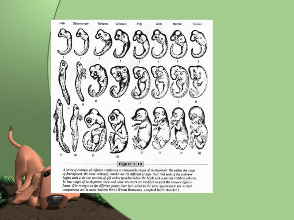
pregnancy, development and lactation chapter 18. species development species can only be perpetuated...
TRANSCRIPT

Pregnancy, Development and Lactation
Chapter 18

Species Development
• Species can only be perpetuated if pregnancy, development of offspring and lactation are appropriately carried out.

Fertilization• Copulation- the act of breeding
allowed by the female during estrous (heat) period.– Usually in a mounted position
• Intromission- the insertion of the penis into the vagina.
• Ejaculation- when the semen is deposited in the upper portion of the vagina– Horse and pig deposit semen directly into
uterus through open cervix.
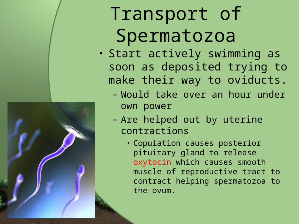
Transport of Spermatozoa
• Start actively swimming as soon as deposited trying to make their way to oviducts. – Would take over an hour under
own power– Are helped out by uterine
contractions• Copulation causes posterior pituitary
gland to release oxytocin which causes smooth muscle of reproductive tract to contract helping spermatozoa to the ovum.

Capacitation• Series of changes that
spermatozoa undergo in the female reproductive tract to increase chances of fertilization. – Changes of ion movement through
cell membranes– Increase in cell’s metabolic rates– Increase in rate of use of simple
sugars for energy production.– Allows acrosome enzymes to be
released.

Fertilization of the Ovum• Spermatozoa are programmed to
seek out something large and round and attempt to penetrate it. – Some try to fertilize non-ovum things.
• Once ovum is found, many spermatozoa may swarm around it and start tunneling through the layers.– Process aided by enzymes of acrosome.
• Once one spermatozoa penetrates ovum, change in membrane prevents any other sperm from entering.


The Zygote• Once ovum is fertilized, it becomes a
zygote. • Male pronucleus- the nucleus of the
male spermatozoan immediately after fertilization.
• Female pronucleus- the nucleus of the ovum immediately after fertilization. – Each pronucleus carries haploid number– Join together to get diploid number
• This joining establishes genetic information for offspring.


Cleavage• The rapid division of the zygote once
single nucleus has been established. • Cell divides rapidly but overall size
remains the same because are dividing so quickly do not have time to grow.
• Once zygote is a solid mass of cells, is in morula stage.
• During this time, zygote is moving down ovum to uterus– Propelled by cilia and muscular contractions

Blastocyst
• Cells of morula stage continued to divide and hollow cavity is formed in center of zygotic cell.
• Once bump of cells on one side is developed, this is now the blastocyst.


Implantation• The means by which the blastocyst
makes itself a home by attaching itself to the lining of the uterus (endometrium).
• Once blastocyst comes to rest beside uterine lining, enzymes produced by blastocyst dissolve away some lining and implants itself into this pit in the lining.
• Placenta begins to form as soon as implantation occurs. – Way of transporting oxygen and nutrients to
blastocyst.


Terminology
• Embryo-what developing offspring is referred to during early part of pregnancy.
• Fetus- What developing offspring is referred to during later part of pregnancy.
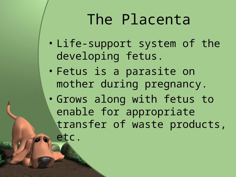
The Placenta
• Life-support system of the developing fetus.
• Fetus is a parasite on mother during pregnancy.
• Grows along with fetus to enable for appropriate transfer of waste products, etc.

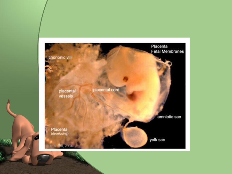
Structure of the Placenta• Multi-layered, fluid-filled
membranous sac.• Develops around embryo
and is connected to it by umbilical cord. – Smaller connections between
outermost layer of placenta and lining of uterus.
• This is where exchange of nutrients and wastes takes place.
• Fetal and maternal blood does not mix but runs in close proximity to one another.

Structure of Placenta Continued…
• 3 layers of placenta– 1. Amnion
• Forms amniotic sac directly around fetus
– 2. Allantois• Outside amniotic sac and forms
allantoic sac.
– 3. Chorion• Outside of allantoic sac and attaches to
uterine lining.• Linked to fetus by umbilical cord.

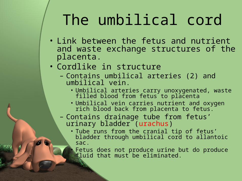
The umbilical cord• Link between the fetus and nutrient
and waste exchange structures of the placenta.
• Cordlike in structure– Contains umbilical arteries (2) and
umbilical vein.• Umbilical arteries carry unoxygenated, waste
filled blood from fetus to placenta• Umbilical vein carries nutrient and oxygen rich
blood back from placenta to fetus.– Contains drainage tube from fetus’ urinary
bladder (urachus)• Tube runs from the cranial tip of fetus’ bladder
through umbilical cord to allantoic sac.• Fetus does not produce urine but do produce
fluid that must be eliminated.


Attachment to the Uterus
• Where chorion attaches to lining of the uterus.
• Type of attachement varies among species and is one of four types:– 1. Diffuse Attachment– 2. Cotyledonary Attachment– 3. Zonary Attachment– 4. Discoid Attachment.


Diffuse Attachment• Means that attachment sites are
spread diffusely over the whole surface of the placenta and the whole lining of the uterus.
• No small, limited areas of attachment.
• Found in pigs and horses• Detaches easily from uterine
lining and is passed after the delivery of the newborn.

Cotyledonary Attachment
• Most complicated type and is somewhat opposite of diffuse attachment.
• Areas of attachment are small, separate, and numerous.
• Placentome- attachment sites.• Cotyledon- area on surface of the placenta• Caruncle-area on surface of uterus
(mushroom-like).– Cotyledon and caruncle interdigitate with one
another.
• Each placentome must separate completely for placenta to pass after birth. – If not completely passed, may be retained and
can cause other problems.


Zonary Attachment
• Placenta attaches to the uterus in belt-shaped area that encircles the placenta.
• Found in dogs and cats• Detaches easily after delivery.


Discoid Attachment
• Area of attachment between placenta and uterus is a single-disk shaped area.
• Found in humans and other primates

Pregnancy• Also called gestation period• Time from implantation to delivery of
the newborn. • Judge time by time since fertlization.• Is divided into three segments called
trimesters – 1st- period of the embryo implanting and
organizing and placental development– 2nd- embryo now called fetus and is fetal
development period. Parts are taking shape and differentiating.
– 3rd- fetal growth. All parts grow dramatically


Gestation PeriodsSpecies Approx Gestation Period• Cat 2 mo (56-69 days)• Dog 2 mo (59-68 days)• Cow 9 mo (271-291 days)• Elephant 21 mo (615-650 days)• Ferret 6 w (42 days)• Goat, Sheep 5 mo (143-155 days)• Hamster 3 w (19-20 days)• Horse 11 mo (321-346 days)• Human 9 mo (280 days)• Pig 3 mo, 3 w, 3 d (110-116
days)• Rabbit 1 mo (30-32 days)

Parturition• Birth process• Lungs of newborn become functional• Goes from parasite to independent being.• Parturition is triggered by size and weight
of uterus and changing hormone levels.– Progesterone of dam declines– Progesterone has kept myometrium from
contracting– Increased levels of glucocorticoid hormones
stimulate rise in estrogen levels– These increase sensitivity to oxytocin, released
from posterior pituitary gland. • Oxytocin stimulates contractions which starts labor
process


3 Stages of Labor• 1. Uterine Contractions
– Presses fetus against uterus– Causes cervix to gradually dilate– Dam may appear restless in this stage.
• 2. Delivery of the Newborn– Combination of uterine and abdominal
muscle contractions– “Water” or amniotic and allantoic sacs
rupture.• 3. Delivery of the placenta
– Placenta separates from wall of the uterus and is expelled by uterine contractions.
– Dam often eats the placenta.
Labor Continued
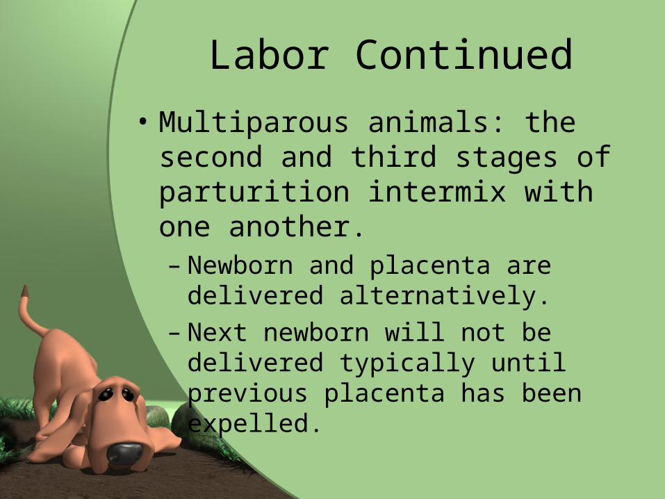
• Multiparous animals: the second and third stages of parturition intermix with one another. – Newborn and placenta are
delivered alternatively.– Next newborn will not be delivered
typically until previous placenta has been expelled.

Parturition: normal presentation

Dystocia
• “difficult birth”• Most common cause is that fetus
is too large to pass or is in wrong orientation for delivery.
• May have to Repell the fetus or deliver through Cesarean section.
• If fetus is dead, may have to be removed in segments- called Embryotomy.

Parturition: abnormal presentations

Delivery

Placental Delivery

Whelping/ Parturition
Normal Presentation Presentation of two sacs

Involution of the Uterus• After parturition is complete, uterus
gradually returns to nonpregnant size.• Process is called involution.• At placental attachment sites,
endometrium sloughs into uterus and areas heal over.
• Myometrium contractions continue slowly, pushing contents through birth canal.
• Will pass from bright red blood, to dead tissue over course of weeks to about a month.

Mammary Glands and Lactation
• Play an important role during neonatal period.
• Mammary glands are specialized skin glands.
• Produce colostrum and milk which are crucial to early life.
• Present in both males and females– Females secrete appropriate
hormones for them to become functional

Species Differences
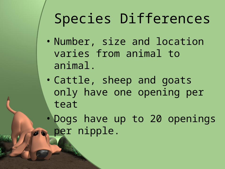
• Number, size and location varies from animal to animal.
• Cattle, sheep and goats only have one opening per teat
• Dogs have up to 20 openings per nipple.

Number of Mammary Glands for Common
animalsSPECIES NUMBER OF
GLANDS
CatsHorses
102
DogsHumans
102
CattlePigs
414

Udder of the Cow• Udder is term used for mammary glands.• Exaggerated in size, but composition is
similar to other animal’s mammary glands• Four mammary glands (quarters)• Quarters are completely separate units
from each other • Each quarter has its own milk-secreting
systems and ducts leading down to separate teats
• Suspended by strong suspensory ligaments that allow it to stretch.– Acts as a shock absorber.


Udder Continued
• Mastitis- infection of the mammary gland– Since are separate, is unlikely to
spread from one quarter to another.
– Can spread through bloodstream.

Alveoli and Duct System• Alveoli- milk-secreting units of the
mammary gland• Alveolar duct- alveoli secrete milk into tube
– Similar in make up to Alveoli found in lungs.
• Ducts empty into large space called gland sinus which is continuous with the teat sinus which is where milk is extracted by suckling young.
• Tip of teat has streak canal- passageway from teat sinus to outside.– Surrounded by elastic fibers and ringlike
sphincter muscle that keeps it closed to prevent leakage.


Mammary Gland Development
• Mammary glands develop in response to hormones produced at puberty– Prolactin and Growth Hormone
directly encourage mammary gland development
– Estrogen and progesterone encourage mammary alveoli and duct systems to develop• Influenced by FSH and LH on ovaries
– Certain drugs may inhibit normal mammary gland development

Lactation
• Process of milk production• Begins at end of pregnancy and
is obvious at time of parturition.• Prolactin and Growth Hormone
from anterior pituitary gland and hormones from adrenal cortex are involved with the starting of lactation.


Colostrum• “First milk” or “premilk”.• Contains large amounts of proteins, lipids, and
amino acids than milk and high levels of essential vitamins.– Antibodies for defense
• Supplies important nutrients and defenses that newborn can not receive elsewhere.– If does not receive within first few hours, body can no
longer process appropriately. • Has laxative effect to clear meconium ( first
feces) from newborn’s intestinal tract. • Involved with passive immunity from dam to
newborn• Those without appropriate colostrum tend to
be weaker and do not grow as rapidly. • Why we wait for vaccines.

Maintenance and Lactation
• Lactation continues as long as mammary gland is emptied regularly.
• Physical stimulation of nipple.• This sends nerve impulses to brain
which continues stimulation of appropriate hormones for milk production.
• When nursing stops, signal stops.– Will lead to involution of the mammary
gland or “drying up”.

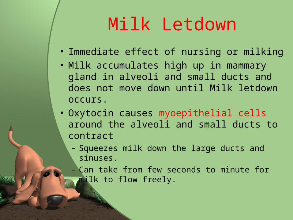
Milk Letdown• Immediate effect of nursing or milking• Milk accumulates high up in mammary
gland in alveoli and small ducts and does not move down until Milk letdown occurs.
• Oxytocin causes myoepithelial cells around the alveoli and small ducts to contract– Squeezes milk down the large ducts and
sinuses.– Can take from few seconds to minute for
milk to flow freely.
