postembryonic development of oribatid mites cepheus cepheiformis and conchogneta traegardhi (acari,...
TRANSCRIPT

ISSN, 0013–8738, Entomological Review, 2012, Vol. 92, No. 1, pp. 112–126. © Pleiades Publishing, Inc., 2012. Original Russian Text © S.G. Ermilov, 2011, published in Zoologicheskii Zhurnal, 2011, Vol. 90, No. 11, pp. 1323–1337.
112
Postembryonic Development of Oribatid Mites Cepheus cepheiformis and Conchogneta traegardhi (Acari, Oribatida)
S. G. Ermilov Consulting Center of the Federal Service for Veterinary and Phytosanitary Inspection, Nizhnii Novgorod, 603107 Russia
e-mail: [email protected] Received January 21, 2009
Abstract—The morphology of all the juvenile stages of Cepheus cepheiformis and Conchogneta traegardhi is de-scribed. The structure of the gnathosoma, ovipositor, and legs is studied in adults of both species. Developmental rates of C. traegardhi in the laboratory conditions are established. DOI: 10.1134/S0013873812010137
Postembryonic development of Cepheus cephei-formis (Nicolet, 1855) (Compactozetidae) and Con-chogneta traegardhi (Forsslund, 1947) (Autognetidae) was examined. The following tasks were set: (1) to describe the morphology of juvenile stages; (2) to compare the obtained data on the morphology of the juvenile stages of C. cepheiformis and C. traegardhi with the available data on other representatives of the corresponding genera; (3) to establish the duration of the development of C. traegardhi in the laboratory.
The oribatid family Compactozetidae comprises 84 species of 15 genera. The genus Cepheus Koch, 1835 (Acari, Oribatida) with 28 species is the most numerous in relation to the number of species in the family (Subías, 2004, online version, 2010). Morphol-ogy of juvenile stages in representatives of Cepheus was not specially examined. Some characters are de-scribed in some publications (Grandjean, 1953; Sit-nikova, 1975a); tritonymphs of 4 species are also de-scribed: C. brachiatus Sitnikova, 1975 (Sitnikova, 1975a), C. cepheiformis (Michael, 1884–1888; Bernini and Nannelli, 1982), and C. dentatus (Michael, 1888) (Michael, 1884–1888), C. latus Koch, 1835 (Sitni-kova, 1975a; Michael, 1879; Willmann, 1931; Bernini S. and Bernini F., 1990).
The oribatid family Autognetidae comprises 28 spe-cies of 7 genera. The genus Conchogneta Grandjean, 1963 (Acari, Oribatida) includes 5 species and a single subspecies (Subías, 2004, online version, 2010). De-velopmental biology in representatives of this family is insufficiently examined. The data on the morphol-ogy of juvenile stages are available for 6 species (Autogneta penicillum Grandjean, 1960, A. longi-
lamellata (Michael, 1885), A. numidiana (Grandjean, 1960a), Conchogneta willmanni willmanni (Dyr-dowska, 1929) (= Autogneta dalecarlica Forsslund, 1947), Cosmogneta impedita Grandjean, 1960, and C. kargi Grandjean, 1963) (Grandjean, 1960a, 1960b, 1963; Lions, 1975); any data on the duration of the development are absent.
MATERAILS AND METHODS
Mites of C. cepheiformis were collected in soils of mixed and coniferous forests; C. traegardhi, in the soil of pine forests of Nizhnii Novgorod Province in 2007–2008. Juveniles of C. cepheiformis were found only in spring collections; juvenile specimens of C. traegardhi were not found in the collected material.
Constant slides were prepared for morphological studies. Body length was measured from the rostrum to the posterior part of the hysterosomal area in lateral view. Body width was measured in the widest part of the hysterosomal area in dorsal view. The length of setae was measured in lateral view. All the measure-ments are given in micrometers. Formulae of leg setae are given in parentheses (trochanter-femur-genu-tibia-tarsus, correspondingly); formulae of solenidia are given in square brackets [genu-tibia-tarsus, corre-spondingly]. Generally accepted designations and terms were used in morphological descriptions.
C. traegardhi mites were cultivated according to the following scheme. Adult mites were kept in groups of 10–15 specimens in five plastic boxes. Emerged larvae were placed into individual Michael’s chambers. Filter paper moistened when necessary was previously placed on the bottom of chambers and weighing bot-

POSTEMBRYONIC DEVELOPMENT OF ORIBATID MITES
ENTOMOLOGICAL REVIEW Vol. 92 No. 1 2012
113
tles. Overwetting of filter paper on chamber bottoms resulted in the slowing of the development and death of some specimens. Therefore, the paper was mois-tened once per 2–3 days. Mites were fed on algae, lichens, moss, half-fermented wood, tree leaves, and raw potatoes. Experiments were performed under sta-ble conditions: humidity 100%, temperature 20ºC.
RESULTS AND DISCUSSION Morphology of the Developmental Stages
of Cepheus cepheiformis Larva. Body discoid, approximately twice as long
as wide. Cuticle colorless, with grayish and yellowish tint. Cerotegument finely granulate. For size of body and setae of prodorsum in larva and subsequent nym-phal stages, see Table 1.
Prodorsum (Fig. 1, 1, 3–6) comparatively short; its length in lateral view constituting approximately 0.4 of length of hysterosomal area. Rostrum rounded. Pro-dorsal setae well-developed, situated on apophyses. Rostral (ro) setae fine and smooth, lamellar setae (le) weakly leaf-shaped, interlamellar (in) and exobothridi-al (ex) thickened and pubescent. Bothridia cup-shaped, their external margin with distinct prominence. Tri-chobothria (ss) club-shaped, covered with small sparse teeth.
Hysterosomal area (Fig. 1, 1, 7–11). Hysterosomal setae constituting 12 pairs, 10 of them visible in dorsal view. Setae large, leaf-shaped, situated on large simple apophyses. Apophyses of setae dp connected. Main “vein” of leaf-shaped setae thick, with long and short dentate branches; small teeth frequently bifurcated at apices. Central dorsal setae (da, dm, dp) longest.
Ventral body side (Fig. 2, 1). Setae h2 long and fine, h3 very short. Slit-shaped organs ih, ip and orifices of opisthosomal glands (gla) distinctly visible. Epimeral formula 3-1-2 (third seta of epimere I represented by protective scale of Claparede’s organ); setae simple, smooth. For changes in body chaetotaxy of larvae and subsequent stages, see Table 2.
Legs (Fig. 3, 1–3). Tarsi with single smooth claw. Majority of setae pubescent. Lateral setae of tibia and genu I strongly thickened. Tibia I with large dorsal apophysis bearing solenidium and protective seta. Solenidia of tibiae II and III and genu I–III very small (slightly swollen apically). Formulae of leg setae and solenidia: I (0-2-3-4-16) [1-1-1], II (0-2-3-3-13) [1-1-1], III (0-2-2-2-13) [1-1-0]. For designations of leg setae and solenidia in larva and subsequent stages, see Table 3.
Nymphs. Proto-, deuto-, and tritonymphs mor- phologically similar to each other and to larva in many
Table 1. Changes in the size of the body and prodorsal setae in Cepheus cepheiformis during development Index Larva Protonymph Deutonymph Tritonymph Adult
Body length 332–348 381–398 464–481 564–647 630–697 Body width 149–166 199–249 332–365 448–481 448–498 Length of setae:
ro 20–24 32–36 41–45 49–57 61–94 le 45–53 53–57 65–73 73–82 77–106 in 24–28 8–12 12–16 16–24 102–143 ss 69–73 82–86 90–98 102–110 77–90 ex 12–16 20–28 20–28 36–45 Absent
Table 2. Changes in body chaetotaxy in Cepheus cepheiformis during development
Index Larva Protonymph Deutonymph Tritonymph Adult Epimeral formula 3-1-2 3-1-2-1 3-1-2-2 3-1-3-3 3-1-4-3 Number of pairs of setae:
hysterosomal 12 12 12 12 10 g 0 1 3 5 6 ag 0 0 1 1 1 ad 0 0 3 3 3 an 0 0 0 2 2

ERMILOV
ENTOMOLOGICAL REVIEW Vol. 92 No. 1 2012
114
Fig. 1. Larva (1, 3–11) and protonymph (2, 12–14) of Cepheus cepheiformis: (1) dorsal view; (2) prodorsum and anterior part of hys-terosoma; (3) lamellar seta; (4) interlamellar seta; (5) trichobothrium; (6) exobothridial seta [(7) c1; (8) c2; (9) dm; (10) lm; (11) h1; (12) c1; (13) c2; (14) c3. Scale (μm): 1, 2—100; 3–8, 9–14—20.

POSTEMBRYONIC DEVELOPMENT OF ORIBATID MITES
ENTOMOLOGICAL REVIEW Vol. 92 No. 1 2012
115
Fig. 2. Ventral side of body (1–4) of juvenile stages and prodorsal setae of tritonymph (5–8) of Cepheus cepheiformis: (1) anogenital area of larva; (2) anogenital area of protonymph; (3) anogenital area of deutonymph; (4) tritonymph, ventral view; (5) lamellar seta; (6) interlamellar seta; (7) trichobothrium; (8) exobothridial seta. Scale (μm): 1—50; 2, 3—100; 4—200; 5–8—20.

ERMILOV
ENTOMOLOGICAL REVIEW Vol. 92 No. 1 2012
116
aspects. Body becoming more and more rounded in each subsequent stage. Hysterosomal area with exuvia. Main morphological stages characteristic of each nymphal stage given below.
Protonymph (Figs. 1, 2, 12–14; 2, 2). Body ap-proximately 1.5–1.7 times as long as wide. Lamellar setae becoming widely leaf-shaped, interlamellar setae becoming smooth and smaller (constituting 0.3–0.5 of length of these setae in larva). Exobothridial setae becoming long, with numerous teeth. Exuvium with cellular ornament and 9 pairs of setae. Actually proto-nymphal hysterosomal setae constituting 12 pairs. Central dorsal setae (da, dm, dp) absent. Additional 3 pairs of setae (p1–p3) appearing. Eight pairs of setae (c3, la, lm, lp, h1–h3, p1) leaf-shaped, situated on large figured apophyses along margins of hysterosomal area. Setae c2 at this stage becoming smaller, possessing sparse pubescence. These setae situated on small fig-ured apophyses with single-sided tooth. Setae p2 and p3 fine and smooth; p2 longer than p3. Slit-shaped or-
gans ips appearing together with pair of fine and smooth genital setae. Epimeral formula 3-1-2-1. All leg solenidia well-developed, simple. Formulae of leg setae and solenidia: I (0-2-3-4-16) [1-1-2], II (0-2-3- 3-13) [1-1-1], III (0-2-2-2-13) [1-1-0], IV (0-0-0-0-7) [0-0-0].
Deutonymph (Fig. 2, 3). Body approximately 1.3–1.4 times as long as wide. Hysterosomal area with two exuvia. Three pairs of adanal setae (ad1–ad3), slit-shaped organs iad and single pair of fine and smooth aggenital setae (ag) appearing. Genital setae constitut-ing 3 pairs. Epimeral formula 3-1-2-2. Formulae of leg setae and solenidia: I (1-4-3-5-16) [1-22], II (1-4-3-4-13) [1-1-2], III (2-3-2-3-13) [1-1-0], IV (0-2-2-2-12) [0-1-0].
Tritonymph (Fig. 2, 4–7). Body approximately 1.2–1.3 times as long as wide. Hysterosomal area with three exuvia. Two pairs of small anal setae (an1, an2) appearing. Genital setae constituting 5 pairs. Epimeral
Table 3. Changes in leg chaetotaxy in Cepheus cepheiformis during development Leg Stage Trochanter Femur Genu Tibia Tarsus
I Larva – d, bv" d, (l), σ d, (l), v', φ1 (ft), (tc), (p), (u), (a), s, (pv), (pl), e, ω1
Protonymph – – – – ω2 Deutonymph v' (l) – v'', φ2 – Tritonymph – – v' – (it) Adult – v'' –d –d –
II Larva – d, v'' d, (l), σ d, l, v', φ (ft), (tc), (p), (u), (a), s,(pv), ω1
Protonymph – – – – – Deutonymph v' (l) – l' ω2 Tritonymph – – v' v'' (it) Adult – – –d –d l"
III Larva – d, v' d, l, σ d, v', φ (ft), (tc), (p), (u), (a), s,(pv)
Protonymph v' – – – – Deutonymph l' l' – l' – Tritonymph – – v' v'' (it) Adult – – –d –d –
IV Protonymph – – – – ft,(p),(u),(pv) Deutonymph – d, v' d, l' d, v', φ (tc), (a), s Tritonymph v' – v' l', v'' – Adult – – v'' –d –
Notes: Latin letters designate setae, Greek letters, solenidia. (e) famulus. A single apostrophe (') designates inner setae; double apo-strophe ('') outer setae. Parentheses designate a pair of setae. Setae are shown only for stages where they appear.

POSTEMBRYONIC DEVELOPMENT OF ORIBATID MITES
ENTOMOLOGICAL REVIEW Vol. 92 No. 1 2012
117
Fig. 3. Legs of larvae and adult of Cepheus cepheiformis: (1–3) legs I–III of larva, respectively; (4–7) legs I–VI of adult mite, respec-tively; (8) solenidia on fore tibia, adult mite. Scale (μm): 1–3—50, 4–7—100, 8—20.

ERMILOV
ENTOMOLOGICAL REVIEW Vol. 92 No. 1 2012
118
formula 3-1-3-3. Formulae of leg setae and solenidia: I (1-4-4-5-18) [1-2-2], II (1-4-4-5-15) [1-1-2], III (2-3-3-4-15) [1-1-0], IV (1-2-3-4-12) [0-1-0].
Adult. Adult specimens are described in literature (Sitnikova, 1975b; Michael, 1884–1888; Weigmann, 2006; etc.); however, no data on structure of gnatho-soma, ovipositor, and legs published; therefore, results of mu studies are given below to fill this gap.
Gnathosoma (Fig. 4, 1–3). Subcapitulum 131–151 × 86–94. Subcapitular setae simple, smooth; ratio be-tween length of setae: a > h > m. Adoral setae absent. Palp length 82–90, setal formula 0-2-1-3-9+1. One femoral seta long, projecting far beyond base of tarsus. Solenidium (ω) baculiform, tightly appressed to surface of tarsus. Chelicerae elongate, their length 159–164, with two pubescent setae; cha longer than chb.
Ovipositor (Fig. 4, 4) 100.0–112.5 × 60–65, semi-transparent, cylindrical, integument with sinuate longi-tudinal folds. Length of base of distal part (bDp) 35.0–42.5, of each lobe, 65–70. Lobes with 12 simple and smooth setae (4 setae on each lobe) of similar length (35.0–37.5). 6 short (5.0–7.5) setae k situated at base of bDp.
Legs (Fig. 3, 4–8). Tarsi with single smooth claw. Majority of setae pubescent. Solenidium φ1 of tarsus I strongly widened at base. Formulae of leg setae and solenidia: I (1-5-3-4-18) [1-2-2], II (1-4-3-4-16) [1-1-2], III (2-3-2-3-15) [1-1-0], IV (1-2-4-3-12) [0-1-0].
Morphological studies of the adult and juvenile stages of C. cepheiformis allow making the following conclusion. Lamellar setae of larvae and nymphs are leaf-shaped, trichobothria are long and club-shaped, they are developed at all the developmental stages.
Fig. 4. Gnathosoma and ovipositor, adult of Cepheus cepheiformis: (1) right part of subcapitulum; (2) palp; (3) chelicera; (4) ovipositor. Scale (μm): 1, 3, 4—50; 2—20.

POSTEMBRYONIC DEVELOPMENT OF ORIBATID MITES
ENTOMOLOGICAL REVIEW Vol. 92 No. 1 2012
119
Juvenile mites are characterized by the presence of 12 pairs of hysterosomal setae, most of which are situ-ated on large figured apophyses and are leaf-shaped. Integument of the nymphal hysterosomal area with exuvia of proceeding stages; therefore, central dorsal setae are absent. Nymphal stages are characterized by the following setal anogenital formulae: genital setae 1-3-5, aggenital 0-1-1, adanal 0-3-3, and anal 0-0-2. The structure of the gnathosoma, ovipositor, and legs are examined in adult mites.
Since juvenile stages of representatives of the genus Cepheus are studied insufficiently, it is impossible to compare ontogenetic development of C. cepheiformis and other representatives of the genus in detail. How-ever, the results of the present study and several avail-able literary data (Michael, 1879, 1884–1888; Will-mann, 1931; Sitnikova, 1975a, 1975b; Bernini S. and Bernini F., 1990) make it possible to note that the tri-tonymph of C. cepheiformis with its leaf-shaped hys-terosomal setae distinctly differs from tritonymphs of C. brachiatus, C. dentatus, and C. latus with their thickened and pubescent setae.
The Rate of the Development of Conchogneta traegardhi
Adult C. traegardhi, placed in weighing bottles, were very agile, actively feeding on pleurococcal algae (Pleurococcus sp.). After 14–25 days, larvae were found in weighing bottles; later, larvae appeared dur-ing the entire experiment that lasted from April to November. In spite of the high population density of adults and their offspring, I failed to find any eggs; therefore, I could not trace the duration of embryonic development.
Larvae and nymphs, similarly to adult mites, were actively moving and feeding only on pleurococcal
algae. Molting occurred mainly on the reverse side of pieces of Pleurococcus.
The following duration (days) of postembryonic de-velopment of C. traegardhi was observed (min–max, average values are given in parentheses):
Larva 6–16 (9.2 ± 0.7)
Prelarval period I 2–4 (3.0 ± 0.1)
Nymph I 2–7 (3.7 ± 0.1)
Prelarval period II 1–3 (2.4 ± 0.1)
Nymph II 1–4 (3.0 ± 0.1)
Prelarval period III 1–3 (1.9 ± 0.1)
Nymph III 6–8 (7.0 ± 0.1)
Prelarval period IV 5–6 (5.4 ± 0.1)
The entire postembryonic development of C. trae-gardhi lasted for 28–38 (33.2 ± 1.0) days. Short postembryonic development was also noted in repre-sentatives of Oppioidea in other families and genera, in particular, in Oppia Koch, 1836 and Oppiella Jacot, 1937 (Oppiidae), Granuloppia Balogh, 1958 (Granu-loppidae) (Chistyakov, 1970; Shereef, 1971, 1972; etc.).
Morphology of the Developmental Stages of Conchogneta traegardhi
Larva. Body oval. Cuticle colorless. For body size and size of setae on prodorsum of larva and subse-quent nymphal stages, see Table 4.
Prodorsum (Fig. 5, 1) triangular, rostrum rounded. Integument with longitudinal and transverse thicken-ings. Rostral and lamellar setae thickened, covered with small teeth, situated on small apophyses. Inter-lamellar setae bushy. Exobothridial setae short, look-
Table 4. Changes in the size of the body and prodorsal setae in Conchogneta traegardhi during development Index Larva Protonymph Deutonymph Tritonymph Adult
Body length 172–188 229–250 270–282 282–365 348–381 Body width 86–102 114–123 127–143 143–166 182–199 Length of setae:
ro 12 12 12–16 16–20 28–32 le 6 6 8–12 8–12 32–41 in 2 2 2–4 2–4 20 ss Rudimental Rudimental Rudimental Rudimental 82–94 ex 2 2 2 2 8–12

ERMILOV
ENTOMOLOGICAL REVIEW Vol. 92 No. 1 2012
120
ing like fine spines. Bothridia developed. True trichobothria absent, represented by rudimental seta with weakly swollen distal part.
Hysterosomal area (Fig. 5, 1). Integument folded. Hysterosomal setae constituting 12 pairs, only 10 of them visible in dorsal view. All setae pubescent; c1, lp,
Fig. 5. Larva (1, 2) and protonymph (3, 4) of Conchogneta traegardhi: (1, 3) dorsal view; (2, 4) ventral view. Scale 50 μm.

POSTEMBRYONIC DEVELOPMENT OF ORIBATID MITES
ENTOMOLOGICAL REVIEW Vol. 92 No. 1 2012
121
and h1 longer than others, narrow leaf-shaped, weakly widened apically; however, their stem setiform and widening formed by longer denticles of pubescence. Slit-shaped organs ia situated between setae c2 and la; lm and orifices of opisthosomal glands (gla), between setae lm and lp.
Ventral body side (Fig. 5, 2). Integument of ano-genital area folded, that of epimeral area smooth. Two pairs of paraproctal setae and also long and pubescent
setae h2 and fine setae h3 situated near anal slit. Slit-shaped organs ih and ip distinctly visible. Epimeral formula 3-1-2 (third seta of epimere I represented by protective scale of Claparede’s organ); setae small. For changes in body chaetotaxy of larvae and subse-quent stages, see Table 5.
Legs (Fig. 7, 1–3). Tarsus with single smooth claw. Tibia I with large apophysis bearing flagelliform solenidium. Distally widened solenidia and short
Table 5. Changes in body chaetotaxy in Conchogneta traegardhi during development Index Larva Protonymph Deutonymph Tritonymph Adult
Epimeral formula 2-1-2 3-1-2-1 3-1-2-2 3-1-3-3 3-1-3-3 Number of pairs of setae:
hysterosomal 12 15 15 15 10 g 0 1 3 5 6
ag 0 0 1 1 1 ad 0 0 3 3 3 an 0 0 0 2 2
Table 6. Changes in leg chaetotaxy in Conchogneta traegardhi during development
Leg Stage Trochanter Femur Genu Tibia Tarsus I Larva – d, bv" d, (l), σ (l), v', φ1 (ft), (tc), (p), (u), (a), s, (pv), (pl),
e, ω1 Protonymph – – – – ω2 Deutonymph v' (l) – φ2 – Tritonymph – – v' – (it) Adult – v'' –d – –
II Larva – d, bv'' d, (l), σ d, (l), v', φ (ft), (tc), (p), (u),(a), s, (pv), ω1
Protonymph – – – – – Deutonymph – (l) – l" ω2 Tritonymph v' – v' v'' (it) Adult – – –d –d –
III Larva – d, v' d, l', σ d, v', φ (ft), (tc), (p), (u),(a), s, (pv)
Protonymph – – – – – Deutonymph l' l' – l' – Tritonymph v' – v' l'' (it) Adult – – –d –d –
IV Protonymph – – – – ft, (p), (u), (pv) Deutonymph – d, v' d, l' d, v', φ (tc), (a), s Tritonymph v' – v' (l) – Adult – – – –d –
Note: for designations, see Table 3.

ERMILOV
ENTOMOLOGICAL REVIEW Vol. 92 No. 1 2012
122
protective setae situated on dorsal surface of tibiae II and III and genue I–III. Formulae of leg setae and so-lenidia: I (0-2-3-3-16) [1-1-1], II (0-2-3-3-13) [1-1-1], III (0-2-2-2-13) [1-1-0]. For designations of leg setae and solenidia in larva and subsequent stages, see Table 6.
Nymphs. Proto-, deuto-, and tritonymphs morpho-logically similar to each other and to larva in many aspects. Nymphs colorless. Foldings of integument remained. Main morphological features characteristic of each nymphal stage are described below.
Protonymph (Fig. 5, 3, 4). Length and shape of pro-dorsal setae similar to those in larva, except for inter-lamellar setae becoming spiniform, smooth, and longer. Hysterosomal setae constituting 15 pairs, setae p1–p3 additionally appearing near anal slit on ventral side of body, setae p1 pubescent, p2 and p3 smooth. Setae c1, lp, h1, and h3 longer than others, widened apically. Other setae significantly shorter, some of them widened medially. Setae h2 becoming noticeably shorter, h3 becoming longer and more strongly pubes-cent in comparison with those in preceding stage. Slit-
Fig. 6. Deuto- and tritonymph of Conchogneta traegardhi: (1) deutonymph, anogenital area; (2) tritonymph, dorsal view; (3) tritonymph, ventral view. Scale (μm): 1—50; 2, 3—100.

POSTEMBRYONIC DEVELOPMENT OF ORIBATID MITES
ENTOMOLOGICAL REVIEW Vol. 92 No. 1 2012
123
Fig. 7. Legs of larvae and adults of Conchogneta traegardhi: (1–3) legs I–III of larva, respectively; (4–7) legs I–VI of adult mite, respec-tively. Scale (μm): 1–3—50; 4–7—100.

ERMILOV
ENTOMOLOGICAL REVIEW Vol. 92 No. 1 2012
124
shaped organs ips appearing together with pair of fine and smooth genital setae. Epimeral formula 3-1-2-1. Formulae of leg setae and solenidia: I (0-2-3-3-16) [1-1-2], II (0-2-3-3-13) [1-1-1], III (0-2-2-2-13) [1-1--0], IV (0-0-0-0-7) [0-0-0].
Deutonymph (Fig. 6, 1). Length of rostral, lamellar, and interlamellar setae slightly increasing, length of exobothridial setae not changed. All setae p pubescent. Three pairs of simple and smooth adanal setae appear-ing together with slit-shaped organs iad and single pair of fine and smooth aggenital setae (ag). Genital setae constituting 3 pairs. Epimeral formula 3-1-2-2. Formu-lae of leg setae and solenidia: I (0-4-3-3-16) [1-2-2], II (0-4-3-4-13) [1-1-2], III (1-3-2-3-13) [1-1-0], IV (0-2-2-2-12) [0-1-0].
Tritonymph (Fig. 6, 2, 3). Length of rostral setae in-creasing; that of lamellar, interlamellar, and exoboth-ridial setae not changing. Anal covers with 2 pairs of short and smooth setae. Genital setae constituting 5 pairs. Epimeral formula 3-1-3-3. Formulae of leg setae and solenidia: I (1-4-4-4-18) [1-22], II (1-4-4-5-15) [1-1-2], III (2-3-3-4-15) [1-1-0], IV (1-2-3-4-12) [0-1-0].
Adult. Adult specimens of C. traegardhi described in literature (Golosova, 1975; Weigmann, 2006; etc.); however, no data on structure of gnathosoma, oviposi-tor, and legs published; therefore, in order to fill this gap, I give below results of my studies.
Gnathosoma (Fig. 8, 1–3). Subcapitulum 73–82 × 49–57. Hypostomal setae (h, a, m) fine and smooth,
Fig. 8. Gnathosoma and ovipositor, adult of Conchogneta traegardhi: (1) subcapitulum; (2) palp; (3) chelicera; (4) ovipositor. Scale (μm): 1, 3, 4—50; 2—20.
POSTEMBRYONIC DEVELOPMENT OF ORIBATID MITES
ENTOMOLOGICAL REVIEW Vol. 92 No. 1 2012
125
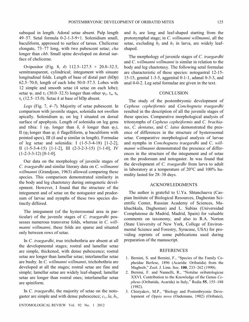
subequal in length. Adoral setae absent. Palp length 49–57. Setal formula 0-2-1-3-9+1. Solenidium small, baculiform, appressed to surface of tarsus. Chelicerae elongate, 73–77 long, with two pubescent setae; cha longer than chb. Small spine developed on dorsal sur-face of chelicerae.
Ovipositor (Fig. 8, 4) 112.5–127.5 × 20.0–32.5, semitransparent, cylindrical; integument with sinuate longitudinal folds. Length of base of distal part (bDp) 62.5–70.0, length of each lobe 50.0–57.5. Lobes with 12 simple and smooth setae (4 setae on each lobe); setae ψ1 and τ1 (30.0–32.5) longer than other ψ2, τa, τb, τc (12.5–15.0). Setae k at base of bDp absent.
Legs (Fig. 7, 4–7). Majority of setae pubescent. In comparison with juvenile stages, solenidia not swollen apically. Solenidium φ2 on leg I situated on dorsal surface of apophysis. Length of solenidia on leg genu and tibia: I (φ1 longer than δ, δ longer than φ2), II (φ2 longer than φ; δ flagelliform, φ baculiform with pointed apex), III (δ and φ similar in length). Formulae of leg setae and solenidia: I (1-5-3-4-18) [1-2-2], II (1-5-3-4-15) [1-1-2], III (2-3-2-3-15) [1-1-0], IV (1-2-3-3-12) [0-1-0].
Our data on the morphology of juvenile stages of C. traegardhi and similar literary data on C. willmanni willmanni (Grandjean, 1963) allowed comparing these species. This comparison demonstrated similarity in the body and leg chaetotaxy during ontogenetic devel-opment. However, I found that the structure of the integument and of setae on the notogaster and prodor-sum of larvae and nymphs of these two species dis-tinctly differed.
The integument (of the hysterosomal area in par-ticular) of the juvenile stages of C. traegardhi pos-sesses numerous transverse folds, whereas in C. will-manni willmanni, these folds are sparse and situated only between rows of setae.
In C. traegardhi, true trichobothria are absent at all the developmental stages; rostral and lamellar setae are simple, thickened, with dense pubescence; rostral setae are longer than lamellar setae; interlamellar setae are bushy. In C. willmanni willmanni, trichobothria are developed at all the stages; rostral setae are fine and simple; lamellar setae are widely leaf-shaped; lamellar setae are longer than rostral ones; interlamellar setae are spiniform.
In C. traegardhi, the majority of setae on the noto-gaster are simple and with dense pubescence; c1, la, h1,
and h3 are long and leaf-shaped starting from the protonymphal stage; in C. willmanni willmanni, all the setae, excluding h2 and h3 in larva, are widely leaf-shaped.
The morphology of juvenile stages of C. traegardhi and C. willmanni willmanni is similar in relation to the body and leg chaetotaxy. The following setal formulae are characteristic of these species: notogastral 12-15-15-15, genital 1-3-5, aggenital 0-1-1, adanal 0-3-3, and anal 0-0-2. Leg setal formulae are given in the text.
CONCLUSION
The study of the postembryonic development of Cepheus cepheiformis and Conchogneta traegardhi resulted in the description of all the juvenile stages of these species. Comparative morphological analysis of tritonymphs of Cepheus cepheiformis and C. brachia-tus, C. dentatus, and C. latus demonstrated the pres-ence of differences in the structure of hysterosomal setae. Comparative morphological analysis of larvae and nymphs in Conchogneta traegardhi and C. will-manni willmanni demonstrated the presence of differ-ences in the structure of the integument and of setae on the prodorsum and notogaster. In was found that the development of C. traegardhi from larva to adult in laboratory at a temperature of 20°C and 100% hu-midity lasted for 28–38 days.
ACKNOWLEDGMENTS
The author is grateful to U.Ya. Shtanchaeva (Cas-pian Institute of Biological Resources, Daghestan Sci-entific Center, Russian Academy of Sciences, Ma-khachkala, Daghestan) and L. Subías (Universidad Complutense de Madrid, Madrid, Spain) for valuable comments on taxonomy, and also to R.A. Norton (State University of New York, College of Environ-mental Science and Forestry, Syracuse, USA) for pro-viding reprints of some publications used during preparation of the manuscript.
REFERENCES 1. Bernini, S. and Bernini, F., “Species of the Family Ce-
pheidae Berlese, 1896 (Acarida: Oribatida) from the Maghreb,” Zool. J. Linn. Soc. 100, 233–262 (1990).
2. Bernini, F. and Nannelli, R., “Notulae oribatologicae XXVI. Contribution to the Knowledge of the Genus Ce-pheus (Oribatida, Acarida) in Italy,” Redia 55, 155–188 (1982).
3. Chistyakov, M.P., “Biology and Postembryonic Deve-lopment of Oppia nova (Oudemans, 1902) (Oribatei),

ERMILOV
ENTOMOLOGICAL REVIEW Vol. 92 No. 1 2012
126
the Dominating Species in Industrial Peatbogs of Gorky Province,” Uchenye Zapiski Gork. Ped. Inst. 114, 51–64 (1970).
4. Golosova, L.D., “Family Autognetidae Grandjean, 1960,” in A Key to Soil Dwelling Mites. Sarcoptiformes (Nauka, Moscow, 1975), pp. 223–225 [in Russian].
5. Grandjean, F., “Essai de classification des Oribates (Acariens),” Bull. Soc. Zool. France 78, 421–446 (1953).
6. Grandjean, F., “Autogneta penicillum n. sp. (Ori-bates),”Acarologia 2, 345–367 (1960a).
7. Grandjean, F., “Les Autognetidae n. fam. (Oribates),” Acarologia 2, 575–609 (1960b).
8. Grandjean, F., “Les Autognetidae (Oribates). Deuxieme partie,” Acarologia 5, 653–689 (1963).
9. Lions, J., “Observations sur l’espèce Cosmogneta kargi Grandjean, 1963: la protonymphe et la tritonymphe compléments a l’etude du gnathosoma,” Acarologia 17, 346–363 (1975).
10. Michael, A.D., British Oribatidae. I–II. (Ray Soc., London, 1884–1888).
11. Shereef, G.M., “Observations on the Feeding, Repro-duction and Faeces Obtained from Oribatids Fed on Dif-ferent Species of Penicillum and Aspergillus,” Proc. IV
Coll. Intern. Zool. Sol., 1970; Ann. Zool. Ecol. An., Paris, 1971, pp. 165–175.
12. Shereef, G.M., Observations on Oribatid Mites in Laboratory Cultures, Vol. 14, pp. 281–291 (1972).
13. Sitnikova, L.G., “A Revision of Mites of the Family Cepheidae Berlese, 1896 (Acarina, Oribatei) with De-scription of New Species from the USSR,” Entomol. Obozr., No. 2, 446–462 (1975a).
14. Sitnikova, L.G., “Family Cepheidae Berlese, 1896, in A Key to Soil Dwelling Mites. Sarcoptiformes (Nauka, Moscow, 1975b), pp. 143–155 [in Russian].
15. Subías, L.S., “Listado sistemático, sinonímico u bio-geográfico de los acaros oribatidos (Acariformes: Oribatida) del mundo (1758–2002),” Graellsia, No. 60, Numero extraordinario, 3–305. Online version accessed in July, 2010, http: // www.ucm.es/info/zoo/Artropodos/ Catalogo.pdf.
16. Weigmann, G., “Hornmilben (Oribatida),” in Die Tier-welt Deutschlands. Teil 76 (Goecke and Evers, Keltern, 2006).
17. Willmann, C., “Moosmilben oder Oribatiden (Oribatei),” in Die Tierwelt Deutschlands. (Gustav Fis-cher, Jena, 1931), Vol. 22, pp. 79–200.