pontifÍcia universidade catÓlica do rio grande do … · faculdade de odontologia programa de...
TRANSCRIPT

PONTIFÍCIA UNIVERSIDADE CATÓLICA DO RIO GRANDE DO SUL
FACULDADE DE ODONTOLOGIA
PROGRAMA DE PÓS-GRADUAÇÃO EM ODONTOLOGIA
NÍVEL: DOUTORADO
ÁREA DE CONCENTRAÇÃO: ENDODONTIA
AVALIAÇÃO DO DESENVOLVIMENTO RADICULAR EM RESPOSTA ÀS PROTEÍNAS
DERIVADAS DA MATRIZ DO ESMALTE E À RESOLVINA E1: ESTUDO
EXPERIMENTAL EM DENTES DE RATOS COM RIZOGÊNESE INCOMPLETA E
NECROSE PULPAR
ASSESSMENT OF ROOT FORMATION IN RESPONSE TO ENAMEL MATRIX DERIVATIVE AND TO RESOLVIN E1: AN EXPERIMENTAL STUDY IN RAT IMMATURE
NECROTIC TEETH
ROBERTA KOCHENBORGER SCARPARO
PORTO ALEGRE
2011

Roberta Kochenborger Scarparo
AVALIAÇÃO DO DESENVOLVIMENTO RADICULAR EM RESPOSTA ÀS PROTEÍNAS
DERIVADAS DA MATRIZ DO ESMALTE E À RESOLVINA E1: ESTUDO
EXPERIMENTAL EM DENTES DE RATOS COM RIZOGÊNESE INCOMPLETA E
NECROSE PULPAR
ASSESSMENT OF ROOT FORMATION IN RESPONSE TO ENAMEL MATRIX DERIVATIVE AND TO RESOLVIN E1: AN EXPERIMENTAL STUDY IN RAT IMMATURE
NECROTIC TEETH
Tese apresentada ao Programa de Pós-Graduação
em Odontologia da Faculdade de Odontologia da
Pontifícia Universidade Católica do Rio Grande do
Sul como requisito para a obtenção do título de
Doutor em Odontologia, na área de concentração
de Endodontia.
Orientador: Prof. Dr. Eraldo Luiz Batista Júnior.
PORTO ALEGRE
2011

Dados Internacionais de
Catalogação na Publicação (CIP)
S286a Scarparo, Roberta Kochenborger Avaliação do desenvolvimento radicular em resposta às
proteínas derivadas da matriz do esmalte e à resolvina E1: estudo experimental em dentes de ratos com rizogênese incompleta e necrose pulpar = Assessment of root formation in response to enamel matrix derivative and to resolvin E1: an experimental study in rat immature necrotic teeth / Roberta Kochenborger Scarparo. – Porto Alegre, 2011. 92 f.
Tese (Doutorado) – Faculdade de Odontologia, Pós-Graduação Odontologia, PUCRS.
Orientador: Prof. Dr. Eraldo Luiz Batista Júnior.
1. Endodontia. 2. Infecção (Odontologia). 3. Dentes - Esmalte. I. Batista Junior, Eraldo Luiz. II. Título.
CDD 617.634
Bibliotecário Responsável
Ginamara Lima Jacques Pinto CRB 10/1204

Agradecimentos

AGRADECIMENTOS
Aos meus pais, Paulo Sergio Scarparo e Helena Beatriz Kochenborger
Scarparo por todo o amor dedicado, apoio e confiança. Por serem meus mais admirados
exemplos na vida.
A Reinaldo Benfica Neto pelo entusiasmo com que participa da minha vida. Pelo
companheirismo, cumplicidade, por me trazer alegrias e me fazer confiar na realização
dos nos nossos sonhos.
A minha avó Lacy Kochenborger, à minha madrinha Letícia Spano e aos meus
irmãos Eduardo Kochenborger Scarparo e Marcelo Kochenborger Scarparo pelo
carinho, amizade e presença que tanto me fazem bem.
À Pontifícia Universidade Católica do Rio Grande do Sul (PUCRS) e ao
Conselho Nacional de Desenvolvimento Científico e Tecnológico (CNPq) pelas
oportunidades oferecidas para o desenvolvimento deste estudo.
Ao meu orientador, Prof. Eraldo Luiz Batista Júnior por toda confiança
demonstrada e pelo incentivo. Meu agradecimento especial pelo convívio, excepcional
aprendizado proporcionado e por compreender e apoiar as frequentes “mudanças de
planos” que caracterizaram essa jornada.
Ao Prof. José Antônio Poli de Figueiredo pelos ensinamentos e oportunidades
oferecidas. Especialmente pelas palavras de incentivo e pelo exemplo de dedicação ao
ensino e à pesquisa.
Às queridas amigas Lenara Dondoni e Daiana Boettcher meu agradecimento
especial pelo carinho, companheirismo e intensa dedicação a esse estudo. Foi
maravilhoso ter dividido com vocês essa etapa da minha vida.
À professora Maria Ivete Bolzan Rockenbach pela disponibilidade e valiosa
colaboração no desenvolvimento da análise radiográfica desse estudo.
A Tiago Giulianni pelos ensinamentos e pela dedicação no processamento
histológico das amostras deste trabalho.
À professora Fernanda Morroni pela disponibilização do Laboratório de
Farmacologia Aplicada.
À professora Maria Martha Campos pela dedicação à construção dos recursos
necessários ao desenvolvimento de pesquisas nesta Universidade, e pela
disponibilização do Laboratório de Toxicologia Pré-Clínica. A ela, e também às
professoras Patrícia Poli Kopper e Fabiana Vier Pelisser, agradeço pelas brilhantes
aulas ministradas, experiências compartilhadas e “sessões de psicoterapia de grupo”.
Aos colegas do Mestrado e Doutorado em Endodontia da PUCRS pela união
demonstrada e excelente convívio. Saibam que foi um imenso prazer ter contado com
vocês nesses anos.
Aos queridos mestres e colegas Régis Burmeister dos Santos, João Ferlini Filho,
Marcus Vinicius Reis Só, Fabiana Soares Grecca e Elaine Freitas Fachin pelos

momentos vividos e pelo crescimento profissional proporcionado. Por manterem as portas
abertas e me incentivarem a seguir lutando.
A todos vocês meus sinceros agradecimentos.

Resumo

RESUMO
Os objetivos deste estudo foram: (a) desenvolver um modelo experimental para
testar estratégias de tratamento em dentes não-vitais com rizogênese incompleta,
utilizando os primeiros molares inferiores de ratos; (b) avaliar, nesse contexto, o efeito da
aplicação intracanal de proteínas de matriz do esmalte (EMD) e da Resolvina E1 (RVE1).
Inicialmente, o método a ser utilizado para interrupção da rizogênese foi testado,
comparando-se dentes hígidos e dentes que sofreram pulpectomia em estágio inicial da
formação das raízes (4 semanas de idade). As avaliações radiográfica e histológica
comprovaram o desenvolvimento de alterações periapicais e a interrupção da rizogênese
após pulpectomia, além de permitirem a adequação de períodos apropriados para testar
estratégias (3 semanas após pulpectomia) e avaliar seus resultados (3 e 6 semanas pós-
tratamento). Em outro grupo de animais, após interrupção da rizogênese os canais foram
irrigados com hipoclorito de sódio e solução salina e foram testadas medicações
intracanal com pasta poliantibiótica, EMD ou RvE1. Para o grupo controle, os dentes
foram mantidos sem tratamento e expostos ao meio oral. Os resultados radiográficos e a
intensidade da inflamação foram comparados por meio de análise de variância (ANOVA)
e posthoc de Bonferroni (p<0,05). Apenas a RvE1 apresentou redução significativa das
lesões periapicais no primeiro período (P<0.05), o que foi corroborado pela menor
resposta inflamatória (P<0.05). Já no segundo período, a pasta poliantibiótica e as EMD
promoveram resultados semelhantes aos da RvE1. Ainda que algumas amostras
apresentassem resultados insatisfatórios, o desenvolvimento radicular às expensas de
tecido cementóide ou osteóide pode ser observado. As EMD promoveram, além da
formação de tecidos mineralizados na região apical e externa das raízes, sua invaginação
para o interior do canal radicular. Tanto a RvE1 como o EMD apresentaram potenciais a
serem explorados para o tratamento de rizogênese incompleta e necrose pulpar. Estudos
adicionais devem otimizar os protocolos, fornecer informações sobre os eventos
moleculares e celulares envolvidos na formação radicular e avaliar resultados em
humanos.
Palavras Chave (termos MeSH):
Endodontia, apicificação, apicigênese, dentes não vitais, inflamação, odontogênese,
proteínas dentárias do esmalte, Resolvina E1
Palavras Chave (DeCS): Endodontia, odonogênese, necrose da polpa dentária, inflamação, mediadores da
inflamação

Abstract

ABSTRACT
The present study aimed at: (a) developing an experimental model for testing
treatment strategies in nonvital immature teeth, using the lower first molars of rats; (b)
evaluating the effects of intracanal medication with enamel matrix proteins (EMD) and
Resolvin E1 (RvE1). At first, the method to be used for arresting root development was
tested, comparing healthy teeth with teeth which underwent pulpectomy and were left
open since the initial stage of root development (four weeks-age). Radiographic and
histological findings proved that induction of periapical lesions and arrest of root
development were achieved. Moreover, these data allowed the definition of appropriate
periods for testing treatment protocols (3 weeks after pulpectomy) and for evaluating its
results (3 and 6 weeks post-treatment). In another group of animals, after arresting root
development, disinfection using sodium hypochlorite and saline solution was carried out
and intracanal medication with either polyantibiotic paste, EMD or RvE1 was tested. At
the control group, no treatment was performed and teeth cavities were left exposed to
the oral environment. Radiographic and histological data were evaluated using two-way
ANOVA and Bonferroni post-hoc (P<0.05). At the first time point, only the teeth
subjected to RvE1 intracanal medication showed reduced periapical lesions (P<0.05),
which was corroborated by the reduced inflammatory response (P<0.05). At the second
time point, polyantibiotic paste, EMD and RvE1 showed similar results. Although some
samples showed unsatisfactory results, root development could be observed, mainly at
the expenses of cementum-like or bone-like tissues. EMD allowed, in addition to hard
tissue formation at the apical and external portion of roots, its ingrowth into the root
canal spaces. RvE1 as EMD presented a potential to be explored in nonvital immature
teeth. Further studies should focus in the optimization protocol, cellular and molecular
events that take part during root formation and treatment outcome in humans.
Keywords (MeSH terms): Endodontics, apexification, apexogenesis, nonvital teeth, inflammation, odontogenesis,
dental enamel proteins, Resolvin E1
Keywords (DeCS): Endodontics, odontogenesis, dental pulp necrosis, inflammation, inflammation mediators

Sumário

SUMÁRIO
1. INTRODUÇÃO GERAL 14
1.1- Proteínas derivadas da matriz do esmalte 17
1.2 - Mediadores lipídicos pró-resolução da resposta inflamatória 19
1.3- Objetivos 21
2. CAPÍTULO I 22
Artigo 1: Response to intracanal medication in immature teeth with pulp
necrosis: an experimental model in rat molars.
3. CAPÍTULO II 40
Artigo 2: Assessment of root formation in response to Resolvin E1 (RvE1) and enamel matrix derivative (Emdogain®): an experimental study in rat immature necrotic teeth.
4. DISCUSSÃO GERAL 63
5. CONCLUSÕES 73
6. REFERÊNCIAS BIBLIOGRÁFICAS 75
7. ANEXOS 90

1. Introdução Geral

15
1. INTRODUÇÃO GERAL
Tradicionalmente, o manejo endodôntico dos dentes com rizogênese incompleta e
necrose pulpar visa à apicificação por meio da utilização de hidróxido de cálcio ou
selamento com agregado trióxido mineral (MTA). Embora apresentem boa previsibilidade
e elevado percentual de redução de lesões periapicais, essas condutas clínicas possuem
limitações importantes, como o não desenvolvimento completo da raiz dentária, mantendo
essa estrutura frágil e aumentado o risco de fraturas (FRANK et al., 1966; CVEK, 1992;
ANDREASSEN, FARIK & MUNKISGAARD, 2002; SIMON et al., 2007).
A relevância dessas limitações fez com que, já a partir da década de 60, alguns
autores buscassem averiguar a possibilidade de desenvolvimento radicular em dentes
imaturos com polpa necrosada (OSTBY, 1961; RULE, 1966; HAM, PATTERSON,
MITCHELL, 1972). Entretanto, falhas decorrentes do pouco conhecimento disponível
acerca de aspectos da regeneração, de materiais inadequados, e de desinfecção
insuficiente produziram resultados insatisfatórios (OSTBY 1961; NYGAARD-OSTBY,
HJORTDAL, 1971; CZVEC, NORD, HOLLANDER, 1976; HORSTED, NYGAARD-OSTBY,
1978; NEVINS et al., 1976; HARGREAVES et al., 2008)
Atualmente, alguns relatos de casos tem afirmado a possibilidade da indução do
desenvolvimento radicular em dentes que tradicionalmente seriam tratados visando à
apicificação (JUNG, LEE, HARGREAVES, 2008; COTTI, MEREU, LUSSO, 2008).
Provavelmente, os protocolos de tratamento descritos favoreçam a manutenção da
viabilidade e a estimulação de células-tronco presentes no tecido pulpar (eventualmente
remanescente), no ligamento periodontal e na região da papila apical (GRONTHOS et al.,
2000; GRONTHOS et al., 2002; SONOYAMA et al., 2008; HUANG et al., 2008).
16
Os protocolos clínicos que visam ao desenvolvimento radicular após necrose
pulpar são bastante variados. De modo geral, é indicada a irrigação com hipoclorito de
sódio e a aplicação de medicação intracanal com uma pasta composta por metronidazol,
ciprofloxacina e minociclina (WINDLEY et al., 2005; CHUEH, HUANG, 2006; BOSE,
NUMMIKOSKI, HARGREAVES, 2009). Após o período de manutenção dessa medicação,
alguns autores sugerem que a pasta seja removida e que sejam induzidos sangramento e
formação de coágulo intracanal, por sobre o qual será realizado selamento do terço
cervical das raízes (IWAYA, IKAWA, KUBOTA, 2001; BANCHS & TROPE, 2004). Por
outro lado, há relatos de desenvolvimento radicular em dentes necrosados sem que seja
necessária a formação do coágulo (COTTI, MEREU & LUSSO, 2008; BOSE,
NUMMIKOSKI & HARGREAVES, 2009).
Os poucos estudos realizados até o momento com o intuito de testar a eficácia
desses protocolos comprovam que a complementação da formação radicular após
necrose pulpar é possível (SHAH et al., 2008; THIBODEAU et al., 2007 ; WANG et al
2010, DA SILVA et al., 2010). Por outro lado, apesar dos protocolos sugeridos visarem à
revascularização do espaço endodôntico, estudos em cães comprovam que na maioria
dos casos ocorre aposição de tecido cementóide ou osteóide permitindo o aumento da
espessura das paredes dentárias e do comprimento radicular (DA SILVA et al., 2010.,
WANG et al., 2010).
Apesar da aposição desses tecidos aumentar a resistência radicular à fratura,
cumprindo seu papel em reduzir perdas dentárias, a previsibilidade dos tratamentos
sugeridos ainda é limitada, não havendo parâmetros estabelecidos para seleção de casos
e percentual de sucesso clínico (DING et al., 2009). Sendo assim, o desenvolvimento de
alternativas que favoreçam de sucesso desses procedimentos é almejado.
A aplicação de mediadores que atuam durante o desenvolvimento embriológico dos

17
dentes é uma hipótese ainda não testada. É sabido que a secreção de proteínas
derivadas da matriz do esmalte (EMD) pela bainha epitelial de Hertwig leva à sinalização
ectomesenquimal recíproca, desencadeando uma cascata de reações que conduzem à
diferenciação de odontoblastos, à formação de dentina, à cementogênese e ao
desenvolvimento de estruturas periodontais de suporte (LINDSKOG, 1982; BROOKES et
al.,1995; HAMMARSTRÖM, 1997; NAKAMURA et al., 2001)
Em razão de seu papel fundamental durante a embriogênese dentária, as proteínas
de matriz do esmalte tem sido testadas com sucesso para diversas aplicações clínicas,
tais como a de regeneração periodontal, capeamento pulpar/pulpotomia e prevenção de
reabsorções dentárias e de anquilose em casos de avulsão (ZETTERSTRÖM et al.,1997;
PONTORIERO, WENNSTROM, LINDHE, 1999; NAKAMURA et al., 2001; FILLIPI, POHL,
VON ARX, 2002; ISHIZAKI et al., 2003; BOSSHARDT & NANCI, 2004; OLSSON et al.,
2005). Entretanto, seu emprego na estimulação do desenvolvimento radicular de dentes
com rizogênese incompleta e necrose pulpar até o momento não foi investigado.
Outro aspecto, ainda não explorado na Endodontia, é a aplicação de mediadores
lipídicos que atuam na resolução da resposta inflamatória. Estudos prévios comprovam
que a aplicação de Resolvina E1 (RvE1) suprime a resposta inflamatória e a perda óssea
induzida por bactérias na doença periodontal, mesmo sem intervenção mecânica sobre o
biofilme bacteriano (HASTURK et al., 2006; HASTURK et al., 2007; SERHAN 2007).
O princípio de atuação desses mediadores merece ser explorado em infecções
endodônticas, especialmente em casos de rizogênese incompleta, quando a
instrumentração do canal é limitada dada a fragilidade das paredes dentárias e pela
necessidade de manter a viabilidade das células-tronco.
1.1 Proteínas derivadas da matriz do esmalte
O Emdogain (Straumann AG, Basel, Suíça) é um gel de propilenoglicol alginato

18
que contém proteínas derivadas da matriz do esmalte secretadas pela bainha epitelial de
Hertwig durante o desenvolvimento dentário. Seu principal componente é a amelogenina,
mas também contém enamelinas, tuftelinas, e ameloblastinas (HAMMARSTRÖM 1997;
ZETTERSTRÖM et al., 1997; PONTORIERO, WENNSTROM, LINDHE, 1999;
NAKAMURA et al., 2001; BOSSHARDT & NANCI, 2004)
A indução promovida por essas proteínas simula parte da odontogênese normal,
facilitando processos regenerativos de tecidos de origem mesenquimal. Acredita-se que
as proteínas da matriz do esmalte participem da sinalização ectomesenquimal recíproca,
a qual controla o desenvolvimento embrionário dos dentes (HAMMARSTRÖM, 1997).
Sendo assim, desempenham papel importante na diferenciação e maturação de células
odontoblásticas, na regulação da mineralização do esmalte e na formação das estruturas
periodontais. Também estimulam a regeneração de tecidos periodontais, como o cemento
acelular, o ligamento periodontal e o osso alveolar, simulando o desenvolvimento dentário
(HAMMARSTRÖM, 1997; ZETTERSTRÖM et al., 1997; PONTORIERO, WENNSTROM,
LINDHE, 1999; NAKAMURA et al., 2001; BOSSHARDT & NANCI, 2004).
Em tratamentos conservadores da polpa, as proteínas da matriz do esmalte
promovem a cascata clássica de regeneração tecidual e reparo de maneira mais intensa
e rápida que o hidróxido de cálcio. A polpa subjacente ao novo tecido formado apresenta-
se livre de inflamação e há diferenciação de odontoblastos. Além disso, a formação de
dentina inicia-se à distância do local onde a polpa foi amputada, havendo também uma
marcada tendência à angiogênese nas regiões mais profundas, o que revela o aumento
do nível do crescimento e metabolismo celular. O tecido duro formado inicialmente é
semenlhante à osteodentina, mas torna-se semelhante à dentina secundária normal, com
odontoblastos e túbulos inseridos (NAKAMURA et al., 2002).
De acordo, alguns autores apontam vantagens na utilização de EMD em
19
detrimento do hidróxido de cálcio em tratamentos pulpares conservadores, tais como não
promover a atresia dos condutos e da câmara pulpar (NAKAMURA et al., 2002; KAIDA et
al., 2008) e reduzir a sintomatologia pós-operatória (OLSSON et al., 2005; KAIDA et al.,
2008).
1.2 - Mediadores lipídicos pró-resolução da resposta inflamatória
O início do processo inflamatório é caracterizado pelo dano tecidual seguido da
liberação de mediadores químicos endógenos (como leucotrienos e citocinas) e exógenos
(como mediadores químicos de origem microbiana) que agem atraindo células
polimorfonucleares. Os neutrófilos atuam na fagocitose de microorganismos e
degradação de restos celulares. Em algumas situações, entretanto, pode ocorrer a
liberação extracelular do conteúdo de grânulos lisossomais dessas células, ricos em
enzimas de degradação e espécies reativas de oxigênio, levando à amplificação do dano
celular e da resposta inflamatória. Paralelamente, mediadores químicos endógenos
podem, inadvertidamente, promover ativação e recrutamento excessivo de neutrófilos,
contribuindo com o dano tecidual ainda que sejam produzidos como parte importante da
defesa do hospedeiro (WEISSMAN, SMOLEN, KORCHACK, 1980; SERHAN, 2008).
Nesse sentido, após a remoção de materiais nocivos por meio da fagocitose, deve
haver a resolução da resposta inflamatória, evitando sua ampliação e conseqüente
cronificação ou manutenção de quadros patológicos. O termo “resolução” refere-se à
autolimitação do quadro inflamatório agudo, o qual é caracterizado pela redução ou
remoção de leucócitos e de restos celulares do sítio inflamatório, permitindo ao tecido o
retorno à homeostase (WEISSMAN, SMOLEN, KORCHACK, 1980; SERHAN, 2007;
SERHAN et al., 2008)
Recentemente, a resolução foi caracterizada como um processo bioquímico e
metabólico ativo, rapidamente iniciado por mecanismos celulares após as fases iniciais da

20
inflamação aguda. Esse processo se dá pela biossíntese de mediadores lipídicos “pró-
resolução”, como lipoxinas, resolvinas e protectinas, os quais atuam como agonistas na
redução do infiltrado inflamatório em tecidos inflamados, além de promover a eliminação
de células apoptóticas e de microrganismos pelos macrófagos (LEVY et al., 2001;
SERHAN et al., 2000; SERHAN, 2007).
Esses mediadores são derivados de ácidos graxos poli-insaturados. O ácido
aracdônico (AA) dá origem às lipoxinas, o ácido eicosapentaenóico (EPA) às resolvinas
da série E, e o ácido docosahexaenóico (DHA) às resolvinas da série D e às protectinas
(SERHAN et al., 2008.).
O termo “resolvinas” foi introduzido com o intuito de explorar as características de
mediadores endógenos biossintetizados durante a fase de resolução do exsudato
inflamatório, os quais processam potentes ações antiinflamatórias e imunorregulatórias.
No presente estudo, foi explorada a ação da Resolvina E1 (RvE1), composta de
5S,12R,18R-trihidroxi-6Z,8E,10E,14Z,16E ácido ecosapentaenóico. Resumidamente, a
RvE1 é derivada da oxigenação do EPA, em um processo que leva a formação produtos
intermediários como o 18R - ácido hidroperoxieicosapentaenóico (18R-HPEPE) o qual
libera o 18R- ácido hidroxieicosapentaenóico (18R-HEPE). Este é rapidamente
transformado pela ação de lipoxigenase-5 de neutrófilos para que seja formado o
composto bioativo (SERHAN et al., 2000; ARITA et al., 2005).
Estudos prévios demonstraram que a RvE1 é um potente regulador da
transmigração de neutrófilos e da inflamação, sendo também atribuído a esse composto
bioativo a estimulação de fagocitose não flogística de neutrófilos apoptóticos pelos
macrófagos (SERHAN et al., 2000; ARITA et al., 2005; ARIEL et al., 2006; ARITA et al.,
2007; SCHUWAB et al., 2007). Ensaios pré-clínicos revelam que a RvE1 apresenta efeito
protetor na doença periodontal, com redução do infiltrado de neutrófilos, evitando a perda

21
óssea e de tecido conjuntivo, promovendo a cicatrização de tecidos danificados e a
regeneração de tecido ósseo e do ligamento periodontal (HASTURK et al., 2006;
HASTURK et al., 2007).
1.3 Objetivos
O presente estudo tem como objetivos:
- Desenvolver um modelo experimental que permita a avaliação de estratégias de
tratamento para dentes com rizogênese incompleta e necrose pulpar, utilizando
molares de ratos.
- Avaliar o efeito da medicação intracanal com proteínas derivadas da matriz do
esmalte (Emdogain) e com o mediador lipídico pró-resolução inflamatória
Resolvina E1 no reparo de lesões periapicais e no desenvolvimento radicular de
dentes de ratos com rizogênese incompleta.

22
2. Capítulo I

23
2. CAPÍTULO I
Artigo 1
Response to intracanal medication in immature teeth with pulp necrosis: an
experimental model in rat molars.
Submetido ao periódico Journal of Endodontics, qualis A1 e fator de impacto 2.953
(Anexo A).

24
Response to intracanal medication in immature teeth with pulp necrosis: an
experimental model in rat molars.
Roberta Kochenborger Scarparo, MSc1, Lenara Dondoni, DDS1, Daiana Elisabeth
Böttcher, DDS2, Fabiana Soares Grecca, PhD2, Maria Ivete Bolzan Rockenbach PhD1,
Eraldo Luiz Batista Júnior, PhD1.
1. Pontifical Catholic University of Rio Grande do Sul – PUCRS
2. Federal University of Rio Grande do Sul - UFRGS
CorrespondingAuthor :
Roberta Kochenborger Scarparo / Eraldo L. Batista Jr.
Av. Ipiranga, 6681 - Prédio 6
Cep.: 90619-900
Porto Alegre - RS - Brazil
(51) 3320-3562/3573
(51) 3320-3626/3609
[email protected] [email protected]
ACKNOWLEDGMENTS This study was supported by grants from Conselho Nacional de Desenvolvimento
Científico e Tecnológico (CNPq), a Brazilian Governmental Institution. The authors are
grateful to Tiago Giulianni for technical assistance. The authors deny any conflicts of
interest.

25
Abstract
Objective: To characterize an experimental model in rats aiming at evaluating treatment
strategies in necrotic immature teeth.
Methods: To define the periods to be adopted in the experimental procedures and to
confirm interruption of root embryogenesis, the left lower first molars of Wistar rats
aging 4-weeks (n=24) underwent pulpectomy and were left open to the oral
environment. Vital teeth were observed on the right lower first molars. In another group
of animals (n=36) the teeth were left open for three weeks, and then received
interventions for disinfection. Changes in root formation were determined based on
radiographic and histological evaluation.
Results: vital teeth showed an increase of root length and hard tissue thickness over the
proposed experimental periods. On the other hand, induction of necrosis arrest root
formation. Teeth subjected to disinfection with sodium hypochlorite and polyantibiotic
paste showed significant reduction of periapical lesions, gain in root length and
increased walls thickness compared to the control (P<0.05).
Conclusion: The protocol tested for root canal disinfection was favorable for periapical
lesions reduction and root development over the experimental periods. The
experimental model presented should contribute to studies that aim at improving
therapeutic strategies for necrotic immature teeth using a rat model.
Keywords rat model, apexogenesis, nonvital teeth, inflammation, odontogenesis
26
Introduction
Pulp necrosis of immature teeth arrest tooth development, resulting in
incompletely formed roots with wide open apices and fragile structure (1). Apexification
is considered efficient for endodontic repair, but still fall short in inducing increase of
root tickness and length (1-4).
Recently, many authors have proposed regenerative therapies that involve the
preservation of stem cells from the apical papilla (5-11). Nevertheless, the ability of
these strategies to promote predictable reconstruction of dental tissues has yet to be
properly addressed. For these reasons, the mechanisms of root development (12-16),
the definition of new treatment modalities (17-18) and the characterization of their
pathways and outcomes (19) have gained great attention.
Ethical and technical issues have a role in restricting investigations using human
subjects, thus requiring implementation of animal models that can reproduce clinical
outcomes (19). Rat molars, including pulp and periodontal tissues, resemble in many
ways those of humans (20-22,24-29). The use of rats offers economical advantages
(20), availability of molecular tools and access to genetic databanks (13,29). This model
has been widely used in orthodontics (21), periodontics (22-23), conservative treatment
of dental pulp (20) and studies of molecular mechanisms involved in teeth
embryogenesis (13), but treatment strategies for immature teeth still have been
investigated mainly in dogs (17-19).
Considering the aforementioned, the present study aimed at developing an
experimental model in rat molars for evaluating treatment strategies in necrotic
immature teeth.

27
Methods
The study protocols were approved by Federal University of Rio Grande do Sul
and Pontifical Catholic University of Rio Grande do Sul Institutional Animal Care and
Use Committees (Protocol 10/00156 and 19001)
The sample consisted of 60 male Wistar rats. Twenty-four animals were used to
define the periods to be adopted in the experimental procedures and to confirm
induction of periapical lesions and interruption of root embryogenesis. The remaining 36
were used for testing methods for the treatment of rat immature first molars .
To perform the experimental procedures, the animals were anesthetized
intraperitoneally with ketamine (0.8 ml/100g) and Xilasine (0.2ml/100g). Mouth opening
was achieved by using a designed device, and the soft tissues were kept away with the
aid of dental forceps. The device was made using approximately 6 cm of a 0.8
orthodontic wire. The wire was first folded in a rectangular shape with sides measuring
1 and 2 cm. Then, the central portion of both sides measuring 2 cm were bent in an
angle of about 45o, forming a convex arc. The sides of the arc measuring 1 cm were
placed on contact with the lingual and palatal surfaces of lower and upper incisors
(Figure 1A).
Definition of experimental periods and confirmation of root embryogenesis arrest
Twenty-four right mandibular first molars were used for the observation of
natural embryogenesis. Pulp necrosis was induced on the left mandibular first molars
during the initial stage of root development (animals aging 4-weeks old). Dental pulps
were exposed by drilling cavities on the central portion of the oclusal face, with a 1011
HL round bur in high speed (KGSorensen, Cotia, SP, Brazil) to a depth nearly equal to
the bur diameter (1 mm). An # 25 endodontic file (Dentsply Maillefer, Ballaigues,
Switzerland) was then used to remove remmants of pulpal tissue. The teeth were left

28
open to the oral environment throughout the course of the experiment. The time needed
for the detection of periapical lesions was confirmed by radiographs taken 1, 2 and 3
weeks after pulpectomy as previously reported (30).
Animals aging 7, 10, 13 and 16 weeks (n=6 per period) were euthanized by
inhalation of isoflurane. The jaws were dissected for radiographic and histological
evaluation.
Methods for implementation of treatment protocols
Pulp necrosis was induced as described above in 36 animals aging 4 weeks-old.
In 18 animals teeth were left open through the course of the experiment. In the other 18,
teeth were left open for three weeks, and then received intervention for disinfection of
the target teeth.
Root canal disinfection
Debris were removed from the cervical third of the roots using a # 25 endodontic
file, inserted to a maximum depth of 2mm to avoid injury to the apical portion of the
canals. The canals were irrigated with of 2.5% sodium hypochlorite followed by 0.9%
sterile saline solution, using anesthetic tubes filled with the solutions, long needles, a
carpule syringe and endodontic suction apparatus. The canals were dried with
absorbent paper points and filled with a polyantibiotic dressing, comprised of
metronidazol, ciprofloxacin and minocycline (50 mg/ml) (Pharma&Cia, Porto Alegre,
RS, Brazil) using an insulin syringe. Teeth were sealed with sterile cotton pellets and
silver amalgam. The animals were divided into three experimental periods (n= 6 per
group) in which the polyantibiotic paste remained for 3, 6 or 9 weeks. After euthanizia
by inhalation of isoflurane, the jaws were dissected for radiographic and histological
evaluation.
29
Radiographs and Image Analysis
The X-ray cylinder was fitted in a way as to form a perpendicular angle with the
buccal surface of the first molar. A focal distance of 30 cm was observed. The X-ray unit
(Gnatus, Ribeirão Preto, SP, Brazil) operated at 7 mA at 70 kVp, with a size 2 phosphor
plate (Gendex, Chicago, IL, USA) and exposure time of 0.2 seconds. Digital x-ray
system (Denoptix/Gendex, Chicago, IL, USA) was used to capture images scanned at
the resolution of 300 d.p.i. and saved in TIFF format.
Image analysis was performed using a software (Image Tool version 3.0,
UTHSCSA, USA). For root length measurements, a linear trace from the pulp chamber
floor to the most apical portion of the mesial root was created. Dental wall thickness at
the apical third was estimated by calculating the percentage of the linear measurement
of the mesial root canal width relative to the linear measurement of the entire mesial
root width. Periapical lesion area at the mesial root was demarcated and measured.
Comparisons of data obtained in each period were performed using two-way
ANOVA, one-way ANOVA and Bonferroni post-hoc. Differences were regarded
significant when P<0.05.
Sample preparation and histological analysis
Samples were fixed with buffered 10% paraformaldehyde for 24 h, decalcified in
17% EDTA for 5 weeks, dehydrated in ascending concentrations of ethanol and
embedded in paraffin. Five-µm serial sections were stained with hematoxylin and eosin.
Three sections were selected for each sample, so the central portion of the roots,
including the apex, was visible. A histological descriptive analysis was performed.
Results
30
Radiographic analysis of vital teeth showed an increase of root length and
thickness over the experimental periods. Pulp exposure to the oral cavity promoted
development of periapical lesions, significant reduction of root length and wall thickness
(Figures 1B and 1C).
Histological evaluation showed progressive apical closure of vital teeth. Root
development was complete in most of the animals aging 13 weeks-old, and continuous
dentin and cementum formation could be observed in internal and external root
surfaces. On the other hand, pulp exposure determined intense inflammatory response
and arrest of root embryogenesis, that lead to open apices and thin dental walls (Figure
2).
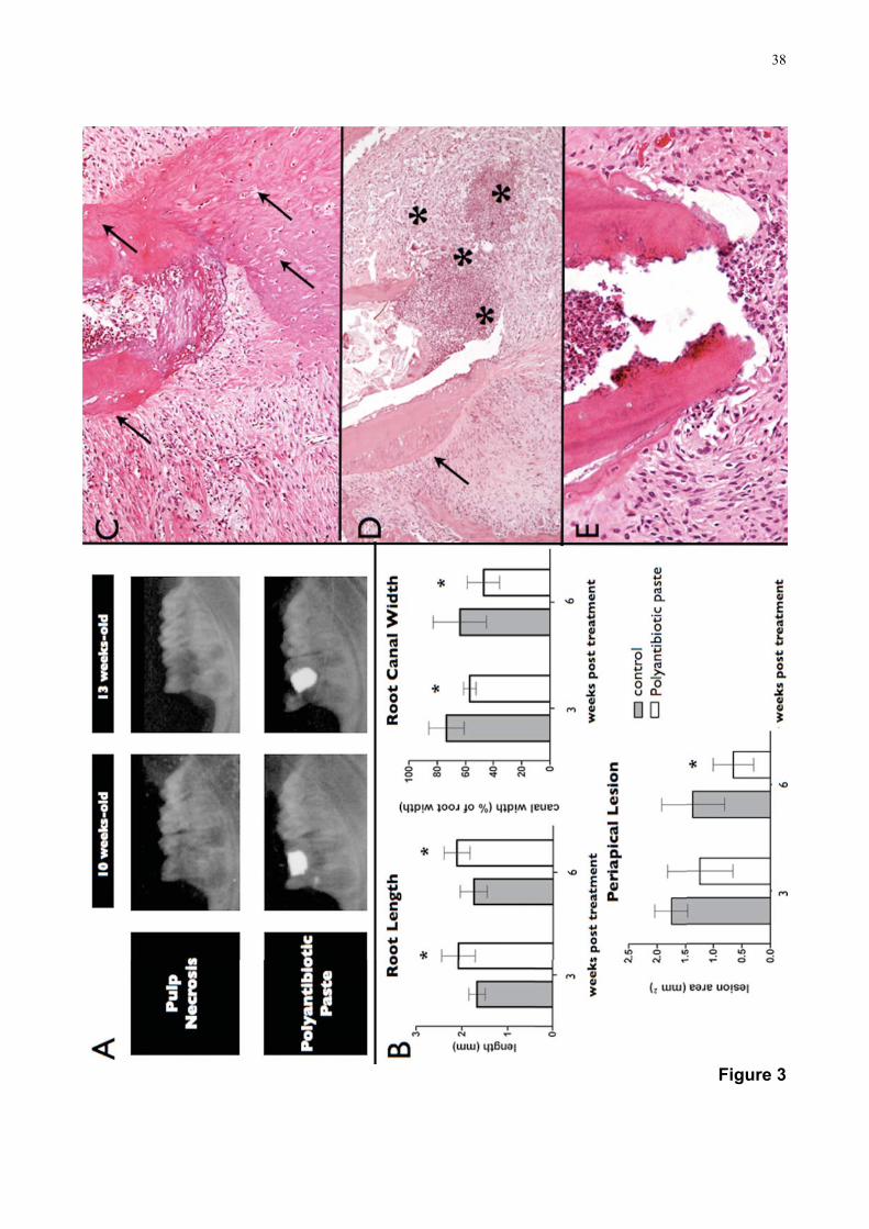
Due to tooth fracture and loss of coronal sealing at the third experimental period
(9 weeks post treatment), teeth subjected to polyantibiotic medication were evaluated 3
and 6 weeks post treatment only. After 6 weeks, teeth presented reduced periapical
lesions and increased root lengths compared to the control (P<0.05). Canal width was
reduced compared to the first period (Figures 3A and 3B), depicting increased wall
thickness (P<0.05).
Histological analysis showed variable inflammatory response to treatment; about
half of the roots showed formation of a cementum-like tissue on its apical portion and
newly formed cementum on the external surfaces (Figures 3C, 3D and 3E).
Discussion
Previous studies showed that pulp exposure of rat molars pulps promotes an
inflammatory reaction identical to that observed in humans (26-28,31), which was
corroborated here. Apart from similar host responses (29), the oral bacteria flora of rats
is more comparable to humans than other commonly used species in research (24-
26,32). Furthermore, biologically, the response progresses faster in rats (31,33), which

31
may be a favorable point when it comes to promptly obtaining data that enable the
continuous development of therapeutic strategies (33). The application of molecular
tools is a routine for this species (13,29), favoring the development of investigations that
focus on biological mechanisms involved in the treatment protocols.
Contamination of root canal after pulp exposure to the oral cavity was critical in
arresting root development as a response to inflammation of apical periodontal tissues.
Radiographic and histological analysis of vital teeth showed that rats aging 7 weeks had
incomplete root development, equivalent to nearly half of its final full length. According
to these data and considering the period needed for periapical lesion development, it
was established that first molars should be endodontically accessed in rats aging 4
weeks-old, and treatment protocols should be applied in animals aging 7 weeks-old.
Some technical problems had to be solved before the teeth could be used in this
experimental model for treatment strategies in immature teeth. The small size of the
teeth and difficulties related to access to the pulp chamber required some adaptations.
Therefore, appropriate training, proper position of rat’s head, mouth opening and soft
tissue removal are critical aspects in order to successfully carry out the procedures (20).
In the present study, complete relaxation of the animal through deep anesthesia (34),
and the use of a device designed to open and stabilize the rat mouth allowed for the
preparation of the operative field. Cavity preparation at the center of the oclusal surface
enabled the access to the three root canals. However, it increased fragility of the dental
structure. Thus, long periods favored tooth fracture, affecting coronal sealing, as
observed during the third experimental period. Moreover, incomplete root development
may offer some advantages related to technical issues; the larger lumen of root canals
and short root length favor location of the canals, adequate irrigation and canal dressing
fillings.

32
The treatment protocol tested promoted root development and was favorable for
periapical lesions repair, similarly to the results observed in studies employing dogs (17-
19). As previously reported (19), root development occurred at the expense of a
cementum-like tissue, corroborating that the rat model reproduces the results observed
in other animals.
On the other hand, investigation of more predictable therapeutic strategies to
obtain the continuity of root development follows as a challenge to be overcome. In
agreement, the experimental model presented herein should contribute to studies that
aim at improving therapeutic strategies for necrotic immature teeth.
References
1. Frank AL. Therapy for the divergent pulpless tooth by continued apical formation. J
Am Dent Assoc 1966;72:78-93.
2. Andreasen JO, Farik B, Munksgaard EC. Long-term calcium hydroxide as a root
canal dressing may increase risc of root fracture. Dent Traumatol 2002;18:134-7.
3. Cvek M. Prognosis of luxated non-vital maxillary incisors treated with calcium
hydroxide and filled with gutta-percha. Endod Dent Traumatol 1992;8:45-55.
4. Simon S, Rilliard F, Berdol A, Machtou P. The use of mineral trioxide aggregate in
one-visit apexification treatment: a prospective study. Int Endod J 2007;46:186-97.
5. Jung IY, Lee SJ, Hargreaves KM. Biollogically based treatment of immature
permanent teeth with pulpal necrosis: a case series. J Endod 2008;34:876-7.
6. Cotti E, Mereu M, Lusso D. Regenerative treatment of na immature, traumatized
tooth with apical periodontitis: a report of a case. J Endod 2008;34:611-6.
7. Chueh LH, Huang GT. Immature teeth with perirradicular periodontitis or abscess

33
undergoing apexogenesis: a paradigm shift. J Endod 2006;32:1205-13.
8. Sonoyama W, Liu Y, Yamaza T, Tuan RS, Wang S, Shi S, Huang GT.
Characterization of the apical papilla and its residing stem cells from human immature
permanent teeth: A pilot study. J Endod 2008;34:166-71.
9. Huang GT, Sonoyama W, Liu Y, Liu H, Wang S, Shi S. The hidden treasure in apical
papilla: the potential role in pulp/dentin regeneration and bioroot engineering. J Endod
2008;34:645-51.
10. Tziafas D, Kodonas K. Differentiation of dental papilla, dental pulp and apical papilla
progenitor cells. J Endod 2010;36:781-9.
11. Hargreaves KM, Giesler T, Henry M, Wang Y. Regeneration potential of the young
permanent tooth: what does the future hold? J Endod 2008;34:51-6.
12. Hosoya A, Kim JY, Cho SW, Jung HS. BMP4 signaling regulates formation of
Hertwig´s epithelial root sheath during tooth root development. Cell Tissue Res
2008;333:503-9.
13. Nakasone N, Yoshie H, Ohshima H. An immunohistohemical study of the
expression of heat-shock protein-25 and cell proliferantion in the dental pulp and
enamel organ during odontogenesis in rat molars. Arch Oral Biol 2006;51:378-86.
14. Madam AK, Kramer B. Immunolocalization of fibroblast growth factor-2 (FGF-2) in
the developing root and supporting structures of the murine tooth. J Mol Histol
2005;36:171-8.
15. Lee DS, Park JT, Kim HM, Ko JS, Son HH, Gronostajski RM, Cho M, Choung PH,
Park JC. Nuclear Factor I is essencial for odontogenic proliferation and odontoblast
differenciation during tooth root development. J Biol Chem 2009;284:17293-303.

34
16. Lee TY, Lee DS, Kim HM, Ko JS, Gronostajski RM, Cho M, Son HH, Park JC.
Disruption of NFIC causes dissociation of odontoblasts by interfering with the formation
of intercellular junctions and aberrant odontoblast differenciation. J Histochem
Cytochem 2009;57:469-76.
17. Thibodeau B, Teixeira F, Yamauchi M, Caplan DJ, Trope M. Pulp revascularization
of immature dog teeth with apical periodontitis. J Endod 2007;33:680-9.
18. da Silva LA, Nelson-Filho P, da Silva RA, Flores DSH, Heilborn C, Johnson JD,
Cohenca N. Revascularization and periapical repair after endodontic treatment using
apical negative pressure irrigation versus conventional irrigation plus triantibiotic
intracanal dressing in dogs´ teeth with apical periodontitis. Oral Surg Oral Med Oral
Pathol Oral Radiol Endod 2010;5:779-87.
19. Wang X, Thibodeau B, Trope M, Lin L, Huang GT. Histologic characterizantion of
regenerate tissues in canal space after the revitalization/revascularization procedure of
immature dog teeth with apical periodontitis. J Endod 2010;36:56-63.
20. Dammaschke T. Rat molar teeth as a study model for direct pulp capping research
in dentistry. Lab Anim 2010;44:1-6.
21. Oikawa T, Nomura Y, Arai C, Noda K, Hanada N, Nakamura Y. Mechanism of
active eruption of molars in adolescent rats. Eur J Orthod 2011; Epud ahead of print.
22. Duarte PM, Tezolin KR, Figueiredo MF, Bastos MF. Microbial profile of ligature-
induced periodontitis in rats. Arch Oral Biol 2010;55:142-7.
23. Okada Y, Hamada N, Kim Y, Takahashi Y, Sasaguri K, Ozono S, Sato S. Blockade
of sympathetic beta-receptors inhibits Porphyromonas gingivalis-induced alveolar bone
loss in an experimental rat periodontitis model. Arch Oral Biol 2010; 55:502-8.
24. Huxley HG. Histology of rat molar fissure plaque. Arch Oral Biol 1971;16:1311-28.

35
25. Huxley HG. The recovery of microorganisms from the fissure of rat molar teeth.
Arch Oral Biol 1972;17:1481-5.
26. Stashenko P, Wang CY, Tani-ishii N, Yu SM. Pathogenesis of induced rat
periapical lesions. Oral Surg Oral Med Oral Pathol 1994;78:494-502.
27. Stanley HR. Criteria for standardizing and increasing credibility of direct pulp
capping studies. Am J Dent 1998;11:S17-34.
28. Kakehashi S, Stanley HR, Fitzgerald RJ. The effects of surgical exposure of dental
pulps in germ-free and conventional laboratory rats. Oral Surg Oral Med Oral Pathol
1965;20:340-9.
29. Rat Genome Sequencing Project Consortium. Genome sequence of the Brown
Norway rat yields insights into mammalian evolution. Nature 2004;428:493-521.
30. Mahl CR, Fontanella V. Evaluation by digital subtraction radiography of induced
changes in the bone density of the female rat mandible. Dentomaxillofac Radiol
2008;37:438-44.
31. Muruzábal M, Eurasquin J. Discussion of: methods and criteria in evaluation of
periapical response. Int Dent J 1970;20:539-54.
32. Wunder JA, Briner WW, Calkins GP. Identification of cultivable bacteria in dental
plaque from the beagle dog. J Dent Res 1976;55:1097-102.
33. Moretton TR, Brown CE, Legan JJ, Kafrawy AH. Tissue reactions after
subcutaneous and intraosseous implantation of mineral trioxide aggregate and
ethoxybenzoic acid cement. J Biomed Mater Res 2000;52:528-33.
34. Maurice CG, Schour I. Experimental cavity preparations in the molar of the rat. J
Dent Res 1955;34:429-34.

36
Figure 1

37
Figure 2
38
Figure 3

39
Figure Legends
Figure 1. (A) Metal device designed for animal mouth opening during experiments.
(B)Radiographic aspects of necrotic and vital teeth in animals aging 7, 10, 13 and 16
weeks-old. (C) Radiographic analysis of root length and canal width showing significant
differences between vital and necrotic teeth during the course of the experiment -* P<0.05;
** P<0.01; ***P<0.001. In vital teeth only, mean differences of root length (◆◆◆P< 0.001)
and canal width (◆◆◆P< 0.001 ◆◆P< 0.01), were also observed among experimental
periods.
Figure 2. Histological aspects of vital and necrotic teeth in animals aging 7, 10, 13 and 16
weeks-old. Vital pulp tissue (P) and the absence of periapical inflammation allowed
formation of dentin (D) and cementum (C) on root internal and external surfaces. Complete
apex formation was observed after 13 weeks of age (arrows). After dental pulp exposure,
root canal (RC) infection lead to periapical inflammation, inducing either abscess (*) or
cystic lesions (CL). Wide open apex and thin dental walls could be observed through the
course of the experiment.
Figure 3. Response to canal disinfection. (A) Radiographic aspects of teeth after exposure
to oral cavity (pulp necrosis) and after disinfection procedures (polyantibiotic paste). (B)
Radiographic analysis of the polyantibiotic paste effects on periapical lesion area, root
length and root canal width showing significant differences relative to the control infected
teeth - *P<0.05. (C) Histologic evaluation showing variable healing outcomes in response
to intracanal medication even after the second experimental period: formation of a
cementum-like tissue on the apical portion and external surfaces (arrows); intense
inflammatory infiltrate (*) and cementum formation detectable only on root external
surfaces and distant from apical opening (arrows) (D); mild inflammatory infiltrate and
absence of detectable hard tissue on the root apex (E).

40
3. Capítulo II

41
3. Capítulo II
Artigo 2
Assessment of root formation in response to Resolvin E1 (RvE1) and enamel matrix derivative (Emdogain®): an experimental study in rat immature necrotic teeth.
Submentido ao periodico International Endodontic Journal, quails A2 e fator de impacto
2.223
(ANEXO B)

42
Introduction
Traditionally, endodontic treatment of immature teeth with pulp necrosis is based on
strategies as apexification with calcium hydroxide or placement of mineral trioxide
aggregate apical plugs. Although considered efficient for endodontic repair, these
strategies keep roots with wide open apices, reduced length and thin dental walls that are
prone to fracture (Simon et al. 2007, Friedlander et al. 2009, Huang 2009).
Recently, other clinical approaches have been suggested, concerning the
preservation and stimulation of stem cells from dental pulp, periodontal ligament and
apical papilla (Gronthos et al.2002, Seo et al. 2005, Sonoyama et al. 2008). Most of them
are based on root canal chemical disinfection, using sodium hypochlorite and a
polyantibiotic paste comprised of metronidazole, ciprofoxacin and minocycline (Windley et
al. 2005, Bose et al. 2009). After disinfection, some authors suggest that, before root
formation occurs, a blood clot should be stimulated by using endodontic hand files inserted
past the canal terminus into the periapical tissues (Banchs & Trope 2004). Case reports
and pre-clinical studies confirm that completion of root development in nonvital teeth is
possible, even in the absence of a blood clot (Bose et al. 2009, da Silva et al. 2010, Wang
et al. 2010). Nevertheless, the suggested therapeutic strategies are limited in their
predictability, and the continuous investigation of new protocols are warranted (Ding et al.
2009).
It is well documented that secretion of enamel matrix derivative proteins (EMD) by
the Hertwig epithelial sheath triggers a cascade of reactions that stimulates odontogenesis
(Sonoyama et al. 2007, Lyngstadaas et al. 2009). EMD, commercially available as
Emdogain (Straumann AG, Basel Switzerland), is well recognized in periodontology for its
regenerative properties (Lyngstadaas et al. 2009). In the conservative treatment of the
dental pulp, EMD induces reparative dentin formation, also protecting the pulp tissue from
inflammation (Nakamura et al. 2002, Igarashi et al. 2003, Olsson et al. 2005). Although the

43
growth factors present in EMD have a role during odontogenesis, its use to stimulate root
development in immature necrotic teeth has not been investigated thus far.
Another promising approach is the use of the lipid pro-resolution compond Resolvin
E1 (RvE1) to assist in the reduction of inflammatory response. Previous studies have
shown that RvE1 controls the inflammatory response and bone loss in periodontal
disease, even without mechanical intervention on the biofilm (Hasturk et al. 2006, Hasturk
et al. 2007). The effect of RvE1 in endodontic infections warrants investigation in vivo,
especially in immature teeth, which have root canal mechanical disinfection limited by the
fragility of dental walls and the need for maintaining dental stem cells viable. The present
study aimed at evaluating the effect of EMD (Emdogain) and Resolvin E1 on root
development of immature teeth with pulp necrosis.
Materials and Methods
Experimental Procedures
The study protocols were approved by Pontifical Catholic University of Rio Grande do
Sul Institutional Animal Care and Use Committees (Protocol 10/00156). Forty-eight (48)
male Wistar rats were used. Experimental procedures were carried out with the animals
anesthetized intraperitoneally with ketamine (0.8ml/100g) and xylasine (0.2ml/100g).
The experimental steps of the study are summarized on Figure 1. Endodontic access
in the lower first molars was performed in four weeks-old animals, in order to induce
periapical lesions and arrest root morophogenesis at this initial stage. Dental pulps were
exposed by drilling cavities on the central portion of the occlusal surface, with a 1011 HL
round bur (KGSorensen, Cotia, SP, Brazil) in high speed, to a depth nearly equal to the
bur diameter (1 mm). A # 25 endodontic file (Dentsply Maillefer, Ballaigues, Switzerland)
was then used to remove remnants of pulpal tissue. Periapical radiographs to verify the
lesion formation were performed as previously described (Mahl & Fontanella 2008).
44
Periapical radiolucency was evident following three weeks of cavities exposure to the oral
environment.
The animals were divided into four groups according to the treatment protocol. In the
control group, teeth were left open to the oral cavity throughout the whole course of the
experiment. In the other groups, teeth were left open to the oral environment for three
weeks and then treatment protocols were applied (7-weeks-old animals). First, for root
canal disinfection, debris was removed from the pulp chamber and cervical third of the
roots using a # 25 endodontic file (Dentsply Maillefer, Ballaigues, Switzerland). During the
implementation of therapeutic procedures, special care was taken in order not to
traumatize the apical portion of the root canals. Canals were irrigated with 2.5% sodium
hypochlorite followed by of 0.9% sterile saline solution and an endodontic suction
apparatus. The pulp chamber and the canals were dried with absorbent paper points and
the canals were filled with either of the following dressings: polyantibiotic paste consisting
of metronidazole, ciprofloxacin and mynocicline - 50 mg of each antibiotic per ml -
(Pharma & Cia, Porto Alegre, RS, Brazil); EMD - 30 mg per ml in propylene glycol alginate
- (Emdogain, Straumann AG, Basel, Switzerland) or Resolvin E1. The latter preparation
was delivered in ethanol and prepared from a stock solution of 50 mg/ml diluted in PBS to
1 μg/ml. The biocompounds were inserted into the canals with the aid of an insulin syringe
and endodontic files until filling root canal spaces. Endodontic access was sealed with
sterile cotton pellets and silver amalgam. The animals were divided into two experimental
periods (n= 6 per group), being euthanized by inhalation of isoflurane at the ages of 10
and 13 weeks (3 and 6 weeks post application of treatments, respectively). The jaws were
dissected for radiographic and histological evaluation.
Image Analysis of radiographs

45
An X-ray cylinder was fitted in a way as to form a perpendicular angle with the buccal
surface of the first molar. A focal distance of 30 cm was observed. The X-ray unit (Gnatus,
Ribeirão Preto, SP, Brazil) operated at 7 mA at 70 kVp, with a size 2 phosphor plate
(Gendex, Chicago, IL, USA) and exposure time of 0.2 seconds. Digital x-ray system
(Gendex, Chicago, IL, USA) was used to capture images scanned at the resolution of 300
d.p.i. and saved in TIFF format. Image analysis was performed by calibrated, blinded
examiners (ICC>0.889 for all analysed variables) using a software (Image Tool version
3.0, UTHSCSA, USA). After a training session explaining the evaluation parameters, two
examiners separately viewed the images and performed the radiographic measurements.
The mean measurements were considered for statistical analysis. For root length
measurements, a linear trace from the pulp chamber floor to the most apical portion of the
mesial root was created. Dental wall thickness at the apical third was estimated by
calculating the percentage of the linear measurement of the mesial root canal width
relative to the linear measurement of the entire mesial root width. Periapical lesion area at
the mesial root was measured by delineating the radiographic image to excluded teeth
structure and healthy bone tissue (Figure 2A).
Sample preparation and histological analysis
The samples were fixed in 10% buffered paraformaldehyde for 24h. Then, the
specimens were decalcified with 17% EDTA for 5 weeks, dehydrated in ascending
concentrations of ethanol and embedded in paraffin. Five-μm serial sections were stained
with hematoxylin and eosin. Three sections were selected for each sample, so the central
portion of the roots, including the apex, was visible. A histological descriptive analysis was
performed by blinded, calibrated examiners (Kappa=0.79; P<0.001), emphasizing the
characteristics of the dental tissues and its surrounding structures. After a training session

46
explaining the gold standard of the evaluation parameters, two examiners separately
scored the intensity of inflammatory response. When there was not agreement between
both evaluators, a discussion was undertaken until a consensus was reached. Periapical
inflammation was classified according to the following scores: (1) absent (inflammatory
cells absent or within vessels; periodontal fibers inserted on dental tissues); (2) mild
(inflammatory cells sparse or restrict to the apex; thickened periodontal ligament and few
fibers arranged irregularly); (3) moderate (inflammatory cells not restricted to the vicinity of
the apex, but yet not dominating the microscopic field; periodontal fibers arranged
irregularly); and (4) intense (inflammatory cells present in the form of infiltrate dominating
the microscopic field; disorganization of the periodontal support structures).
Statistical Analysis
Sample size estimation was obtained using SPSS® 16 for Mac (SPSS, Chicago IL,
USA). Statistical analysis was performed using GraphPad Prism version 4.00 for Windows
(GraphPad Software, San Diego California, USA). Radiographic and histological data were
evaluated using two-way ANOVA and Bonferroni post-hoc. Differences were regarded
significant when P<0.05.
Results
Digital radiographs showed that at the first experimental period (3 weeks), only RvE1
promoted significant reduction of periapical lesion when compared to the control; root
lengths were larger for RvE1 and the polyantibiotic paste; root canal was significantly
wider in the control group. At the second experimental period (6 weeks), all groups
presented reduced periapical lesions, larger root lengths and narrower canals related to
the control. Emdogain showed narrower canals compared to polyantibiotic paste (Figure
2B). Histological evaluation (Figure 3) showed that teeth left open to the oral environment

47
presented moderate or intense inflammation. At the first time point (3 weeks post
treatment), periapical inflammatory reaction was significantly lower for RvE1 (inflammation
was absent or mild in all specimens) compared to the other groups. Animals treated with
polyantibiotic paste or EMD presented a variable inflammatory response, and about half of
the roots showed moderate or intense periapical inflammation.
At 6 weeks the three test groups presented lower inflammatory response related to the
control. Furthermore, in samples that presented absence or only mild inflammatory
response the three treatment protocols promoted root development to some extent (Figure
4). Overall, RvE1 and EMD treated specimens presented a root morphology that closely
resembled a complete root formation.The polyantibiotic paste and RvE1 promoted root
formation at the expense of a bone-like and/or cementum-like tissue deposition on its
apical portion, and newly formed cementum on its external surfaces. In some samples,
ingrowth of periodontal ligament into root canals was detected. For EMD treated samples,
the continuity of root development was mainly due to the formation of a cementum-like
tissue. Additionally to the tissue deposition on root external surfaces and apical region,
ingrowth of the newly formed hard tissues into root canal could be observed.
Discussion
The present study confirms that resolution of bacterial-triggered inflammation is
crucial for obtaining root development in nonvital teeth, and that the population of
precursor cells is able to respond even after intense bacterial challenge. Previous studies
have shown that growth factors related to the inflammatory response arrest events that are
necessary for root embryogenesis (Shiba et al.1998). In agreement, the current results
show that reduction of periapical lesion was associated with gain in root length and
thickness, and the absence or mild inflammation was associated with hard tissue
formation. It is feasible that the treatment strategies adopted here favored the viability of

48
precursor cells, also stimulating their differentiation into mineralized-tissue forming cells.
Apart from the intracanal medication tested, the apical portion of root canals was not
instrumented, and irrigation with sodium hypochlorite was used, thus reducing infection in
immature teeth. On the other hand, canal irrigation alone is unable to produce an
environment that is consistently free of bacteria (Windley et al. 2005).
Through different pathways, the protocols tested aimed at overcoming the harm
potentially caused by the maintenance of inflammatory process. Based on previous results
of others, the polyantibiotic paste enhance the disinfection achieved by the irrigation
regimen (Windley et al. 2005), RvE1 regulates host inflammatory response to microbial
challenge (Serhan & Chiang, 2008) and EMD induces regenerative processes by
regulating signals that are altered during infections (Suzuki et al. 2005). The ingrowth of
connective tissue within the root canal and the stimulation of hard tissues in the apex and
on external root surfaces could be observed in these groups, corroborating the results of
other studies with dogs (Wang et al.2010, da Silva et al. 2010).
RvE1 lead to a significantly lower inflammatory reaction, especially at three weeks
post-treatment. RvE1 was initially discovered in resolving inflammatory exudates and
identified as a potent regulator of resolution of acute inflammation (Serhan et al. 2000,
Arita et al. 2005). Our results showed that RvE1 reduced neutrophil infiltration, thus
hastening inflammation resolution. Differently from the other groups, at the first time point,
periapical lesions were reduced in teeth treated with RvE1 when compared to the control
specimens, i.e., RvE1 was more effective in controlling a pre-induced inflammation faster
than the other treatments. The property of rapidly downregulating inflammatory cell
recruitment and activation is highly desirable since it could very effectively diminish and
even abrogate the damage caused by the early inflammatory events, thus protecting and
activating precursor cells. In agreement, previous studies support the effect of RvE1 in
directly modulating osteoclast differentiation and consequently bone resorption, as well as
49
inflammatory cell recruitment (Hasturk et al. 2006, Herrera et al. 2008), which also may
have an impact in the periapical lesion size.
At 6 weeks some samples treated with RvE1 showed a moderate inflammatory
infiltrate, an unexpected finding given the results at three weeks, which were significantly
superior to the other treatments. The single-dose regimen, the characteristics of the root
canal environment and, very likely, the leakage through the occlusal access restoration,
certainly played a role in this outcome. Previous studies demonstrating the beneficial
effects of the topical application of RvE1 have used the drug at least daily in
periodontology and ophtalmology (Hasturk et al. 2006, Hasturk et al. 2007, Li et al. 2010),
something that was not feasible to reproduce in the protocol described herein, since
repeated access to the rat teeth frequently lead to crown fracture. Another critical issue to
address in order to improve its beneficial effects within the canal is the working
concentration; we worked with only one concentration based on the fact that RvE1 seems
to be very effective in doses as low 300 ng when used intraperitoneally in murine models
(Schwab et al. 2007). Therefore, it was used topically in a secluded environment - tooth
crown and canal - a lower concentration was chosen. Noteworthy, in the concentration
used RvE1 was very effective it controlling inflammation within the first 3 weeks, with
obvious advantages over the other treatments. Nevertheless, it is likely that more
concentrated preparations will lead to improved results in this model. Also noteworthy,
structural aspects of the endodontic environment favor the maintenance of
microorganisms in empty canals (Menezes et al. 2004), especially in young teeth, which
have a higher number of infected dentinal tubules and deeper bacterial penetration (Kakoli
et al. 2009).
Differently from RvE1, the polyantibiotic medication and EMD have a gel-like
consistency, thus, their physical characteristics seemingly allowed for better stability of the
material during the course of the experiment, which may have reduced endodontic
50
reinfection and favored root development at the second time point. Teeth subjected to
treatment with EMD presented different patterns of root formation; while the other drugs
increased dentinal thickness mainly at the expense of hard-tissue deposition onto the root
external surfaces, EMD promoted, additionally, the reduction of root canal width due to the
ingrowth of a cementum-like tissue, which may have enhanced tooth structure resistance.
These characteristics corroborate the differences of canal width observed on the
radiographs. As a matter of fact, the expression of cementum attachment protein (CAP)
and cementum protein-23 (CP-23), two putative cementoblast markers, has been
previously detected in EMD-stimulated whole dental folicule and in cultured human dental
folicle cells (Kémoun et al. 2007).
Several investigations confirmed the efficiency of EMD proteins in promoting
osteogenesis and cementogenesis (Hammarström 1997, Boyan et al. 2000). The activity
of growth factors including Transforming growth factor β1 (TGF-β1) and bone
morphogenetic proteins (BMPs) (Suzuki et al. 2005), the increase of phagocytic activity of
monocytic cells (Kedhmat et al. 2010) and the inhibition of tumor necrosis factor-α (TNF-α)
(Sato et al. 2008) support the biological significance EMD for wound healing and
periodontal regeneration (Lyngstadaas et al. 2009). Accordingly, these potential
mechanisms may induce biological features and exclude factors that could negatively
affect root development and wound healing. Although EMD have been reported to
suppress the growth of microorganisms (Spahr et al. 2002), Porphyromonas gingivalis
infection was found to hamper wound closure in EMD-stimulated periodontal ligament cells
(Inaba et al. 2004). Since anaerobic microorganisms are found in endodontic infections,
the hypothesis that infection has affected repair may reflect the heterogeneity of
responses observed mainly during the first experimental period.
The results presented suggest that RvE1 and EMD have the potential to increase
root development in necrotic immature teeth in the rat model as proposed. Further
51
investigations should focus in the optimization protocol for use as an intracanal
medication, cellular and molecular events that take part during root formation and
treatment outcome in humans.
Conclusion
RvE1 hastened periapical inflammation resolution and promoted a more favorable
environment for root development earlier than other treatments. In later stages of the
healing process EMD promoted, in addition to hard-tissue deposition onto the root apical
portion and external surfaces, the ingrowth of a cementum-like tissue into root canal
spaces. Optimization of delivery systems and concentrations of RvE1 may enhance the
results presented herein.
References
Arita M, Bianchini F, Alberti J et al. (2005) Stereochemical assignment, antiinflammatory
properties, and receptor for the omega-3 lipid mediator resolvin E1. The Journal of
Experimental Medicine 201, 713-22.
Banchs F, Trope M (2004) Revascularization of immature permanent teeth with apical
periodontitis: new treatment protocol? Journal of Endodontics 304, 196-200.
Bose R, Nummikoski P, Hargreaves K (2009) A retrospective evaluation of radiographic
outcomes in immature teeth with necrotic root canal systems treated with regenerative
endodontic procedures. Journal of Endodontics 35, 1343-9.
Boyan BD, Weesner TC, Lohmann CH et al. (2000) Porcine fetal enamel matrix derivative
enhances bone formation induced by demineralized freeze dried bone allograft in vivo.
Journal of Periodontology 71, 1278-86.

52
Ding RY, Cheung GS, Chen J, Yin XZ, Wang QQ, Zhang CF (2009) Pulp revascularization
of immature teeth with apical periodontitis: a clinical study. Journal of Endodontics 35, 745-
9.
Friedlander LT, Cullinan MP, Love RM (2009) Dental stem cells and their potential role in
apexogenesis and apexification. International Endododontic Journal 42, 955–62.
Gronthos S, Brahin J, Li W et al. (2002) Stem cells propreties of human dental pulp stem
cells. Journal of Dental Research 81, 531-5.
Hammarström L (1997) Enamel matrix, cementum development and regeneration. Journal
of Clininical Periodontology 24, 658-68.
Hasturk H, Kantarci A, Ohira T et al. (2006) RvE1 protects from local inflammation and
osteoclast-mediated bone destruction in periodontitis. The FASEB Journal 20, 401-3.
Hasturk H, Kantarci A, Goguet-Surmenian E et al. (2007) Resolvin E1 regulates
inflammation at the cellular and tissue level and restores tissue homeostasis in vivo.
Journal of Immunology 179, 7021-9.
Herrera BS, Ohira T, Gao L et al. (2008) An endogenous regulator of inflammation,
resolvin E1, modulates osteoclast differentiation and bone resorption. British Journal of
Pharmacology 155, 1214–23.
Huang GT (2009) Apexification: the beginning of its end. International Endodontic Journal
42, 855-66.
Igarashi R, Shahara T, Shimizu-Ishiura M, Sasaki T (2003) Porcine enamel matrix
derivative enhances the formation of reparative dentin and dentine bridges during wound
healing of amputed rat molars. Journal of Electron Mycroscopy (Tokyo) 52, 227-36.

53
Inaba H, Kawai S, Nakayama K, Okahashi N, Amano A (2004) Effect of enamel matrix
derivative on periodontal ligament cells in vitro is diminished by Porphyromonas gingivalis.
Journal of Periodontololgy 75, 858-65.
Kakoli P, Nandakumar R, Romberg E, Arola D, Foaud AF (2009) The effect of age on
bacterial penetration of radicular dentin. Journal of Endodontics 35, 78-81.
Kémoun P, Laurencin-Dalicieux S, Rue J et al. (2007) Human dental folicule cells acquire
cementoblast features under stimulation by BMP-2/-7 and enamel matrix derivatives
(EMD) in vitro. Cell and Tissue Research 329, 283-94.
Khedmat S, Hadjati J, Iravani A, Nourizadeh M (2010) Effects of enamel matrix derivative
on the viability, cytokine secretion, and phagocytic activity of human monocytes. Journal of
Endodontics 36, 1000-3.
Li N, He J, Schwartz CE, Gjorstrup P, Bazan HE (2010) Resolvin E1 Improves Tear
Production and Decreases Inflammation in a Dry Eye Mouse Model. Journal of Ocular
Pharmacology and Therapheutics 26, 431-9.
Lyngstadaas SP, Wohlfahrt JC, Brookes SJ, Paine ML, Snead ML, Reseland JE (2009)
Enamel matrix proteins; old molecules for new applications. Orthodontics & Craniofacial
Reserach 12, 243-53.
Mahl CR, Fontanella V (2008) Evaluation by digital subtraction radiography of induced
changes in the bone density of the female rat mandible. Dentomaxillofacial Radiology
37, 438-44.
Menezes MM, Valera MC, Jorge AO, Koga-Ito CY, Camargo CH, Mancini MN (2004) In
vitro evaluation of the effectiveness of irrigants and intracanal medicaments on
microorganisms within root canals. International Endodontic Journal 37, 311-9.

54
Nakamura Y, Hammarstrom L, Matsumoto K, Lyngstadaas SP (2002) The induction of
reparative dentin by enamel proteins. International Endododontic Journal 35, 407-17.
Olsson H, Davies JR, Holst KE, Schroder U, Petterson K (2005) Dental Pulp capping:
effect of Emdogain gel on experimentally exposed human pulps. International
Endododontic Journal 38, 186-94.
Sato S, Kitagawa M, Sakamoto K et al. (2008) Enamel matrix derivative exhibits anti-
inflammatory properties in monocytes. Journal of Periodontology 79, 535-40.
Seo BM, Miura M, Sonoyama W, Coppe C, Stanyon R, Shi S (2005) Recovery of stem
cells from cryopreserved periodontal ligament. Journal of Dental Research 84, 907-12.
Serhan CN, Clish CB, Brannon J, Colgan SP, Chiang N, Gronert K (2000) Novel functional
sets of lipid-derived mediators with anti-inflammatory actions generated from omega-3
fattyacids via cyclooxygenase-2-NSAIDs and transcellular processing. The Journal of
Experimental Medicine 192, 1197-1204.
Serhan CN, Chiang N (2008) Endogenous pro-resolving and anti-inflammatory lipid
mediators: a new pharmacologic genus. British Journal of Pharmacology 153, S200-15.
Shiba H, Fugita T, Doi N et al. (1998) Differential effects of various growth factors and
cytokines on the synthesis of DNA type I, collagen, laminin, fibronectin,
osteonectin/secreted protein, acid and rich cystein and alkaline phosphatase by human
pulp cells in culture. Journal of Cellular Physiology 174, 194-205.
Schwab JM, Chiang N, Arita M, Serhan, CN (2007) Resolvin E1 and protectin D1 activate
inflammation-resolution programmes. Nature 447, 869–874.
da Silva LA, Nelson-Filho P, da Silva RA et al. (2010) Revascularization and periapical
repair after endodontic treatment using apical negative pressure irrigation versus

55
conventional irrigation plus triantibiotic intracanal dressing in dogs' teeth with apical
periodontitis. Oral Surgery Oral Medicine Oral Pathology Oral Radiology and
Endododontics 109, 779-87.
Simon S, Rilliard F, Berdol A, Machtou P (2007) The use of mineral trioxide aggregate in
one-visit apexification treatment: a prospective study. International Endododontic Journal
46, 186-97.
Sonoyama W, Seo BM, Yamaza T, Shi S (2007) Human Hertwig´s epithelial root sheath
cells play crucial roles in cementum formation. Journal of Dental Reserach 86, 594-9.
Sonoyama W, Liu Y, Yamaza T et al. (2008) Characterization of the apical papilla and its
residing stem cells from human immature permanent teeth: A pilot study. Journal of
Endodontics 34, 166-71.
Spahr A, Lyngstadaas SP, Boeckh C, Andersson C, Podbielski A, Haller B (2002) Effect of
the enamel matrix derivative Emdogain on the growth of periodontal pathogens in vitro.
Journal of Clinical Periodontology 29, 62-72.
Suzuki S, Nagano T, Yamakoshi Y et al. (2005) Enamel matrix derivative gel stimulates
signal transduction of BMP and TGF-{beta} Journal of Dental Research 84, 510-4.
Wang X, Thibodeau B, Trope M, Lin LM, Huang GT (2010) Histologic characterization of
regenerated tissues in canal space after the revitalization/revascularization procedure of
immature dog teeth with apical periodontitis. Journal of Endodontics 36, 56-63.
Windley W 3rd, Teixeira F, Levin L, Sigurdsson S, Trope M (2005) Disinfection of
immature teeth with a triple antibiotic paste. Journal of Endodontics 31, 439-43.

56
Acknowledgments This study was supported by grants from Conselho Nacional de Desenvolvimento
Científico e Tecnológico (CNPq), a Brazilian Governmental Institution. The authors are
grateful to Tiago Giuliani for technical assistance. Eraldo Luiz Batista Jr. is a research
career awardee of the National Council for Scientific and Technological Development of
Brazil (CNPq) #303175/2009-5.
Conflict of Interests
Dr. Van Dyke holds patents at Boston University that are subject to royalty payments

57
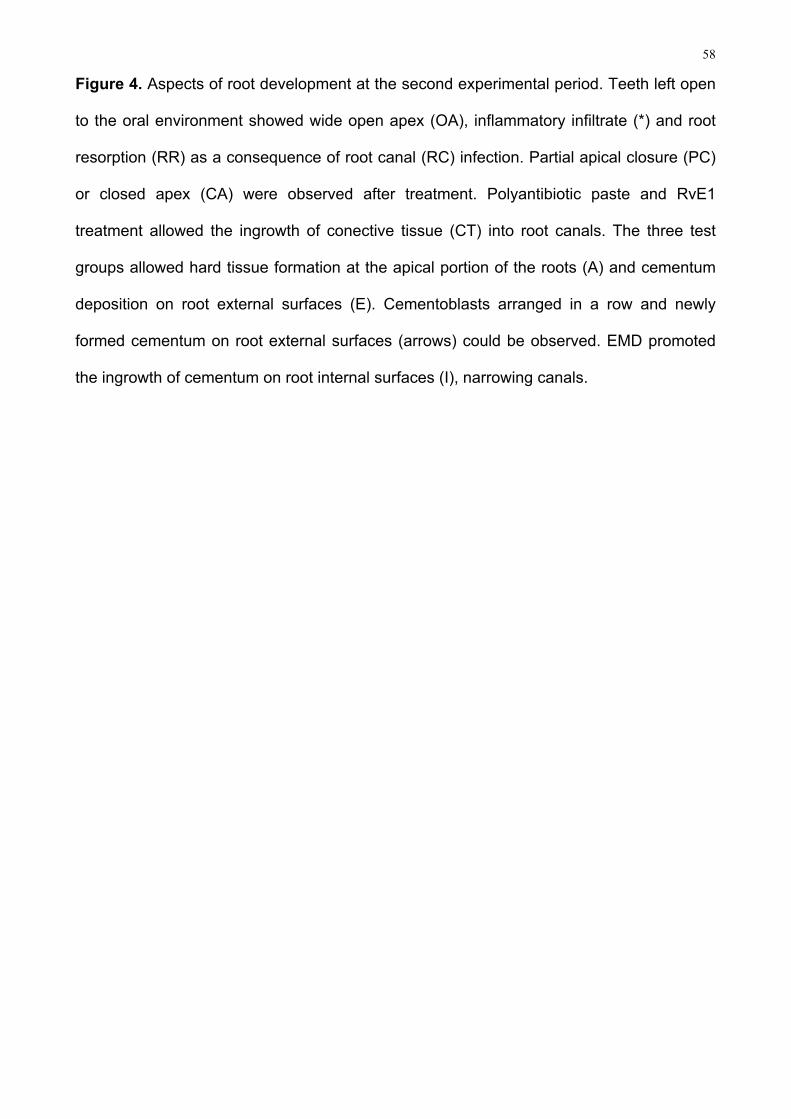
Figure Legends
Figure 1. Diagram summarizing the steps of experimental procedures on lower first molars
of rats: at 4 weeks-old, endodontic access was performed on the central portion of oclusal
surface (arrow), creating a 1mm deep cavity (*). The bur diameter and the crown length -
nearly 1.5mm (**) – guided the extent of drilling (A); teeth of the control group (baseline for
apical periodontitis) were left open to the oral cavity throughout the experiment (B1); At the
other groups, after 3 weeks debris were removed from the pulp chamber and cervical third
of the roots in an extent of about 2mm (***) and then were irrigated with NaOCl and saline
solution, and filled with polyantibiotic paste, EMD or RvE1 (B2).
Figure 2. Radiographic parameters for root length, canal width (percentage of “b” relative
to a”) and periapical lesion area measurements (A); Mesial roots (arrows) analysis of
periapical lesion area, length and canal width for the four groups and two experimental
periods. Means differ significantly related to control (*P<0.05; **P<0.01). EMD promoted
narrower canals related to polyantibiotic paste (■ P<0.05) (B).
Figure 3. Histological analysis of periapical inflammation at the two experimental periods.
For the three test groups, mean scores differ significantly related to control (*P<0.05;
**P<0.01; ***P<0.001). At the first experimental period, Resolvin E1 promoted milder
inflammation related to the other drugs (■ P<0.05). Periapical aspects after the first
experimental period: teeth left open to the oral cavity during the course of the experiment
showing intense periapical inflammation; Inflammatory cells were observed next to apical
opening (*) and also dominating the microscopic field in distant areas (arrows) (A); Teeth
subjected to intracanal medication with polyantibiotic paste (B), EMD (C) showed a
variable inflammatory response to treatment. In some samples, cystic lesions (CL) and
inflammatory infiltrate (*) were detected. All specimens treated with RvE1 (D) showed
either absent or mild inflammatory response 3 weeks after treatmant.
58
Figure 4. Aspects of root development at the second experimental period. Teeth left open
to the oral environment showed wide open apex (OA), inflammatory infiltrate (*) and root
resorption (RR) as a consequence of root canal (RC) infection. Partial apical closure (PC)
or closed apex (CA) were observed after treatment. Polyantibiotic paste and RvE1
treatment allowed the ingrowth of conective tissue (CT) into root canals. The three test
groups allowed hard tissue formation at the apical portion of the roots (A) and cementum
deposition on root external surfaces (E). Cementoblasts arranged in a row and newly
formed cementum on root external surfaces (arrows) could be observed. EMD promoted
the ingrowth of cementum on root internal surfaces (I), narrowing canals.

59
Figure 1
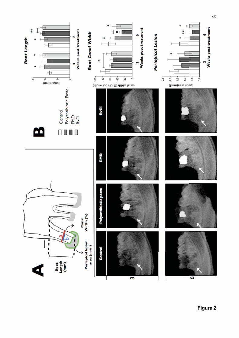
60
Figure 2
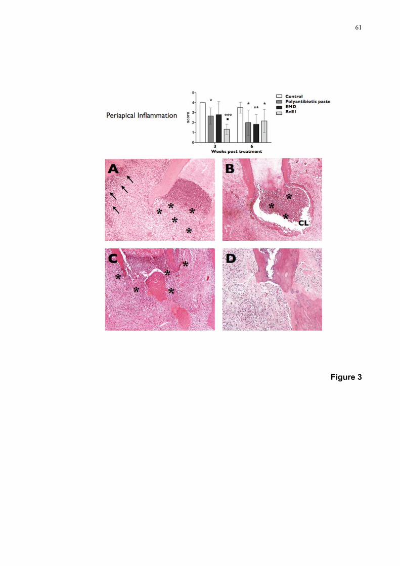
61
Figure 3

62

63
4. Discussão Geral
64
4. DISCUSSÃO GERAL
O presente estudo avaliou, em ratos, alternativas de tratamento para dentes com
rizogênese incompleta e necrose pulpar. Apesar de investigações nessa área comumente
utilizarem cães (WANG et al., 2010; da SILVA et al. 2010), o modelo em ratos apresenta
vantagens técnicas e financeiras (DAMMASCHKE, 2010), além de proporcionar maior
rapidez na progressão de respostas biológicas a serem investigadas (MORETTON et al.,
2000) e apresentar microbiota oral e respostas do hospedeiro semelhantes às observadas
em humanos (KAKEHASHI et al., 1965 HUXLEY, 1971; STASHENKO et al., 1994).
Estudos prévios demonstraram que exposições pulpares em molares de ratos
produzem o desenvolvimento de alterações periapicais inflamatórias semelhantes às
observadas em humanos (KAKEHASHI et al., 1965; MURUZÁBAL e EURASQUIN, 1970;
STASHENKO et al., 1994). Por outro lado, a validação do modelo para dentes com
rizogênese incompleta ainda não havia sido realizada. O corrente estudo, além de
confirmar o desenvolvimento de processos periapicais inflamatórios após a contaminação
do sistema de canais radiculares, comprovou a interrupção do desenvolvimento radicular
neste modelo animal. Contudo, as excepcionais resiliência e capacidade de reparo que
caracterizam o tecido pulpar de molares de ratos (MAURICE e SCHOUR, 1955) tornam
necessárias a sua desorganização por meios mecânicos e a manutenção das cavidades
expostas por período não inferior a três semanas.
A avaliação das características normais do desenvolvimento embriológico em
molares de ratos, somada aos aspectos técnicos necessários para indução de necrose
pulpar e interrupção da formação radicular, permitiram a adequação do desenho
experimental a ser executado na segunda etapa deste trabalho. Sendo assim,
procedimentos para indução da necrose foram realizados em animais com quatro
65
semanas de idade, levando à contaminação do espaço endodôntico no início da
embriogênese radicular. Além disso, a aplicação dos protocolos de tratamento sugeridos
foi realizada em animais com 7 semanas de idade, proporcionando a verificação das
respostas em situações de rizogênese incompleta e presença de patologias periapicais
inflamatórias. Os resultados da primeira etapa do estudo também justificam os períodos
pós-operatórios de três e seis semanas escolhidos, uma vez que o desenvolvimento
natural das raízes está completo em grande parte dos animais com 13 semanas de idade.
Por outro lado, a observação de períodos mais longos foi prejudicada pela fragilização da
estrutura dentária após execução dos procedimentos, o que determinou fratura dentária e
falhas no selamento coronário em animais com 16 semanas de idade, os quais foram
excluídos da avaliação.
Além desta, outras dificuldades técnicas tiveram de ser superadas para a execução
dos procedimentos operatórios. A dimensão reduzida e o posicionamento anatômico dos
dentes tornam imprescindível, além de adequadas anestesia e posicionamento dos
animais, a adaptação de meios que favoreçam o acesso ao campo operatório (MAURICE
e SCHOUR, 1955, DAMMASCHKE, 2010), como o aparato desenvolvido no presente
estudo para abertura de boca.
Por outro lado, características próprias da rizogênese incompleta favoreceram a
aplicação técnica dos experimentos. O amplo diâmetro dos canais radiculares e o
comprimento reduzido das raízes facilitaram a localização, irrigação e aplicação de
medicações intracanal. Além disso, os canais não precisaram ser instrumetados e/ou
obturados, proporcionando de maneira simples a padronização dos protocolos.
A aplicabilidade do modelo para avaliar estratégias de tratamento foi confirmada
com base em achados semelhantes aos dos estudos que testaram a aplicação da
pasta poliantibiótica em cães (THIBODEAU et al., 2007; da SILVA et al., 2010; WANG

66
et al., 2010). Conforme previamente relatado (da SILVA et al., 2010; WANG et al.,
2010), a aplicação de pasta poliantibiótica permitiu a redução do quadro inflamatório
periapical em relação ao grupo controle, e o desenvolvimento radicular ocorreu
principalmente às expensas de tecido osteóide ou cementóide. Também de forma
semelhante aos achados da literatura (da SILVA et al., 2010), no segundo período
experimental, aproximadamente um terços das amostras avaliadas não apresentou
formação de tecidos dentários mineralizados, denotando a relevância de que outras
alternativas de tratamento, como a aplicação de EMD ou RvE1, fossem investigadas.
Além da análise radiográfica quantitativa e da descrição das características
histológicas dos tecidos formados, o processo inflamatório foi classificado de acordo
com sua extensão e intensidade em quatro escores, os quais permitiram a comparação
entre os grupos de estudo por meio de análise de variância (ANOVA). Embora este
teste tenha sido inicialmente recomendado para a comparação de variáveis
paramétricas, o mesmo também tem sido empregado quando do uso de valores
categóricos (escores), pois, ao contrário do que se possa pensar, a inferência na
análise de dados de variáveis ordinais podem envolver procedimentos paramétricos
(CHILTON,1982; MONTGOMERY, 1984; ZAR, 1996). De acordo, Campbell e Machin
(1993) afirmam que, se os dados são ordinais categóricos, podem ser atribuídos
escores, como 1, 2, 3 e 4 e calculada a média. Essa afirmação é compartilhada por
Snedecor & Cochran (1980) e Montgomery (1984) e respaldada em diversos estudos
(TROIAN et al., 2006; GOMES et al., 2007; GUERRERO et al., 2011).
Independentemente da medicação intracanal empregada, a resolução do
processo inflamatório apresentou caráter fundamental para a obtenção do
desenvolvimento radicular em dentes não vitais. Por outro lado, a população de células
precursoras presentes foi capaz de responder aos estímulos induzidos mesmo após
intenso desafio microbiano.

67
Estudos prévios confirmam que fatores de crescimento relacionados com o
processo inflamatório inibem ou impedem eventos indispensáveis à embriogênese
dentária (SHIBA et al., 1998). Sendo assim, as estratégias de tratamento adotadas,
além de visarem à preservação da viabilidade de células tronco remanescentes na
polpa, ligamento periodontal e papila apical, buscaram reduzir o processo inflamatório
decorrente da contaminação do espaço endodôntico. O regime de irrigação com
hipoclorito de sódio, por si, já favorece a redução da contaminação do sistema de
canais radiculares. Por outro lado, especialmente por não ser indicada a
instrumentação da região apical dos canais, mantém-se algum nível de contaminação
microbiana (WINDLEY et al., 2005).
Por diferentes meios, os protocolos testados buscaram superar o dano causado
pela provável manutenção de estímulos microbianos. Estudos prévios atestam que a
pasta poliantibiótica apresenta capacidade de complementar a desinfecção promovida
pelo hipoclorito de sódio em dentes com rizogênese incompleta (WINDLEY et al., 2005); a
RvE1 apresenta capacidade de regular a resposta inflamatória do hospedeiro a estímulos
nocivos, como o microbiano (SERHAN e CHIANG 2008); e as proteínas derivadas da
matriz do esmalte induzem processos regenerativos a partir da regulação de mecanismos
alterados durante o processo inflamatório (SUZUKI et al., 2005).
A invaginação de tecido conjuntivo para o interior dos canais radiculares e a
estimulação da deposição de tecido mineralizado nas paredes radiculares externas e na
região apical puderam ser observados em amostras submetidas à medicação intracanal
com a pasta poliantibiótica. De acordo, estudos prévios confirmam que a eliminação do
processo inflamatório permite que, em alguns casos, o espaço endodôntico seja ocupado
por tecido conjuntivo proveniente do ligamento periodontal (WANG et al., 2010; da SILVA
et al., 2010).

68
Por outro lado, a resolução do processo inflamatório foi significativamente mais
rápida nas amostras tratadas com RvE1, o que fica claro nos resultados histológicos do
primeiro período experimental. De acordo, estudos prévios demonstram que a RvE1 é
potente reguladora da transmigração de neutrófilos e da inflamação in vivo, sendo
também atribuído a esse composto bioativo a estimulação de fagocitose não flogística de
neutrófilos apoptóticos pelos macrófagos, o bloqueio da produção de interleucina-12 por
células dendríticas e a regulação da expressão de CCR5 em células T (SERHAN et al.,
2000; ARITA et al., 2005; ARIEL et al., 2006; ARITA et al., 2007; SCHUWAB et al., 2007)
Até o momento, dois receptores foram reconhecidos na atuação da RvE1. O GPRC
chemokime-like receptor (CMKLR1) está presente em monócitos e células dendríticas e
atenua a ativação de NFκB estimulada pelo TNF (mediador-chave nas fases iniciais do
processo inflamatório) quando ligado à RvE1 (SERHAN et al., 2000; ARITA et al., 2005).
Por outro lado, outro receptor GPCR, o leucotriene B4 receptor (BLT1), se expressa em
neutrófilos e interage com a RvE1 como um receptor antagonista, o qual atenua os
processos inflamatórios dependentes da sinalização do leucotrieno B4 (ARITA et al.,
2007). No presente estudo, a redução clara do infiltrado inflamatório neutrofílico pode ser
observada, resultando na resolução do processo inflamatório.
Também de forma diversa dos outros grupos, no primeiro período experimental
apenas a RvE1 promoveu a redução da área de lesão periapical em relação ao grupo
controle. A RvE1 foi mais eficaz que os outros tratamentos no controle da inflamação
previamente induzida, o que é bastante favorável por reduzir os danos causados pelo
processo inflamatório. De acordo, estudos prévios confirmam o potencial deste mediador
em modular a diferenciação de osteoclastos e consequentemente os processos de
reabsorção óssea , assim como o recrutamento de células inflamatórias (HARSTUK et al.,
2006; HERRERA et al., 2008), o que pode ter impacto na extensão das lesões
periapicais.

69
No segundo período experimental, algumas amostras tratadas com RvE1
apresentaram infiltrado inflamatório moderado, sendo constatado que a resposta
inflamatória, a área da lesão periapical, o comprimento radicular e a espessura dos canais
radiculares foram semelhantes as das amostras tratadas com os outros medicamentos.
Nesse sentido, considerações acerca das características dos protocolos empregados
merecem ser feitas.
Nos dentes tratados com RvE1, uma única aplicação tópica da medicação
(veiculada em etanol) foi admnistrada. Este fato, somado às características do ambiente
endodôntico podem ter contribuído para que no segundo período avaliado não fossem
observadas as diferenças favoráveis ao mediador lipídico verificadas no primeiro período
experimental. Estudos prévios que atestam os efeitos benéficos da aplicação tópica da
RvE1 na periodontia e oftalmologia realizaram aplicações pelo menos a cada 24 horas,
(HASTURK et al., 2006; HASTURK et al., 2007; LI et al., 2010), o que apesar de favorecer
a atuação constante do mediador, não é exequível em protocolos endodônticos. Além
disso, o modelo aqui adotado não permite testar protocolos que exijam repetidos acessos
ao espaço endodôntico, uma vez que a fragilidade da estrutura dentária poderia resultar
em fraturas e comprometer o selamento coronário.
Outro aspecto importante a ser aprimorado é a concentração de RvE1
preconizada. Com base em estudos prévios que comprovam a eficácia sistêmica desse
mediador em doses reduzidas (SCHWAB et al., 2007), e considerando a aplicação tópica
do medicamento em um ambiente isolado, no presente estudo, uma concentração
bastante baixa foi escolhida. Ainda assim, os resultados do primeiro período experimental
mostram clara vantagem da RvE1 em controlar o processo inflamatório em comparação
aos demais medicamentos. É provável que maiores concentrações aprimorem os
resultados obtidos.
70
Por outro lado, aspectos estruturais que caracterizam o sistema de canais
radiculares podem favorecer a manutenção de microrganismos em canais vazios
(MENEZES et al., 2004), especialmente em dentes jovens, os quais apresentam maior
número de túbulos dentinários infectados e penetração mais profunda de microrganismos
(KAKOLI et al., 2009). Ao contrário da RvE1, a pasta poliantibiótica e as EMD apresentam
consistência semelhante a de um gel. Dessa forma, suas características físicas permitem
maior estabilidade do material durante o curso do experimento, o que pode ter reduzido a
reinfecção do sistema de canais radiculares e favorecido o desenvolvimento radicular no
segundo período experimental.
Os padrões de formação radicular nas amostras tratadas com EMD diferiram em
relação aos promovidos pelas outras duas medicações. Além da deposição de tecido
mineralizado na região apical e nas paredes radiculares externas, este grupo apresentou
a invaginação de tecido cementóide para o interior do canal radicular, o que pode explicar
a menor relação de espessura do canal radicular observada nos dados da análise
radiográfica. Provavelmente, essas características contribuam com uma maior resistência
da estrutura dentária, o que é desejável a fim de reduzir os riscos de fratura.
Diversos estudos confirmam a capacidade das EMD em induzir osteogênese e
cementogênese (HAMMASTROM, 1997; BOYAN et al., 2000). Nesse sentido, a atividade
de alguns fatores de crescimento podem estar relacionados com a resposta observada
após medicação com EMD. Estudos prévios confirmam que essas proteínas atuam de
maneira semelhante ao TGF-β1 (com efeito mitogênico sobre células do ligamento
periodontal, inibição da proliferação epitelial e estímulo à diferenciação e proliferação de
osteoblastos) à IL-6 (estimulando a proliferação de odontoblastos e de osteoblastos) e às
BMPs (simulando a osteogênese e a cementogênese) (SUZUKI et al., 2005; SONOYAMA
et al., 2007; LINDE, GOLDBERG, 1993; NAKAMURA et al., 2002). Por outro lado, as
EMD promovem o aumento da atividade fagocitária de monócitos (KHEDMAT et al.,

71
2010), e inibem o fator de necrose tumoral-α (TNF-α) (SATO et al., 2008).
Também é atribuído às proteínas da matriz do esmalte efeito semelhante ao do
TGF-β na inibição da indução de apoptose promovida pelo TNF-α em células
osteoblásticas. Esses achados levam à especulação de que o mesmo efeito protetor
possa ocorrer na diferenciação e cementoblastos, e nas reabsorções ósseas (HE et al.,
2005).
Nos últimos anos, o papel das EMD na diferenciação de cementoblastos tem sido
explorado, sendo a elas atribuída a regulação da diferenciação de células do folículo
dentário de camundongos em fenótipos osteo-cementoblásticos (HAKKI et al., 2001). De
acordo, Bosshardt et al., (2005) identificaram o papel dessas proteínas na estimulação da
expressão de marcadores específicos de cementoblastos, como cementum attachment
protein (CAP) e cementum protein-23 (CP-23). A expressão desses marcadores também
foi identificada após estimulação de células do folículo dentário pelas proteínas da matriz
do esmalte e pelas BMPs (bone morphometric proteins) 2 e 7 (KÉMOUM et al., 2007)
Os mecanismos acima mencionados suportam a significancia biológica das EMD para
o reparo e regeneração periodontal (LYNGSTADAAS et al., 2009). De acordo, esses
potenciais mecanismos podem ter contribuído para a eliminação de estímulos nocivos e
estimulação das respostas observadas especialmente após o segundo período.
Características semelhantes as do cemento puderam ser observadas nos tecidos
neoformados, o que sugere que os mecanismos já elucidados nos processos de
regeneração periodontal possam estar envolvidos no desenvolvimento radicular.
Embora alguns estudos atribuam atividade antimicrobiana às EMD (SPAHR et al.,
2002), outros demonstram que a infecção Porphyromonas gingivalis inibe efeitos
benéficos dessa medicação em relação a regeneração periodontal (INABA et al., 2004).
Uma vez que as infecções endodônticas são predominantemente anaeróbias, a hipótese

72
de que efeito semelhante tenha retardado o reparo é consistente com a heterogeneidade
das respostas observadas no primeiro período experimental.
Pelo exposto, sugere-se que tanto a RvE1 como as EMD apresentaram potenciais
a serem explorados para a obtenção do desenvolvimento radicular em dentes com
necrose pulpar. Estudos adicionais devem ter foco na superação das limitações dos
protocolos, no esclarecimento os eventos celulares e moleculares envolvidos na formação
radicular, e em resultados clínicos em humanos.

73
5. Conclusões

74
5. CONCLUSÕES
A partir dos resultados do presente estudo pode-se concluir:
- A metodologia desenvolvida permitiu a avaliação de protocolos de tratamento para
casos de rizogênese incompleta e necrose pulpar, utilizando um modelo em ratos;
- O mediador lipídico Resolvina E1, aplicado como medicação intracanal, acelerou a
resolução do processo inflamatório e o reparo ósseo, apresentando resultados
favoráveis em todas as amostras no primeiro período experimental.
- No segundo período experimental, a medicação intracanal com EMD favoreceu o
aumento da espessura das paredes radiculares, dada a invaginação de tecido
cementóide para o interior do espaço endododôntico.
- Especialmente no segundo período avaliado, a desinfecção química dos canais
radiculares associada à aplicação das medicações testadas promoveram o
desenvolvimento radicular em dentes não vitais, estando esse condicionado à redução
da inflamação periapical. Estudos adicionais devem ter foco na superação das limitações
dos protocolos testados, no esclarecimento os eventos celulares e moleculares
envolvidos na formação radicular, e em resultados clínicos em humanos.

75
6. Referências Bibliográficas

76
6. REFERÊNCIAS BIBLIOGRÁFICAS
ANDREASEN, J.O.; FARIK, B.; MUNKSGAARD, E.C. Long-term calcium hydroxide as a
root canal dressing may increase risk of root fracture. Dent Traumatol, v.18, n.3, p.134-
7,2002.
ARIEL, A.; FREDMAN G.; SUN Y.P.; VAN DYKE T.E.; LUSTER A.D.; SERHAN C.N.
Apoptotic neutrophils and T cells sequester chemokines during immune response
resolution via modulation of CCR5 expression. Nature Immunol, v.7, n.11, p.1209-16,
2006.
ARITA, M.; BIANCHINI, F.; ALIBERTI, J.; SHER, A.; CHIANG, N.; HONG, S.; YANG, R.;
PETASIS, N.A.; SERHAN, C.N. Stereochemical assignment, anti-inflammatory properties
and receptor for the omega-3 lipid mediator Resolvin E1. J Exped Med, v.201, n.5, p.713-
22, 2005.
ARITA, M.; OHIRA T.; SUN Y.P.; ELANGOVAN S.; CHIANG N.; SERHAN C.N. Resolvin
E1 selectively interacts with leucotriene B4 receptor BLT1 and ChemR23 to regulate
inflammation. J Immunol, v.178, n.6, p.3912-17, 2007.
BANCHS, F.; TROPE, M. Revascularization of immature teeth with apical periodontitis:
new treatment protocol? J Endod, v.30, n.4 , p.196-200, 2004.
BROOKES, S.J; ROBINSON, C.; KIRKHAN, J.; BONASS, W.A. Biochemistry and
molecular biology of amelogenin proteins of developing dental enamel. Arch Oral Biol ,
v.40, n. , p.1-14, 1995.
BOSE R.; NUMMIKOSKI, P.; HARGREAVES, K. A retrospective evaluation of
radiographic outcomes in immature teeth with necrotic root canal systems treated with
regenerative endodontic procedures. J Endod v.35, n.10, p.1343-9, 2009.
BOSSHARDT, D.D.; NANCI, A. Hertwig´s epithelial root sheath, enamel matrix proteins

77
and initiation of cementogenesis in porcine teeth. J Clin Periodontol v.31, n.3, p.184-92,
2004.
BOSSHARDT, D.D. Are Cementoblasts a Subpopulation of Osteoblasts or a Unique
Phenotype? J Dent Res, v.84, n.5, p.390-406, 2005.
BOYAN, B.D.; WEESNER, T.C.; LOHMANN, C.H.; ANDREACCHIO, D.;CARNES, D.L.;
CAMPBELL, M.J.; MACHIN, D. Medical Statistics: A common sense approach. 2nd ed.
Nova York: John Willey and Sons,1993. p.56.
CHILTON, N.M. Design and analysis in dental and oral research. 2nd.ed. Nova York:
Praeger, 1982. P.6, p.354.
CHUEH, L.H.; HUANG, G.T. Immature teeth with perirradicular periodontitis or abscess
undergoing apexogenesis: a paradigm shift. J Endod, v.32, n.12, p.1205-13, 2006.
CHUEH, L.H.; HO, Y.C.; KUO, T.C.; LAI W.H.; CHEN, Y.H.; CHIANG, C.P. Regenerative
endodontic treatment for necrotic immature permanent teeth. J Endod, v.35, n.2 , p.160-4,
2009.
COTTI, E.; MEREU, M.; LUSSO, D. Regenerative treatment of an immature, traumatized
tooth with apical periodontitis: report of a case. J Endod, v.34, n.5, p.611-6, 2008.
CZVEK, M. Prognosis of luxated non-vital maxillary incisors treated with calcium hydroxide
and filled with gutta-percha. Endod Dent Traumatol, v.8, n., p.45-55, 1992.
CZVEK, M.; NORD, C.E.; HOLLENDER, L. Antimicrobial effect of root canal debridment in
teeth with immature root: a clinical and microbiological study. Odontol Revy, v.27, n.1, p.1-
10, 1976.
DAMMASCHKE T. Rat molar teeth as a study model for direct pulp capping research in
dentistry. Lab Anim, v.44, n.1, p.1-6, 2010.
78
DEAN D.D.; COCHRAN, D.L.; SCHWARTZ, Z.Porcine fetal enamel matrix derivative
enhances bone formation induced by demineralized freeze dried bone allograft in vivo.J
Periodontol v.71, n.8, p.1278-86, 2000.
DING, R.Y.; CHEUNG, G.S.; CHEN, J.; YIN, X.Z.; WANG, Q.Q.; ZHANG, C.F. Pulp
revascularization of immature teeth with apical periodontitis: a clinical study. J Endod, v.
35, n.5, p.745-9, 2009.
DUARTE, P.M.; TEZOLIN, K.R.; FIGUEIREDO, M.F.; BASTOS M.F. Microbial profile of
ligature-induced periodontitis in rats. Arch Oral Biol, v.55, n.1, p.142-7, 2010.
FILIPPI, A.; POHL, Y.; VON ARX, T. Treatment of replacement resorption with Emdogain -
a prospective clinical study. Dent Traumatol, v.18, n.3, p.138-43, 2002.
FRANK, A.L. Therapy for the divergent pulpless tooth by continued apical formation. J Am
Dent Assoc, v.7, n.1, p.78-93, 1966.
FRIEDLANDER L.T.; CULLIAN, M.P.; LOVE, R.M. Dental stem cells and their potential
role in apexogenesis and apexification. Int Endod J, v.42, n.11, p.955-62, 2009.
GRONTHOS, S.; MANKANI, M.; BRAHIM, J.; ROBEY, P.G.; SHI, S. Postnatal human
dental pulp stem cells (DPSCs) in vitro and n vivo. Proc Natl Acad Sci USA, v.97, n.25,
p.13625-30, 2000.
GRONTHOS, S.; BRAHIM, J.; LI, W.; FISHER, L.W.; CHERMAN, N.; BOYDE, A.;
DENBESTEN, P.; ROBEY, P.G.; SHI, S. Stem cells propreties of human dental pulp stem
cells. J Dent Res, v.81, n.8, p.531-5, 2002.
GOMES, M.S.; BARLETTA, F.B.; DELLA BONA, A.; VANNI, J.R.; PEREIRA, C.C.;
FIGUEIREDO, J.A.P. Microbial leakage and apical inflammatory response in dog`s teeth
after root canal filling with different sealers, post space preparation and exposure to the
79
oral environment. J Appl Oral Sci, v.15, n.5, p.429-36, 2007.
GUERRERO, T.G.; MAKARA, M.A.; KATIOFSKY, K.; FLUCKIGER, M.A.; MORGAN, J.P.;
HAESSIG, M.; MONTAVON, P.M. Comparison of healing of the osteotomy gap after tibial
tuberosity advancement with and without use of an autogenous cancellous bone graft. Vet
Surg, v.40, n.1, p.27-33, 2011.
HAKKI, S.S.; BERRY, J.E.; SOMERMAN, M.J. The effect of enamel matrix protein
derivative on follicle cells in vitro. J Periodontol, v.72, n.5, p.679–687, 2001.
HAM, J.W.; PATTERSON, S.S.; MITCHELL, D.F.; Induced apical closure of immature
pulpless teeth in monkeys. Oral Surg Oral Med Oral Pathol, v.33, n.3, p.438-49, 1972.
HAMMARSTRÖM, L. Enamel matrix, cementum development and regeneration. J Clin
Periodontol, v.24, n.9, p.658-68, 1997.
HARGREAVES K.M.; GEISLER, T.; HENRY, M.; WANG, Y.; Regeneration potential of the
young permanent tooth: what does the future hold? J Endod, v.34, supl. 7, p.S51-6, 2008.
HASTURK, H.; KANTARCI, A.; OHIRA, T.; ARITA, M.; EBRAHIMI, N.; CHIANG, N.;
PETASIS, N.A.; LEVY, B.D.; SERHAN, C.N.; VAN DYKE, T.E. RvE1 protects from local
inflammation and osteoclast mediated bone destruction in periodontitis. FASEB J, v.20,
n.2 , p.401-3, 2006.
HASTURK, H.; KANTARCI, A.; GOGUET-SURMENIAN, E.; BLACKWOOD, A.; ANDRY,
C.; SERHAN, C.N.; VAN DYKE, T.E. Resolvin E1 regulates inflammation at the cellular
and tissue level and restores tissue homeostasis in vivo. J Immunol, v.179, n.10, p.7021-
29, 2007.
HE, J.; KING, Y.; JING, J.; SAFAVI, K.E, SPANGBERG, L.S., ZHU, Q. Enamel matrix
derivative inhibits TNF-α induced apoptosis in osteoblastic MC3T3-E1 cells. Oral Surg

80
Oral Med Oral Pathol Oral Radiol Endod, v.99, n.6, p.761-7, 2005.
HERRERA, B.S.; OHIRA, T.; GAO, L.; OMORI, K.; YANG, R.; ZHU, M.; MUSCARA, M.N.;
SERHAN, C.N.; VAN DYKE, T.E.; GYURKO, R. An endoenous regulator of inflammation,
resolvin E1, modulates osteoclast differentiation and bone resorption. Br J Pharmacol
v.55, n.8, p.1214-23, 2008.
HORSTED, P.; NYGAARD-OSTBY, B. Tissue formation in the root canal after total
pulpectomy and partial root filling. Oral Surg Oral Med Oral Pathol, v.46, n.2, p.275-82,
1978.
HOSOYA, A.; KIM, J.Y.; CHO, S.W.; JUNG, H.S. BMP4 signaling regulates formation of
Hertwi´s epithelial root sheath during tooth root development. Cell Tissue Res v.333, n.3,
p.503-9, 2008.
HUANG, G. T.; SONOYAMA, W.; LIU, Y.; LIU, H.; WANG, S.; SHI, S. The hidden treasure
in apical papilla: the potential role in pulp/dentin regeneration and bioroot engineering. J
Endod v.34, n.6, p.645-51, 2008.
HUANG, G.T. Apexification: the beginning of its end. Int Endod J; v.42, n.10, p.855-66,
2009.
HUXLEY, H.G. The histology of rat molar fissure plaque. Arch Oral Biol v.16, n.11, p.1311-
28, 1971.
HUXLEY, H.G. The recovery of microorganisms from the fissure of rat molar theeth.
Arch Oral Biol v.17, n.10, p.1481-5, 1972.
IGARASHI, R.; SAHARA, T.; SHIMIZU-ISHIURA M, SASAKI T. Porcine enamel matrix
derivative enhances the formation of reparative dentine and dentine bridges during
wound healing of amputated rat molars. J Electron Microsc (Tokyo) v.52, n.2, p.227-36,
2003.
81
INABA, H.; KAWAI, S.; NAKAYAMA, K.; OKAHASHI, N.; AMANO, A. Effect of enamel
matrix derivative on periodontal ligament cells in vitro is diminished by Porphyromonas
gingivalis. J Periodontol v.75, n.6, p.858-865, 2004.
ISHIZAKI, N.T.; MATSUMOTO, K.; KIMURA, Y.; WANG, X.; YAMASHITA, A.;
Histopathological study of dental pulp tissue capped with enamel matrix derivative. J
Endod; v.29, n.3, p.176-9, 2003.
IWAYA, S.I.; IKAWA, M.; KUBOTA, M. Revascularization of a immature permanent tooth
with apical periodontitis and sinus tract. Dental Traumatol, v.17, n.4, p.185-7, 2001.
JUNG I.Y.; LEE, S.J.; HARGREAVES, K.M. Biollogically based treatment of immature
permanent teeth with pulpal necrosis: a case series. J Endod, v.34, n.7, p.876-7, 2008.
KAIDA, H.; HAMACHI, T.; ANAN, H.; MAEDA, K. Wound healing process of injured pulp
tissues with Emdogain Gel. J Endod, v.34, n.1, p.26-30, 2008.
KAKEHASHI, S.; STANLEY, H.R.; FITZGERALD, R.J. The effects of surgical exposure of
dental pulps in germ-free and conventional laboratory rats. Oral Surg Oral Med Oral Pathol
v.20, p.340-9, 1965.
KAKOLI, P.; NANDAKUMAR, R.; ROMBERG, E.; AROLA, D.; FOUAD, A.F. The effect of
age on bacterial penetration of radicular dentin. J Endod v.35, n.1, p.78-81, 2009.
KÉMOUN, P.;LAURENCIN-DALICIEUX, S.; RUE, J.; FARGES, J.C.; GENNERO, I.;
CONTE-AURIOL, F.; BRIAND-MESANGE, F.; GADELORGE M.; ARZATE H.;
NARAYANAN S.; BRUNEL, G.; SALLES, J. Human dental follicle cells acquire
cementoblast features under stimulation by BMP-2/-7 and enamel matrix derivatives
(EMD) in vitro. Cell Tissue Res, v. 329, n.2, p.283–294, 2007.
82
KHEDMAT, S.; HADJATI, J.; IRAVANI, A.; NOURIZADEH, M. Effects of enamel matrix
derivative on the viability, cytokine secretion, and phagocytic activity of human monocytes.
J Endod v.36, n.6, p.1000-1003, 2010.
LEE, A.Z.; JIANG, J.; HE, J.; SAFAVI, K.E.; SPANGBERG, L.S; ZHU, Q. Stimulation of
cytokines in osteoblasts cultured on enamel matrix derivative. Oral Surg Oral Med Oral
Pathol Oral Radiol Endod, v.106, n.1, p.133-8, 2008.
LEE, D.S.; PARK, J.T.; KIM, H.M.; KO, J.S.; SON, H.H.; GRONOSTAJSKI, R.M.; CHO,
M.I.; CHOUNG, P.H.; PARK, J.C. Nuclear Factor I is essencial for odontogenic
proliferation and odontoblast differenciation during tooth root development. J Biol Chem
v.284, n.25, p.17293-17303, 2009.
LEE, T.Y.; LEE, D.S.; KIM, H.M.; KO, J.S.; GRONOSTAJSKI, R.M.; CHO, M.I.; SON, H.H.;
PARK, J.C. Disruption of NFIC causes dissociation of odontoblasts by interfering with the
formation of intercellular junctions and aberrant odontoblast differenciation. J Histochem
Cytochem v.57, n.5, p.469-76, 2009.
LEVY, B.D.; CLISH, C.B.; SCHMIDT, B.; GRONERT, K.; SERHAN, C.N. Lipid mediator
class switching during acute inflammation: signs in resolution. Nature Immunol,v.2,
n.7,p.612-9, 2001.
LI, N.; HE, J.; SCHWARTZ, C.E.; GJORSTRUP, P.; BAZAN, H.E. Resolvin e1 improves
tear production and decreases inflammation in a dry eye mouse model. J Ocul Pharmacol
Ther v.26, n.5, p.431-439, 2010.
LINDE, A.; GOLDBERG, M. Dentinogenesis. Crit Rev Oral Biol Med, v.4, n.5, p.679-728,
1993.
LINDSKOG, S. Formation of intermediate cementum II: a sccaning electron microscopic
study of the epithelial root sheath of Hertwig in monkey. J Craniofac Genet Dev Biol, v.2,

83
n.2, p.161-9, 1982.
LYNGSTADAAS, S.P.; WOHLFAHRT, J.C.; BROOKES, S.J.; PAINE, M.L.; SNEAD, M.L.;
RESELAND, J.E. Enamel matrix proteins; old molecules for new applications. Orthod
Craniofac Res v.12, n.3, p.243-253, 2009.
MADAM, A.K.; KRAMER, B. Immunolocalization of fibroblast growth factor-2 (FGF-2) in
the developing root and supporting structures of the murine tooth. J Mol Histol v.36, n.3,
p.171-8, 2005.
MAHL, C.R.; FONTANELLA, V. Evaluation by digital subtraction radiography of induced
changes in the bone density of the female rat mandible. Dentomaxillofac Radiol v.37
n.8, p.438-44, 2008.
MAURICE, C.G.; SCHOUR, I. Experimental cavity preparations in the molar of the rat. J
Dent Res v.34, n.3, p.429-34, 1955.
MENEZES, M.M.; VALERA, M.C.; JORGE, A.O.; KOGA-ITO, C.Y.; CAMARGO, C.H.;
MANCINI, M.N. In vitro evaluation of the effectiveness of irrigants and intracanal
medicaments on microorganisms within root canals. Int Endod J v.37, n.5, p.311-319,
2004.
MESAROS, S.V.; TROPE, M. Revascularization of traumatized teeth assessed by laser
Doppler flowmetry: case report. Endod Dent Traumatol v.13, n.1, p.24-30, 1997.
MONTGOMERY, D.C. Design and analysis of experiments. 2nd ed. Nova York: John
Wiley e Sons, 1984. P.118.
MORETTON, T.R.; BROWN, C.E.; LEGAN, J.J.; KAFRAWY, A.H. Tissue reactions after
subcutaneous and intraosseous implantation of mineral trioxide aggregate and
ethoxybenzoic acid cement. J Biomed Mater Res v.52: n.3, p.528-33, 2000.

84
MURUZÁBAL, M.; EURASQUIN, J. Discussion of; methods and criteria in evaluation of
periapical response. Int Dent J v.20, n.3, p.539-54, 1970.
NAKAMURA, Y.; HAMMARSTRÖM, L.; LUNDBERG, E.; EKDAHL, H.; MATSUMOTO, K.;
GESTRELIUS, S.; LYNGSTADAAS, S.P. Enamel matrix derivative promotes reparative
processes in the dental pulp. Advances in Dental Research, v.15, p.105-7, 2001.
NAKAMURA, Y.; HAMMARSTRÖM, L.; MATSUMOTO, K.; LYNGSTADAAS, S.P. The
induction of reparative dentin by enamel proteins. Int Endod J, v.35, n.5, p.407-17, 2002.
NAKASONE, N.; YOSHIE, H.; OHSHIMA, H. An immunohistohemical study of the
expression of heat-shock protein-25 and cell proliferantion in the dental pulp and enamel
organ during odontogenesis in rat molars. Arch Oral Biol v.51, n.5, p.378-86, 2006.
NEVINS, A.J.; FINKELSTEIN, F.; BORDEN, B.G.; LAPORTA, R. Revitalization of pulpless
open apex teeth in rhesus monkeys using collagen-calcium phosphate gel. J Endod, v.2,
n.6, p.159-65, 1976.
NYGAARD-OSTBY, B.; HJORTDAL, O. Tissue formation in the root canal following pulp
removal. Scand J Dent Res, v.79, n.5, p.333-49, 1971.
OSTBY, B.N The role of the blod clot in endodontic therapy: an experimental histologic
study. Acta Odontol Scand; v.19, p.324-53, 1961.
OIKAWA, T.; NOMURA, Y.; ARAI, C.; NODA, K.; HANADA, N.; NAKAMURA, Y.
Mechanism of active eruption of molars in adolescent rats. Eur J Orthod 2011, article in
press.
OKADA, Y.; HAMADA, N.; KIM, Y.; TAKAHASHI, Y.; SASAGURI, K.; OZONO, S.;
SATO, S. Blockade of sympathetic beta-receptors inhibits Porphyromonas gingivalis-
induced alveolar bone loss in an experimental rat periodontitis model. Arch Oral Biol
v.55, n.7, p.502-8, 2010.

85
OLSSON, H.; DAVIES, J.R.; HOLST, K.E.; SCHRÖDER, U.; PETERSSON, K. Dental Pulp
capping: effect of Emdogain gel on experimentally exposed human pulps. Int Endod J,
v.38, n.3, p.186-94, 2005.
PONTORIERO, R.; WENNSTROM, J.; LINDHE, J. The use of barrier membranes and
enamel matrix proteins in the treatment of angular bone deffects. A prospective controlled
clinical study. J Clin Periodontol, v.26, n.12, p.833-40. 1999
RAT GENOME SEQUENCING PROJECT CONSORTIUM. Genome sequence of the
Brown Norway rat yields insights into mammalian evolution. Nature v.1;428(6982), p.493-
521, 2004.
RULE, D.C.; WINTER, G.B.; Root growth and apical repair subsequent to pulpal necrosis
in children. Br Dent J, v.120, n.12, p.586-90, 1966.
SATO, S.; KITAGAWA, M.; SAKAMOTO, K.; IIZUKA, S.; KUDO, Y.; OGAWA, I.;
MIYAUCHI, M.; CHU, E.Y.; FOSTER, B.L.; SOMERMAN, M.J.; TAKATA,T. Enamel matrix
derivative exhibits anti-inflammatory properties in monocytes. J Periodontol v.79, n.3,
p.535-540, 2008.
SCHUWAB, J.M.; CHIANG, N.; ARITA, M.; SERHAN, C.N.; Resolvin E1 and Protectin D1
activate inflammation-resolution programmes. Nature, v.14;447(7146) p.869-74, 2007.
SEO, B.M.; MIURA, M.; SONOYAMA, W.; COPPE, C.; STANYON, R.; SHI, S.. Recovery
of stem cells from cryopreserved periodontal ligament. J Dent Res v.84, n.10, p.907-912,
2005.
SNEDECOR, G.W.; COCHRAN, W.G. Statistical Methods. 7nded. Ames: Iowa States
University Press, 1980. P. 204-206.
SERHAN, C.N.; CLISH, C.B.; BRANNON, J.; COLGAN, S.P.; CHIANG, N.; GRONERT, K.
Novel functional sets of lipid-derived mediators with anti-inflammatory actions generated

86
from omega-3 fatty acids via cicloxigenase 2-nonsteroidal anti-inflammatory drugs and
transcellular processing. J Exp Med, v.192, n.8, p.1197-204, 2000.
SERHAN, C.N. Resolution phase of inflammation: novel endogenous anti-inflammatory
and proresolving lipid mediators and pathways. Annu Rev Immunol, v.25, p.101-37, 2007.
SERHAN, C.N.; CHIANG, N.; VAN DYKE, T.E. Resolving inflammation: dual anti-
inflammatory and pro-resolution lipid mediators. Nat Rev Immunol v.8, n.5, p.349-61,
2008.
SERHAN, C. N.; CHIANG, N. Endogenous pro-resolving and anti-inflammatory lipid
mediators: a new pharmacologic genus. Br J Pharmacol v.153, Suppl.1, p.S200-215,
2008.
SERHAN, C.N.; Controlling the resolution of acute inflammation: a new genus of dual anti-
inflammatory and proresolving mediators. Periodontol, v.79, n.8, p.1520-6, 2008.
SHAH, N.; LOGANI, A.; BHASKAR, U.; AGARWALL, V. Efficacy of revascularization to
induce apixification/apixogenesis in infected nonvital immature teeth: a pilot clinical study.
J Endod, v.34, n.8, p.919-25, 2008.
SHIBA, H.; FUJITA, T.; DOI, N.; NAKAMURA, S.; NAKANISHI, K.; TAKEMOTO, T.; HINO,
T.; NOSHIRO, M.; KAWAMOTO, T.; KURIHARA, H.; KATO, Y. Differential effects of
various growth factors and cytokines on the syntheses of DNA, type I collagen, laminin,
fibronectin, osteonectin/secreted protein, acidic and rich in cysteine (SPARC), and alkaline
phosphatase by human pulp cells in culture. J Cell Physiol v.174, n.2, p.194-205, 1998.
DA SILVA, L.A.; NELSON-FILHO, P.; DA SILVA, R.A.; FLORES, D. S. H.; HEILBORN,
C.;JOHNSON, J.D.; COHENCA, N. Revascularization and periapical repair after
endodontic treatment using apical negative pressure irrigation versus conventional
irrigation plus triantibiotic intracanal dressing in dogs’ teeth with apical periodontitis. Oral
87
Surg Oral Med Oral Pathol Oral Radiol Endod, v.109, n.5, p.779-787, 2010.
SIMON, S.; RILLIARD, F.; BERDOL, A.; MACHTOU, P. The use of mineral trioxide
aggregate in one-visit apexification treatment: a prospective study. Int Endod J, v. 46, n.
p.186-97, 2007.
SONOYAMA, W.; LIU, Y.; FANG, D.; YAMAZA, T.; SEO, B.M.; ZHANG, C.; LIU, H.;
GRONTHOS, S.; WANG, C.Y.; WANG, S.; SHI, S. Mesenchymal stem cell-mediated tooth
regeneration in swine. PLoS ONE v.20, n.1, p.e79, 2006.
SONOYAMA, W.; SEO, B.M.; YAMAZA, T.; SHI, S. Human Hertwig´s epithelial root
sheath cells play crucial roles in cementum formation. J Dent Res, v.86, n.7 p.594-9, 2007.
SONOYAMA, W.; LIU, Y.; YAMAZA, T.; TUAN, R.S.; WANG, S.; SHI, S.; HUANG, J. T-G.
Characterization of the apical papilla and its residing stem cells from human immature
permanent teeth: A pilot study. J Endod, v.34, n.2, p.166-171, 2008.
SPAHR, A.S.; LYNGSTADAAS, P.; BOECKH, C.; ANDERSSON, C.; PODBIELSKI, A.;
HALLER, B. Effect of the enamel matrix derivative Emdogain on the growth of periodontal
pathogens in vitro. J Clin Periodontol v.29, n.1, p.62-72, 2002.
STASHENKO, P.; WANG, C.Y.; TANI-ISHII, N.; YU, S.M. Pathogenesis of induced rat
periapical lesions. Oral Surg Oral Med Oral Pathol v.78, n.4, p.494-502, 1994.
STANLEY HR. Criteria for standardizing and increasing credibility of direct pulp capping
studies. Am J Dent v.11, Spec No, p.S17-34, 1998.
SUZUKI, S.; NAGANO, T.; YAMAKOSHI, Y.; GOMI, K.; ARAI, T.; FUKAE, M.; KATAGIRI,
T.; OIDA, S. Enamel matrix derivative gel stimulates signal transduction of BMP e TGF-β.
J Dent Res, v.84, n.6, p.510-14, 2005.
THIBODEAU, B.; TEIXEIRA, F.; YAMAUCHI, M.; CAPLAN, D.J.; TROPE, M. Pulp

88
revascularization of immature dog teeth with apical periodontitis. J Endod, v.33, n.6,
p.680-9, 2007.
TROIAN, C.H.; SÓ, M.V.R.; FIGUEIREDO, J.A.P.; OLIVEIRA, E.P.M. Deformation and
fracture of RaCe and K3 endodontic instruments according to the number of uses. Int
Endod J, v.39, p.616–625, 2006.
TZIAFAS, D.; KODONAS, K. Differentiation of dental papilla, dental pulp and apical papilla
progenitor cells. J Endod v.36, n.5, p.781-89, 2010.
WANG, X.; THIBODEAU, B.; TROPE, M.; LIN, L.; HUANG, G.T. Histologic
characterizantion of regenerate tissues in canal space after the
revitalization/revascularization procedure of immature dog teeth with apical
periodontitis. J Endod v. 36 n.1, p.56-63, 2010.
WEISSMAN, G.; SMOLEN, J.E.; KORCHACK, H.M. Release of inflammatory mediators
from stimulated neutrophils. N Engl J Med, v.303, n.1, p.27-34, 1980.
WINDLEY, W.; TEIXEIRA, F.; LEVIN, L.; SIGURDSSON, S.; TROPE, M. Disinfection of
immature teeth with a triple antibiotic paste. J Endod, v.31, n.6, p.439-3, 2005.
WUNDER, J.A.; BRINER, W.W.; CALKINS, G.P. Identification of cultivable bacteria in
dental plaque from the beagle dog. J Dent Res v.55, n.6, p.1097-102, 1976.
ZAR, J.H. Biostatistical Analysis. 3thed. Upper Saddle River: Prentice Hall, 1996. p.269-
270.
ZETTERSTRÖM, O.; ANDERSSON, C.; ERIKSSON, L.; FREDRIKSSON, A.; FRISKOPP,
J.; HEDEN, G.; JANSSON, B.; LUNDGREN, T.; NILVEUS, R.; OLSSON, A.; RENVERT,
S.; SALONEN, L.; SJÖSTRÖM, L.; WINELL, A.; OSTGREN, A.; GESTRELIUS, S. Clinical
safety of enamel matrix derivative (EMDOGAIN®) in the treatment of periodontal defects.

89
J Clin Periodontol, v.24, n.9, p.697-704, 1997.

90
7. Anexos
91
Anexo A: Submissão do artigo “Response to intracanal medication in immature teeth
with pulp necrosis: an experimental model in rat molars” periódico Journal of
Endodontics
Dear Dr. Batista, Jr., Your submission entitled "Response to intracanal medication in immature teeth with pulp necrosis: an experimental model in rat molars." has been received by the Journal of Endodontics. You will be able to check on the progress of your paper by logging on to the Journal of Endodontics web site as an author. The URL is http://ees.elsevier.com/joe/ Your username is: Eraldo Batista If you need to retrieve password details, please go to: http://ees.elsevier.com/joe/automail_query.asp Your manuscript will be given a reference number once an Editor has been assigned. Thank you for submitting your work to the Journal of Endodontics. Kind regards, Journal of Endodontics

92
Anexo B: Submissão do artigo “Assessment of root formation in response to Resolvin E1 (RvE1) and Enamel Matrix Derivative (Emdogain®): an experimental study in rat immature necrotic teeth.” ao periódico International Endodontic Journal
Manuscript ID: IEJ-11-00042
Title: Assessment of Root Formation in Response to Resolvin E1 (RvE1) And Enamel Matrix Derivative (Emdogain®). An Experimental Study in Rat Immature Necrotic Teeth.
Authors: Scarparo, Roberta Dondoni, Lenara BÖttcher, Daiana Grecca, Fabiana Van Dyke, Thomas Kantarci, Alpdogan, Figueiredo JAP, Batista, Jr., Eraldo
Date Submitted: 24-Jan-2011

93