polymorphism of 2-(5-benzyl-6-oxo-3-phenyl-1,6-dihydro
TRANSCRIPT
432 https://doi.org/10.1107/S2056989020002406 Acta Cryst. (2020). E76, 432–437
research communications
Received 22 January 2020
Accepted 19 February 2020
Edited by M. Weil, Vienna University of
Technology, Austria
Keywords: crystal structure; polymorphism;
pyridazine; Hirshfeld surface analysis.
CCDC references: 1985197; 1985196
Supporting information: this article has
supporting information at journals.iucr.org/e
Polymorphism of 2-(5-benzyl-6-oxo-3-phenyl-1,6-dihydropyridazin-1-yl)acetic acid with twomonoclinic modifications: crystal structures andHirshfeld surface analyses
Said Daoui,a* Cemile Baydere,b* Tarik Chelfi,a Fouad El Kalai,a Necmi Dege,b
Khalid Karrouchic and Noureddine Benchata
aLaboratory of Applied Chemistry and Environment (LCAE), Faculty of Sciences, Mohamed I University, 60000 Oujda,
Morocco, bDepartment of Physics, Faculty of Arts and Sciences, Ondokuz Mayıs University, 55139-Samsun, Turkey, andcLaboratory of Medicinal Chemistry, Faculty of Medicine and Pharmacy, University, Mohammed V, Rabat, Morocco.
*Correspondence e-mail: [email protected], [email protected]
Two polymorphs of the title compound, C19H16N2O3, were obtained from
ethanolic (polymorph I) and methanolic solutions (polymorph II), respectively.
Both polymorphs crystallize in the monoclinic system with four formula units
per cell and a complete molecule in the asymmetric unit. The main difference
between the molecules of (I) and (II) is the reversed position of the hydroxy
group of the carboxylic function. All other conformational features are found to
be similar in the two molecules. The different orientation of the OH group
results in different hydrogen-bonding schemes in the crystal structures of (I) and
(II). Whereas in (I) intermolecular O—H� � �O hydrogen bonds with the
pyridazinone carbonyl O atom as acceptor generate chains with a C(7) motif
extending parallel to the b-axis direction, in the crystal of (II) pairs of inversion-
related O—H� � �O hydrogen bonds with an R22(8) ring motif between two
carboxylic functions are found. The intermolecular interactions in both crystal
structures were analysed using Hirshfeld surface analysis and two-dimensional
fingerprint plots.
1. Chemical context
Pyridazin-3(2H)-ones are an important family of heterocycles
because of their great chemical reactivity (Chelfi et al., 2015;
Zarrouk et al., 2010), with new products reported recently
(Chakraborty et al., 2018; El Kalai et al., 2019a). In addition,
the importance of pyridazinones in medicinal chemistry has
increased in recent years thanks to their pharmacological
properties, including anticancer (Yarden & Caldes, 2013), anti-
hypertensive (Siddiqui et al., 2011), antibacterial (Akhtar et
al., 2016), anti-HIV (Livermore et al., 1993), anti-inflammatory
(Singh et al., 2017), antidepressant (Boukharsa et al., 2016),
anti-convulsant (Partap et al., 2018) and cardiotonic (Costas et
al., 2015) activities. Several pyridazinone-based products are
already present in the pharmaceutical market such as
Minaprine (Sotelo et al., 2003), Azanrinone (Mahmoodi et al.,
2014), Indolidan (Abouzid et al., 2008) and Levosimendan
(Archan & Toller, 2008).
In a continuation of our recent work on the synthesis and
crystal structures of new pyridazin-3(2H)-one derivatives (El
Kalai et al., 2019b; Daoui et al., 2019a,b), we report here the
synthesis, crystal structure and polymorphism of 2-(5-benzyl-
6-oxo-3-phenylpyridazin-1(6H)-yl)acetic acid, which is going
to be subjected to further pharmacological investigations.
ISSN 2056-9890

2. Structural commentary
The title compound is dimorphic with two monoclinic poly-
morphs. The molecular structure of polymorph (I) is shown in
Fig. 1 and that of polymorph (II) in Fig. 2. The differences in
the conformations of the two molecules is shown in the
structural overlap drawing (Fig. 3). The main difference
between (I) and (II) pertains to the OH function of the
carboxyl group, which is reversed in the two molecules. All
other conformational features are quite similar in the mol-
ecules of the two polymorphs. In (I), the phenyl ring (C1–C6)
and the pyridazine ring (N1/N2/C10–C7) are nearly co-planar,
making a dihedral angle of 5.92 (2)� whereas the phenyl ring
of the benzyl group (C14–C19) is perpendicular to the pyri-
dazine ring, with a dihedral angle of 89.91 (1)� (Fig. 1). In (II),
the corresponding values are 15.44 (2) and 89.13 (1)�,
respectively. In the molecule of (I), the carboxyl group has a
C12—O2 bond length of 1.277 (2) A between the C atom and
the OH function, and the C12 O3 bond length of the
carbonyl group is 1.187 (2) A. The corresponding values in
(II) are 1.3057 (16) and 1.2108 (18) A. The differences in the
research communications
Acta Cryst. (2020). E76, 432–437 Daoui et al. � C19H16N2O3 and C19H16N2O3 433
Figure 2The molecular structure of (II) with displacement ellipsoids drawn at the30% probability level.
Figure 1The molecular structure of (I) with displacement ellipsoids drawn at the30% probability level.
Figure 3Structural overlap of molecules (I) and (II).

bond lengths of the two carboxylic groups can be attributed to
their different roles in intermolecular hydrogen bonding (see
below). In both molecules, weak intramolecular hydrogen
bonds [C—H� � �N for (I) and C—H� � �O for (II); Figs. 1 and 2,
Tables 1 and 2] stabilize the molecular conformation.
3. Supramolecular features
In the crystal structure of (I), molecules are linked by O2—
H2� � �O1i hydrogen bonds between the carboxylic OH func-
tion and the pyridazinone carbonyl O1 atom of a neighbouring
molecule, generating C(7) chains extending parallel to the b-
axis direction (Fig. 4, Table 1). A weak �–� stacking inter-
action occurs between the pyridazinone rings of inversion-
related molecules [Cg1� � �Cg1(1 � x, 1 � y,1 � z)], with a
centroid–to–centroid distance of 3.8437 (12) A and a slippage
of 1.690 (Cg1 is the centroid of the N1/N2/C10–C7 ring)
(Fig. 4). As a result of the reversed orientation of the carb-
oxylic hydroxy function, in the crystal structure of (II) the
hydrogen-bonding scheme is different. Here molecules are
linked by pairs of O3—H3� � �O2i hydrogen bonds between the
carboxylic groups of neighbouring molecules, forming inver-
sion dimers with an R22(8) ring motif. The dimers are linked by
weak C5—H5� � �O2ii and C11—H11A� � �O1iii hydrogen bonds,
forming C(8) chains extending parallel to the b-axis direction
(Table 2, Fig. 5). The crystal packing of (II) also features weak
�–� interactions involving the centroids of the N1/N2/C7–C10
(Cg1) and C14–C19 (Cg3) rings, with Cg1� � �Cg3(x, 12 � y,
�12 + z) = 4.3830 (12) A.
4. Database survey
A search of the Cambridge Structural Database (CSD, version
5.40, update August 2019; Groom et al., 2016) using 2-[6-
oxopyridazin-1(6H)-yl]acetic acid as the main skeleton
revealed the presence of three structures similar to the title
compound, but with different substituents on the pyridazione
ring, viz. ethyl 2-[6-oxo-3,4-diphenyl-1,6-dihydropyridazin-1-
yl]acetic acid acetate (CIPTOL; Aydın et al., 2007), ethyl
3-methyl-6-oxo-5-[3-(trifluoromethyl)phenyl]-1,6-dihydro-1-
pyridazineacetate (QANVOR; Xu et al., 2005) and ethyl {4-
[(5-chloro-1-benzofuran-2-yl)methyl]-3-methyl-6-oxopyrida-
zin-1(6H)-yl}acetate (XULSEE; Boukharsa et al., 2015). Like
in (I) and (II), the packing within the crystal structures of
these compounds is dominated by O—H� � �O hydrogen bonds
and consolidated by C—H� � �O interactions. In CIPTOL, the
pyridazinone ring and two phenyl rings are inclined to each
other by 72.73 (11) and 49.97 (10)� compared to the corres-
ponding dihedral angles of 5.92 (2), 89.91 (1) and 15.44 (2)�,
89.13 (1)� in (I) and (II), respectively. In QANVOR, the
434 Daoui et al. � C19H16N2O3 and C19H16N2O3 Acta Cryst. (2020). E76, 432–437
research communications
Table 1Hydrogen-bond geometry (A, �) for I.
D—H� � �A D—H H� � �A D� � �A D—H� � �A
O2—H2� � �O1i 0.82 1.82 2.593 (2) 156C1—H1� � �N1 0.93 2.47 2.780 (3) 100
Symmetry code: (i) �xþ 32; y� 1
2;�zþ 12.
Table 2Hydrogen-bond geometry (A, �) for II.
D—H� � �A D—H H� � �A D� � �A D—H� � �A
C11—H11B� � �O1 0.97 2.39 2.7325 (19) 100O2—H3� � �O3i 0.82 1.84 2.6599 (16) 177C5—H5� � �O3ii 0.93 2.40 3.280 (2) 159C11—H11A� � �O1iii 0.97 2.47 3.2814 (19) 141
Symmetry codes: (i) �xþ 2;�y þ 1;�zþ 1; (ii) �xþ 1;�yþ 1;�zþ 1; (iii)x;�y þ 1
2; z� 12.
Figure 4The crystal packing of (I). The O—H� � �O hydrogen bonds are shown asblue dotteded lines, and �–� contacts are represented by green dottedlines. For clarity, only H atoms involved in hydrogen bonding (whitesticks) were included.
Figure 5The crystal packing of (II), with O—H� � �O and C—H� � �O interactionsshown as blue and black dotted lines, respectively.
3-(trifluoromethyl)phenyl and pyridazinone rings are
approximately coplanar with a dihedral angle of 4.84 (13)�. In
XULSEE, the dihedral angle between the benzofuran ring
system [maximum deviation 0.014 (2) A] and the pyridazinone
ring is 73.33 (8)�.
5. Hirshfeld surface analysis
Hirshfeld surface analysis was applied to quantify the inter-
molecular contacts in (I) and (II), using CrystalExplorer17.5
(Turner et al., 2017). A standard (high) surface resolution with
the three-dimensional dnorm surfaces plotted over a fixed
colour scale of�0.7266 (red) to 1.4843 (blue) a.u. was used for
(I) and of �0.7232 (red) to 1.3047 (blue) a.u. for (II). The
bright-red spots on the Hirshfeld surface mapped over dnorm
show the presence of O—H� � �O interactions with neigh-
bouring molecules in (I) (Fig. 6a) and (II) (Fig. 7a), respec-
tively. The presence of red and blue triangles on the shape-
index map [Fig. 6b (I) and 7b (II)] are indicative for the
presence of �–� stacking interactions. The curvedness plots
show flat surface patches characteristic of planar stacking
(Fig. 6c and 7c). The complete two-dimensional fingerprint
plots are shown in Fig. 8a and 9a for (I) and (II). The H� � �H,
H� � �O, C� � �H, C� � �C, C� � �N, N� � �H and C� � �O interactions
are illustrated in Fig. 8b–h for (I), and H� � �H, C� � �H, H� � �O,
N� � �H, C� � �C and C� � �O interactions are illustrated in Fig. 9b–
g for (II). In both crystal structures, H� � �H interactions make
the largest contributions to the overall Hirshfeld surfaces
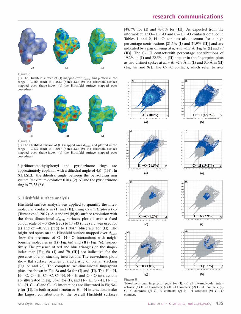
[48.7% for (I) and 43.6% for (II)]. As expected from the
intermolecular O—H� � �O and C—H� � �O contacts detailed in
Tables 1 and 2, H� � �O contacts also account for a high
percentage contributions [21.5% (I) and 21.9% (II)] and are
indicated by a pair of wings at de + di�1.7 A [Fig. 8c (I) and 9d
(II)]. The C� � �H contacts,with percentage contributions of
19.2% in (I) and 22.5% in (II) appear in the fingerprint plots
as two distinct spikes at de + di �2.9 A in (I) and 3.0 A in (II)
(Fig. 8d and 9c). The C� � �C contacts, which refer to �–�
research communications
Acta Cryst. (2020). E76, 432–437 Daoui et al. � C19H16N2O3 and C19H16N2O3 435
Figure 6(a) The Hirshfeld surface of (I) mapped over dnorm, and plotted in therange �0.7266 (red) to 1.4843 (blue) a.u.; (b) the Hirshfeld surfacemapped over shape-index; (c) the Hirshfeld surface mapped overcurvedness.
Figure 7(a) The Hirshfeld surface of (II) mapped over dnorm, and plotted in therange �0.7232 (red) to 1.3047 (blue) a.u.; (b) the Hirshfeld surfacemapped over shape-index, (c) the Hirshfeld surface mapped overcurvedness.
Figure 8Two-dimensional fingerprint plots for (I): (a) all intermolecular inter-actions; (b) H� � �H contacts; (c) H� � �O contacts; (d) C� � �H contacts; (e)C� � �C contacts; (f) C� � �N contacts; (g) N� � �H contacts; (h) C� � �Ocontacts.
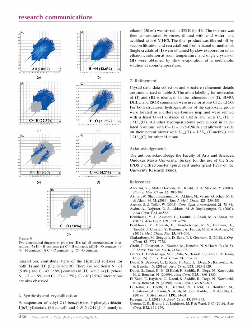
interactions, contribute 4.2% of the Hirshfeld surfaces for
both (I) and (II) (Fig. 8e and 9f). There are additional N� � �H
(5.0%) and C� � �O (2.8%) contacts in (II), while in (I) (where
N� � �H = 1.8% and C� � �O = 1.7%), C� � �N (2.9%) interactions
are also observed.
6. Synthesis and crystallization
A suspension of ethyl 2-(5-benzyl-6-oxo-3-phenylpyridazin-
1(6H)-yl)acetate (3.6 mmol), and 6 N NaOH (14.4 mmol) in
ethanol (50 ml) was stirred at 353 K for 4 h. The mixture was
then concentrated in vacuo, diluted with cold water, and
acidified with 6 N HCl. The final product was filtered off by
suction filtration and recrystallized from ethanol or methanol.
Single crystals of (I) were obtained by slow evaporation of an
ethanolic solution at room temperature, and single crystals of
(II) were obtained by slow evaporation of a methanolic
solution at room temperature.
7. Refinement
Crystal data, data collection and structure refinement details
are summarized in Table 3. The atom labelling for molecules
of (I) and (II) is identical. In the refinement of (I), SIMU,
DELU and ISOR commands were used for atoms C12 and O3.
For both structures, hydrogen atoms of the carboxylic group
were located in a difference-Fourier map and were refined
with a fixed O—H distance of 0.82 A and with Uiso(H) =
1.5Ueq(O). All other hydrogen atoms were placed in calcu-
lated positions, with C—H = 0.93–0.96 A and allowed to ride
on their parent atoms with Uiso(H) = 1.5Ueq(C-methyl) and
1.2Ueq(C) for other H atoms.
Acknowledgements
The authors acknowledge the Faculty of Arts and Sciences,
Ondokuz Mayıs University, Turkey, for the use of the Stoe
IPDS 2 diffractometer (purchased under grant F.279 of the
University Research Fund).
References
Abouzid, K., Abdel Hakeem, M., Khalil, O. & Maklad, Y. (2008).Bioorg. Med. Chem. 16, 382–389.
Akhtar, W., Shaquiquzzaman, M., Akhter, M., Verma, G., Khan, M. F.& Alam, M. M. (2016). Eur. J. Med. Chem. 123, 256–281.
Archan, S. & Toller, W. (2008). Curr. Opin. Anaesthesiol. 21, 78–84.Aydın, A., Dogruer, D. S., Akkurt, M. & Buyukgungor, O. (2007).
Acta Cryst. E63, o4522.Boukharsa, Y., El Ammari, L., Taoufik, J., Saadi, M. & Ansar, M.
(2015). Acta Cryst. E71, o291–o292.Boukharsa, Y., Meddah, B., Tiendrebeogo, R. Y., Ibrahimi, A.,
Taoufik, J., Cherrah, Y., Benomar, A., Faouzi, M. E. A. & Ansar, M.(2016). Med. Chem. Res. 25, 494–500.
Chakraborty, M., Sengupta, D., Saha, T. & Goswami, S. (2018). J. Org.Chem. 83, 7771–7778.
Chelfi, T., Elaatioui, A., Koudad, M., Benchat, N. & Hacht, B. (2015).J. Mater. Environ. Sci, 6, 2174–2178.
Costas, T., Costas-Lago, M. C., Vila, N., Besada, P., Cano, E. & Teran,C. (2015). Eur. J. Med. Chem. 94, 113–122.
Daoui, S., Baydere, C., El Kalai, F., Mahi, L., Dege, N., Karrouchi, K.& Benchat, N. (2019a). Acta Cryst. E75, 1925–1929.
Daoui, S., Cınar, E. B., El Kalai, F., Saddik, R., Dege, N., Karrouchi,K. & Benchat, N. (2019b). Acta Cryst. E75, 1880–1883.
El Kalai, F., Baydere, C., Daoui, S., Saddik, R., Dege, N., Karrouchi,K. & Benchat, N. (2019b). Acta Cryst. E75, 892–895.
El Kalai, F., Chelfi, T., Benchat, N., Hacht, B., Bouklah, M.,Elaatiaoui, A., Daoui, S., Allali, M., Ben Hadda, T. & Almalki, F.(2019a). J. Mol. Struct. 1191, 24–31.
Farrugia, L. J. (2012). J. Appl. Cryst. 45, 849–854.Groom, C. R., Bruno, I. J., Lightfoot, M. P. & Ward, S. C. (2016). Acta
Cryst. B72, 171–179.
436 Daoui et al. � C19H16N2O3 and C19H16N2O3 Acta Cryst. (2020). E76, 432–437
research communications
Figure 9Two-dimensional fingerprint plots for (II): (a) all intermolecular inter-actions; (b) H� � �H contacts; (c) C� � �H contacts; (d) H� � �O contacts; (e)N� � �H contacts; (f) C� � �C contacts; (g) C� � �O contacts.

Livermore, D. G. H., Bethell, R. C., Cammack, N., Hancock, A. P.,Hann, M. M., Green, D. V. S., Lamont, R. B., Noble, S. A., Orr, D. C.& Payne, J. J. (1993). J. Med. Chem. 36, 3784–3794.
Macrae, C. F., Sovago, I., Cottrell, S. J., Galek, P. T. A., McCabe, P.,Pidcock, E., Platings, M., Shields, G. P., Stevens, J. S., Towler, M. &Wood, P. A. (2020). J. Appl. Cryst. 53, 226–235.
Mahmoodi, N. O., Safari, N. & Sharifzadeh, B. (2014). Synth.Commun. 44, 245–250.
Partap, S., Akhtar, M. J., Yar, M. S., Hassan, M. Z. & Siddiqui, A. A.(2018). Bioorg. Chem. 77, 74–83.
Sheldrick, G. M. (2015a). Acta Cryst. A71, 3–8.Sheldrick, G. M. (2015b). Acta Cryst. C71, 3–8.Siddiqui, A. A., Mishra, R., Shaharyar, M., Husain, A., Rashid, M. &
Pal, P. (2011). Bioorg. Med. Chem. Lett. 21, 1023–1026.Singh, J., Sharma, D. & Bansal, R. (2017). J. Heterocycl. Chem. 54,
2935–2945.
Sotelo, E., Coelho, A. & Ravina, E. (2003). Tetrahedron Lett. 44,4459–4462.
Spek, A. L. (2020). Acta Cryst. E76, 1–11.Stoe & Cie (2002). X-AREA and X-RED32. Stoe & Cie GmbH,
Darmstadt, Germany.Turner, M. J., McKinnon, J. J., Wolff, S. K., Grimwood, D. J.,
Spackman, P. R., Jayatilaka, D. & Spackman, M. A. (2017).CrystalExplorer17. University of Western Australia. http://hirsh-feldsurface.net.
Westrip, S. P. (2010). J. Appl. Cryst. 43, 920–925.Xu, H., Song, H.-B., Yao, C.-S., Zhu, Y.-Q., Hu, F.-Z., Zou, X.-M. &
Yang, H.-Z. (2005). Acta Cryst. E61, o1561–o1563.Yarden, Y. & Caldes, C. (2013). Eur. J. Cancer, 49, 2619–2620.Zarrouk, A., Chelfi, T., Dafali, A., Hammouti, B., Al-Deyab, S. S.,
Warad, I., Benchat, N. & Zertoubi, M. (2010). Int. J. Electrochem.Sci. 5, 696–705.
research communications
Acta Cryst. (2020). E76, 432–437 Daoui et al. � C19H16N2O3 and C19H16N2O3 437
Table 3Experimental details.
I II
Crystal dataChemical formula C19H16N2O3 C19H16N2O3
Mr 320.34 320.34Crystal system, space group Monoclinic, P21/n Monoclinic, P21/cTemperature (K) 296 296a, b, c (A) 10.5500 (8), 9.3679 (6), 16.5606 (15) 10.5976 (6), 15.5500 (7), 10.3731 (7)� (�) 93.886 (7) 109.120 (5)V (A3) 1632.9 (2) 1615.11 (17)Z 4 4Radiation type Mo K� Mo K�� (mm�1) 0.09 0.09Crystal size (mm) 0.58 � 0.43 � 0.34 0.77 � 0.70 � 0.59
Data collectionDiffractometer Stoe IPDS 2 STOE IPDS 2Absorption correction Integration (X-RED32; Stoe & Cie, 2002) Integration (X-RED32; Stoe & Cie, 2002)Tmin, Tmax 0.961, 0.981 0.950, 0.966No. of measured, independent and observed [I >
2�(I)] reflections12987, 4603, 1989 12114, 4562, 2560
Rint 0.039 0.037(sin �/�)max (A�1) 0.698 0.699
RefinementR[F 2 > 2�(F 2)], wR(F 2), S 0.053, 0.158, 0.89 0.049, 0.131, 0.98No. of reflections 4603 4562No. of parameters 217 218No. of restraints 19 0H-atom treatment H-atom parameters constrained H-atom parameters constrained�max, �min (e A�3) 0.35, �0.34 0.21, �0.21
Computer programs: X-AREA and X-RED32 (Stoe & Cie, 2002), SHELXT2017/1 (Sheldrick, 2015a), SHELXL2018/3 (Sheldrick, 2015b), Mercury (Macrae et al., 2020), WinGX(Farrugia, 2012), PLATON (Spek, 2020) and publCIF (Westrip, 2010).
supporting information
sup-1Acta Cryst. (2020). E76, 432-437
supporting information
Acta Cryst. (2020). E76, 432-437 [https://doi.org/10.1107/S2056989020002406]
Polymorphism of 2-(5-benzyl-6-oxo-3-phenyl-1,6-dihydropyridazin-1-yl)acetic
acid with two monoclinic modifications: crystal structures and Hirshfeld
surface analyses
Said Daoui, Cemile Baydere, Tarik Chelfi, Fouad El Kalai, Necmi Dege, Khalid Karrouchi and
Noureddine Benchat
Computing details
For both structures, data collection: X-AREA (Stoe & Cie, 2002); cell refinement: X-AREA (Stoe & Cie, 2002); data
reduction: X-RED32 (Stoe & Cie, 2002); program(s) used to solve structure: SHELXT2017/1 (Sheldrick, 2015a);
program(s) used to refine structure: SHELXL2018/3 (Sheldrick, 2015b); molecular graphics: Mercury (Macrae et al.,
2020) and PLATON (Spek, 2020); software used to prepare material for publication: WinGX (Farrugia, 2012), PLATON
(Spek, 2020) and publCIF (Westrip, 2010).
2-(5-Benzyl-6-oxo-3-phenyl-1,6-dihydropyridazin-1-yl)acetic acid (I)
Crystal data
C19H16N2O3
Mr = 320.34Monoclinic, P21/na = 10.5500 (8) Åb = 9.3679 (6) Åc = 16.5606 (15) Åβ = 93.886 (7)°V = 1632.9 (2) Å3
Z = 4
F(000) = 672Dx = 1.303 Mg m−3
Mo Kα radiation, λ = 0.71073 ÅCell parameters from 9543 reflectionsθ = 1.9–29.8°µ = 0.09 mm−1
T = 296 KPrism, colorless0.58 × 0.43 × 0.34 mm
Data collection
Stoe IPDS 2 diffractometer
Radiation source: sealed X-ray tube, 12 x 0.4 mm long-fine focus
Plane graphite monochromatorDetector resolution: 6.67 pixels mm-1
rotation method scansAbsorption correction: integration
(X-RED32; Stoe & Cie, 2002)
Tmin = 0.961, Tmax = 0.98112987 measured reflections4603 independent reflections1989 reflections with I > 2σ(I)Rint = 0.039θmax = 29.7°, θmin = 2.4°h = −12→14k = −13→12l = −23→23
Refinement
Refinement on F2
Least-squares matrix: fullR[F2 > 2σ(F2)] = 0.053wR(F2) = 0.158
S = 0.894603 reflections217 parameters19 restraints

supporting information
sup-2Acta Cryst. (2020). E76, 432-437
Hydrogen site location: inferred from neighbouring sites
H-atom parameters constrained
w = 1/[σ2(Fo2) + (0.0772P)2]
where P = (Fo2 + 2Fc
2)/3(Δ/σ)max < 0.001Δρmax = 0.35 e Å−3
Δρmin = −0.34 e Å−3
Special details
Geometry. All esds (except the esd in the dihedral angle between two l.s. planes) are estimated using the full covariance matrix. The cell esds are taken into account individually in the estimation of esds in distances, angles and torsion angles; correlations between esds in cell parameters are only used when they are defined by crystal symmetry. An approximate (isotropic) treatment of cell esds is used for estimating esds involving l.s. planes.
Fractional atomic coordinates and isotropic or equivalent isotropic displacement parameters (Å2)
x y z Uiso*/Ueq
N2 0.63345 (14) 0.43785 (17) 0.40115 (9) 0.0550 (4)N1 0.56137 (14) 0.34515 (16) 0.44077 (9) 0.0539 (4)O1 0.66753 (13) 0.60906 (17) 0.31011 (10) 0.0811 (5)O2 0.76216 (13) 0.27628 (19) 0.30333 (10) 0.0914 (6)H2 0.804328 0.226747 0.274357 0.137*C7 0.43814 (16) 0.35514 (19) 0.42800 (10) 0.0508 (4)C6 0.36108 (18) 0.2513 (2) 0.47105 (11) 0.0536 (4)C8 0.38202 (17) 0.4630 (2) 0.37706 (11) 0.0567 (5)H8 0.294010 0.471278 0.371699 0.068*C10 0.58990 (18) 0.5384 (2) 0.34591 (11) 0.0589 (5)C12 0.83446 (19) 0.3288 (2) 0.36076 (12) 0.0612 (5)C9 0.45351 (17) 0.5535 (2) 0.33636 (11) 0.0594 (5)O3 0.94446 (16) 0.3027 (3) 0.37082 (12) 0.1213 (7)C11 0.77005 (17) 0.4220 (2) 0.41864 (12) 0.0634 (5)H11A 0.785002 0.382843 0.472680 0.076*H11B 0.808847 0.515829 0.418553 0.076*C14 0.26299 (19) 0.7017 (2) 0.29161 (12) 0.0638 (5)C5 0.2306 (2) 0.2435 (2) 0.45719 (13) 0.0677 (6)H5 0.189656 0.304979 0.419821 0.081*C15 0.2264 (2) 0.7924 (3) 0.35035 (15) 0.0794 (7)H15 0.288323 0.838285 0.383550 0.095*C4 0.1600 (2) 0.1465 (3) 0.49763 (14) 0.0780 (6)H4 0.072279 0.143929 0.487252 0.094*C13 0.4010 (2) 0.6709 (3) 0.28207 (15) 0.0822 (7)H13A 0.411644 0.644798 0.226260 0.099*H13B 0.449726 0.757252 0.293614 0.099*C17 0.0092 (2) 0.7521 (3) 0.31546 (16) 0.0888 (8)H17 −0.075763 0.769037 0.323863 0.107*C19 0.1676 (2) 0.6361 (3) 0.24455 (14) 0.0836 (7)H19 0.188376 0.573613 0.203863 0.100*C3 0.2156 (3) 0.0557 (3) 0.55173 (15) 0.0859 (7)H3 0.167270 −0.008738 0.579307 0.103*C1 0.4167 (2) 0.1565 (3) 0.52585 (16) 0.0904 (8)H1 0.504386 0.157532 0.536391 0.109*
supporting information
sup-3Acta Cryst. (2020). E76, 432-437
C16 0.1008 (2) 0.8173 (3) 0.36152 (17) 0.0958 (8)H16 0.078911 0.880366 0.401664 0.115*C18 0.0408 (2) 0.6619 (3) 0.25690 (16) 0.0901 (8)H18 −0.022580 0.616712 0.224545 0.108*C2 0.3444 (3) 0.0599 (3) 0.56547 (18) 0.1078 (10)H2A 0.384238 −0.003519 0.602205 0.129*
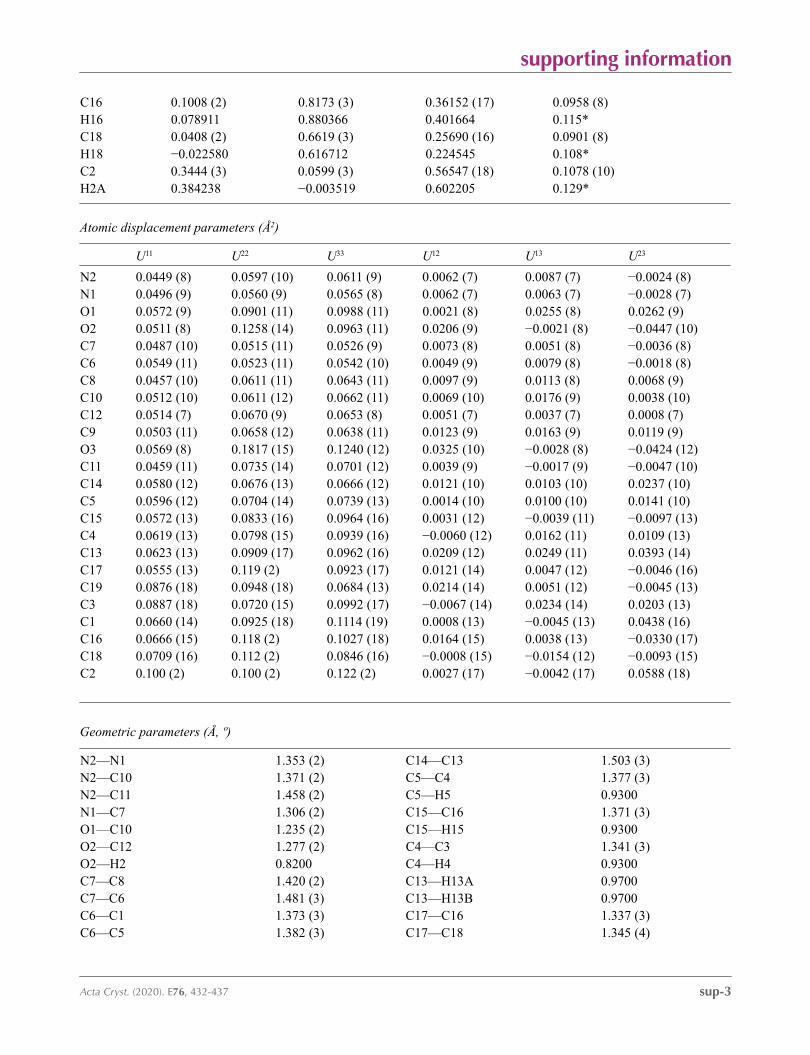
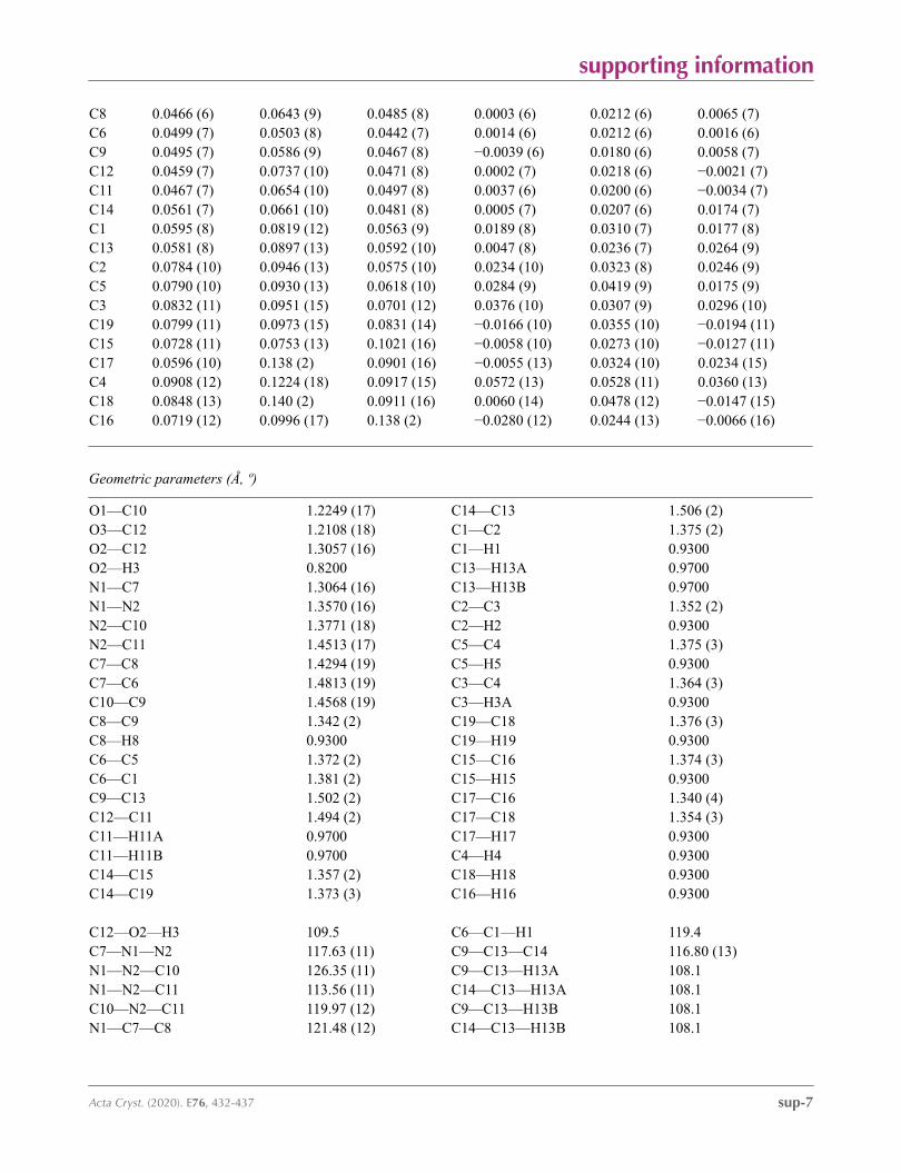
Atomic displacement parameters (Å2)
U11 U22 U33 U12 U13 U23
N2 0.0449 (8) 0.0597 (10) 0.0611 (9) 0.0062 (7) 0.0087 (7) −0.0024 (8)N1 0.0496 (9) 0.0560 (9) 0.0565 (8) 0.0062 (7) 0.0063 (7) −0.0028 (7)O1 0.0572 (9) 0.0901 (11) 0.0988 (11) 0.0021 (8) 0.0255 (8) 0.0262 (9)O2 0.0511 (8) 0.1258 (14) 0.0963 (11) 0.0206 (9) −0.0021 (8) −0.0447 (10)C7 0.0487 (10) 0.0515 (11) 0.0526 (9) 0.0073 (8) 0.0051 (8) −0.0036 (8)C6 0.0549 (11) 0.0523 (11) 0.0542 (10) 0.0049 (9) 0.0079 (8) −0.0018 (8)C8 0.0457 (10) 0.0611 (11) 0.0643 (11) 0.0097 (9) 0.0113 (8) 0.0068 (9)C10 0.0512 (10) 0.0611 (12) 0.0662 (11) 0.0069 (10) 0.0176 (9) 0.0038 (10)C12 0.0514 (7) 0.0670 (9) 0.0653 (8) 0.0051 (7) 0.0037 (7) 0.0008 (7)C9 0.0503 (11) 0.0658 (12) 0.0638 (11) 0.0123 (9) 0.0163 (9) 0.0119 (9)O3 0.0569 (8) 0.1817 (15) 0.1240 (12) 0.0325 (10) −0.0028 (8) −0.0424 (12)C11 0.0459 (11) 0.0735 (14) 0.0701 (12) 0.0039 (9) −0.0017 (9) −0.0047 (10)C14 0.0580 (12) 0.0676 (13) 0.0666 (12) 0.0121 (10) 0.0103 (10) 0.0237 (10)C5 0.0596 (12) 0.0704 (14) 0.0739 (13) 0.0014 (10) 0.0100 (10) 0.0141 (10)C15 0.0572 (13) 0.0833 (16) 0.0964 (16) 0.0031 (12) −0.0039 (11) −0.0097 (13)C4 0.0619 (13) 0.0798 (15) 0.0939 (16) −0.0060 (12) 0.0162 (11) 0.0109 (13)C13 0.0623 (13) 0.0909 (17) 0.0962 (16) 0.0209 (12) 0.0249 (11) 0.0393 (14)C17 0.0555 (13) 0.119 (2) 0.0923 (17) 0.0121 (14) 0.0047 (12) −0.0046 (16)C19 0.0876 (18) 0.0948 (18) 0.0684 (13) 0.0214 (14) 0.0051 (12) −0.0045 (13)C3 0.0887 (18) 0.0720 (15) 0.0992 (17) −0.0067 (14) 0.0234 (14) 0.0203 (13)C1 0.0660 (14) 0.0925 (18) 0.1114 (19) 0.0008 (13) −0.0045 (13) 0.0438 (16)C16 0.0666 (15) 0.118 (2) 0.1027 (18) 0.0164 (15) 0.0038 (13) −0.0330 (17)C18 0.0709 (16) 0.112 (2) 0.0846 (16) −0.0008 (15) −0.0154 (12) −0.0093 (15)C2 0.100 (2) 0.100 (2) 0.122 (2) 0.0027 (17) −0.0042 (17) 0.0588 (18)
Geometric parameters (Å, º)
N2—N1 1.353 (2) C14—C13 1.503 (3)N2—C10 1.371 (2) C5—C4 1.377 (3)N2—C11 1.458 (2) C5—H5 0.9300N1—C7 1.306 (2) C15—C16 1.371 (3)O1—C10 1.235 (2) C15—H15 0.9300O2—C12 1.277 (2) C4—C3 1.341 (3)O2—H2 0.8200 C4—H4 0.9300C7—C8 1.420 (2) C13—H13A 0.9700C7—C6 1.481 (3) C13—H13B 0.9700C6—C1 1.373 (3) C17—C16 1.337 (3)C6—C5 1.382 (3) C17—C18 1.345 (4)
supporting information
sup-4Acta Cryst. (2020). E76, 432-437
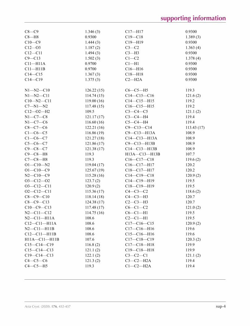
C8—C9 1.346 (3) C17—H17 0.9300C8—H8 0.9300 C19—C18 1.389 (3)C10—C9 1.444 (3) C19—H19 0.9300C12—O3 1.187 (2) C3—C2 1.363 (4)C12—C11 1.494 (3) C3—H3 0.9300C9—C13 1.502 (3) C1—C2 1.378 (4)C11—H11A 0.9700 C1—H1 0.9300C11—H11B 0.9700 C16—H16 0.9300C14—C15 1.367 (3) C18—H18 0.9300C14—C19 1.375 (3) C2—H2A 0.9300
N1—N2—C10 126.22 (15) C6—C5—H5 119.3N1—N2—C11 114.74 (15) C14—C15—C16 121.6 (2)C10—N2—C11 119.00 (16) C14—C15—H15 119.2C7—N1—N2 117.48 (15) C16—C15—H15 119.2C12—O2—H2 109.5 C3—C4—C5 121.1 (2)N1—C7—C8 121.17 (17) C3—C4—H4 119.4N1—C7—C6 116.60 (16) C5—C4—H4 119.4C8—C7—C6 122.21 (16) C9—C13—C14 113.43 (17)C1—C6—C5 116.86 (19) C9—C13—H13A 108.9C1—C6—C7 121.27 (18) C14—C13—H13A 108.9C5—C6—C7 121.86 (17) C9—C13—H13B 108.9C9—C8—C7 121.38 (17) C14—C13—H13B 108.9C9—C8—H8 119.3 H13A—C13—H13B 107.7C7—C8—H8 119.3 C16—C17—C18 119.6 (2)O1—C10—N2 119.04 (17) C16—C17—H17 120.2O1—C10—C9 125.67 (19) C18—C17—H17 120.2N2—C10—C9 115.28 (16) C14—C19—C18 120.9 (2)O3—C12—O2 123.7 (2) C14—C19—H19 119.5O3—C12—C11 120.9 (2) C18—C19—H19 119.5O2—C12—C11 115.36 (17) C4—C3—C2 118.6 (2)C8—C9—C10 118.14 (18) C4—C3—H3 120.7C8—C9—C13 124.38 (17) C2—C3—H3 120.7C10—C9—C13 117.48 (17) C6—C1—C2 121.0 (2)N2—C11—C12 114.75 (16) C6—C1—H1 119.5N2—C11—H11A 108.6 C2—C1—H1 119.5C12—C11—H11A 108.6 C17—C16—C15 120.9 (2)N2—C11—H11B 108.6 C17—C16—H16 119.6C12—C11—H11B 108.6 C15—C16—H16 119.6H11A—C11—H11B 107.6 C17—C18—C19 120.3 (2)C15—C14—C19 116.8 (2) C17—C18—H18 119.9C15—C14—C13 121.1 (2) C19—C18—H18 119.9C19—C14—C13 122.1 (2) C3—C2—C1 121.1 (2)C4—C5—C6 121.3 (2) C3—C2—H2A 119.4C4—C5—H5 119.3 C1—C2—H2A 119.4
supporting information
sup-5Acta Cryst. (2020). E76, 432-437
Hydrogen-bond geometry (Å, º)
D—H···A D—H H···A D···A D—H···A
O2—H2···O1i 0.82 1.82 2.593 (2) 156C1—H1···N1 0.93 2.47 2.780 (3) 100
Symmetry code: (i) −x+3/2, y−1/2, −z+1/2.
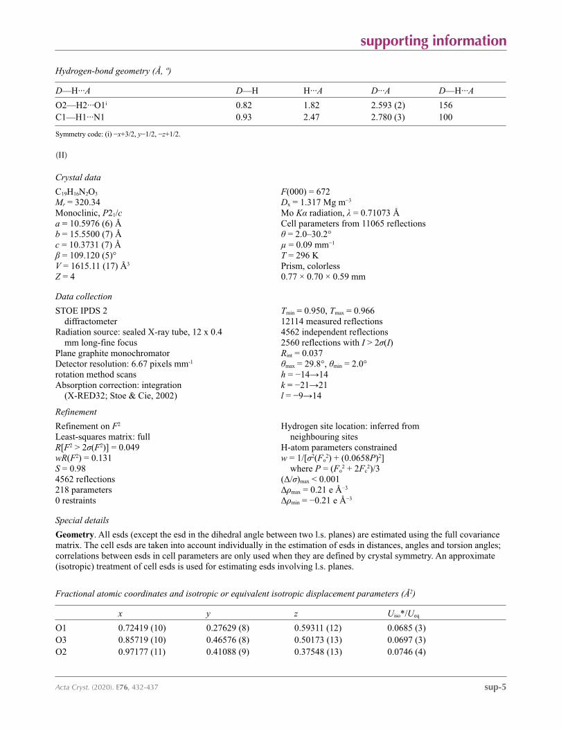
(II)
Crystal data
C19H16N2O3
Mr = 320.34Monoclinic, P21/ca = 10.5976 (6) Åb = 15.5500 (7) Åc = 10.3731 (7) Åβ = 109.120 (5)°V = 1615.11 (17) Å3
Z = 4
F(000) = 672Dx = 1.317 Mg m−3
Mo Kα radiation, λ = 0.71073 ÅCell parameters from 11065 reflectionsθ = 2.0–30.2°µ = 0.09 mm−1
T = 296 KPrism, colorless0.77 × 0.70 × 0.59 mm
Data collection
STOE IPDS 2 diffractometer
Radiation source: sealed X-ray tube, 12 x 0.4 mm long-fine focus
Plane graphite monochromatorDetector resolution: 6.67 pixels mm-1
rotation method scansAbsorption correction: integration
(X-RED32; Stoe & Cie, 2002)
Tmin = 0.950, Tmax = 0.96612114 measured reflections4562 independent reflections2560 reflections with I > 2σ(I)Rint = 0.037θmax = 29.8°, θmin = 2.0°h = −14→14k = −21→21l = −9→14
Refinement
Refinement on F2
Least-squares matrix: fullR[F2 > 2σ(F2)] = 0.049wR(F2) = 0.131S = 0.984562 reflections218 parameters0 restraints
Hydrogen site location: inferred from neighbouring sites
H-atom parameters constrainedw = 1/[σ2(Fo
2) + (0.0658P)2] where P = (Fo
2 + 2Fc2)/3
(Δ/σ)max < 0.001Δρmax = 0.21 e Å−3
Δρmin = −0.21 e Å−3
Special details
Geometry. All esds (except the esd in the dihedral angle between two l.s. planes) are estimated using the full covariance matrix. The cell esds are taken into account individually in the estimation of esds in distances, angles and torsion angles; correlations between esds in cell parameters are only used when they are defined by crystal symmetry. An approximate (isotropic) treatment of cell esds is used for estimating esds involving l.s. planes.
Fractional atomic coordinates and isotropic or equivalent isotropic displacement parameters (Å2)
x y z Uiso*/Ueq
O1 0.72419 (10) 0.27629 (8) 0.59311 (12) 0.0685 (3)O3 0.85719 (10) 0.46576 (8) 0.50173 (13) 0.0697 (3)O2 0.97177 (11) 0.41088 (9) 0.37548 (13) 0.0746 (4)
supporting information
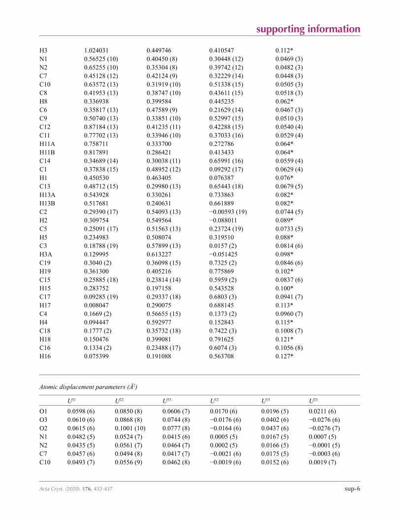
sup-6Acta Cryst. (2020). E76, 432-437
H3 1.024031 0.449746 0.410547 0.112*N1 0.56525 (10) 0.40450 (8) 0.30448 (12) 0.0469 (3)N2 0.65255 (10) 0.35304 (8) 0.39742 (12) 0.0482 (3)C7 0.45128 (12) 0.42124 (9) 0.32229 (14) 0.0448 (3)C10 0.63572 (13) 0.31919 (10) 0.51338 (15) 0.0505 (3)C8 0.41953 (13) 0.38747 (10) 0.43611 (15) 0.0518 (3)H8 0.336938 0.399584 0.445235 0.062*C6 0.35817 (13) 0.47589 (9) 0.21629 (14) 0.0467 (3)C9 0.50740 (13) 0.33851 (10) 0.52997 (15) 0.0510 (3)C12 0.87184 (13) 0.41235 (11) 0.42288 (15) 0.0540 (4)C11 0.77702 (13) 0.33946 (10) 0.37033 (16) 0.0529 (4)H11A 0.758711 0.333700 0.272786 0.064*H11B 0.817891 0.286421 0.413433 0.064*C14 0.34689 (14) 0.30038 (11) 0.65991 (16) 0.0559 (4)C1 0.37838 (15) 0.48952 (12) 0.09292 (17) 0.0629 (4)H1 0.450530 0.463405 0.076387 0.076*C13 0.48712 (15) 0.29980 (13) 0.65443 (18) 0.0679 (5)H13A 0.543928 0.330261 0.733863 0.082*H13B 0.517681 0.240631 0.661889 0.082*C2 0.29390 (17) 0.54093 (13) −0.00593 (19) 0.0744 (5)H2 0.309754 0.549564 −0.088011 0.089*C5 0.25091 (17) 0.51563 (13) 0.23724 (19) 0.0733 (5)H5 0.234983 0.508074 0.319510 0.088*C3 0.18788 (19) 0.57899 (13) 0.0157 (2) 0.0814 (6)H3A 0.129995 0.613227 −0.051425 0.098*C19 0.3040 (2) 0.36098 (15) 0.7325 (2) 0.0846 (6)H19 0.361300 0.405216 0.775869 0.102*C15 0.25885 (18) 0.23814 (14) 0.5959 (2) 0.0837 (6)H15 0.283752 0.197158 0.543528 0.100*C17 0.09285 (19) 0.29337 (18) 0.6803 (3) 0.0941 (7)H17 0.008047 0.290075 0.688145 0.113*C4 0.1669 (2) 0.56655 (15) 0.1373 (2) 0.0960 (7)H4 0.094447 0.592977 0.152843 0.115*C18 0.1777 (2) 0.35732 (18) 0.7422 (3) 0.1008 (7)H18 0.150476 0.399081 0.791625 0.121*C16 0.1334 (2) 0.23488 (17) 0.6074 (3) 0.1056 (8)H16 0.075399 0.191088 0.563708 0.127*
Atomic displacement parameters (Å2)
U11 U22 U33 U12 U13 U23
O1 0.0598 (6) 0.0850 (8) 0.0606 (7) 0.0170 (6) 0.0196 (5) 0.0211 (6)O3 0.0610 (6) 0.0868 (8) 0.0744 (8) −0.0176 (6) 0.0402 (6) −0.0276 (6)O2 0.0615 (6) 0.1001 (10) 0.0777 (8) −0.0164 (6) 0.0437 (6) −0.0276 (7)N1 0.0482 (5) 0.0524 (7) 0.0415 (6) 0.0005 (5) 0.0167 (5) 0.0007 (5)N2 0.0435 (5) 0.0561 (7) 0.0464 (7) 0.0002 (5) 0.0166 (5) −0.0001 (5)C7 0.0457 (6) 0.0494 (8) 0.0417 (7) −0.0021 (6) 0.0175 (5) −0.0003 (6)C10 0.0493 (7) 0.0556 (9) 0.0462 (8) −0.0019 (6) 0.0152 (6) 0.0019 (7)
supporting information
sup-7Acta Cryst. (2020). E76, 432-437
C8 0.0466 (6) 0.0643 (9) 0.0485 (8) 0.0003 (6) 0.0212 (6) 0.0065 (7)C6 0.0499 (7) 0.0503 (8) 0.0442 (7) 0.0014 (6) 0.0212 (6) 0.0016 (6)C9 0.0495 (7) 0.0586 (9) 0.0467 (8) −0.0039 (6) 0.0180 (6) 0.0058 (7)C12 0.0459 (7) 0.0737 (10) 0.0471 (8) 0.0002 (7) 0.0218 (6) −0.0021 (7)C11 0.0467 (7) 0.0654 (10) 0.0497 (8) 0.0037 (6) 0.0200 (6) −0.0034 (7)C14 0.0561 (7) 0.0661 (10) 0.0481 (8) 0.0005 (7) 0.0207 (6) 0.0174 (7)C1 0.0595 (8) 0.0819 (12) 0.0563 (9) 0.0189 (8) 0.0310 (7) 0.0177 (8)C13 0.0581 (8) 0.0897 (13) 0.0592 (10) 0.0047 (8) 0.0236 (7) 0.0264 (9)C2 0.0784 (10) 0.0946 (13) 0.0575 (10) 0.0234 (10) 0.0323 (8) 0.0246 (9)C5 0.0790 (10) 0.0930 (13) 0.0618 (10) 0.0284 (9) 0.0419 (9) 0.0175 (9)C3 0.0832 (11) 0.0951 (15) 0.0701 (12) 0.0376 (10) 0.0307 (9) 0.0296 (10)C19 0.0799 (11) 0.0973 (15) 0.0831 (14) −0.0166 (10) 0.0355 (10) −0.0194 (11)C15 0.0728 (11) 0.0753 (13) 0.1021 (16) −0.0058 (10) 0.0273 (10) −0.0127 (11)C17 0.0596 (10) 0.138 (2) 0.0901 (16) −0.0055 (13) 0.0324 (10) 0.0234 (15)C4 0.0908 (12) 0.1224 (18) 0.0917 (15) 0.0572 (13) 0.0528 (11) 0.0360 (13)C18 0.0848 (13) 0.140 (2) 0.0911 (16) 0.0060 (14) 0.0478 (12) −0.0147 (15)C16 0.0719 (12) 0.0996 (17) 0.138 (2) −0.0280 (12) 0.0244 (13) −0.0066 (16)
Geometric parameters (Å, º)
O1—C10 1.2249 (17) C14—C13 1.506 (2)O3—C12 1.2108 (18) C1—C2 1.375 (2)O2—C12 1.3057 (16) C1—H1 0.9300O2—H3 0.8200 C13—H13A 0.9700N1—C7 1.3064 (16) C13—H13B 0.9700N1—N2 1.3570 (16) C2—C3 1.352 (2)N2—C10 1.3771 (18) C2—H2 0.9300N2—C11 1.4513 (17) C5—C4 1.375 (3)C7—C8 1.4294 (19) C5—H5 0.9300C7—C6 1.4813 (19) C3—C4 1.364 (3)C10—C9 1.4568 (19) C3—H3A 0.9300C8—C9 1.342 (2) C19—C18 1.376 (3)C8—H8 0.9300 C19—H19 0.9300C6—C5 1.372 (2) C15—C16 1.374 (3)C6—C1 1.381 (2) C15—H15 0.9300C9—C13 1.502 (2) C17—C16 1.340 (4)C12—C11 1.494 (2) C17—C18 1.354 (3)C11—H11A 0.9700 C17—H17 0.9300C11—H11B 0.9700 C4—H4 0.9300C14—C15 1.357 (2) C18—H18 0.9300C14—C19 1.373 (3) C16—H16 0.9300
C12—O2—H3 109.5 C6—C1—H1 119.4C7—N1—N2 117.63 (11) C9—C13—C14 116.80 (13)N1—N2—C10 126.35 (11) C9—C13—H13A 108.1N1—N2—C11 113.56 (11) C14—C13—H13A 108.1C10—N2—C11 119.97 (12) C9—C13—H13B 108.1N1—C7—C8 121.48 (12) C14—C13—H13B 108.1
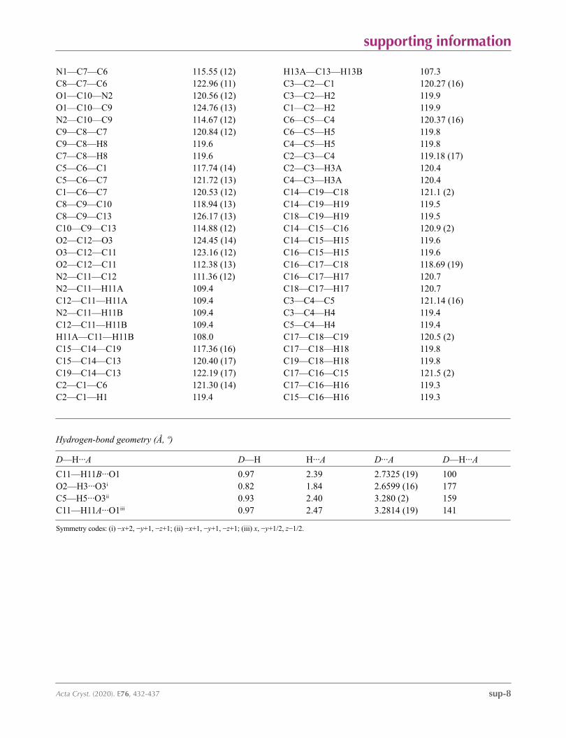
supporting information
sup-8Acta Cryst. (2020). E76, 432-437
N1—C7—C6 115.55 (12) H13A—C13—H13B 107.3C8—C7—C6 122.96 (11) C3—C2—C1 120.27 (16)O1—C10—N2 120.56 (12) C3—C2—H2 119.9O1—C10—C9 124.76 (13) C1—C2—H2 119.9N2—C10—C9 114.67 (12) C6—C5—C4 120.37 (16)C9—C8—C7 120.84 (12) C6—C5—H5 119.8C9—C8—H8 119.6 C4—C5—H5 119.8C7—C8—H8 119.6 C2—C3—C4 119.18 (17)C5—C6—C1 117.74 (14) C2—C3—H3A 120.4C5—C6—C7 121.72 (13) C4—C3—H3A 120.4C1—C6—C7 120.53 (12) C14—C19—C18 121.1 (2)C8—C9—C10 118.94 (13) C14—C19—H19 119.5C8—C9—C13 126.17 (13) C18—C19—H19 119.5C10—C9—C13 114.88 (12) C14—C15—C16 120.9 (2)O2—C12—O3 124.45 (14) C14—C15—H15 119.6O3—C12—C11 123.16 (12) C16—C15—H15 119.6O2—C12—C11 112.38 (13) C16—C17—C18 118.69 (19)N2—C11—C12 111.36 (12) C16—C17—H17 120.7N2—C11—H11A 109.4 C18—C17—H17 120.7C12—C11—H11A 109.4 C3—C4—C5 121.14 (16)N2—C11—H11B 109.4 C3—C4—H4 119.4C12—C11—H11B 109.4 C5—C4—H4 119.4H11A—C11—H11B 108.0 C17—C18—C19 120.5 (2)C15—C14—C19 117.36 (16) C17—C18—H18 119.8C15—C14—C13 120.40 (17) C19—C18—H18 119.8C19—C14—C13 122.19 (17) C17—C16—C15 121.5 (2)C2—C1—C6 121.30 (14) C17—C16—H16 119.3C2—C1—H1 119.4 C15—C16—H16 119.3
Hydrogen-bond geometry (Å, º)
D—H···A D—H H···A D···A D—H···A
C11—H11B···O1 0.97 2.39 2.7325 (19) 100O2—H3···O3i 0.82 1.84 2.6599 (16) 177C5—H5···O3ii 0.93 2.40 3.280 (2) 159C11—H11A···O1iii 0.97 2.47 3.2814 (19) 141
Symmetry codes: (i) −x+2, −y+1, −z+1; (ii) −x+1, −y+1, −z+1; (iii) x, −y+1/2, z−1/2.