kinetics,structure,andmechanismof8-oxo-7,8-dihydro-2...
TRANSCRIPT

Kinetics, Structure, and Mechanism of 8-Oxo-7,8-dihydro-2�-deoxyguanosine Bypass by Human DNA Polymerase �*�
Received for publication, January 22, 2014, and in revised form, April 14, 2014 Published, JBC Papers in Press, April 23, 2014, DOI 10.1074/jbc.M114.551820
Amritraj Patra‡, Leslie D. Nagy‡, Qianqian Zhang‡, Yan Su‡, Livia Muller§, F. Peter Guengerich‡, and Martin Egli‡1
From the ‡Department of Biochemistry and Center in Molecular Toxicology, Vanderbilt University School of Medicine, Nashville,Tennessee 37232 and the §Laboratory of Food and Nutrition Toxicology, Eidgenossische Technische Hochschule-Zentrum,CH-8092 Zurich, Switzerland
Background: 8-OxoG is a major oxidative lesion in DNA and is associated with cancer.Results: Kinetic and mass spectrometric studies demonstrate that human polymerase � bypasses 8-oxoG in a largely error-freemanner.Conclusion: Arginine 61 from the finger domain plays a key role in error-free bypass at the insertion stage.Significance: In addition to photo-adducts and cisplatinated DNA, polymerase � might also be involved in accurate bypass of8-oxoG in vivo.
DNA damage incurred by a multitude of endogenous andexogenous factors constitutes an inevitable challenge for thereplication machinery. Cells rely on various mechanisms toeither remove lesions or bypass them in a more or less error-prone fashion. The latter pathway involves the Y-familypolymerases that catalyze trans-lesion synthesis across sitesof damaged DNA. 7,8-Dihydro-8-oxo-2�-deoxyguanosine(8-oxoG) is a major lesion that is a consequence of oxidativestress and is associated with cancer, aging, hepatitis, andinfertility. We have used steady-state and transient-statekinetics in conjunction with mass spectrometry to analyze invitro bypass of 8-oxoG by human DNA polymerase � (hpol �).Unlike the high fidelity polymerases that show preferentialinsertion of A opposite 8-oxoG, hpol � is capable of bypassing8-oxoG in a mostly error-free fashion, thus preventingGC3AT transversion mutations. Crystal structures of ter-nary hpol �-DNA complexes and incoming dCTP, dATP, ordGTP opposite 8-oxoG reveal that an arginine from the fingerdomain assumes a key role in avoiding formation of the nas-cent 8-oxoG:A pair. That hpol � discriminates against dATPexclusively at the insertion stage is confirmed by structures ofternary complexes that allow visualization of the extensionstep. These structures with G:dCTP following either8-oxoG:C or 8-oxoG:A pairs exhibit virtually identical activesite conformations. Our combined data provide a detailedunderstanding of hpol � bypass of the most common oxida-tive DNA lesion.
7,8-Dihydro-8-oxo-2�-deoxyguanosine (8-oxoG)2 is one ofthe most common DNA lesions resulting from oxidative stressduring normal aerobic cellular metabolism (1–3). It is foundin both lower organisms and eukaryotes and is relevantbecause of its association with cancer (4, 5), aging (6), hepa-titis (7), and infertility (8). Compared with G and the canon-ical G:C pairing mode, the presence of oxygen at the C8-position in 8-oxoG alters the nucleoside conformationalequilibrium between the anti and syn modes in favor of thelatter, thus enabling pairing with A. This effect constitutesthe basis for the mutagenicity of 8-oxoG due to the resultingG:C to T:A transversion mutations (9, 10). Among the cellu-lar responses dealing with oxidative damage such as 8-oxoG,mismatch repair (MMR) assumes an important role (11). Inyeast, the Ogg1 glycosylase is known to remove the 8-oxoGlesion opposite C in double-stranded DNA (12, 13). Con-versely, when A is replicated opposite 8-oxoG in yeast, it isrecognized and excised by MutS� (14).
Lesions that evade recognition and repair prior to replicationcan elicit trans-lesion synthesis (TLS) (15–18). TLS involves theY-family of DNA polymerases (pols) (19, 20), with four mem-bers identified in humans, pol �, pol �, pol �, and Rev 1, whichexhibit various propensities and fidelities in the bypass of indi-vidual lesions (20 –23). Detailed structural and functionalinvestigations have shed light on the characteristics of theseTLS pols and revealed clear differences among them in terms oftheir mechanisms of adduct bypass (21, 24). Although Y-familypols share the right-handed arrangement of palm, thumb, andfinger subdomains (25) with high fidelity pols (i.e. A-family),they also feature a distinct “little finger” (LF) or pol-associateddomain that is crucial for lesion bypass (24, 26 –29). TLS polsgenerally exhibit lower processivity, catalytic efficiency, and
* This work was supported, in whole or in part, by National Institutes of HealthGrants ES010375 (to F. P. G. and M. E.), P01 CA160032 (to M. E.), and P30ES000267 (to F. P. G. and M. E.).
� This article was selected as a Paper of the Week.The atomic coordinates and structure factors (codes 4O3N, 4O3O, 4O3P, 4O3Q, 4O3R,
and 4O3S) have been deposited in the Protein Data Bank (http://wwpdb.org/).1 To whom correspondence should be addressed: Dept. of Biochemistry and
Center in Molecular Toxicology, Vanderbilt University School of Medicine,868A Robinson Research Bldg., Nashville, TN 37232-0146. Tel.: 615-343-8070; Fax: 615-322-7122; E-mail: [email protected].
2 The abbreviations used are: 8-oxoG, 7,8-dihydro-8-oxo-2�-deoxyguanosine;pol, (DNA) polymerase; hpol, human polymerase; Dpo4, S. solfataricus DNApolymerase IV; TLS, trans-lesion synthesis; MMR, mismatch repair; LF, littlefinger; CID, collision-induced dissociation; dNMPNPP, 2�-deoxynucleoside-5�-[(�,�)-imido]triphosphate; dTMPNPP, 2�-deoxythymidine-5�-[(�,�)-imido]triphosphate; dCMPNPP, 2�-deoxycytidine-5�-[(�,�)-imido]triphosphate; dAMPNPP, 2�-deoxyadenosine-5�-[(�,�)-imido]triphosph-ate; dGMPNPP, 2�-deoxyguanosine-5�-[(�,�)-imido]triphosphate.
THE JOURNAL OF BIOLOGICAL CHEMISTRY VOL. 289, NO. 24, pp. 16867–16882, June 13, 2014© 2014 by The American Society for Biochemistry and Molecular Biology, Inc. Published in the U.S.A.
JUNE 13, 2014 • VOLUME 289 • NUMBER 24 JOURNAL OF BIOLOGICAL CHEMISTRY 16867
at Vanderbilt U
niversity - Biom
edical & Science/E
ngineering Libraries on June 13, 2014
http://ww
w.jbc.org/
Dow
nloaded from

copying fidelity on undamaged DNA than high fidelity pols.However, we observed that the pol � homolog Dpo4 from Sul-folobus solfataricus bypasses 8-oxoG with high fidelity (19:1ratio of dCTP:dATP incorporation opposite 8-oxoG) and effi-ciency (pre-steady-state kinetics revealed faster rates of dCTPincorporation opposite 8-oxoG than G) (30) that could beattributed to a significant extent to Arg-332 from the LFdomain (31). By comparison, we established that human pol �(hpol �) bypasses 8-oxoG in an error-prone fashion, with dATPrather than dCTP preferentially incorporated opposite thelesion (32). Its proclivity to insert A opposite 8-oxoG resemblesthe action of replicative polymerases (33, 34).
Among the eukaryotic TLS pols, pol � is considered to playan important role in 8-oxoG bypass, although initial assaysusing human large-cell carcinoma cell line H1299 and xero-derma pigmentosum variant (defective in pol �) cells showedthat the nucleotide inserted opposite 8-oxoG was the correct Cat frequencies of 81 and 77%, respectively (35). However, muta-tion frequencies were increased in mammalian GM637 and inxeroderma pigmentosum variant cells in the presence of siRNAtargeting pol � (36). Interestingly, in the same system knock-down of MMR glycosylase OGG1 also triggered an increase inmutagenesis, providing support for the hypothesis that both theMMR and TLS systems combat mutagenic damage due to8-oxoG. Overlaps between MMR and TLS were confirmed by arecent in vivo study in yeast. The A residues opposite 8-oxoGwere removed by MMR, subsequently triggering re-replicationand, in the absence of MMR, by pol � TLS, with an accuracy ofinsertion of dCTP opposite 8-oxoG of 94% (37).
Other investigators using yeast-based assays and/or theenzyme from yeast observed accurate and efficient bypass of8-oxoG by pol � (38), whereby C, A, or G was incorporatedopposite 8-oxoG with a relative efficiency of 100:56:14 (39).Moreover, pol � binds dCTP opposite both G and 8-oxoG withsimilar affinities and inserts the correct nucleotide oppositeboth G and 8-oxoG with similar rates (40). Structural data forthe complexes between yeast pol � with 8-oxoG:C (bound atthe insertion and extension stages) were in line with the rela-tively accurate bypass of 8-oxoG by this enzyme but allowed nocomparison with insertion of dATP opposite 8-oxoG or exten-sion from 8-oxoG:A (41). An investigation entailing six humanB-, X-, and Y-family pols established that (relative to dATP)correct incorporation of dCTP opposite 8-oxoG in the presenceof the proliferating cell nuclear antigen and replication proteinA auxiliary proteins is 1,200- and 68-fold more efficient for pol� and pol �, respectively (42). An important role of pol � incombination with pol � for bypass of 8-oxoG was also estab-lished in vitro using mouse embryonic fibroblasts and HeLacells (43).
The somewhat inconsistent observations regarding the accu-racy of 8-oxoG bypass catalyzed by pol � in cells are mirrored bythe results from in vitro primer extension experiments probingits ability to correctly insert dCTP instead of dATP. Yung et al.(44) tested the bypass of 8-oxoG by human pol � (hpol �) invitro in four different sequence contexts and found that theidentity of the nucleotide 5�-adjacent to 8-oxoG had a signifi-cant effect on the incorporation efficiency and accuracy. Thus,for the sequences 5�-oligo-CGX, -CCX, -CAX, and -CTX (X �
8-oxoG), the dC:dA ratio varied between 1.1 and 2.9 (lowest forCAX and highest for CCX). However, the efficiency was lowestfor the CCX and highest for the CAX oligonucleotide. Con-versely, the dC:dG ratio was lowest for the CAX (2.0) and high-est for the CGX oligonucleotide (7.7). Using a 30-mer oligonu-cleotide with a single 8-oxoG for in vitro bypass, Zhang et al.(45) observed relatively error-prone insertion opposite 8-oxoGby hpol � (dC:dA �1:1 based on Vmax/Km) as well as similarefficiencies for extension from either 8-oxoG:C or 8-oxoG:A(5:3). Using oligonucleotides corresponding to a region of thehuman c-Ha-ras gene and containing either one or two 8-oxoGresidues, Jaloszynski et al. (46) found that the ratios between theefficiencies (kcat/Km) of dC and dA incorporation opposite theadduct varied between 8.5 and 2.4 (C � A). The lower ratio issimilar to the results obtained by McCulloch et al. (47), whoreported that the pol � enzymes from mouse and human showerror rates for in vitro bypass of 8-oxoG that approach 50%, inline with the data by others that support similar efficiencies forinsertion of either dC or dA opposite 8-oxoG as well as for theextension of the resulting pairs by the human enzyme (42, 45).Clearly, these results are inconsistent with the error-free bypassof 8-oxoG by pol �. Thus, the enzyme appears (based on thisliterature) to have inherently less fidelity than Dpo4, which syn-thesizes past the lesion with high accuracy and efficiency.
pol � displays an unmatched ability to replicate past UV-induced cyclic pyrimidine dimers (48), and a deficiency in thepolymerase is the basis for a variant form of the human syn-drome xeroderma pigmentosum, characterized by a highly ele-vated occurrence of skin malignancies (49, 50). pol � also facil-itates TLS past cancer drug adducts such as cisplatin (51) andthus lowers the cellular sensitivity to treatment. Structuralstudies on hpol � have focused on complexes of the catalyticcore with DNA template-primer constructs containing cis-syncyclobutane thymine dimers and have disclosed how thepolymerase accommodates two nucleotides at the active site(52). Elegant work by Yang and co-workers (53) has providedinsight into the step-by-step mechanism of pol � bypass andphosphodiester bond formation. Crystal structures of theenzymes from yeast (54) and human (55), in complex with cis-platin-modified DNA, have also provided a better understand-ing of the correct insertion of dCTP opposite the distortedcross-linked G dimer and the efficient extension after thelesion. No structures of hpol � in complex with DNA contain-ing the 8-oxoG adduct have been reported to date.
To evaluate the efficiency and accuracy of in vitro bypass of8-oxoG by hpol �, we carried out a comprehensive analysis ofthe bypass activity of the catalytic core of the enzyme, includingsteady-state and pre-steady-state kinetics of primer extensionopposite and beyond 8-oxoG and LC-MS/MS assays of full-length extension products. To gain a better understanding ofthe mechanism of 8-oxoG bypass by hpol �, we determinedcrystal structures of the native hpol � complex (G:dCTP; refer-ence structure), three insertion-stage complexes with 8-oxoGpaired to dCTP, dATP, or dGTP, and two complexes capturingthe extension steps following 8-oxoG:C or 8-oxoG:A. Our anal-ysis demonstrates that hpol � inserts almost exclusively dCTPopposite template 8-oxoG, with Arg-61 from the finger domainplaying a crucial role in the discrimination against dATP, and
Kinetics, Structure, and Mechanism of 8-OxoG Bypass by hpol �
16868 JOURNAL OF BIOLOGICAL CHEMISTRY VOLUME 289 • NUMBER 24 • JUNE 13, 2014
at Vanderbilt U
niversity - Biom
edical & Science/E
ngineering Libraries on June 13, 2014
http://ww
w.jbc.org/
Dow
nloaded from

that the polymerase achieves error-free bypass of the 8-oxoGlesion at the insertion stage.
EXPERIMENTAL PROCEDURES
Materials—All unlabeled dNTPs were obtained from Amer-sham Biosciences, and [-32P]ATP was purchased fromPerkinElmer Life Sciences. All nonhydrolyzable dNMPNPPswere obtained from Jena Bioscience (Jena, Germany). All oligo-nucleotides used in this work were synthesized by Midland Cer-tified Reagent Co. (Midland, TX), TriLink Biotechnologies (SanDiego), or Integrated DNA Technologies (Coralville, IA), and insome cases they were purified using HPLC by the manufac-turer, with analysis by matrix-assisted laser desorption time-of-flight MS.
hpol � Catalytic Core Protein Expression and Purification—The hpol � plasmid (pET28a) comprising residues 1– 432 was agenerous gift from Dr. Wei Yang, NIDDK, National Institutesof Health. The polymerase was expressed in Escherichia coliand purified as described previously (52). The protein solutionwas concentrated to 5 mg/ml.
Steady-state Kinetics—Steady-state incubation typicallyincluded 2.5–10 nM hpol �, 5 M template-primer oligonucleo-tide mixture as substrate (the primer was 5�-6-carboxyfluores-cein-labeled, and template-primer duplexes (1:1 molar ratio)were pre-annealed by heating and slow cooling), 40 mM Tris-HCl buffer (pH 7.5), 100 mM KCl, 5 mM MgCl2, 10 mM DTT, 5%glycerol (v/v), 100 g/ml bovine serum albumin (BSA), andvarying concentrations of a single dNTP (added last to start thereaction). The incubation temperature was 37 °C, and reactionswere typically run for 5 min (or less to keep product formation�20% of the oligonucleotide substrate concentration). Reac-tions were quenched by the addition of a quench reagent con-taining formamide, EDTA, bromphenol blue, and xylene cyanol(56), and aliquots were applied to an 18% (w/v) acrylamide, 7.5M urea gel and separated by electrophoresis. Fluorescence in thesubstrate and product primer bands was scanned using aTyphoon system (GE Healthcare) and quantified by ImageJ(National Institutes of Health), and the data were fit to hyper-bolic plots (Michaelis-Menten equation) using the programGraphPad Prism (GraphPad, La Jolla, CA).
Pre-steady-state Kinetics—A KinTek RP-3 instrument wasused, utilizing general procedures described previously (56). Ina typical experiment, 50 nM hpol � was incubated with 500 nM
oligonucleotide (5�-32P-primer/unlabeled template) and rap-idly mixed with 0.5 mM dNTP in the same buffer system used inthe steady-state kinetic studies (see above), incubated forvarying lengths of time (10 ms to 5 s), and quenched by theaddition of 0.5 mM EDTA (from the quench line). Radioac-tivity in the substrate and product primer bands was quan-tified using a phosphorimager system (Bio-Rad), and thedata were fit with the log-linear relationship y � Aekpt � kss,where kp is the first-order rate for the first catalytic cycle; kssis the steady-state rate, and A is the extent of the productburst (GraphPad Prism).
LC-MS/MS Analysis of Full-length Extension Products—Sixdifferent template-primer complexes were used, based uponliterature precedents for work with hpol � and 8-oxoG (X) asfollows: 5�-TCGTAAGCGTCUT-3� and 3�-AGCATTCG-
CAGTAXTACT-5� (this work); 5�-CGGCCTCCGGAUC-3�and 3�-GCCGGAGGCCTAGXACGT-5� (45); 5�-TTGCCCA-CACCUC-3� and 3�-AACGGGTGTGGCGXCCGC-5� (46);5�-GCCATGGCCCAUT-3� and 3�-CGGTACCGGGTTAXC-CTT-5� (47); 5�-TGCCGAATTCAUA-3� and 3�-ACGGCT-TAAGTGTXGCAG-5� (42); and 5�-GCCCAGGGTTTUC-3�and 3�-CGGGTCCCAAAAGXGTCA-5� (35). In all cases, ura-cil (U) was included in the primer to facilitate cleavage of theproduct to a shorter form that could be analyzed (followingcleavage with uracil DNA glycosylase and then hot piperidine)in an ion-trap mass spectrometer (57–59). hpol � (1.6 nmol)was reacted with each (5�-5/6-carboxyfluorescein-labeled)template-primer complex (2 nmol) and a mixture of 1 mM eachof dATP, dCTP, dGTP, and dTTP at 37 °C for 4 h in 50 mM
Tris-HCl buffer (pH 7.5). The reactions were terminated byspin column separations to extract dNTPs and Mg2�. Theextent of the reaction was checked by electrophoresis/fluorog-raphy prior to LC-MS analysis. The resulting product wastreated with 25 units of uracil DNA glycosylase and 0.25 M
piperidine following a previous protocol (57–59). To identifythe products, the resulting reactions were analyzed byLC-MS/MS using an Acquity UPLC system (Waters) interfacedto a Thermo-Finnigan LTQ mass spectrometer (Thermo Sci-entific, San Jose, CA) equipped with a negative ion electrospraysource. Chromatographic separation was achieved with theAcquity UPLC BEH octadecylsilane (C18) column (1.0 � 100mm, 1.7 m). The LC conditions were as follows: mobile phaseA, 10 mM NH4CH3CO2 in 98% H2O; mobile phase B, 10 mM
NH4CH3CO2 in 95% CH3CN (v/v). The following gradient pro-gram (v/v) was used with a flow rate of 200 l min1: the gra-dient started with 2% B (v/v), increased to 10% B (v/v) at 5 min,increased to 20% B (v/v) at 9 min, and held at 30% B (v/v) for 1min. The column was re-equilibrated for 3 min with 5% B (v/v).The temperature of the column was maintained at 50 °C. MSconditions were as follows: source voltage, 4 kV; source current100 A; capillary voltage, 49 V; capillary temperature, 350 °C;tube lens voltage, 90 V. Product ion spectra were acquiredover the range m/z 300 –2,000, and the most abundant species(2 charge) was used for collision-induced dissociation (CID)analysis. The most abundant species (2 or 3 charged) wereused for CID analysis; calculation of the CID fragmentation ofthe oligonucleotide sequence was done using a program linkedto the Mass Spectrometry Group of Medicinal Chemistry at theUniversity of Utah; the Mongo Oligo Mass Calculator (version2.6) from the University of Utah was used to calculate the the-oretical CID spectra of the candidate oligonucleotidesequences. The relative yields of various products were calcu-lated based on the peak areas of extracted ion chromatogramsfrom LC-MS analyses. The sum of the peak areas was used formulticharged species.
Crystallizations—Primer and template sequences used in thecrystallization experiments are listed in Table 6. 8-OxoG-mod-ified DNA templates were purchased from TriLink (San Diego),and unmodified 8-mer primers were obtained from IntegratedDNA Technologies (Coralville, IA). Template and primerstrands were mixed in a 1:1 molar ratio and were annealed inthe presence of 10 mM sodium HEPES buffer (pH 8.0), 0.1 mM
EDTA, and 50 mM NaCl by heating for 10 min at 85 °C followed
Kinetics, Structure, and Mechanism of 8-OxoG Bypass by hpol �
JUNE 13, 2014 • VOLUME 289 • NUMBER 24 JOURNAL OF BIOLOGICAL CHEMISTRY 16869
at Vanderbilt U
niversity - Biom
edical & Science/E
ngineering Libraries on June 13, 2014
http://ww
w.jbc.org/
Dow
nloaded from

by slow cooling to room temperature. For crystallization, theDNA duplex was mixed with the protein in a 1.2:1 molar ratio inthe presence of excess 50 mM Tris-HCl buffer (pH 7.5) contain-ing 450 mM KCl and 3 mM DTT. After adding 5 l of 100 mM
MgCl2, the complex was concentrated to a final concentrationof 2–3 mg/ml by ultrafiltration. Nonhydrolyzable nucleotidetriphosphates were added last to form ternary complexes. Crys-tallization experiments were performed by the hanging dropvapor diffusion technique at 18 °C using a sparse matrix screen(Hampton Research, Aliso Viejo, CA) (60). One l of the com-plex solution was mixed with 1 l of reservoir solution andequilibrated against 500-l reservoir wells. Crystals appeared indroplets containing 0.1 M MES (pH 5.5) buffer containing 5 mM
MgCl2 and 15–17% (w/v) PEG 2000 MME within 1 day andgrew to maximum size within a week.
X-ray Diffraction Data Collection, Structure Determination,and Refinement—Crystals were mounted in nylon loops, cryo-protected in reservoir solution containing 25% glycerol (v/v),and frozen in liquid nitrogen. Diffraction data were collectedeither on the 21-ID-D or the 21-ID-F beamline of the Life Sci-ences Collaborative Access Team at the Advanced PhotonSource, Argonne National Laboratory (Argonne, IL). Data wereintegrated and scaled with the program HKL2000 (61). Thestructures were determined by the molecular replacementtechnique using the program MOLREP (62, 63) and the hpol �structure with Protein Data Bank code 4ECQ (protein alone) asthe search model. Structure refinement and model buildingwere carried out with PHENIX (64) and COOT (65), respec-tively. Illustrations were generated with the program UCSFChimera (66).
RESULTS
Kinetics of Incorporation of dCTP and dATP Opposite Tem-plate G and 8-OxoG by hpol �—For all kinetic and structuralassays, we used the catalytic core construct of hpol � thatincludes amino acids 1– 432 (52). In the steady-state kineticanalysis, hpol � preferably inserted dCTP opposite G with�99% fidelity, relative to dATP (Fig. 1 and Table 1). In the caseof template 8-oxoG, dCTP was inserted 3.5-fold more effi-ciently than dATP. The catalytic efficiency of dCTP insertionopposite 8-oxoG was thus significantly higher than dATPinsertion (kcat/Km 0.52 versus 0.15 M1 s1, Table 1). By com-parison, the efficiency of insertion of dGTP opposite 8-oxoGwas drastically lower but clearly elevated (16-fold) relative toinsertion of dGTP opposite template G (Table 1).
The enhanced ability of hpol � to incorporate dCTP oppositetemplate 8-oxoG (relative to G) was also observed in pre-steady-state kinetic experiments (Fig. 2 and Table 2). Burstswere seen for incorporation of dCTP opposite G or 8-oxoG, butthis was not the case for insertion of dATP opposite 8-oxoG
FIGURE 1. Steady-state kinetic analysis of dNTP incorporation by hpol �. The reaction contained a template with G or 8-oxoG at position X as follows:5�-CGGTCGTAAGCGTCAT-3� and 3�-GCCAGCATTCGCAGTAXTACT-5�. hpol � (2.5–10 nM) was used with an oligonucleotide concentration of 5 M and varyingconcentrations of dATP or dCTP. Incubation proceeded for 5 min at 37 °C, and products formed were seperated by PAGE. All reactions were done in duplicate,and the indicated data points are shown as means � standard deviation. A, dCTP incorporation opposite G; B, dCTP incorporation opposite 8-oxoG; C, dATPincorporation opposite G. D, dATP incorporation opposite 8-oxo G. See Table 1 for kcat and Km values (estimated using fit to a hyperbolic equations in Prism,GraphPad, San Diego).
TABLE 1Steady-state kinetics of incorporation of dCTP, dATP, and dGTP oppo-site G and 8-oxoG by hpol �
Templatebase dNTP kcat Km kcat/Km f a
s1 M M1 s1
G dCTP 1.33 � 0.05 1.3 � 0.2 1.0 � 0.16G dATP 0.10 � 0.01 92 � 23 0.0011 � .0003 0.001G dGTP 0.15 � 0.02 80 � 34 0.0019 � 0.0008 0.00198-OxoG dCTP 1.20 � 0.03 2.3 � 0.2 0.52 � 0.058-OxoG dATP 0.78 � 0.03 5.4 � 0.6 0.15 � 0.02 0.288-OxoG dGTP 0.43 � 0.02 27 � 5 0.016 � 0.003 0.031
a f � (kcat/Km)dNTP/(kcat/Km)dCTP.
Kinetics, Structure, and Mechanism of 8-OxoG Bypass by hpol �
16870 JOURNAL OF BIOLOGICAL CHEMISTRY VOLUME 289 • NUMBER 24 • JUNE 13, 2014
at Vanderbilt U
niversity - Biom
edical & Science/E
ngineering Libraries on June 13, 2014
http://ww
w.jbc.org/
Dow
nloaded from
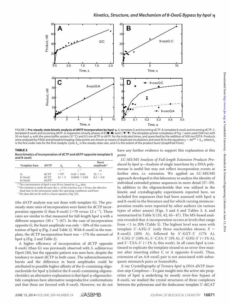
(the dATP analysis was not done with template G). The pre-steady-state rates of incorporation were faster for dCTP incor-poration opposite G than 8-oxoG (�70 versus 12 s1). Theserates are similar to that measured for full-length hpol � with adifferent sequence (30 s1) (67). In the case of incorporationopposite G, the burst amplitude was only 50% of the concen-tration of hpol � (Fig. 2 and Table 2). With 8-oxoG in the tem-plate, the dCTP incorporation burst was 17% the amount ofhpol � (Fig. 2 and Table 2).
A higher efficiency of incorporation of dCTP opposite8-oxoG (than G) was previously observed with S. solfataricusDpo4 (30), but the opposite pattern was seen here, despite thetendency to insert dCTP in both cases. The substoichiometricbursts and the difference in burst amplitudes could beattributed to possible higher affinity of the G-containing oligo-nucleotide for hpol � (relative the 8-oxoG-containing oligonu-cleotide); an alternative explanation is that hpol �-oligonucleo-tide complexes have alternative nonproductive conformationsand that these are favored with 8-oxoG. However, we do not
have any further evidence to support this explanation at thispoint.
LC-MS/MS Analysis of Full-length Extension Products Pro-duced by hpol �—Analysis of single insertions by a DNA poly-merase is useful but may not reflect incorporation events atfurther sites, i.e. extension. We applied an LC-MS/MSapproach developed in this laboratory to analyze the identity ofindividual extended primer sequences in more detail (57–59).In addition to the oligonucleotide that was utilized in thekinetic and crystallography experiments reported here, weincluded five sequences that had been assessed with hpol �and 8-oxoG in the literature and for which varying misincor-poration results were reported by other authors (in varioustypes of other assays) (Figs. 3 and 4 and Tables 3, 4, andsummarized in Table 5) (35, 42, 45– 47). The MS-based anal-ysis revealed that A incorporation occurs at levels that rangefrom �1 to 20% (Table 5). The highest level was seen withtemplate 5�-AXG-3� (only three nucleotides shown; X �8-oxoG) (20% A), followed by 5�-GXT-3� (17% A),5�-CXG-3� (10% A), 5�-CXA-3� (3% A), 5�-GXG-3� (�1% A),and 5�-TXA-3� (�1% A; this work). In all cases hpol � con-tinued to replicate the template strand in an error-free man-ner after inserting either C or A opposite 8-oxoG. Thus,extension of an A:8-oxoG pair is not associated with subse-quent mismatch pairs or frameshifts.
X-ray Crystallography of Ternary hpol �-DNA-dNTP Inser-tion-step Complexes—To gain insight into the active site prop-erties of hpol � underlying its mostly error-free bypass of8-oxoG, we studied the crystal structures of three complexesbetween the polymerase and the dodecamer template 5�-d(CAT
FIGURE 2. Pre-steady-state kinetic analysis of dNTP incorporation by hpol �. A, template G and incoming dCTP. B, template 8-oxoG and incoming dCTP. C,template 8-oxoG and incoming dATP. D, expansion of early phases of B (F–F) and C (�–�). The template-primer complexes of Fig. 1 were used (500 nM) with50 nM hpol �, with the same buffer system (37 °C) and 0.5 mM dCTP or dATP, for the indicated times, and quenched by the addition of 500 mM EDTA. Productswere analyzed by PAGE and phosphorimaging. Data points are shown as means of duplicate incubations and were fit to the equation y � Aekpt � kss, where kpis the first-order rate for the first catalytic cycle; kss is the steady-state rate, and A is the extent of the product burst (GraphPad Prism).
TABLE 2Burst kinetics of incorporation of dCTP and dATP opposite template Gand 8-oxoG
Template base dNTP kp kss
Burstamplitudea
s1 s1 nM
G dCTP �70b 0.45 � 0.03 25 � 18-OxoG dCTP 12 � 5 0.0045 � 0.05 8.5 � 0.88-OxoG dATPc
a The concentration of hpol � was 50 nM, based on A280 data.b The estimate is made because the t1⁄2 of the reaction was �10 ms, the effective
dead time in the instrument under the operating conditions used here.c The data did not fit well to a burst equation (Fig. 2D).
Kinetics, Structure, and Mechanism of 8-OxoG Bypass by hpol �
JUNE 13, 2014 • VOLUME 289 • NUMBER 24 JOURNAL OF BIOLOGICAL CHEMISTRY 16871
at Vanderbilt U
niversity - Biom
edical & Science/E
ngineering Libraries on June 13, 2014
http://ww
w.jbc.org/
Dow
nloaded from

(8-oxoG)AT GAC GCT)-3� paired to primer 5�-d(AGC GTCAT)-3� and dCMPNPP, dAMPNPP, or dGMPNPP opposite8-oxoG (Ci, Ai, and Gi complexes, respectively; i indicatesinsertion). For a full list of oligonucleotide constructs used,please see Table 6. To prevent the primer from being extendedduring crystallization in the presence of Mg2�, we useddNMPNPP analogs instead of dNTPs. The crystal structure ofthe complex with the same template-primer duplex and incom-
ing dCMPNPP opposite native G served as the reference (Cncomplex; n indicates native). The crystal structures of the Cn,Ci, Ai, and Gi complexes were all determined at resolutions ofbetween 1.5 and 1.8 Å (Table 7). In the structures, all residues ofthe catalytic core of hpol � were visible in the electron densitywith very few exceptions (Table 7). The 5�-terminal C and Anucleotides in the template strand are disordered in the struc-tures, and the T adjacent to the lesion exhibits high conforma-tional flexibility. Examples of the quality of the final electrondensity and views of the active sites are depicted in Fig. 5.
The structure of the G:dCMPNPP pair at the active site of theCn complex reveals the expected Watson-Crick geometry (Fig.6A). A very similar configuration of the active site is seen inthe Ci complex with template 8-oxoG opposite incomingdCMPNPP, with the nucleoside lesion in the anti conformationand thus establishing three H-bonds with cytosine (Fig. 6B). Inboth cases, amino acids from the hpol � finger domain engagein contacts to the nascent base pair. On the template side,Gln-38 N� donates in an H-bond to N3 of 8-oxoG (or G in thenative template) from the minor groove side. On the primerside, the side chain of Arg-61 adopts a coiled conformation andstacks on top of the base of the incoming nucleotide (Fig. 6B,right). In addition to the stacking interaction, Arg N� forms anH-bond to the �-phosphate group of dCMPNPP. The distances
FIGURE 3. LC-MS analysis of full-length extension products by hpol � in the presence of all four dNTPs. A, sample reconstructed extracted ion chromat-ogram for m/z 727.1 (3) for product with sequence 5�-TCATGAA-3�. B, mass spectrum of peak at retention time 4.42 min. See Table 3 for full list of productsand respective m/z assignments.
FIGURE 4. LC-MS analysis of full-length extension products by human pol � in the presence of all four dNTPs. A, sample reconstructed extracted ionchromatogram for m/z 714.1 (3) for product with sequence 5�-CCTGCAA-3�. B, mass spectrum of peak at retention time 4.42 min. See Table 4 for full list ofproducts and respective m/z assignments.
TABLE 3Observed and theoretical m/z for all products in primer-template com-plex shown in Fig. 3
Product m/z observed m/z theoretical
5�-pTAATGA-3� Not observed (1) 1894.24, (2) 946.625�-pTCATGA-3� (2) 934.64 (1) 1870.22, (2) 934.605�-pTCATGAG-3� (2) 1099.27, (3) 732.27 (2) 1099.21, (3) 732.475�-pTCATGAA-3� (2) 1091.27, (3) 727.00 (2) 1091.21, (3) 727.14
TABLE 4Observed and theoretical m/z for all products in primer-template com-plex shown in Fig. 4
Product m/z observed m/z theoretical
5�-pCATGCA-3� (2) 926.64 (1) 1855.20, (2) 927.105�-pCATGCAG-3� (2) 1091.64, (3) 727.55 (2) 1091.70, (3) 727.475�-pCATGCAA-3� (2) 1083.09, (3) 722.09 (2) 1083.70, (3) 722.135�-pCCTGCA-3� (2) 915.00 (1) 1831.18, (2) 915.085�-pCCTGCAG-3� (2) 1079.64, (3) 719.27 (2) 1079.69, (3) 719.465�-pCCTGCAA-3� (2) 1071.64, (3) 713.91 (2) 1071.69, (3) 714.12
Kinetics, Structure, and Mechanism of 8-OxoG Bypass by hpol �
16872 JOURNAL OF BIOLOGICAL CHEMISTRY VOLUME 289 • NUMBER 24 • JUNE 13, 2014
at Vanderbilt U
niversity - Biom
edical & Science/E
ngineering Libraries on June 13, 2014
http://ww
w.jbc.org/
Dow
nloaded from

between O3� from the 3�-terminal T of the primer and P� of theincoming nucleotide triphosphate in the Cn and Ci complexesare 3.27 and 3.24 Å, respectively. The angle (pT)O3����P�–N(P�) between nucleophile and scissile bond in the Cn com-plex was 177.9°, and in the Ci complex the corresponding anglewas 176.8°.
Opposite both incoming dAMPNPP in the Ai complex anddGMPNPP in the Gi complex, 8-oxoG is in the syn conforma-tion and thus presents its Hoogsteen edge to the nucleotidetriphosphates. Gln-38 N� now donates in an H-bond to O8 of8-oxoG that is pointing into the minor groove. The 8-oxoG:dAMPNPP pair is stabilized by H-bonds between N6(H)2 (A)
and O6 (8-oxoG) and between N1 (A) and N7(H) (8-oxoG) (Fig.6C). Arg-61 has shifted its position relative to the active sites inthe Cn and Ci complexes, and there is no overlap between itsside chain and adenine consistent with stacking (Fig. 6C, right).However, Arg N� still forms an H-bond with the �-phosphateof dAMPNPP. Compared with the Ci complex, the distancebetween O3� from the 3-terminal T of the primer and P� ofdAMPNPP is slightly shorter (3.13 Å). The (pT)O3����P�–N(P�) angle was 176.0°.
In the active site harboring 8-oxoG:dGMPNPP, the sidechain of Arg-61 no longer adopts the coiled conformation seenin the structures with dAMPNPP and dCMPNPP opposite theadduct. Instead, the side chain adopts a more or less extendedconformation, thus allowing the N� groups of the guanidinomoiety to establish H-bonds to O6 of both 8-oxoG anddGMPNPP in the major groove (Fig. 6D). The latter interactionis possible because the base of the incoming nucleotide hasbeen pushed into the minor groove relative to both the struc-tures with incoming dCMPNPP or with incoming dAMPNPP(Fig. 7). As a result, one of the two H-bonds established between8-oxoG and dGMPNPP involves the 8-oxygen of the former(Fig. 4D), whereas this oxygen is not involved in an H-bond inthe 8-oxoG:dAMPNPP pair (Fig. 6C). Along with this move-ment of the incoming nucleotide, the 3�-terminal T of theprimer has also changed its orientation relative to the struc-tures of the Cn, Ci, and Ai complexes (Figs. 6D, and 7). Thedistance between O3� (T) and P� is considerably longer (3.71Å), and the 3�-hydroxyl group is no longer positioned more orless in line with the scissile P–O bond as in the other complexes,but rather it assumes an adjacent orientation (angle 89.5°; Fig.6D).
X-ray Crystallography of Ternary hpol �-DNA-dNTP Exten-sion-step Complexes—To visualize the active site geometriesand relative orientations of incoming nucleotide triphosphateand DNA primer at the extension stage, following insertion ofeither dCTP or dATP opposite 8-oxoG, we determined twoadditional hpol � crystal structures. The structures of the ter-nary complexes between the polymerase, the template-primerduplex 5�-d(CAT G(8-oxoG)T GAC GCT)-3�:5�-d(AGC GTCAX)-3� (X � dC or dA), and dCMPNPP were determined at1.72 and 1.62 Å resolution, respectively. Selected crystal dataand refinement parameters for these Ce (extension of 8-oxoG:Cpair) and Ae (extension of 8-oxoG:A) complex structures aregiven in Table 7. Illustrations of the active site configurations inthe complexes and the quality of the final electron density aredepicted in Fig. 5, E (Ce complex) and F (Ae complex). In the Cecomplex, the nascent dG:dCMPNPP pair stacks on top of anti8-oxoG:dC, and in the Ae complex the same nascent pair stackson top of syn 8-oxoG:dA. The orientation of Arg-61 from thefinger domain relative to the base plane of the incoming nucle-otide is very similar to that observed in the structure of the Cnand Ci complexes. Thus, the curled conformation of the argi-nine side chain results in extensive overlap with the cytosine.On the side of the template strand, Gln-38 contacts the N3atom of G in the minor groove, but it is too short to reach downto 8-oxoG at the 1 position either in the anti conformationopposite dC or in the syn conformation opposite dA (Fig. 8).The superimposition of the two complexes reveals closely sim-
TABLE 5LC-MS/MS analysis of full-length extension products
Primer/template % of total % C % A Ref.
Product 5�-TAATGA-3� 0 100 �1 This workProduct 5�-TCATGA-3� 15Product 5�-TCATGAG-3� 21Product 5�-TCATGAA-3� 64Template 3�-AXTACT-5�Product 5�-CATGCA-3� 2 80 20 45Product 5�-CATGCAG-3� 8Product 5�-CATGCAA-3� 10Product 5�-CCTGCA-3� 1Product 5�-CCTGCAG-3� 29Product 5�-CCTGCAA-3� 50Template 3�-GXACGT-5�Product 5�-CAGGCG-3� 4 90 10 46Product 5�-CAGGCGG-3� 1Product 5�-CAGGCGA-3� 5Product 5�-CCGGCG-3� 46Product 5�-CCGGCGG-3� 9Product 5�-CCGGCGA-3� 28Product 5�-CCGGCGC-3� 7Template 3�-GXCCGC-5�Product 5�-TAGGAA-3� 0 97 3 47Product 5�-TAGGAAA-3� 3Product 5�-TCGGAA-3� 25Product 5�-TCGGAAG-3� 18Product 5�-TCGGAAA-3� 46Product 5�-TCGGAAC-3� 8Template 3�-AXCCTT-5�Product 5�-AACGTC-3� 1 83 17 42Product 5�-AACGTCG-3� 7Product 5�-AACGTCA-3� 9Product 5�-ACCGTC-3� 1Product 5�-ACCGTCG-3� 20Product 5�-ACCGTCA-3� 58Product 5�-ACCGTCC-3� 4Template 3�-TXGCAG-5�Product 5�-CACAGT-� 0 100 �1 35Product 5�-CCCAGT-3� 3Product 5�-CCCAGTG-3� 40Product 5�-CCCAGTA-3� 53Product 5�-CCCAGTC-3� 4Template 3�-GXGTCA-5�
TABLE 6DNA sequences used in crystallization of hpol �-DNA complexes
Structure name DNA sequenceIncomingnucleotide
G:dCTP reference (Cn) 3�-TCG CAG TAG TAC-5� dCMPNPP5�-AGC GTC AT-3�
8-OxoG:dCTP (Ci) 3�-TCG CAG TA(8OG) TAC-5� dCMPNPP5�-AGC GTC AT-3�
8-OxoG:dATP (Ai) 3�-TCG CAG TA(8OG) TAC-5� dAMPNPP5�-AGC GTC AT-3�
8-OxoG:dGTP (Gi) 3�-TCG CAG TA(8OG) TAC-5� dGMPNPP5�-AGC GTC AT-3�
8-OxoG:C extension (Ce) 3�-TCG CAG T(8OG)G TAC-5� dCMPNPP5�-AGC GTC AC-3�
8-OxoG:A extension (Ae) 3�-TCG CAG T(8OG)G TAC-5� dCMPNPP5�-AGC GTC AA-3�
Kinetics, Structure, and Mechanism of 8-OxoG Bypass by hpol �
JUNE 13, 2014 • VOLUME 289 • NUMBER 24 JOURNAL OF BIOLOGICAL CHEMISTRY 16873
at Vanderbilt U
niversity - Biom
edical & Science/E
ngineering Libraries on June 13, 2014
http://ww
w.jbc.org/
Dow
nloaded from

TA
BLE
7C
ryst
ald
ata,
dat
aco
llect
ion
par
amet
ers
and
stru
ctu
rere
fin
emen
tst
atis
tics
Com
plex
G:d
CM
PNPP
(Cn)
8-O
xoG
:dC
MPN
P(C
i)8-
Oxo
G:d
AM
PNPP
(Ai)
8-O
xoG
:dG
MPN
PP(G
i)8-
Oxo
G:C
(Ce)
8-O
xoG
:A(A
e)
Dat
aco
llect
ion
Wav
elen
gth
Å1.
0781
0Å
0.97
872
Å1.
0781
0Å
0.97
872
Å0.
9787
2Å
0.97
872
ÅSp
ace
grou
pP6
1P6
1P6
1P6
1P6
1P6
1Re
solu
tion
42.2
4to
1.58
Å(1
.64
to1.
58Å
)a42
.52
to1.
72Å
(1.7
8to
1.72
Å)
42.6
8to
1.70
Å(1
.76
to1.
70Å
)42
.59
to1.
72Å
(1.7
8to
1.72
Å)
42.5
7to
1.72
Å(1
.78
to1.
72Å
)42
.70
to1.
62Å
(1.6
8to
1.62
Å)
Uni
tcel
la�
b,c
98.6
6,81
.79
Å98
.20,
81.5
5Å
98.5
7,81
.56
Å98
.37,
81.7
0Å
98.3
1,81
.60
Å98
.61,
81.8
4Å
Uni
que
refle
ctio
ns61
,923
(6,1
48)
47,4
24(4
,714
)49
,367
(4,8
54)
47,6
75(4
,752
)47
,720
(4,7
25)
57,3
91(5
,696
)C
ompl
eten
ess�
%
99.9
(99.
7)99
.9(9
9.7)
99.5
(98.
6)99
.9(9
9.9)
99.7
(99.
0)10
0.0
(100
.0)
I/
(I)
20.8
6(2
.33)
23.7
3(4
.29)
15.6
5(1
.92)
18.3
5(2
.93)
26.3
2(3
.94)
15.3
3(3
.21)
Wils
onB-
fact
or19
.4Å
219
.1Å
223
.7Å
221
.0Å
221
.1Å
217
.1Å
2
R-m
erge
0.06
5(0
.863
)0.
070
(0.6
47)
0.06
4(0
.948
)0.
082
(0.8
27)
0.06
5(0
.755
)0.
099
(0.8
06)
Redu
ndan
cy7.
4(7
.2)
7.3
(6.7
)7.
4(6
.9)
7.5
(7.4
)7.
2(6
.6)
7.4
(6.5
)
Ref
inem
ent
R-w
ork
0.17
0(0
.233
)0.
163
(0.2
11)
0.17
2(0
.244
)0.
185
(0.2
71)
0.17
2(0
.228
)0.
162
(0.1
99)
R-fr
ee0.
215
(0.2
69)
0.20
8(0
.253
)0.
219
(0.2
94)
0.23
2(0
.351
)0.
210
(0.2
70)
0.20
4(0
.238
)N
o.of
atom
spr
otei
n/D
NA
3,45
9/40
93,
470/
410
3,44
1/41
03,
517/
430
3,38
0/40
93,
445/
413
dNM
PNPP
/Mg2�
28/2
28/2
30/2
31/2
28/2
28/2
Wat
er/s
olut
e47
5/6
466/
1242
4/6
462/
645
2/6
532/
12Pr
otei
nre
sidu
es45
045
145
145
044
744
9A
vera
geB-
fact
or26
.024
.430
.726
.628
.624
.4Pr
otei
n/D
NA
24.9
/27.
323
.1/2
6.8
29.7
/31.
525
.2/3
1.3
27.4
/31.
423
.1/2
6.4
dNM
PNPP
/Mg2�
15.2
/13.
612
.1/1
0.6
18.8
/13.
516
.7/1
4.8
16.4
/17.
813
.0/1
9.6
Wat
er/s
olut
e33
.0/1
6.7
32.7
/21.
738
.0/2
3.1
34.3
/18.
136
.2/1
7.9
31.7
/22.
4r.m
.s.b
(bon
ds)
0.01
90.
015
0.01
70.
014
0.01
20.
015
r.m.s.
(ang
les)
1.8
1.4
1.5
1.2
1.2
1.4
Ram
acha
ndra
n,fa
vore
d98
%98
%97
%97
%98
%98
%
Ram
acha
ndra
n,ou
tlier
s0.
23%
0.23
%0.
46%
0.22
%0.
47%
0.46
%
aSt
atis
ticsf
orth
ehi
ghes
tres
olut
ion
shel
lare
show
nin
pare
nthe
ses.
br.m
.s.is
root
mea
nsq
uare
.
Kinetics, Structure, and Mechanism of 8-OxoG Bypass by hpol �
16874 JOURNAL OF BIOLOGICAL CHEMISTRY VOLUME 289 • NUMBER 24 • JUNE 13, 2014
at Vanderbilt U
niversity - Biom
edical & Science/E
ngineering Libraries on June 13, 2014
http://ww
w.jbc.org/
Dow
nloaded from

ilar active site geometries relative to orientations between the3-terminal residue of the primer and the incoming dCMPNPPand positions of Mg2� ions (Fig. 6C). The distance between O3�(T) and P� in the Ce complex amounts to 3.38 Å, and the cor-responding distance in the Ae complex is 3.35 Å. Together,these structures demonstrate convincingly that, unlike duringthe insertion steps, once hpol � has incorporated either dC ordA opposite the 8-oxoG adduct, the different pairing modes ofthe resulting 8-oxoG:C and 8-oxoG:A pairs are of little conse-quence for the relative orientations of primer strand and thenext incoming nucleotide.
DISCUSSIONAlthough 8-oxoG is the most common oxidative damage
product in DNA and pol � is considered important for TLS pastthe adduct, the human enzyme has hitherto not been subjectedto a detailed kinetic (i.e. pre-steady-state level) and three-di-mensional structural investigation in the context of 8-oxoGbypass. Our analysis addresses this void and provides a com-plete kinetic and structural framework for hpol � bypass of thismajor oxidative damage product. The steady-state results indi-cate a somewhat lower efficiency (kcat/Km) of dCTP incorpora-
tion opposite 8-oxoG relative to the corresponding processopposite G (30%). The efficiency of dATP incorporationopposite 8-oxoG is increased 136-fold relative to dATP:G, adifference that translates into a 280-fold change in the fre-quency of insertion in favor of dATP:8-oxoG. A further find-ing of the steady-state kinetic study is that hpol � has anincreased tendency to insert dGTP opposite 8-oxoG relativeto template G (8-fold at the level of efficiency kcat/Km),although both processes are negligible compared with bothdCTP and dATP insertion (the frequency of dGTP:8-oxoGinsertion is reduced 10-fold compared with dATP:8-oxoG).The steady-state results support the notion that hpol �bypasses 8-oxoG efficiently and with limited error. The pre-steady-state results confirm this conclusion in that the inser-tions of dCTP opposite both G and 8-oxoG exhibit burstphases with the burst amplitude and burst rate (kp) being infavor of insertion opposite G. By comparison, the insertionof dATP opposite 8-oxoG did not display a true burst, andthe apparent rate was reduced (Fig. 2D). These data are con-sistent with a clear kinetic advantage of dCTP incorporationopposite 8-oxoG over dATP by hpol �.
FIGURE 5. Quality of the final electron density in the insertion- and extension-step complexes between hpol � and 8-oxoG-adducted template-primerduplexes. The Fourier (2Fo Fc) sum electron density around 8-oxoG and neighboring base pairs in the Cn (A), Ci (B), Ai (C), Gi (D), Ce (E), and Ae (F) complexes(see Table 6 for DNA sequences) is drawn at the 1 threshold. The two depicted polymerase side chains are from Gln-38 (top left in panels) and Arg-61 (top rightin panels), and Mg2� ions are light green spheres.
Kinetics, Structure, and Mechanism of 8-OxoG Bypass by hpol �
JUNE 13, 2014 • VOLUME 289 • NUMBER 24 JOURNAL OF BIOLOGICAL CHEMISTRY 16875
at Vanderbilt U
niversity - Biom
edical & Science/E
ngineering Libraries on June 13, 2014
http://ww
w.jbc.org/
Dow
nloaded from

The results of the kinetic analysis here can be compared withthose obtained previously from the kinetics of 8-oxoG bypasscatalyzed by the pol Dpo4 from S. solfataricus (30). Dpo4 wasfound to exhibit a favorable efficiency of dCTP incorporationopposite 8-oxoG relative to dATP:8-oxoG, and the kp value ofdCTP incorporation opposite 8-oxoG at the pre-steady-statelevel was nearly double that of the kp value for dCTP incorpo-ration opposite G (2.3 s1 versus 1.3 s1, respectively; both wereconsiderably lower than the values reported here for hpol �). Aswell, the activation energy for dCTP incorporation opposite theadduct by Dpo4 was considerably lower than that for incorpo-ration of dCTP opposite the native template G (30). These
observations support the general view that Dpo4 displays abehavior that is more similar to that of hpol � but is clearlydistinct from its genetic homolog, hpol �. We reported earlierthat hpol � bypasses 8-oxoG in an error-prone fashion (32). Forexample, the kp values of insertions of dATP and dCTP oppo-site the adduct were 8.2 and 0.4 s1, respectively, and only theformer process was consistent with a burst phase. The LC-MS/MS-based analyses of the full-length extension products of invitro primer bypass past 8-oxoG carried out for all three poly-merases (Table 5) (this work and Refs. 30, 32) are also support-ive of the similar behaviors of hpol � and Dpo4, i.e. nearly error-free bypass of 8-oxoG and relatively error-prone bypass by hpol
FIGURE 6. Active site conformations and base pairing configurations in hpol �-DNA-dNMPNPP insertion-step complexes. Views are into the active sitesfrom the major groove side (panels on the left) and rotated by 90° around the horizontal axis and looking roughly along the normal to the nucleobase planeof the incoming dNMPNPP (panels on the right) for four hpol � complexes. The polymerase is shown as a schematic, and the DNA template-primerduplex and selected hpol � side chains are shown in stick form (e.g. finger residues Gln-38 and Arg-61; carbon atoms colored in beige). Oxygen, nitrogen,and DNA phosphorus atoms are colored in red, blue, and orange, respectively, and Mg2� ions are drawn as light green spheres. Hydrogen bonds aredashed lines. A, complex with template G paired to incoming dCMPNPP (reference structure). DNA carbon atoms are green. B, complex with template8-oxoG paired to dCMPNPP. DNA carbon atoms are light blue except for 8-oxoG carbons that are highlighted in magenta. Arg-61 and the primer3�-terminal T adopt two alternative conformations. C, complex with template 8-oxoG paired to dAMPNPP. DNA carbon atoms are lilac except for 8-oxoGcarbons that are highlighted in pink. D, complex with template 8-oxoG paired to dGMPNPP. DNA carbon atoms are brown except for 8-oxoG carbons thatare highlighted in purple.
Kinetics, Structure, and Mechanism of 8-OxoG Bypass by hpol �
16876 JOURNAL OF BIOLOGICAL CHEMISTRY VOLUME 289 • NUMBER 24 • JUNE 13, 2014
at Vanderbilt U
niversity - Biom
edical & Science/E
ngineering Libraries on June 13, 2014
http://ww
w.jbc.org/
Dow
nloaded from

�. Dpo4 preferred dCTP over dATP with a ratio of 19:1 (30),and hpol � preferred dATP over dCTP with a ratio of 4:1 (32). Inthe work presented here, we found that hpol � has an evenhigher preference for dCTP than Dpo4, i.e. the analysis of full-length extension products by LC-MS/MS is consistent with�1–3% incorporation of dATP opposite 8-oxoG in three of thesix sequences examined and 10 –20% in the other three.
The mass spectrometric analysis of the extension products(not only of the sequence used for the kinetic and structural
studies here but also five further sequences previously investi-gated by others and featuring different nearest neighbors of8-oxoG) largely attests to the ability of hpol � to bypass 8-oxoGin a largely error-free fashion (Table 5). Accuracies of bypassvaried between basically error-free in this study and two previ-ous investigations relying on different sequences (35, 47) andabout 4:1 in favor of dCTP (42, 45). In the case of the sequenceemployed in this study for kinetics and x-ray crystallography(first one shown under “Experimental Procedures” and in Table5), the LC-MS/MS analysis of full-length extension productsdemonstrated convincingly that hpol � discriminates againstdATP opposite 8-oxoG at both the insertion and extensionsteps. Thus, no single oligonucleotide product contained amajor fraction of A as a consequence of insertion opposite theadduct, supporting the idea that discrimination occurs duringboth insertion and extension and is consistent with higher fidel-ity in LC-MS/MS relative to single nucleoside triphosphateinsertion assays (Figs. 1 and 2 and Tables 1 and 2). The maininsights gained from the kinetic and LC-MS/MS results are thatdCTP is overwhelmingly preferred over dATP by hpol � oppo-site 8-oxoG and that this bias is kinetically controlled, poten-tially as a result of an active site configuration that guaranteespreferred accommodation of the incoming dCTP.
Although our steady-state kinetic analysis cannot shed lighton the relative accuracies of the insertion and extension steps asit was done with individual dNTPs and thus provides informa-tion only on the insertion of each (opposite 8-oxoG), the massspectrometric analysis does. The LC-MS analysis is of the fullyextended products, which is a measure of misincorporationopposite the adduct (8-oxoG) and the proclivity to extend var-ious pairs (e.g. 8-oxoG:C, 8-oxoG:A, and 8-oxoG:G). Thus, apolymerase could efficiently insert a base opposite 8-oxoG butbe incapable of extension, leading to differences. Apparentlythat happens with 8-oxoG:A pairing.
In addition to a reference structure (template G:dCMPNPP;Cn), we determined three crystal structures of hpol � withdCMPNPP (Ci), dAMPNPP (Ai), or dGMPNPP (Gi) oppositetemplate 8-oxoG. Moreover, the structures of complexes witheither 8-oxoG:C (Ce) or 8-oxoG:A (Ae) at the 1 position weredetermined at a similar resolution. The two latter complexesdisplay nearly identical active site configurations (Fig. 8C),thereby corroborating the earlier observations and conclusionsfrom kinetics and the LC-MS/MS analysis. Thus, the structuraldata indicate that the polymerase may extend equally well fromeither 8-oxoG:C or 8-oxoG:A. Therefore, the absence of Arevealed by the mass spectrometric dissection of the full-lengthextension products needs to be attributed to a less favorableinteraction with dATP relative to dCTP during the insertionstep of 8-oxoG TLS. The insertion-step structures show thatthe nascent base pair is probed by two amino acids from thefinger domain, Gln-38 and Arg-61 (Fig. 6). Both were identifiedas conserved residues unique to pol � (28) and were found toplay important roles in the bypass by the polymerase of cyclicpyrimidine dimer (52) and cisplatinated DNA (55). However, asfar as bypass of 8-oxoG by hpol � is concerned, Gln-38 is notlikely to influence accuracy in a decisive manner. This isbecause the finger residue can interact either with N3 of8-oxoG in the anti conformation and thus be ready to receive
FIGURE 7. Superimpositions of individual hpol �-DNA-dNMPNPP inser-tion-step complexes. All views are into the major groove, and color codesmatch those used in Fig. 6 except that the protein is colored in pink, and thecoloring of protein side chains in the individual structures matches that of theincoming dNMPNPP. The template thymidine residue 5�-adjacent to 8-oxoGand the 1 and 2 base pairs are shown in gray. A, superimposition of thecomplexes with G:dCMPNPP (green carbons) and 8-oxoG:dCMPNPP (light bluecarbons; 8-oxoG carbons are highlighted in magenta). B, superimposition ofthe complexes with 8-oxoG:dCMPNPP (light blue carbons; 8-oxoG carbonshighlighted in magenta) and 8-oxoG:dAMPNPP (lilac carbons; 8-oxoG carbonsare highlighted in pink). C, superimposition of the complexes with 8-oxoG:dAMPNPP (lilac carbons; 8-oxoG carbons highlighted in pink) and 8-oxoG:dGMPNPP (brown carbons; 8-oxoG carbons are highlighted in purple).
Kinetics, Structure, and Mechanism of 8-OxoG Bypass by hpol �
JUNE 13, 2014 • VOLUME 289 • NUMBER 24 JOURNAL OF BIOLOGICAL CHEMISTRY 16877
at Vanderbilt U
niversity - Biom
edical & Science/E
ngineering Libraries on June 13, 2014
http://ww
w.jbc.org/
Dow
nloaded from

FIGURE 8. Active site conformations and base pairing configurations in hpol �-DNA-dCMPNPP extension-step complexes. Views into the active sitesfrom the major groove side (panels on the left) and rotated by 90° around the horizontal axis and looking roughly along the normal to the nucleobase plane ofthe incoming dCMPNPP (right-hand side panels in A and B) and 30° rotation around the horizontal axis in C for two hpol � complexes. Drawing mode and colorcodes match those in Figs. 6 and 7, i.e. protein backbone and carbon atoms of selected side chains are colored in beige in A and B and in pink (backbone) withside chains matching the coloring of primer C opposite 8-oxoG in the 1 base pair in C. The thymidine residue 5�-adjacent to template dG at the active site andthe 2 base pairs are shown in gray on the right and in C. Hydrogen bonds are dashed lines. A, view of the active site with 8-oxoG:dC at the 1 position andtemplate G opposite incoming dCMPNPP. Carbon atoms are light blue except for 8-oxoG carbons that are highlighted in magenta. B, view of the active site with8-oxoG:dA at the 1 position and template G opposite incoming dCMPNPP. Carbon atoms are lilac except for 8-oxoG carbons that are highlighted in pink. C,superimposition of the two complexes shown in A and B.
FIGURE 9. Schematic of the relative orientations of dNMPNPPs and 8-oxoG as well as Arg-61 in the Ci, Ai, and Gi insertion-step hpol � complexes.Arrows indicate the movements of dGMPNPP (brown) and dAMPNPP (lilac) into the minor groove relative to dCMPNPP (light blue), the movements of Arg-61 inthe Gi (brown carbons) and Ai (lilac carbons) complexes into the major groove relative to the Ci complex (light blue carbons), and the movement of the3�-terminal T of the primer in the Gi complex into the major groove relative to the Ai and Ci complexes. The relative movements of the nucleotide triphosphatesand Arg-61 in the Gi and Ai complexes result in the loss of stacking between the nucleobase of the incoming dNMPNPP and the guanidino group. Themovement of the 3�-terminal T in the Gi complex results in an unfavorable orientation of the 3�-OH for an attack at the �-phosphate group.
Kinetics, Structure, and Mechanism of 8-OxoG Bypass by hpol �
16878 JOURNAL OF BIOLOGICAL CHEMISTRY VOLUME 289 • NUMBER 24 • JUNE 13, 2014
at Vanderbilt U
niversity - Biom
edical & Science/E
ngineering Libraries on June 13, 2014
http://ww
w.jbc.org/
Dow
nloaded from

dCTP or O8 of the adducted nucleoside in the syn conforma-tion, giving rise to dATP incorporation. Therefore, hpol � doesnot maintain accuracy of bypass by way of suppressing thetendency of the 8-oxoG nucleoside to adopt the syn conforma-tion. Rather it appears to fall to Arg-61 to facilitate accommo-dation of dCTP in the active site and thus endow it with akinetic advantage over dATP insertion. Geometric consider-ations, i.e. the position of O3� from the incoming dNTP relativeto the P� phosphate group, are not suggestive of the preferencefor dCTP being the result of a faster chemical step comparedwith dATP. However, comparison between the active sites inthe structures of the Ci and Ai complexes brings to light a moreoptimal interaction between Arg-61 and the cytosine base,involving both the aliphatic portion of the arginine side chain aswell as the guanidino moiety (Figs. 7 and 9). Thus, Arg-61 could
direct dCTP more optimally into the active site, stabilize itsorientation opposite the adduct, and enhance the residencetime. Suarez et al. (68), using a bacterial forward mutagenesisassay with a 1–511 hpol � R61A construct, found that thismutation has only a limited effect on the bypass efficiency.However, in preliminary site-directed mutagenesis work, weobserved that the mutation R61K significantly decreased thefidelity, i.e. f(dATP/dCTP catalytic selectivity ratio, Table 1)increased from 0.28 to 0.74. Further studies are planned withmore mutants.
Contrary to dCTP and dATP, which exhibit only moderatelydifferent orientations relative to the primer and alternativeArg-61 side chain rotamers, dGTP is significantly shifted andtherefore in a considerably more unfavorable position for the3�-OH nucleophile to carry out its attack on the �-phosphate
FIGURE 10. Similar thymine-arginine-guanine interactions in the major grooves of the DNA complexes of yeast sporulation regulator Ndt80 andhpol �. A, sporulation regulator Ndt80 (69) (Protein Data Bank code 1MNN). B, human pol � in complex with 8-oxoG adducted template-primer duplex (Gicomplex, this work). Carbon atoms of the arginine side chain and the thymine are highlighted in yellow and are labeled along with dG (Ndt80) and dGTP (hpol�) and terminal residues; hydrogen bonds are solid lines in black and the guanidino-thymine cation-� stacking interaction is indicated with a thin dashed linein magenta. Rather than Arg interacting with the major groove edge of dG that is seen in virtually all structures of DNA-protein complexes, the illustrationsconvey the particular sequence context and the cation-� stacking interaction in the hpol � complex that are reminiscent of the recognition of 5�-TGTG bytandem arginines by Ndt80 (only one Arg shown in A). Differences of course are that in the pol � complex, dG(TP) and T are not covalently bound and thatArg-61 protrudes from the finger domain above, rather than being inserted into the major groove more or less within the guanine plane as in the Ndt80complex. But just like in the Ndt80-DNA complex, Arg-61 from hpol � pulls out T from the stack, with the consequence that the 3�-hydroxyl group of T is furtherremoved from the �-phosphate of dGTP than in the complexes with dCTP or dATP. In the case of the Ndt80 complex, this particular interaction provides ameans of indirect readout; the interaction in the pol � complex is detrimental to activity and explains the low efficiency of dGTP incorporation opposite 8-oxoG(and probably also opposite G).
FIGURE 11. Individual amino acids are key to bypass activity by Y-family DNA polymerases. Composite of the active sites of S. solfataricus Dpo4 (graycarbons), hpol � (magenta carbons), and hpol � (cyan carbons) with template 8-oxoG opposite incoming dCTP (Dpo4), dCMPNPP (hpol �), or dATP (hpol �) isshown. Arg-332 in Dpo4 forms an H-bond with O8 of 8-oxoG, and Met-135 in hpol � and Arg-61 in hpol � engage in stacking interactions with the templateadduct and the incoming nucleotide, respectively.
Kinetics, Structure, and Mechanism of 8-OxoG Bypass by hpol �
JUNE 13, 2014 • VOLUME 289 • NUMBER 24 JOURNAL OF BIOLOGICAL CHEMISTRY 16879
at Vanderbilt U
niversity - Biom
edical & Science/E
ngineering Libraries on June 13, 2014
http://ww
w.jbc.org/
Dow
nloaded from

group (Figs. 7 and 9). The cation-� interaction between Arg-61,now in an extended conformation, and the 3�-T of the primer isreminiscent of the arrangement of arginine pulling T fromunder the adjacent G in the DNA major groove of the complexwith the yeast sporulation regulator Ndt80 (Fig. 10) (69). Asimilar displacement of the terminal primer base was also seenin the structure of hpol � with a T:dGTP mismatch at the activesite (70). These observations underscore the versatile nature ofthe roles played by the arginine residue in bypass catalysis byhpol �, involving H-bonding, electrostatics, and/or stacking.
Although both hpol � and S. solfataricus Dpo4 are capable ofbypassing 8-oxoG efficiently and accurately, they rely onentirely different strategies to avoid misincorporation of dATPand subsequent transversion mutation. Unlike pol �, whichdoes not recruit a residue from the LF domain to support accu-rate 8-oxoG bypass, Dpo4 uses LF residue Arg-332 to prevent8-oxoG at the 0 (insertion) and 1 (extension) locations fromadopting the preferred syn conformation by establishing anH-bond between the Arg-332 guanidino moiety and the O8atom of 8-oxoG (Fig. 11). Therefore, it appears that either inter-acting with the template strand and the adduct itself or exertinginfluence on the choice of the incoming nucleotide triphos-phate can be successfully used by Y-family pols to catalyzeaccurate TLS. Both pol � and Dpo4 are likely the polymerases ofchoice for warding off the negative consequences of oxidativedamage in the respective organisms. Conversely, hpol �bypasses 8-oxoG in an error-prone fashion (32). Like hpol �,hpol � uses a residue from the finger domain to interact with thenascent base pair (Fig. 11). However, the particular arrange-ment between Met-135 and template 8-oxoG favors the synconformation of the adducted nucleoside (32, 71). In eukaryoticorganisms, the recruitment of pol � for bypassing the 8-oxoGadduct is deleterious.
Acknowledgments—We are grateful to Dr. L. Lei for help with proteinexpression and purification, Dr. Z. Wawrzak for assistance with x-raydiffraction data collection at the Advanced Photon Source, Dr. W.Yang for helpful discussions, and K. Trisler for assistance in prepara-tion of the manuscript. Vanderbilt University is a member institutionof the Life Sciences Collaborative Access Team at sector 21 of theAdvanced Photon Source, Argonne, IL. Use of the Advanced PhotonSource at Argonne National Laboratory was supported by the UnitedStates Department of Energy, Office of Science, Office of Basic EnergySciences, under Contract DE-AC02-06CH11357.
REFERENCES1. Beckman, K. B., and Ames, B. N. (1997) Oxidative decay of DNA. J. Biol.
Chem. 272, 19633–196362. Helbock, H. J., Beckman, K. B., Shigenaga, M. K., Walter, P. B., Woodall,
A. A., Yeo, H. C., and Ames, B. N. (1998) DNA oxidation matters: theHPLC-electrochemical detection assay of 8-oxo-deoxyguanosine and8-oxo-guanine. Proc. Natl. Acad. Sci. U.S.A. 95, 288 –293
3. Kamiya, H. (2004) Mutagenicities of 8-hydroxyguanine and 2-hydroxyad-enine produced by reactive oxygen species. Biol. Pharm. Bull. 27, 475– 479
4. Degan, P., Shigenaga, M. K., Park, E. M., Alperin, P. E., and Ames, B. N.(1991) Immunoaffinity isolation of urinary 8-hydroxy-2�-deoxyguanosineand 8-hydroxyguanine and quantitation of 8-hydroxy-2-deoxyguanosinein DNA by polyclonal antibodies. Carcinogenesis 12, 865– 871
5. Malins, D. C., and Haimanot, R. (1991) Major alterations in the nucleotidestructure of DNA in cancer of the female breast. Cancer Res. 51,
5430 –54326. Fraga, C. G., Shigenaga, M. K., Park, J. W., Degan, P., and Ames, B. N.
(1990) Oxidative damage to DNA during aging: 8-hydroxy-2�-deox-yguanosine in rat organ DNA and urine. Proc. Natl. Acad. Sci. U.S.A. 87,4533– 4537
7. Shimoda, R., Nagashima, M., Sakamoto, M., Yamaguchi, N., Hirohashi, S.,Yokota, J., and Kasai, H. (1994) Increased formation of oxidative DNAdamage, 8-hydroxydeoxyguanosine, in human livers with chronic hepati-tis. Cancer Res. 54, 3171–3172
8. Fraga, C. G., Motchnik, P. A., Shigenaga, M. K., Helbock, H. J., Jacob, R. A.,and Ames, B. N. (1991) Ascorbic acid protects against endogenous oxida-tive DNA damage in human sperm. Proc. Natl. Acad. Sci. U.S.A. 88,11003–11006
9. Beard, W. A., Batra, V. K., and Wilson, S. H. (2010) DNA polymerasestructure-based insight on the mutagenic properties of 8-oxoguanine.Mutat. Res. 703, 18 –23
10. van Loon, B., Markkanen, E., and Hubscher, U. (2010) Oxygen as a friendand enemy: how to combat the mutational potential of 8-oxo-guanine.DNA Repair 9, 604 – 616
11. Earley, M. C., and Crouse, G. F. (1998) The role of mismatch repair in theprevention of base pair mutations in Saccharomyces cerevisiae. Proc. Natl.Acad. Sci. U.S.A. 95, 15487–15491
12. Nash, H. M., Bruner, S. D., Scharer, O. D., Kawate, T., Addona, T. A.,Spooner, E., Lane, W. S., and Verdine, G. L. (1996) Cloning of a yeast8-oxoguanine DNA glycosylase reveals the existence of a base excisionDNA-repair protein superfamily. Curr. Biol. 6, 968 –980
13. van der Kemp, P. A., Thomas, D., Barbey, R., de Oliveira, R., and Boiteux,S. (1996) Cloning and expression in Escherichia coli of the OGG1 gene ofSaccharomyces cerevisiae, which codes for a DNA glycosylase that excises7,8-dihydro-8-oxoguanine and 2,6-diamino-4-hydroxy-5-N-methylfor-mamidopyrimidine. Proc. Natl. Acad. Sci. U.S.A. 93, 5197–5202
14. Ni, T. T., Marsischky, G. T., and Kolodner, R. D. (1999) MSH2 and MSH6are required for removal of adenine misincorporated opposite 8-oxo-gua-nine in S. cerevisiae. Mol. Cell 4, 439 – 444
15. Friedberg, E. C., Walker, G. C., Siede, W., Wood, R. D., Shultz, R. A., andEllenberger, T. (eds) (2006) DNA Repair and Mutagenesis, 2nd Ed, Amer-ican Society for Microbiology, Washington, D. C.
16. Bridges, B. A. (2005) Error-prone DNA repair and translesion DNA syn-thesis II: the inducible SOS hypothesis. DNA Repair 4, 725–726
17. Edmunds, C. E., Simpson, L. J., and Sale, J. E. (2008) PCNA ubiquitinationand REV1 define temporally distinct mechanisms for controlling transle-sion synthesis in the avian cell line DT40. Mol. Cell 30, 519 –529
18. Fu, Y., Zhu, Y., Zhang, K., Yeung, M., Durocher, D., and Xiao, W. (2008)Rad6-Rad18 mediates a eukaryotic SOS response by ubiquitinating the9-1-1 checkpoint clamp. Cell 133, 601– 611
19. Lehmann, A. R. (2006) New functions for Y family polymerases. Mol. Cell24, 493– 495
20. Sale, J. E., Lehmann, A. R., and Woodgate, R. (2012) Y-family DNA poly-merases and their role in tolerance of cellular DNA damage. Nat. Rev. CellMol. Biol. 13, 141–152
21. Prakash, S., Johnson, R. E., and Prakash, L. (2005) Eukaryotic translesionsynthesis DNA polymerases: specificity of structure and function. Annu.Rev. Biochem. 74, 317–353
22. Guengerich, F. P. (2006) Interactions of carcinogen-bound DNA with in-dividual DNA polymerases. Chem. Rev. 106, 420 – 452
23. Eoff, R. L., Egli, M., and Guengerich, F. P. (2010) in The Chemical Biologyof DNA Damage (Geacintov, N. E., and Broyde, S., eds) pp. 299 –330,Wiley-VCH, Weinheim, Germany
24. Yang, W., and Woodgate, R. (2007) What a difference a decade makes:insights into translesion DNA synthesis. Proc. Natl. Acad. Sci. U.S.A. 104,15591–15598
25. Steitz, T. A. (1999) DNA polymerases: structural diversity and commonmechanisms. J. Biol. Chem. 274, 17395–17398
26. Boudsocq, F., Iwai, S., Hanaoka, F., and Woodgate, R. (2001) Sulfolobussolfataricus P2 DNA polymerase IV (Dpo4): an archaeal DinB-like DNApolymerase with lesion-bypass properties akin to eukaryotic pol�. NucleicAcids Res. 29, 4607– 4616
27. Ling, H., Boudsocq, F., Woodgate, R., and Yang, W. (2001) Crystal struc-
Kinetics, Structure, and Mechanism of 8-OxoG Bypass by hpol �
16880 JOURNAL OF BIOLOGICAL CHEMISTRY VOLUME 289 • NUMBER 24 • JUNE 13, 2014
at Vanderbilt U
niversity - Biom
edical & Science/E
ngineering Libraries on June 13, 2014
http://ww
w.jbc.org/
Dow
nloaded from

ture of a Y-family DNA polymerase in action: a mechanism for error-prone and lesion-bypass replication. Cell 107, 91–102
28. Ling, H., Boudsocq, F., Plosky, B. S., Woodgate, R., and Yang, W. (2003)Replication of a cys-syn thymine dimer at atomic resolution. Nature 424,1083–1087
29. Boudsocq, F., Kokoska, R. J., Plosky, B. S., Vaisman, A., Ling, H., Kunkel,T. A., Yang, W., and Woodgate, R. (2004) Investigating the role of the littlefinger domain of Y-family DNA polymerases in low fidelity synthesis andtranslesion replication. J. Biol. Chem. 279, 32932–32940
30. Zang, H., Irimia, A., Choi, J.-Y., Angel, K. C., Loukachevitch, L. V., Egli, M.,and Guengerich, F. P. (2006) Efficient and high fidelity incorporation ofdCTP opposite 7,8-dihydro-8-oxodeoxyguanosine by Sulfolobus solfatari-cus DNA polymerase Dpo4. J. Biol. Chem. 281, 2358 –2372
31. Eoff, R. L., Irimia, A., Angel, K. C., Egli, M., and Guengerich, F. P. (2007)Hydrogen bonding of 7,8-dihydro-8-oxodeoxyguanosine with a chargedresidue in the little finger domain determines miscoding events in Sulfolo-bus solfataricus DNA polymerase Dpo4. J. Biol. Chem. 282, 19831–19843
32. Irimia, A., Eoff, R. L., Guengerich, F. P., and Egli, M. (2009) Structural andfunctional elucidation of the mechanism promoting error-prone synthesisby human DNA polymerase � opposite the 7,8-dihydro-8-oxo-2�-de-oxyguanosine adduct. J. Biol. Chem. 284, 22467–22480
33. Shibutani, S., Takeshita, M., and Grollman, A. P. (1991) Insertion of spe-cific bases during DNA synthesis past the oxidation-damaged base 8-oxodG. Nature 349, 431– 434
34. Hogg, M., Wallace, S. S., and Doublie, S. (2005) Bumps in the road: howreplicative DNA polymerases see DNA damage. Curr. Opin. Struct. Biol.15, 86 –93
35. Avkin, S., and Livneh, Z. (2002) Efficiency, specificity and DNA polymer-ase-dependence of translesion replication across the oxidative DNA lesion8-oxoguanine in human cells. Mutat. Res. 510, 81–90
36. Lee, D. H., and Pfeifer, G. P. (2008) Translesion synthesis of 7,8-dihydro-8-oxo-2-deoxyguanosine by DNA polymerase � in vivo. Mutat. Res. 641,19 –26
37. Rodriguez, G. P., Song, J. B., and Crouse, G. F. (2013) In vivo bypass of8-oxoG. PLoS Genet. 9, e1003682
38. Haracska, L., Yu, S. L., Johnson, R. E., Prakash, L., and Prakash, S. (2000)Efficient and accurate replication in the presence of 7,8-dihydro-8-oxoguanine by DNA polymerase �. Nat. Genet. 25, 458 – 461
39. Yuan, F., Zhang, Y., Rajpal, D. K., Wu, X., Guo, D., Wang, M., Taylor, J.-S.,and Wang, Z. (2000) Specificity of DNA lesion bypass by the yeast poly-merase �. J. Biol. Chem. 275, 8233– 8239
40. Carlson, K. D., and Washington, M. T. (2005) Mechanism of efficient andaccurate nucleotide incorporation opposite 7,8-dihydro-8-oxoguanine bySaccharomyces cerevisiae DNA polymerase �. Mol. Cell Biol. 25,2169 –2176
41. Silverstein, T. D., Jain, R., Johnson, R. E., Prakash, L., Prakash, S., andAggarwal, A. K. (2010) Structural basis for error-free replication of oxida-tively damaged DNA by yeast DNA polymerase �. Structure 18,1463–1470
42. Maga, G., Villani, G., Crespan, E., Wimmer, U., Ferrari, E., Bertocci, B., andHubscher, U. (2007) 8-Oxo-guanine bypass by human DNA polymerasesin the presence of auxiliary proteins. Nature 447, 606 – 608
43. Markkanen, E., Castrec, B., Villani, G., and Hubscher, U. (2012) A switchbetween polymerases � and � promotes error-free bypass of 8-oxo-G le-sions. Proc. Natl. Acad. Sci. U.S.A. 109, 20401–20406
44. Yung, C., Suzuki, T., Okugawa, Y., Kawakami, A., Loakes, D., Negishi, K.,and Negishi, T. (2007) Nucleotide incorporation against 7,8-dihydro-8-oxoguanine is influenced by neighboring base sequences in TLS DNApolymerase reaction. Nucleic Acids Symp. Ser. 51, 49 –50
45. Zhang, Y., Yuan, F., Wu, X., Wang, M., Rechkoblit, O., Taylor, J.-S., Gea-cintov, N. E., and Wang, Z. (2000) Error-free and error-prone lesion by-pass by human DNA polymerase � in vitro. Nucleic Acids Res. 28,4138 – 4146
46. Jaloszynski, P., Masutani, C., Hanaoka, F., Perez, A. B., and Nishimura, S.(2003) 8-Hydroxyguanine in a mutational hotspot of the c-Ha-ras genecauses misreplication, ‘action-at-a-distance’ mutagenesis and inhibitionof replication. Nucleic Acids Res. 31, 6085– 6095
47. McCulloch, S. D., Kokoska, R. J., Garg, P., Burgers, P. M., and Kunkel, T. A.
(2009) The efficiency and fidelity of 8-oxo-guanine bypass by DNA poly-merases � and �. Nucleic Acids Res. 37, 2830 –2840
48. Johnson, R. E., Prakash, S., and Prakash, L. (1999) Efficient bypass of athymine-thymine dimer by yeast DNA polymerase Pol�. Science 283,1001–1004
49. Masutani, C., Kusumoto, R., Yamada, A., Dohmae, N., Yokoi, M., Yuasa,M., Araki, M., Iwai, S., Takio, K., and Hanaoka, F. (1999) The XPV (xero-derma pigmentosum variant) gene encodes human DNA polymerase �.Nature 399, 700 –704
50. Johnson, R. E., Kondratick, C. M., Prakash, S., and Prakash, L. (1999)hRAD30 mutations in the variant form of xeroderma pigmentosum. Sci-ence 285, 263–265
51. Vaisman, A., Masutani, C., Hanaoka, F., and Chaney, S. G. (2000) Efficienttranslesion replication past oxaliplatin and cisplatin GpG adducts by hu-man DNA polymerase �. Biochemistry 39, 4575– 4580
52. Biertumpfel, C., Zhao, Y., Kondo, Y., Ramon-Maiques, S., Gregory, M.,Lee, J. Y., Masutani, C., Lehmann, A. R., Hanaoka, F., and Yang, W. (2010)Structure and mechanism of human DNA polymerase �. Nature 465,1044 –1048
53. Nakamura, T., Zhao, Y., Yamagata, Y., Hua, Y. J., and Yang, W. (2012)Watching DNA polymerase � make a phosphodiester bond. Nature 487,196 –201
54. Alt, A., Lammens, K., Chiocchini, C., Lammens, A., Pieck, J. C., Kuch, D.,Hopfner, K. P., and Carell, T. (2007) Bypass of DNA lesions generatedduring anticancer treatment with cisplatin by DNA polymerase �. Science318, 967–970
55. Zhao, Y., Biertumpfel, C., Gregory, M. T., Hua, Y. J., Hanaoka, F., andYang, W. (2012) Structural basis of human polymerase �-mediatedchemoresistance to cisplatin. Proc. Natl. Acad. Sci. U.S.A. 109, 7269 –7274
56. Lowe, L. G., and Guengerich, F. P. (1996) Steady-state and pre-steady-state kinetic analysis of dNTP insertion opposite 8-oxo-7,8-dihydrogua-nine by Escherichia coli polymerases I exo and II exo. Biochemistry 35,9840 –9849
57. Zang, H., Goodenough, A. K., Choi, J. Y., Irimia, A., Loukachevitch, L. V.,Kozekov, I. D., Angel, K. C., Rizzo, C. J., Egli, M., and Guengerich, F. P.(2005) DNA adduct bypass polymerization by Sulfolobus solfataricusDNA polymerase Dpo4. Analysis and crystal structures of multiple base-pair substitution and frameshift products with the adduct 1, N2-ethe-noguanine. J. Biol. Chem. 280, 29750 –29764
58. Christov, P. P., Angel, K. C., Guengerich, F. P., and Rizzo, C. J. (2009)Replication past the N5-methyl-formamidopyrimidine lesion of deox-yguanosine by DNA polymerases and an improved procedure for se-quence analysis of in vitro bypass products by mass spectrometry. Chem.Res. Toxicol. 22, 1086 –1095
59. Chowdhury, G., and Guengerich, F. P. (2011) in Current Protocols NucleicAcid Chemistry (Egli, M., Herwijn, P., Matsuda, A., and Sanghvi, Y., eds)pp. 7.16.1–7.16.11, John Wiley and Sons, Inc., Hoboken, NJ
60. Jancarik, J., and Kim, S. H., (1991) Sparse matrix sampling: a screeningmethod for crystallization of proteins. J. Appl. Crystallogr. 24,409 – 411
61. Otwinowski, Z., and Minor, W. (1997) Processing of x-ray diffractiondata collected in oscillation mode. Methods Enzymol. 276, 307–326
62. Vagin, A., and Teplyakov, A. (2010) Molecular replacement withMOLREP. Acta Crystallogr. D Biol. Crystallogr. 66, 22–25
63. Collaborative Computational Project No. 4 (1994) The CCP4 suite: pro-grams for protein crystallography. Acta Crystallogr. D Biol. Crystallogr.50, 760 –763
64. Adams, P. D., Afonine, P. V., Bunkoczi, G., Chen, V. B., Davis, I. W., Echols,N., Headd, J. J., Hung, L. W., Kapral, G. J., Grosse-Kunstleve, R. W., Mc-Coy, A. J., Moriarty, N. W., Oeffner, R., Read, R. J., Richardson, D. C.,Richardson, J. S., Terwilliger, T. C., and Zwart, P. H. (2010) PHENIX: acomprehensive Python-based system for macromolecular structure solu-tion. Acta Crystallogr. D Biol. Crystallogr. 66, 213–221
65. Emsley, P., and Cowtan, K. (2004) Coot: model-building tools for molec-ular graphics. Acta Crystallogr. D Biol. Crystallogr. 60, 2126 –2132
66. Pettersen, E. F., Goddard, T. D., Huang, C. C., Couch, G. S., Greenblatt,D. M., Meng, E. C., and Ferrin, T. E. (2004) UCSF Chimera-a visualizationsystem for exploratory research and analysis. J. Comput. Chem. 25,
Kinetics, Structure, and Mechanism of 8-OxoG Bypass by hpol �
JUNE 13, 2014 • VOLUME 289 • NUMBER 24 JOURNAL OF BIOLOGICAL CHEMISTRY 16881
at Vanderbilt U
niversity - Biom
edical & Science/E
ngineering Libraries on June 13, 2014
http://ww
w.jbc.org/
Dow
nloaded from
1605–161267. Choi, J. Y., and Guengerich, F. P. (2005) Adduct size limits efficient and
error-free bypass across bulky N2-guanine DNA lesions by human DNApolymerase �. J. Mol. Biol. 352, 72–90
68. Suarez, S. C., Beardslee, R. A., Toffton, S. M., and McCulloch, S. D. (2013)Biochemical analysis of active site mutations of human polymerase �.Mutat. Res. 745, 46 –54
69. Lamoureux, J. S., Maynes, J. T., and Glover, J. N. (2004) Recognition of5�-YpG-3� sequences by coupled stacking/hydrogen bonding interactions
with amino acid residues. J. Mol. Biol. 335, 399 – 40870. Zhao, Y., Gregory, M. T., Biertumpfel, C., Hua, Y.-J., Hanaoka, F., and
Yang, W. (2013) Mechanism of somatic hypermutation at the WA motifby human DNA polymerase �. Proc. Natl. Acad. Sci. U.S.A. 110,8146 – 8151
71. Vasquez-Del Carpio, R., Silverstein, T. D., Lone, S., Swan, M. K., Choud-hury, J. R., Johnson, R. E., Prakash, S., Prakash, L., and Aggarwal, A. K.(2009) Structure of human DNA polymerase � inserting dATP oppositean 8-oxoG DNA lesion. PLoS ONE 4, e5766
Kinetics, Structure, and Mechanism of 8-OxoG Bypass by hpol �
16882 JOURNAL OF BIOLOGICAL CHEMISTRY VOLUME 289 • NUMBER 24 • JUNE 13, 2014
at Vanderbilt U
niversity - Biom
edical & Science/E
ngineering Libraries on June 13, 2014
http://ww
w.jbc.org/
Dow
nloaded from

Papers of the Week
How DNA Polymerase � Deals with DNA Damage�
� See referenced article, J. Biol. Chem. 2014, 289, 16867–16882
Kinetics, Structure, and Mechanism of 8-Oxo-7,8-dihydro-2�-deoxyguanosine Bypass byHuman DNA Polymerase �
DNA damage can happen in many ways, and the DNA replication machinery has to come upwith ways to deal with the damage. 7,8-Dihydro-8-oxo-2�-deoxyguanosine (8-oxoG) is a form ofDNA damage brought on by oxidative stress that is associated with cancer, aging, hepatitis,and infertility. In this Paper of the Week, a team led by Martin Egli at the VanderbiltUniversity School of Medicine demonstrated how human DNA polymerase � recognizes 8-oxoGand corrects for it in vitro. The investigators applied pre-steady state kinetics and a massspectrometric approach to determine that DNA polymerase � can insert the correct nucleotide,dCTP, opposite 8-oxoG and avoid mutations in the genetic code. The polymerase uses one of itsarginine residues to guide the dCTP into place while favoring the appropriate anti conforma-tion for the template base. The authors state, “Our analysis demonstrates that hpol � insertsalmost exclusively dCTP opposite template 8-oxoG, with Arg-61 from the finger domainplaying a crucial role in the discrimination against dATP, and that the polymerase achieveserror-free bypass of the 8-oxoG lesion at the insertion stage.”
DOI 10.1074/jbc.P114.551820
16883
Individual amino acids are key to bypass activity by Y-family DNA polymerases, which are involved in trans-lesionsynthesis of DNA.
at Vanderbilt U
niversity - Biom
edical & Science/E
ngineering Libraries on June 13, 2014
http://ww
w.jbc.org/
Dow
nloaded from