plasticity of connections underlying locomotor recovery...
TRANSCRIPT
doi: 10.1098/rstb.2006.1889, 1647-1671361 2006 Phil. Trans. R. Soc. B
Serge Rossignol central and/or peripheral lesions in the adult mammalsPlasticity of connections underlying locomotor recovery after
References
http://rstb.royalsocietypublishing.org/content/361/1473/1647.full.html#related-urls Article cited in:
http://rstb.royalsocietypublishing.org/content/361/1473/1647.full.html#ref-list-1
This article cites 191 articles, 77 of which can be accessed free
Email alerting service hereright-hand corner of the article or click Receive free email alerts when new articles cite this article - sign up in the box at the top
http://rstb.royalsocietypublishing.org/subscriptions go to: Phil. Trans. R. Soc. BTo subscribe to
on September 4, 2012rstb.royalsocietypublishing.orgDownloaded from

Phil. Trans. R. Soc. B (2006) 361, 1647–1671
doi:10.1098/rstb.2006.1889
on September 4, 2012rstb.royalsocietypublishing.orgDownloaded from
Plasticity of connections underlying locomotorrecovery after central and/or peripheral lesions
in the adult mammals
Published online 4 August 2006
Serge Rossignol*
One con
*serge.r
Department of Physiology, Centre for Research in Neurological Sciences, Faculty of Medicine, Universite deMontreal, PO Box 6128, Station Centre-Ville, Montreal, Quebec, Canada H3C 3J7
This review discusses some aspects of plasticity of connections after spinal injury in adult animalmodels as a basis for functional recovery of locomotion. After reviewing some pitfalls that must beavoided when claiming functional recovery and the importance of a conceptual framework for thecontrol of locomotion, locomotor recovery after spinal lesions, mainly in cats, is summarized. It isconcluded that recovery is partly due to plastic changes within the existing spinal locomotornetworks. Locomotor training appears to change the excitability of simple reflex pathways as well asmore complex circuitry. The spinal cord possesses an intrinsic capacity to adapt to lesions of centraltracts or peripheral nerves but, as a rule, adaptation to lesions entails changes at both spinal andsupraspinal levels. A brief summary of the spinal capacity of the rat, mouse and human to expressspinal locomotor patterns is given, indicating that the concepts derived mainly from work in the catextend to other adult mammals. It is hoped that some of the issues presented will help to evaluate howplasticity of existing connections may combine with and potentiate treatments designed to promoteregeneration to optimize remaining motor functions.
Keywords: locomotion; pharmacology; spinal sections; plasticity; reflexes; training
1. INTRODUCTIONThis review summarizes multiple observations relatedto the functional recovery of locomotion after spinalinjury, especially in animal models and humans withspinal cord injuries, and discusses putative mechanismsof this recovery. Functional recovery of locomotionafter central nervous system lesions in the adultsuggests that the properties of circuits and cellsnormally implicated in the generation and control oflocomotion have been modified to optimize itsre-expression through a reduced circuitry. However,we are generally still far from establishing how newanatomical connections made by regenerating axons orsprouting from undamaged pathways or how plasticchanges in the transmission within existing circuitscorrelate with functional recovery of locomotion.Nevertheless, it is worthwhile to report and discussexperiments demonstrating such recovery becausesome of the conclusions reached can be helpful inevaluating the recovery of function in paradigms aimedat fostering regeneration or axonal sprouting.
(a) Miraculous locomotor recoveries
(i) False resurrections versus miraculous functionallocomotor recoveryIn an excellent paper addressing issues of anatomicalregeneration (Steward et al. 2003), the authorsprovided several anatomical guidelines to distinguish,in spinal lesion studies, true regenerating axons from
tribution of 13 to a Theme Issue ‘The regenerating brain’.
1647
spared axons. Indeed, there are several possible pitfallsto avoid before claiming that true regeneration hasoccurred. This is even more important since some ofthese pitfalls may obfuscate the claim of functionalrecovery in the same studies. As detailed later, theneural controls of locomotion are widely distributedand partial recovery of function does not necessarilyimply regeneration or the acquisition of new function.Therefore, distinguishing recovery of function due toregeneration from the re-expression of partial remnantfunction is as important as differentiating true ana-tomical regeneration from sprouting.
(ii) PreparationsChoice of preparation is important when studying therecovery of locomotion. It is often argued that lesionsbest approximating clinical situations in humansshould be studied in animals (i.e. contusion). Whilethis argument is entirely valid when assessing potentialtherapeutic approaches to spinal lesion, it might blurthe issue on the mechanisms of recovery themselves.Indeed, when studying the recovery of locomotion inthe hindlimbs after a spinal lesion, it is crucial to beaware that the spinal cord caudal to the injury hasautonomous intrinsic capabilities to generate loco-motor movements, when sensory afferents are stimu-lated or pharmacological agents are applied. This willbe detailed later for various species. Using completesurgical section of the cord might not be the mostclinically relevant preparation but it is the only one thatcan clearly identify these intrinsic spinal capabilitiesthat must be taken into account when claiming thereturn of function after partial spinal cord injuries.
This journal is q 2006 The Royal Society
1648 S. Rossignol Plasticity of connections in adult mammals
on September 4, 2012rstb.royalsocietypublishing.orgDownloaded from
Although it is not always feasible to make a secondspinal section to assess that the recovered function isdependent on regenerating or sprouting axons, itshould be strongly encouraged. In some studies,such second complete spinal lesions were performedin spinal rats implanted with 5-hydroxytryptamine(5-HT; serotonin) cells below the transection todemonstrate that locomotor recovery was entirely dueto spinal mechanisms and not potential re-growth ofaxons through the spinal lesion (Ribotta et al. 2000).
The level of the complete spinal section is importantbecause some segments of the spinal cord are crucialfor the expression of locomotion. In the rat, upperlumbar segments are important for locomotion eitherbecause they essentially contribute to pattern gener-ation (Cazalets 2000; Bertrand & Cazalets 2003) or aremore excitable and prone to generate locomotion whenactivated (Kjaerulff & Kiehn 1996). Similar findings inthe cat indicate that midlumbar segments (L3–L4)have profound effects on locomotion since restrictedchemical activation or inhibition of these segmentsinduces or inhibits locomotion (Marcoux & Rossignol2000). Furthermore, spinal cats cannot walk after alesion at caudal L4, which remains rostral to the mainhindlimb motor neuron pools (Langlet et al. 2005).
The notion that all spinal segments are notequivalent applies not only to the organization ofcellular elements but also to the distribution of multipletransmitter receptors at different spinal levels. Forexample, 5-HT receptors of various subtypes aredistributed non-uniformly along the thoraco-lumbarcord so that the presence or absence of locomotorresponse to drugs may entirely depend on the level ofthe cord accessible to the drugs (damaged or unda-maged; Schmidt & Jordan 2000). This point wasimportant in the earlier mentioned studies on 5-HTcell grafts in adult spinal rats (Gimenez y Ribotta et al.2000) since only those grafts that re-innervatedappropriate levels (namely L2 in the rat) inducedlocomotor recovery.
(iii) Scales and other measurementsVarious techniques can be used to assess the recovery oflocomotion after lesion. In rats, the Basso, Beattie andBresnahan (BBB) scale (Basso et al. 1995) evaluatesseveral aspects of locomotion on a 22-point scale(including the 0 level) of open-field locomotion. Such ascale has obviously been very useful to screen animalswith partial spinal lesions and correlate the recovery offunction with the amount of spinal damage after spinalcontusion utilizing mechanical devices or controlledweight drops (Basso et al. 1996; Young 2002).However, it is based on qualitative assessment and itslinear scale does not account for the nonlinearity oflocomotor recovery. Moreover, the lowest and highestscores are not necessarily used (the worst and the bestperformances), which may distort statistics (see,however, the correction suggested by the originatorsof this scale; Ferguson et al. 2004).
However, as detailed elsewhere (Antri et al. 2002,2003), the BBB score is severely limited when dealingwith animalmodelswith complete spinal section. Indeed,after such complete section, there are no neuralmechanisms for fore- and hindlimb coordination. This
Phil. Trans. R. Soc. B (2006)
means that on the BBB scale the maximum attainablescore is 11 since this is the highest possible scorewithout fore- and hindlimb coordination. However, inthe absence of such coordination, the quality oflocomotor movements produced by the spinal cordbelow the lesion still evidently improves. Thus, thisamelioration cannot be reflected with the BBB scale.This led some researchers (Antri et al. 2002, 2003) touse a modified 23-point scale whereas scores above 11reflect improvements in body weight support, plantarfoot contact and left–right coordination of the hindlimblocomotor movements.
Another concern with the BBB scale is the relianceon visual observation for fore- and hindlimb coordi-nation over 4–5 cycles. The definition of coordinationfor this scale is a one-to-one correspondence betweensteps of the fore- and hindlimbs. Two independentoscillators at the same frequency can be ‘coordinated’,although they are not coupled. Fore- and hindlimboscillators, which have evolved biologically to operateat close frequencies because they are usually coupled,will oscillate at their natural frequency if allowed tooscillate independently of each other and may appearcoupled even though they are not. Such apparentcoordination is well exemplified by studies in the cat inwhich extensive but partial lesions of the cordwere performed at the last thoracic segment (T13;Brustein & Rossignol 1998). In such animals, fore- andhindlimb coordination could be maintained for severalcycles, but precise measurements indicated a gradualshift of one limb pair with the other since both walkedat a slightly different mean frequency. A casualobservation over 4–5 cycles would miss this. Similarfindings were reported for stepping in spinal mice(Leblond et al. 2003).
Therefore, it is most advisable to use electromyo-graphic (EMG) recordings and kinematic measure-ments (Belanger et al. 1996; Brustein & Rossignol1998; Ribotta et al. 2000; Merkler et al. 2001; Leblondet al. 2003) to better characterize changes observedover several cycles after different types of lesions andexperimental manipulations designed to improverecovery of locomotion. These quantitative methodsensure, for instance, that real and not apparentinterlimb coordination has been regained or the jointmovement and muscle activation have been restoredclose to normal. Obviously, when skilled locomotormovement must be assessed other methods, asreviewed by others (Basso 2004; Fouad & Pearson2004), should also be complementary.
(b) Conceptual framework of locomotor control
The conceptual framework of the organization andcontrol of locomotion is also a key to the interpretationof locomotor recovery after spinal lesion. It has beenknown for a long time (Sherrington 1910) that spinalanimals have the potential to generate well-organizedbilateral locomotor movements of the hindlimbs asreviewed elsewhere (Delcomyn 1980; Grillner 1981;Rossignol 1996; Rossignol et al. 1996; Rossignol et al.2002). Although not much is yet known concerningneural elements of the spinal circuitry, we know for afact that it can operate in isolation from supraspinalcommands and afferent feedback. The latter has been

(b)(a)
F
E
F E
spinalcord
forebrain
CPG
IN IN
brain stem
F
E
F E
forebrain
CPG
IN IN
brain stem
Figure 1. General scheme of the normal control of locomotion. (a) In this simplified scheme, essential elements of the control oflocomotion are represented. At the core and within the spinal cord is a network of interneurons capable on its own to generatelocomotion (central pattern generator, CPG). For the sake of simplification, the CPG has only two main phases: F (flexion) andE (extension). This CPG is presumed to project to groups of interneurons (IN), which in turn contact various pools of flexor (F)and extensor (E) motor neurons innervating flexor and extensor muscles, represented here in a human leg. Afferent feedback isrepresented generically by a single type of afferent originating from the muscles and should be interpreted to mean variousproprioceptive afferents as well as cutaneous afferents. These afferents form intraspinal circuits (monosynaptic, disynaptic,trisynaptic and polysynaptic) that can reach the motor neurons, the interneurons or the CPG. Similarly, descending inputs fromthe forebrain or the brainstem can modify locomotion through actions on the motor neurons, interneurons or the CPG eitherdirectly or through the brainstem descending pathways. All these descending pathways are lumped for the sake of simplicity andinclude pathways with fast conduction (corticospinal, rubrospinal, reticulospinal and vestibulospinal) as well as slowlyconducting and neurochemically defined pathways originating from the brainstem and releasing for instance noradrenaline orserotonin. (b) In this scheme, all the descending pathways are cut and degenerate (dashed lines). In this situation, the control oflocomotion must rely on internal changes of connectivity within the spinal cord (nor represented) and on an increasedimportance of sensory pathways from the periphery represented here as thicker lines that can reach the cord and influencelocomotion by acting at several sites (motor neurons, interneurons or CPG).
Plasticity of connections in adult mammals S. Rossignol 1649
on September 4, 2012rstb.royalsocietypublishing.orgDownloaded from
elegantly demonstrated using ‘fictive locomotion’ in
which spinal rhythms are recorded with peripheral
nerve or ventral root discharges in the absence of any
overt movement or phasic afferent inputs as a result of
the curarization (Grillner & Zangger 1975, 1979). This
is a key concept having far reaching consequences for
the interpretation of animal paradigms to study
locomotor recovery after spinal lesions (Grillner
2002). The concept of central pattern generation
suggests that the basic locomotor pattern is generated
within the spinal cord and that descending commands
as well as sensory information interact with this
circuitry to initiate, stop or modulate its characteristics
(frequency, output amplitude and coordination; see
figure 1a). This basic spinal circuitry is genetically
determined since kittens spinalized prior to having
actually walked develop walking capabilities of the
hindlimbs (Grillner 1973; Forssberg et al. 1980a,b).
Phil. Trans. R. Soc. B (2006)
Therefore, locomotion is a genetically determined
spinal circuitry with which afferent feedback and
descending inputs interact. Thus, eliminating some
afferent feedbacks (e.g. nerve sections, deafferentation)
or sectioning descending pathways with surgical or
mechanical methods removes inputs to the spinal
locomotor generating circuits but does not affect the
main spinal circuitry, which is normally involved in the
generation of rhythmicity. After such lesions, however,
a new equilibrium must be achieved between con-
trollers to optimize the remaining locomotor capabili-
ties. This reorganization suggests a great deal of
plasticity in the connectivity of pathways controlling
locomotor functions.
This sturdy but flexible design is important to ensure
that locomotion, a basic essential function in animals, is
optimized despite various types of central or peripheral
nerve injuries that deprive it of some controls.
1650 S. Rossignol Plasticity of connections in adult mammals
on September 4, 2012rstb.royalsocietypublishing.orgDownloaded from
However, it raises the complexity for the experimenterwanting to show that a given treatment improveslocomotor function after spinal cord injury (SCI).Indeed, what is due to the treatment and what resultsfrom spontaneous recovery of an intrinsic spinalfunction? The usual argument is that if controluntreated animals do not show the same improvementthen the latter is due to the treatment. Withregeneration, this becomes problematic since partialregeneration of a given pathway might be accompaniedby sprouting in undamaged pathways and the creationof new circuits that may reach spinal circuitry viareticulospinal, rubrospinal or propriospinal pathways(Raineteau et al. 2002; Steward et al. 2003; Bareyreet al. 2004) or even to neurochemically definedpathways that may change the overall excitability ofthe spinal circuitry. Therefore, the functional improve-ment resulting from a given treatment may in part bedue to changes in undamaged pathways or the intrinsicspinal circuitry.
The latter point is important since amelioration oflocomotion might not be due to a point-to-pointreconnection between descending pathways and thespinal circuitry but rather to a generalized effect thatmay increase the overall excitability of the circuit. Aswill be seen later, intravenous, epidural or intrathecalinjections of some neurotransmitter agonists can excitespinal locomotor circuits even in the absence of anydescending connections. This is true even in chronicanimals in which the terminals of aminergic descendingpathways remaining below the lesion have disappeared.The implication here is that re-growth of descendingpathways that can increase neurotransmitter release(e.g. 5-HT) can lead to some recovery of function evenwithout a point-to-point re-growth of descending fibreswith specific neural elements. Indeed, such neuro-transmitter release could re-establish essential mem-brane properties crucial for rhythmicity. Thisconstitutes the basis for studies using chronic drugadministration to maintain or regain membraneproperties involved in rhythm generation (Antri et al.2002, 2003; Orsal et al. 2002).
It was felt necessary to address these generalconsiderations in the introduction to better evaluatethe experimental observations made in animal prep-arations and in humans with various types of spinallesions. Because of the main theme of this review, moreobservations from animal experiments will be reportedto highlight the plasticity of connections. In thiscontext, the expression ‘plasticity of connections’ wastaken to include changes in locomotor behaviour,modifications of reflexes or modulation of pharma-cological responsiveness, which must implicate theplasticity of some connections.
2. FUNCTIONAL LOCOMOTOR RECOVERYAFTER SPINAL LESIONS(a) Studies in animals
(i) CatSpontaneous recoveryComplete spinal section. After a complete section at T13,cats gradually recover hindlimb locomotion over atreadmill. Although initially well studied in kittens
Phil. Trans. R. Soc. B (2006)
(Grillner 1973; Forssberg et al. 1980a,b), this recoveryis also prominent in the adult cat (Barbeau & Rossignol1987; Belanger et al. 1996; de Leon et al. 1998a,b;Rossignol et al. 2000, 2002) suggesting a great deal ofplasticity even as adult. Figure 1b illustrates how, after acomplete spinal section in the adult cat, intrinsic spinalmechanisms (CPG) coupled to increased sensory feed-back may cooperate in the re-expression of locomotion.
Two to three days after spinalization, the cat isplaced with its forelimb standing on an immobileplatform above the treadmill while the hindlimbs areheld over the moving treadmill belt. At this very earlystage, the experimenter has to hold the hindquarters ofthe animal. Pinching the perineum or the base of thetail (sometimes the abdomen), some small alternatesteps of the hindlimbs can be observed especially inmore proximal joints (hip and knee movements) butthe animal is incapable of plantar foot placement andwalks on the dorsum of the foot. After 2–3 weeks ofdaily training on the treadmill, cats can walk withhindquarter weight support while making plantar footcontacts and generating much larger steps where thefoot is placed in front of a line projecting from the hipjoint to the ground. Perineal stimulation is no longerneeded, but the animal must be prevented from fallingon the side by holding the tail since all vestibulospinaland reticulospinal control of equilibrium has been lost.
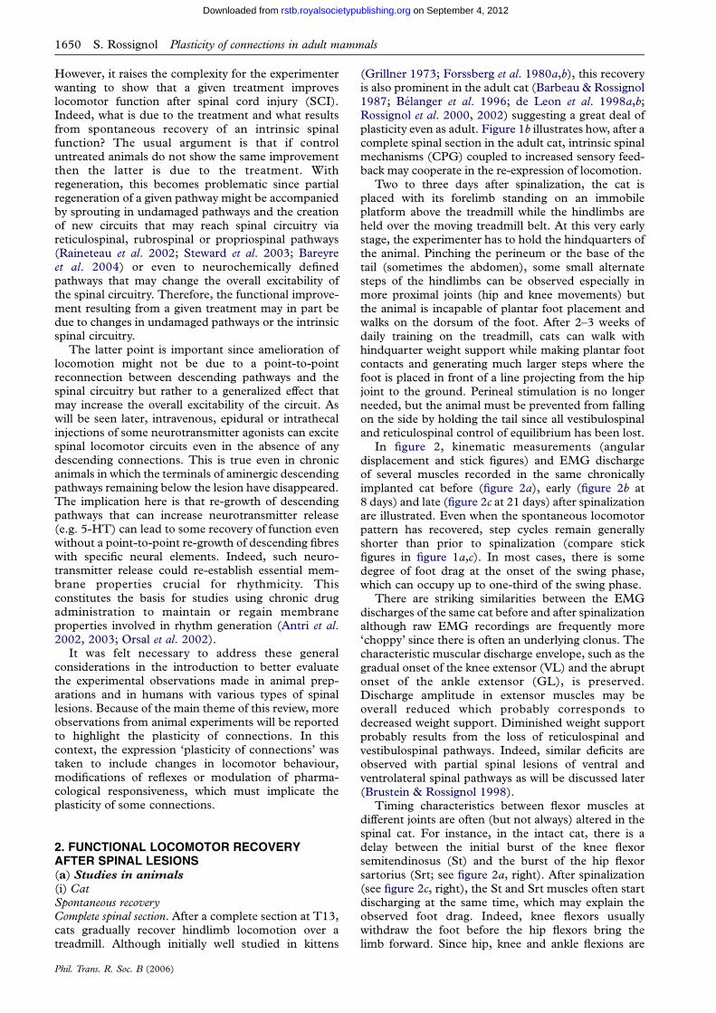
In figure 2, kinematic measurements (angulardisplacement and stick figures) and EMG dischargeof several muscles recorded in the same chronicallyimplanted cat before (figure 2a), early (figure 2b at8 days) and late (figure 2c at 21 days) after spinalizationare illustrated. Even when the spontaneous locomotorpattern has recovered, step cycles remain generallyshorter than prior to spinalization (compare stickfigures in figure 1a,c). In most cases, there is somedegree of foot drag at the onset of the swing phase,which can occupy up to one-third of the swing phase.
There are striking similarities between the EMGdischarges of the same cat before and after spinalizationalthough raw EMG recordings are frequently more‘choppy’ since there is often an underlying clonus. Thecharacteristic muscular discharge envelope, such as thegradual onset of the knee extensor (VL) and the abruptonset of the ankle extensor (GL), is preserved.Discharge amplitude in extensor muscles may beoverall reduced which probably corresponds todecreased weight support. Diminished weight supportprobably results from the loss of reticulospinal andvestibulospinal pathways. Indeed, similar deficits areobserved with partial spinal lesions of ventral andventrolateral spinal pathways as will be discussed later(Brustein & Rossignol 1998).
Timing characteristics between flexor muscles atdifferent joints are often (but not always) altered in thespinal cat. For instance, in the intact cat, there is adelay between the initial burst of the knee flexorsemitendinosus (St) and the burst of the hip flexorsartorius (Srt; see figure 2a, right). After spinalization(see figure 2c, right), the St and Srt muscles often startdischarging at the same time, which may explain theobserved foot drag. Indeed, knee flexors usuallywithdraw the foot before the hip flexors bring thelimb forward. Since hip, knee and ankle flexions are
cat Bstance swing
stance swing
stance swing
120
90110
170
240
1700 0.5 1.0 1.5 2.0
phase of step cycle
80
110
left contact0.3 m s–1
hip
knee
angle
mtp
intact
lesion T138 days
21 days
150 LSt
RSt
RVL
LSrt
RSrt
LVL
LGL
LSt
RSt
RVL
LSrt
RSrt
LVL
LGL
LSt
RSt
RVL
LSrt
RSrt
LVL
LGL
80160
150
join
t ang
le (
degr
ees)
250
170
80
80
0.2 m s–1
C16t307
1s
C16t602
C16t802
0.3 m s–1
150
80160
150
250
170
80
80
5 cm
5 cm
hip
(a)
(b)
(c)
knee
mtpankle
Figure 2. Comparison of treadmill locomotion of a cat in the intact condition and at two different days after a T13 spinal section.(a) Locomotor pattern in the intact state. (b) Locomotor pattern 8 days after spinalization. (c) Recovery of locomotion after21 days of treadmill training. Left column: stick figures reconstructed from a video sequence of one-step cycle generated from aframe-by-frame video analysis displaying the swing and the stance phase separately. The orientations of joint measurements aregiven. Note that the calibration of the x-axis is twice that of the y-axis (see calibration in (c), left part of the figure). Middle column:angular excursion of the four joints (hip, knee, ankle and metatarsophalangeal (mtp)) averaged over 10 cycles. Paw contact(down arrow); paw lift (up arrow); stance phase (horizontal line). Flexion always corresponds to downward deflections of theangular traces. The treadmill speed is indicated in metres per second (m sK1) and is 0.3 m sK1 except for (b), which is at0.2 m sK1 but the cat was not walking. Right column: raw EMG activity of the flexor and extensor muscles of the left (L) and right(R) hindlimb during locomotion. LSt, left semitendinosus; LSrt, left sartorius; LVL, left vastus lateralis; LGL, leftgastrocnemius lateralis; RSt, right semitendinosus; RSrt, right sartorius; RVL, right vastus lateralis; and RGL,right gastrocnemius lateralis. The scale bar equals 1 s and applies to all EMG traces. Modified with permission from Langletet al. (2005).
Plasticity of connections in adult mammals S. Rossignol 1651
on September 4, 2012rstb.royalsocietypublishing.orgDownloaded from
initiated more or less at the same time, the foot moves
forward on the treadmill belt before it is lifted. Such
specific deficits in the timing of hindlimb flexors may
result from damage to corticospinal pathways since
they are also seen after lesions of the dorsolateral tracts
in the cat ( Jiang & Drew 1996).
Incomplete spinal sections. In the context of locomotor
plasticity, it is of great interest to perform restricted
lesions of the spinal cord to study early deficits, which
are related mainly to the destruction of some pathways
and then to assess the time course and extent of
locomotor compensation that results from the action of
remnant pathways. The results of lesions of major
tracts on the recovery of locomotion will be briefly
reviewed and the reader is referred to previous reviews
for more complete details (Grillner 1981; Armstrong
1986, 1988; Rossignol 1996; Grillner et al. 1997;
Jordan 1998; Drew et al. 2004). It will be seen that none
of the spinal quadrants, and, therefore, none of the
descending pathways plays an indispensable role in the
Phil. Trans. R. Soc. B (2006)
basic generation of locomotion in the cat, although
severe deficits may be observed as a result of deficient
supraspinal controls.
Ventrolateral pathways. It is generally believed that
locomotion is initiated via the activation of the
mesencephalic locomotor region (MLR; Shik et al.
1966; Orlovsky & Shik 1976), which activates reticu-
lospinal cells projecting down to the spinal cord and thus
the central pattern generator (CPG) (see figure 1a).
Various diencephalic and telencephalic structures
project directly to the MLR and reticular formation
(Grillner et al. 1997; Jordan 1998). Besides initiating
locomotion, the reticular formation also plays a major
role in the control of locomotion and associated posture
(Shimamura et al. 1982; Drew et al. 1986; Mori 1987;
Drew & Rossignol 1990a,b; Drew 1991; Perreault et al.1993, 1994; Kably & Drew 1998a,b; Matsuyama &
Drew 2000a,b; Prentice & Drew 2001; Drew et al. 2004;
Matsuyama et al. 2004a,b). Thus, it should be expected
that a lesion affecting reticulospinal pathways should

1652 S. Rossignol Plasticity of connections in adult mammals
on September 4, 2012rstb.royalsocietypublishing.orgDownloaded from
greatly impact locomotion. Although it is difficult tocompletely section the reticulospinal pathways, theconsequences of major damage can be observed usingventral and ventrolateral lesions of the cord.
Based on the literature cited earlier, it was initiallysuggested that medial and mediolateral pathways wereessential for locomotion (Eidelberg 1981). For loco-motion to recover, a small part of a ventrolateralquadrant had to be spared (Afelt 1974; Eidelberg et al.1981a,b; Contamin 1983). However, other experi-ments suggested that cats (Gorska et al. 1990, 1993a,b;Zmyslowski et al. 1993; Bem et al. 1995; Brustein &Rossignol 1999; Rossignol et al. 1999) and monkeys(Vilensky et al. 1992) could walk with the hindlimbseven after large lesions of these pathways at the lastthoracic segment (T13). Similarly, humans who had asurgical section of ventral pathways for intractable painretained walking ability (Nathan 1994).
In cats chronically implanted with EMG electrodes,ventral/ventrolateral pathways were sectioned bilater-ally (Brustein & Rossignol 1998, 1999). With smalllesions, cats could walk voluntarily at speeds of up to0.7 m sK1 with all four limbs, 1–3 days after the lesion.However, with large lesions, which spared only part ofthe dorsal columns and various amounts of thedorsolateral quadrant, cats initially behaved ascomplete spinal cats. In open field, the forelimbspropelled the body but they dragged their hindquartersaround over ground for a period of 3–6 weeks. Withregular treadmill training, all cats regained voluntarylocomotion of all the four limbs, although with thelargest lesions, animals could not walk faster than0.4 m sK1. Hindlimb coupling remained stable ataround 50% of the cycle. These cats walked with amore crouched position denoting a reduction in weightsupport ability. The hindlimb–forelimb coordinationwas at times unstable and cats often adopted a pacinggait, i.e. the fore- and hindlimbs on one side were inswing or in stance at the same time and alternated withthe contralateral limbs. The fore- and hindlimb couldeven walk at slightly different mean frequencies leadingto occasional stumbling. Precise measurements of theforelimb–hindlimb coupling showed a gradual driftover several cycles that led to stumbling. When walkingup-slope there was an increase in the amplitude of theforelimb elbow extensors to compensate for increasedload. The amplitude of extensor burst in the hindlimbsdid not compensate during up-slope walking due to thelack of supraspinal compensatory signals. On forceplatforms, the forelimbs became propulsive in contrastto the normal situation where the hindlimbs arepropulsive. Despite some walking instability on thetreadmill, it is remarkable that at this stage cats couldvoluntarily stand up, walk around and overcomenatural obstacles on the ground or treadmill.
Horseradish peroxidase (HRP) was injected belowthe spinal lesion at the conclusion of the experiment toevaluate the number and location of spinally projectingcells. In the pontine reticular and medullary reticularformations, labelled cells accounted for 5–48% ofnormal values depending on the size of the spinallesion. Vestibulospinal neurons were virtually wipedout. Counts of rubrospinal cells were either normal orsomewhat decreased since some lesions may have
Phil. Trans. R. Soc. B (2006)
encroached rubrospinal axons. The remaining reticu-lospinal and rubrospinal cells may have participated inthe recovery of locomotion. Although propriospinalneurons were not studied, they could be strategicallyplaced to participate in such compensation assuggested by others ( Jordan & Schmidt 2002; Bareyreet al. 2004). However, preliminary evidence suggeststhat the number of HRP-labelled cells was higher in themotor cortex of lesioned cats compared with controlcats and their distribution was somewhat more lateral(Rossignol et al. 1999), suggesting that a significantcompensation from corticospinal pathways maycontribute to locomotor recovery.
Dorsolateral pathways. Cats can walk over groundafter large lesions of the dorsolateral white matter(Gorska et al. 1993b; Zmyslowski et al. 1993; Bemet al. 1995). In more quantitative studies of treadmilllocomotion after lesions of the dorsolateral funiculus,which included the dorsal columns ( Jiang & Drew1996), it was shown that voluntary quadrupedallocomotion is impaired for 3–10 days. Cats adopteda more crouched posture during walking for a periodof 2–3 weeks and step-cycle duration was increaseddue to a prolongation of stance, which is contrary tocats with ventrolateral lesions where step cycles ofindividual hindlimbs close to normal were observed.On the contrary, cats with dorsolateral lesions hadchanges in intra-cycle characteristics. For instance,there was simultaneous onset of the knee flexor Stwith the hip flexor Srt. As mentioned earlier, therenormally exists a delay between the two with Stdischarging before Srt, which potentially underscorespersistent foot drag. Contrary to ventrolaterallesions, cats with dorsolateral lesions are unable tovoluntarily modify their gait to step over an obstacleon a treadmill (Drew et al. 1996). Finally, in catswith dorsolateral lesions, both fore- and hindlimbsparticipate in compensating for treadmill slopes.
Hemisections. Hemisections of the cord have beenstudied in various animal preparations, but there are anumber of inconsistent reports on the deficits andrecovery of locomotion. Nevertheless, there is anagreement that the hindlimb ipsilateral to the lesiongradually recovers locomotion within approximatelyone month (Eidelberg et al. 1986; Basso et al. 1994;Kuhtz-Buschbeck et al. 1996). Remaining deficitsfollowing this period are not consistent among authors.In one study using precise kinematic measures(Kuhtz-Buschbeck et al. 1996), the permanent deficitis a shortening of stance and a prolongation of swing onthe side of the lesion leading to some asymmetrical gait.A reduction in stance is compatible with an inability tosupport weight (as seen in the complete spinal cat).The slowing of the swing phase might in turn be relatedto the lesion of the corticospinal tract (CST), althoughthe timing abnormalities seem to differ compared withdorsolateral tract lesions (Jiang & Drew 1996). There isalso a variable degree of spasticity manifested as areduced yield during the E2 phase of locomotion(Kuhtz-Buschbeck et al. 1996). Although the role ofdescending intact tracts were considered important forthe recovery of locomotion after hemisection, sproutingof afferents was proposed as a major player in thisrecovery (Basso et al. 1994). However, other work

Plasticity of connections in adult mammals S. Rossignol 1653
on September 4, 2012rstb.royalsocietypublishing.orgDownloaded from
suggests that afferent fibre sprouting caudal to thelesion does not greatly differ between the lesioned andintact sides (Nacimiento et al. 1993).
PharmacologyPrevious sections indicated a functional locomotorrecovery resulting from a reorganization of pathways atseveral levels. However, the plasticity in connectionscan also stem from changes in responsiveness topharmacological agents and/or changes in the densityof various neurotransmitter receptors.
Neurotransmitter agonists. Early work using thenoradrenaline precursor 3,4-dihydroxy-L-phenyl-alanine (L-DOPA) in acute spinal cats ( Jankowskaet al. 1967a,b) led to the concept of a central patterngenerator for locomotion (Grillner & Zangger 1979).Indeed, after an i.v. injection of L-DOPA, thecharacteristic discharge pattern recorded in flexor andextensor nerves (electroneurograms) during fictivelocomotion shared many similarities with EMGpatterns recorded in the walking cat. Therefore,specific chemicals can trigger the activity in an extantautonomous spinal circuitry since cats were bothspinalized and curarized. As mentioned earlier (seefigure 1a), this central pattern generator concept iscentral to our understanding of locomotor control.This seminal work was followed by other studiesattempting to determine which receptors of whichneurotransmitter systems activate the CPG with theaim of applying such pharmacological tools in therehabilitation of locomotion after spinal injury inanimals and particularly humans.
First, agonists of different subtypes of adrenergicreceptors were used. The a-2 noradrenergicagonist, clonidine, was first used in acute spinal cats(Forssberg & Grillner 1973) and it was found that a well-developed bilateral hindlimb walking pattern could beevoked within minutes of the clonidine injection inabout one-third of cats. It is important to realize thatwith appropriate neurochemical stimulation, even afteracute spinalization, cats can walk with the hindlimbs.Therefore, recovery of spontaneous locomotion inchronic spinal cats must then represent plastic changesin the pathways leading to the activation of this extantspinal pattern generator more than in the CPG itself.
Following this work, we have found that, in adultspinal cats chronically implanted with EMG electro-des, only the a-2 adrenergic agonists, such as clonidine,injected intraperitoneally (Barbeau et al. 1987a), orsimilar agonists (tizanidine, oxymethazoline) injectedintrathecally (Chau et al. 1998b) induce locomotionshortly after spinalization. Later on, when cats haveregained spontaneous locomotion, these noradrenergicagonists still exert potent effects on the locomotorpattern by increasing EMG burst duration and overallstep length. There are, however, smaller changes in theburst amplitude. In the present context of plasticity ofconnection, it is worth mentioning that the effects ofclonidine differ, whether the cats have an intact spinalcord, a complete or a partial spinal lesion (Rossignolet al. 1998, 2001). Whereas in the intact stateintrathecal injection of clonidine has little effect, ithas, in the same cat but early after spinal section thestriking effect of evoking locomotion (Giroux et al.
Phil. Trans. R. Soc. B (2006)
2001). Furthermore, in cats with a large ventral andventrolateral lesion, an intrathecal clonidine injectioncan altogether stop voluntary quadrupedal locomotion(Brustein & Rossignol 1999). Therefore, the state ofreceptivity of receptors as well as the presence of pre-and postsynaptic receptors in various neural elementsmay differ in different models of spinal lesion, and thiswill determine the effects of the drugs. This isimportant when assessing drugs in humans since theextent of the lesion is not known most of the time(Remy-Neris et al. 1999).
Serotonergic agonists suchasquipazine,5-methoxy-N,N-dimethyltryptamine or the precursor 5-hydroxytry-ptophan do not initiate locomotion in acute spinal cats(Barbeau & Rossignol 1990). As will be seen later, thisis a major difference between cats and rodents. Thereason for the inability of 5-HT agonists in evokinglocomotion might be related to the level of the spinalsection as suggested by others (Schmidt & Jordan2000) on the basis of the segmental distribution of5-HT receptor subunits important for locomotion.Therefore, the level of section may be important inevaluating the effects of stimulation by certain drugssince the receptors on which they are acting as well astheir state of responsiveness may differ in differentpreparations (intact cord, complete or incompletespinal section). Although 5-HT agonists do not initiatelocomotion in spinal cats, they increase the outputamplitude of activity of hindlimb muscles (especiallyextensors) and paraxial muscles (Barbeau & Rossignol1990). In cats with ventrolateral spinal lesions,5-HT agonists increased weight support as well asthe endurance of the cats to walk uninterruptedly(Brustein & Rossignol 1999). Importantly, thesepharmacological effects were well integrated in anotherwise voluntarily generated locomotor pattern,suggesting that voluntary commands could make bestuse of the increased spinal excitability provided by thepharmacological stimulation.
Shortly after spinal section in cats, intrathecalinjections of N-methyl-D-aspartate (NMDA), contraryto in vitro neonatal rats or lampreys and in contrastwith decerebrate cats (Douglas et al. 1993), does notinduce locomotion but generates tremor and toefanning (Chau et al. 2002; Giroux et al. 2003).However, when injected intrathecally in spinal catsthat just started to generate small steps (around6–7 days), NMDA could boost emergent locomotorpatterns for several hours (Chau et al. 2002). NMDAhad little effects per se on the spontaneously recoveredlocomotor pattern several weeks after spinalization.
Neurotransmitter antagonists. The use of variousneurotransmitter antagonists is of interest not only toblock the effects of previously described effects ofagonists in the spinal cat but also in determining therole that certain receptors may play in the spontaneouslygenerated locomotor pattern in the intact or spinal cat.
Yohimbine, an a-2 adrenergic blocker, reverses theeffect of clonidine on the initiation of locomotion or theclonidine-induced change in the step cycle (Barbeauet al. 1987a; Giroux et al. 2001). However, yohimbinehas no effect in the chronic spinal cat walking on atreadmill. Although this might appear obvious sincethe neurotransmitter is no longer present following
1654 S. Rossignol Plasticity of connections in adult mammals
on September 4, 2012rstb.royalsocietypublishing.orgDownloaded from
spinalization, it is important to block these receptors toshow that residual noradrenaline and other moleculesthat could potentially activate these receptors arenot responsible for the ability of the cat to walk.However, yohimbine induces, in the intact cat, animportant incoordination of the fore- and hindlimbsand an inability to adequately control the trunkand hindquarters that can often walk sideways relativeto the forelimbs (Giroux et al. 2001) suggesting thatin normal locomotion, noradrenergic neurotrans-mission is important for interlimb coordination(McDearmid et al. 1997).
Since 5-HT does not induce locomotion in the cat,less evidence is available on 5-HT blockers. However,one study showed that cyproheptadine greatly reducesthe enhanced muscular output induced by 5-HTagonists in spinal cats (Barbeau & Rossignol 1990,1991). No data were obtained yet on intact cats.
Aminophosphonopentanoic acid (AP-5), an NMDAblocker, was shown to influence locomotion in the intactcat by reducing its weight support. However, the cat hadno difficulty in compensating for this deficit and couldcontinue walking on the treadmill. In the very same cats,which had received AP-5 in the intact state withouteffect, the same drug completely blocked the spon-taneously generated locomotion after spinal transection(Giroux et al. 2003). This suggests that NMDAreceptors play a crucial role in generating spinallocomotion and that minor effects observed in the intactcat after AP-5 injection probably result from compen-sation by other neurotransmitters released by intactdescending pathways, which are absent in the spinal cat.
Thus, in the weeks after spinalization, one of themajor changes that occur is a basic transformation ofthe central spinal circuitry which normally operatesthrough a balance of various neuromodulators(including monoamines) to a more restricted set oftransmitters/modulators acting on glutamatergicreceptors. Furthermore, the key role played by afferentinputs in spinal locomotion (see later) may be related totheir ability to release neurotransmitters capable ofactivating the locomotor spinal circuitry.
Evolution of receptors after spinalization. In an attemptto study the plastic changes in the cord after spinaliza-tion, the distribution of a1-, a2-noradrenergic andserotonin1A (5-HT1A) receptors was studied in thespinal cords of normal cats as well as in cats spinalized atT13 a few weeks or months earlier (Giroux et al. 1999).Binding densities of a-1 and a-2 receptors significantlyincreased in the lumbar segments at 15–30 days afterspinalization. At longer survival times (three or moremonths), binding densities returned to near controlvalues. The 5-HT1A receptors also followed the sameprofile of upregulation and returned to control values.The marked upregulation of various monoaminergicreceptors observed after spinal section is therefore aclear example of the plasticity of connections occurringin the adult. However, although the timing appearsappropriate, the links between these receptor changesand recovery of locomotor capabilities have not yet beenestablished. This might explain why larger doses ofclonidine are usually needed to induce locomotion inthe acute stage of spinalization. When receptors areupregulated, smaller doses of clonidine are efficacious.
Phil. Trans. R. Soc. B (2006)
However, even after several months, clonidine is stillvery effective at very low doses suggesting that otherintracellular mechanisms that may increase receptorcoupling with intracellular events become importantwith time (Reader et al. 2001).
Preliminary work on a-amino-5-hydroxy-3-methyl-4-isoxazole propionic acid (AMPA) receptors shows anupregulation but, contrary to aminergic receptors, itappears that this upregulation is maintained (Chauet al. 2001), consistent with the fact that glutamatergicreceptors are important, for long-term maintenance ofspinal locomotion.
Preliminary work has been done to determine thesegmental and laminar distribution of various types ofreceptors (Rossignol et al. 2002). There are indicationsthat the upregulation differs according to segments(being highest in the midlumbar segments; Rossignolet al. 2002) but this must be confirmed because it hasimplications for determining the importance of thelevel of spinal transection for locomotor recovery.
Level of spinal sectionThe level of spinalization in the recovery of locomotionis important on several counts. A better understandingof spinal levels implicated in the generation oflocomotion might be extremely useful in optimizingstrategies when using spinal electrical stimulation,pharmacological stimulation through intrathecaldelivery systems or through grafts of cells releasingbiogenic amines. In the context of plasticity ofconnections, it might also suggest how intersegmentalreconnections are important for the re-expression oflocomotion.
Our initial work using intrathecal delivery of drugsindicated that application of drugs at rostral segmentsof the spinal cord in cats (at around L4) were veryeffective in triggering or modulating locomotion (Chauet al. 1998a,b; Giroux et al. 2001). Further workshowed that, in cats spinalized one week earlier, anintraspinal injection of clonidine or yohimbine,restricted to the L4 segment, initiated or blocked thespinal locomotion, respectively (Marcoux & Rossignol2000). It should be recalled that this spinal level in thecat is rostral to the main hindlimb motor neuron pools(Vanderhorst & Holstege 1997). In such experiments,it was also shown that a lesion at caudal L4 abolished allspinal locomotion even after i.v. injections of clonidine.It was unclear whether this constituted a second spinalshock or a disconnection of specific spinal segmentscritical for locomotion. Indeed, previous work byothers suggested that neural elements in these seg-ments are critical for locomotion (Shefchyk et al. 1990;Jankowska & Edgley 1993; Davies & Edgley 1994;Jankowska et al. 2003).
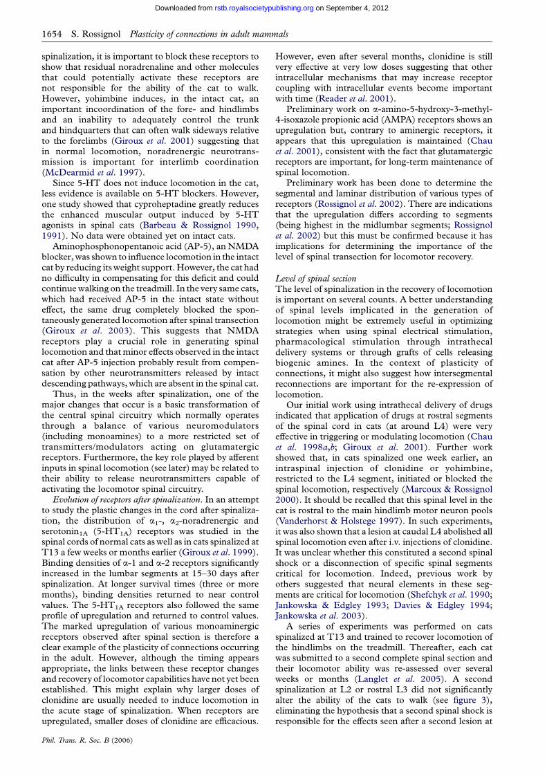
A series of experiments was performed on catsspinalized at T13 and trained to recover locomotion ofthe hindlimbs on the treadmill. Thereafter, each catwas submitted to a second complete spinal section andtheir locomotor ability was re-assessed over severalweeks or months (Langlet et al. 2005). A secondspinalization at L2 or rostral L3 did not significantlyalter the ability of the cats to walk (see figure 3),eliminating the hypothesis that a second spinal shock isresponsible for the effects seen after a second lesion at
stance swing
stance
cat C
swing120
90120
140
230
180
0 0.5 1.0 1.5 2.0phase of step cycle
90
110
110
11080
90140
200
170
100
first lesion T1317 days
second lesion caudal L22 days
LSt
RSt
RVL
LSrt
RSrt
LVL
LGL
LSt
RSt
RVL
LSrt
RSrt
LVL
LGL
0.3 m s–1
c17t808
c17t1001
0.3 m s–1
5 cm
5 cm
(a)
(b)
Figure 3. Comparison of recovery of locomotion after a second spinal lesion at the caudal part of L2 segment. Stick diagrams,angular displacement and raw EMG traces of hindlimb flexor and extensor muscles are as displayed in figure 3. (a) Recovery oflocomotion after a first transection at T13 after 17 days of training. (b) Locomotor performance 2 days after the second lesion atthe caudal L2 level in the same cat. Modified with permission from Langlet et al. (2005).
Plasticity of connections in adult mammals S. Rossignol 1655
on September 4, 2012rstb.royalsocietypublishing.orgDownloaded from
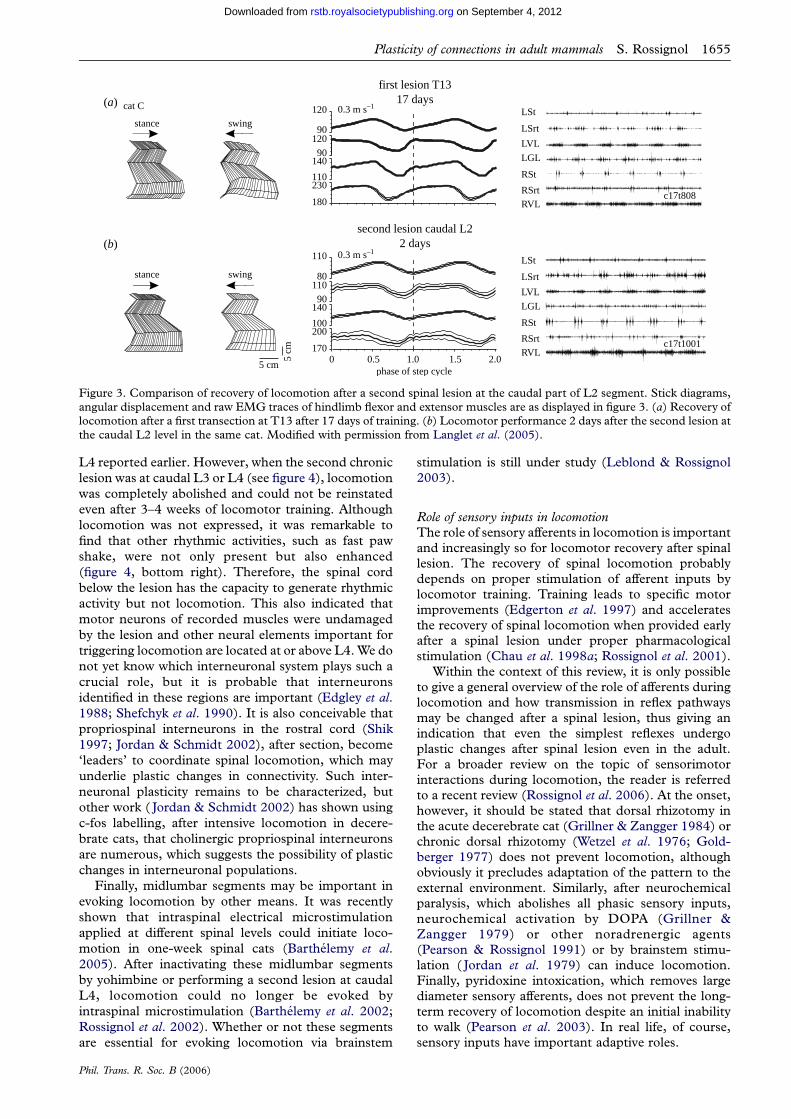
L4 reported earlier. However, when the second chronic
lesion was at caudal L3 or L4 (see figure 4), locomotion
was completely abolished and could not be reinstated
even after 3–4 weeks of locomotor training. Although
locomotion was not expressed, it was remarkable to
find that other rhythmic activities, such as fast paw
shake, were not only present but also enhanced
(figure 4, bottom right). Therefore, the spinal cord
below the lesion has the capacity to generate rhythmic
activity but not locomotion. This also indicated that
motor neurons of recorded muscles were undamaged
by the lesion and other neural elements important for
triggering locomotion are located at or above L4. We do
not yet know which interneuronal system plays such a
crucial role, but it is probable that interneurons
identified in these regions are important (Edgley et al.1988; Shefchyk et al. 1990). It is also conceivable that
propriospinal interneurons in the rostral cord (Shik
1997; Jordan & Schmidt 2002), after section, become
‘leaders’ to coordinate spinal locomotion, which may
underlie plastic changes in connectivity. Such inter-
neuronal plasticity remains to be characterized, but
other work ( Jordan & Schmidt 2002) has shown using
c-fos labelling, after intensive locomotion in decere-
brate cats, that cholinergic propriospinal interneurons
are numerous, which suggests the possibility of plastic
changes in interneuronal populations.
Finally, midlumbar segments may be important in
evoking locomotion by other means. It was recently
shown that intraspinal electrical microstimulation
applied at different spinal levels could initiate loco-
motion in one-week spinal cats (Barthelemy et al.2005). After inactivating these midlumbar segments
by yohimbine or performing a second lesion at caudal
L4, locomotion could no longer be evoked by
intraspinal microstimulation (Barthelemy et al. 2002;
Rossignol et al. 2002). Whether or not these segments
are essential for evoking locomotion via brainstem
Phil. Trans. R. Soc. B (2006)
stimulation is still under study (Leblond & Rossignol2003).
Role of sensory inputs in locomotionThe role of sensory afferents in locomotion is importantand increasingly so for locomotor recovery after spinallesion. The recovery of spinal locomotion probablydepends on proper stimulation of afferent inputs bylocomotor training. Training leads to specific motorimprovements (Edgerton et al. 1997) and acceleratesthe recovery of spinal locomotion when provided earlyafter a spinal lesion under proper pharmacologicalstimulation (Chau et al. 1998a; Rossignol et al. 2001).
Within the context of this review, it is only possibleto give a general overview of the role of afferents duringlocomotion and how transmission in reflex pathwaysmay be changed after a spinal lesion, thus giving anindication that even the simplest reflexes undergoplastic changes after spinal lesion even in the adult.For a broader review on the topic of sensorimotorinteractions during locomotion, the reader is referredto a recent review (Rossignol et al. 2006). At the onset,however, it should be stated that dorsal rhizotomy inthe acute decerebrate cat (Grillner & Zangger 1984) orchronic dorsal rhizotomy (Wetzel et al. 1976; Gold-berger 1977) does not prevent locomotion, althoughobviously it precludes adaptation of the pattern to theexternal environment. Similarly, after neurochemicalparalysis, which abolishes all phasic sensory inputs,neurochemical activation by DOPA (Grillner &Zangger 1979) or other noradrenergic agents(Pearson & Rossignol 1991) or by brainstem stimu-lation ( Jordan et al. 1979) can induce locomotion.Finally, pyridoxine intoxication, which removes largediameter sensory afferents, does not prevent the long-term recovery of locomotion despite an initial inabilityto walk (Pearson et al. 2003). In real life, of course,sensory inputs have important adaptive roles.
Cl3t161
Cl3t1701
1s
LSt
LSrt
LVL
LGL
RSt
RVL
RSrt
LSt
LSrt
LVL
LGL
RSt
RVL
RSrt
(b)
(a)
(c)
second lesion caudal L36 days
20 days
Cl3t12001
LSt
LSrt
LVL
LGL
RSt
RVL
RSrt
FPS
first lesion T1335 days
0.3 m s–1
Cat E
Figure 4. Abolition of locomotion after a second spinal transaction at caudal L3. Figurines drawn from the video recordingsshow the hindlimb position. (a) Spinal locomotion after 35 days of training following a first transection at T13. (b) Absence oflocomotion 6 days after a second lesion at caudal L3. Only tonic activity of the flexors (St and Srt) was observed. (c) Absence oflocomotion following 20 days of training after the caudal L3 lesion. Some small movements of flexion and extension wereobserved but no locomotion. Note the presence of spontaneous fast paw shakes (FPS), a rhythmic pattern which has a muchfaster frequency than locomotion. Modified with permission from Langlet et al. (2005).
1656 S. Rossignol Plasticity of connections in adult mammals
on September 4, 2012rstb.royalsocietypublishing.orgDownloaded from
Proprioceptive afferents (groups Ia, Ib and II)
appear to regulate in part the duration of various
sub-phases of the step cycle (step frequency and speed)
as well as the discharge amplitude of muscles.
For instance, protracting the hip joint or the shoulder
joint generally lengthens the step cycle and stops
walking if it reaches the maximum limit (Pearson &
Rossignol 1991; Saltiel & Rossignol 2004a).
A retraction has the opposite effect and speeds up the
cycle. Phasic protraction of the shoulder during fictive
or real locomotion (Rossignol et al. 1993; Saltiel &
Rossignol 2004b) or imposed protraction of the
hindlimb during swing (Lam & Pearson 2001; McVea
et al. 2005) shortens the swing phase during real
locomotion whereas protraction during the stance
phase of fictive locomotion prolongs swing. This
indicates very well that although fictive locomotor
rhythms can be evoked in spinalized and curarized
animals, when present, proprioceptive afferent feed-
back participates in the regulation of the step cycle.
Similarly, the amplitude of muscle discharge can be
regulated by proprioceptive afferent feedback. It has
Phil. Trans. R. Soc. B (2006)
been proposed that 40–60% (Donelan & Pearson
2004a) of the amplitude of ankle extensor discharge
depends on afferent feedback. Thus, during unloading
a human subject walking on a treadmill, there is a major
reduction in EMG amplitude (Harkema et al. 1997;
Dietz & Colombo 1998; Dietz & Duysens 2000). If the
ankle extensors are unloaded during stance by
mechanically extending the ankle through an external
ankle brace, a 50% decrease in EMG amplitude of the
ankle extensor muscle ensues (Sinkjaer et al. 2000). In
cats, elegant experiments have been performed to
evaluate unloading during locomotion, in particular
‘foot-in-the hole’ experiments in which cats suddenly
make one step through a trap (Gorassini et al. 1994) or
walk on a peg, which is lower than expected (Donelan &
Pearson 2004b). In all these cases, where sensory
feedback is reduced presumably because of a decrease
in ankle extensor load, there is a short latency reduction
in ankle extensor muscle activity.
Cutaneous inputs, on the other hand, appear to have
a major role in positioning the foot during locomotion.
In humans (Zehr et al. 1998a,b; Zehr & Stein 1999),

E
IAIB
stim
Ext Mn
2 disynaptic IB inhibition
3
polysynaptic excitation
1
monosynaptic excitation
CDP
1 ms
1m
v
Pl (1p 1.8T)
LGS Mn
CDP
10 ms
1 m
v
Pl (6p 1.8T)
LGS Mn
MG Mn
CDP
10 ms
1 m
v
LGS (6p 1.8T)
shamtrained
(a)
(b)
(c)
(d)
Figure 5. Evaluation of the effect of locomotor training onload pathways. (a) Spinal proprioceptive pathways understudy. A schematic of three sensory pathways transmittinginputs from muscle group I afferents to ExtMn is shown tothe left: the monosynaptic (stretch reflex) pathway (fromgroup Ia afferents originating in muscle spindles ofextensors), the disynaptic inhibitory pathway (from groupIb afferents of extensors originating in Golgi-tendon organsplus some group Ia fibres) and the polysynaptic excitatorypathway (from groups Ib and Ia afferents of extensors). Inthe acute spinal cat, this latter pathway shares interneuronswith the network generating the excitatory locomotor drivein extensors (box E). Sample records of motor neuronalpostsynaptic potentials used for measurements are on theright. (b) The amplitude of monosynaptic EPSPs wasmeasured at a latency of 1.4 ms (rising phase in thisexample, i.e. just before the onset of possible disynapticcomponents). Note that compared to the sham (green),the monosynaptic reflex is decreased after locomotortraining (black). (c) The disynaptic Ib inhibition wasevoked by a short train of stimuli (six pulses, 1.4–2.0 T,200–300 Hz), and the inhibitory post-synaptic potential(IPSP) amplitude was measured at the maximal negativedeflection in the intracellular trace. Note that there wereoften monosynaptic EPSPs (six positive humps) overridingthe inhibitory trough (dotted line). It is clear thatlocomotor training reduces the maximal disynaptic inhi-bition. Afferent volley was monitored by recording the corddorsum potential (CDP). (d ) Polysynaptic excitation wasevoked by a similar short train of stimuli, and theamplitude was measured at the maximal positive deflection(dotted line) underlying monosynaptic EPSPs. Modifiedwith permission from Cote et al. (2003).
Plasticity of connections in adult mammals S. Rossignol 1657
on September 4, 2012rstb.royalsocietypublishing.orgDownloaded from
cats (Rossignol et al. 1988, 2006; Rossignol 1996;
Bouyer & Rossignol 2003a) and rats (Schouenborg2003), an exquisite number of reflexes are used to
correctly place the foot after electrical or mechanicalstimulation. Given the multisynaptic nature of
cutaneous pathways, it can be expected that theseare highly plastic and considerably modified after
spinal lesions.Changes in reflex pathways after chronic spinalization.
After chronic spinal hemisection (Hultborn & Malmsten1983a,b) or complete spinal section (Hochman &
McCrea 1994a), the spasticity observed has beenattributed to changes in reflex pathways although precise
underlying mechanisms are unclear and appear to differ
in various motor neuron pools and even in various typesof motor neurons (Hochman & McCrea 1994c). In the
anaesthetized cat, six weeks after complete cord section,homonymous monosynaptic Ia EPSPs in lateral gastro-
cnemius (LG) can almost double, whereas in other ankleextensors, changes in amplitude are non-existent or
minimal. Heteronymous excitatory post-synapticpotentials (EPSPs) are also markedly increased not only
in LG but also in medial gastrocnemius (MG),suggesting that these changes are not due to changes in
membrane properties of motor neurons since homon-ymousEPSPs inMGmotor neurons are unchanged. The
rise time and half width of these EPSPs are alsodiminished and these changes cannot be wholly attrib-
uted to the minimal changes in membraneproperties seen in motor neurons after spinalization
(Hochman & McCrea 1994b) and must therefore beattributed to changes in other control mechanisms such
as presynaptic inhibition or to changes in the efficacy ofsome synapses close to the soma. However, it was more
recently shown in rats which develop tail spasticity after a
low sacral spinal section that plateau potentials return inmotor neurons and might in part be responsible for the
spasticity and increase in some reflex responses (Bennettet al. 2004; Li et al. 2004; Heckmann et al. 2005).
Changes in other pathways have been documentedafter spinalization in animals and humans such as
an increase in recurrent inhibition (Hultborn &Malmsten 1983b; Shefner et al. 1992) and a decrease
in reciprocal inhibition (Xia & Rymer 2005). Flexionresponses following spinalization are increased
(Sherrington 1910).Changes in reflex pathways after locomotor training.
Interesting studies were performed in spinal cats thatwere either trained or not (shams) on a treadmill to
determine if training influenced transmission in reflexpathways. Monosynaptic excitation, disynaptic inhi-
bition and polysynaptic excitation were studied in aterminal experiment in trained and sham chronic spinal
cats (Cote et al. 2003). The changes were first
quantified at rest in the absence of fictive locomotion.It was found that locomotor training significantly
decreased monosynaptic excitation and disynapticinhibition evoked by 1b afferent stimulation. Disynap-
tic inhibition could even be reversed by clonidine to apolysynaptic excitation even without locomotion.
During fictive locomotion, similar changes wereobserved. Figure 5 gives a summary of these changes
in various pathways.
Phil. Trans. R. Soc. B (2006)
It is believed that observed changes could reducespasticity (reduction in 1a afferent transmission) andincrease the recruitment of extensor motor neurons(ExtMn) for the recovery of weight-bearing loco-motion. Again these changes are not due to alterationsin membrane properties of the motor neurons since nocorrelation could be established between the changes
stim
(a)
(b)
(c)
MnR1
R2
R3
R2
R1R3
R1: early excitationR2: middle inhibitionR3: late excitation
10 ms
R1
R1
R1
R2
R2
R2
R3
R3
R3
shamtrained
2
–3–4
0
4
–2
6
–1
8
0
10
1
–3–2–101
2
ampl
itude
(m
v)am
plitu
de (
mv)
ampl
itude
(m
v)
shamtrained
shamtrained
**
**
*
*
**
MG Mn
MG Mn
MG Mn
CDP
CDP
CDP
1 m
v1
mv
1 m
v
CCS (1p2T)
SP (1p 2T)
MPL (1p 2T)
(i) CCS-MG
MPL-MG
SP-MG(iii)
(ii)
10 ms
10 ms
10 ms
trained
trained
trained
sham
47%
61%
sham89%
46%
54%
67%
sham
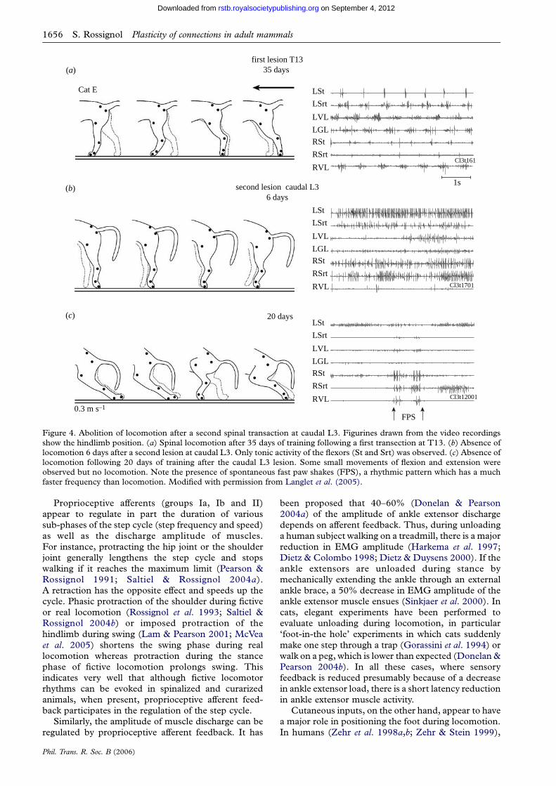
Figure 6. Evaluation of the effect of locomotor training on cutaneous pathways. (a) Hypothetical cutaneous pathways leading toresponses of various latencies (R1, R2 and R3) are illustrated. (b) Type of responses to cutaneous stimulation recorded in motorneurons. Representative averaged post-synaptic potential patterns (nZ62) evoked by cutaneous afferents (caudal cutaneoussural (CCS), medial plantar (MPL) or superficial peroneal (SP)) recorded in extensor or flexor/bifunctional motor neurons areshown. The initial depolarization is referred to as R1 (early excitation), the subsequent hyperpolarization as R2 (middleinhibition) and the following depolarization as R3 (late excitation). (c) Training specifically modified transmission fromcutaneous afferents to the MG motor pool. Left: PSPs (nR40) evoked by stimulation of (i) CCS, (ii) MPL and (iii) SP afferentsrecorded in MG motor neurons with similar AHPs (range 70–91 ms) in a sham (grey) and a trained cat (black). Right:,histograms of the mean amplitude of responses evoked by (i) CCS, (ii) MPL and (iii) SP afferents recorded in all MG motorneurons in shams (grey) and trained cats (black). Six of the nine pathways tested in MG motor neurons were modified bytraining. Significant differences are indicated as follows: �p!0.05; ��p!0.01. Overall, training (i) decreased both CCS-MG-R1(by 47%) and CCS-MG-R3 (by 61%) amplitudes, (ii) increased MPL-MG-R1 amplitude (by 89%), decreased MPL-MG-R2(by 54%) and MPL-MG-R3 (by 46%) amplitudes and (iii) decreased SP-MG-R2 amplitude (by 67%). Mn, motor neuron.Modified with permission from Cote & Gossard (2004).
1658 S. Rossignol Plasticity of connections in adult mammals
on September 4, 2012rstb.royalsocietypublishing.orgDownloaded from
observed and the after-hyperpolarization (AHP) ofmotor neurons, which reflects the membrane timeconstant and input resistance. The changes are ratherattributed to excitability changes of interneuronsreceiving group I inputs to explain the reducedautogenic inhibition and to increases in the output ofpresynaptic interneurons on group Ia afferent term-inals, which can reduce monosynaptic excitation.
Interestingly, reflexes evoked by so-called flexorreflex afferents (FRAs) generated crossed activation offlexor motor neurons rather than ExtMn, suggesting animportant reorganization of specific reflex pathways orat least a significant change in bias since both crossedextensor and crossed flexor pathways exist (Safyants1970; Rossignol & Gauthier 1980; Gauthier &Rossignol 1981).
Furthermore, cutaneous pathways show reducedexcitability specifically from foot pads (from the medialplantar nerve) after locomotor training (Cote &
Phil. Trans. R. Soc. B (2006)
Gossard 2003), again suggesting that trainingdiminishes hyperreflexia from the foot pads afterspinalization. Figure 6 schematically illustrates thepathways involved, the various responses observedand the changes after locomotor training.
The relationship between the changes in proprio-ceptive and cutaneous pathways induced by loco-motor training and the actual improvement oflocomotor performance is an important questionand may justify major efforts in attempting tonormalize the gain of reflex pathways after SCI inorder to achieve the best possible locomotorperformance.
Plastic changes after neurectomies. The functionalrecovery of locomotion after spinal lesions suggestschanges occurring within the spinal cord to optimizethe remaining circuitry controlling locomotion. Theabove reflex changes occurring during locomotortraining suggest a great deal of plasticity in the spinal
Plasticity of connections in adult mammals S. Rossignol 1659
on September 4, 2012rstb.royalsocietypublishing.orgDownloaded from
cord after lesion and the possibility of influencing thisplasticity. In the last decade or so, we have performedvarious experiments to demonstrate spinal plasticity,i.e. a functional compensation achieved solely by thespinal cord in absence of descending inputs. Whereasstudies on locomotion demonstrate some type ofplasticity, the fact that there exists a spinal patterngenerator (Grillner & Zangger 1979) and spinal kittenscan express locomotion even if the spinalization hasoccurred before having had the chance to walk clearlysuggests a largely hardwired spinal pattern generator(Forssberg & Svartengren 1983). Thus, the expressionof locomotion after spinalization is not the result oflearning a new pattern but the result of re-expressing aspinal component of the overall locomotor program.That specific training can promote aspects of thismotor control (bias on alternation or bias on posturalcontrol; Hodgson et al. 1994) reveals that the patterngenerator, although hardwired, can still be modulatedby biasing some of its components and this training canhave long-term effects (de Leon et al. 1999).
To demonstrate spinal plasticity more directly, westudied how cats adapt their locomotor pattern after anerve section either in the intact state or afterspinalization and we have compared the compensationoccurring in both states in the same cat. To this end wehave severed muscle nerves and cutaneous nerves ofthe hindlimbs.
Muscle nerve section. Although sectioning a periph-eral muscle nerve removes both sensory and motorfunctions for that particular muscle, such sections areuseful in evaluating the capacity of cats in the intact orspinal state to adapt its locomotor pattern when specificmuscles are removed. Nerves to ankle flexors (tibialisanterior, TA and extensor digitorum longus, EDL)were cut in one leg while cats had an otherwise intactspinal cord and had been chronically implanted withEMG electrodes (Carrier et al. 1997). Remarkably,following such a neurectomy, locomotor movementswere very similar to control after only a few days. Therewas obviously some reduction in the ankle flexion andthe amplitude of hip and knee flexor EMGs wereincreased to compensate for the reduced ankle flexion.When the cats had recovered a stable symmetricallocomotion they were spinalized, and walking becameasymmetric and included increased hip flexions duringswing on the denervated side, which were largelydysfunctional. However, when the neurectomy wasperformed in a spinal cat that had already recoveredlocomotion, there was a reduced ankle flexion but noabnormal dysfunctional hyperflexions as observedwhen the neurectomy was performed before spinaliza-tion. It can thus be concluded that the removal of amuscle usually active during locomotion results inplastic changes occurring both at the spinal andsupraspinal levels. The spinal cord alone does notseem capable in this case to compensate functionallybut there are undoubtedly changes occurring in thecord manifested as hyperflexions. How supraspinal andspinal changes combine to produce an almost normallocomotion when the neurectomy is performed beforespinalization is still being investigated.
Similarly, others have developed a model of muscleneurectomy of the gastrocnemius lateralis–soleus
Phil. Trans. R. Soc. B (2006)
(GLS; Whelan et al. 1995; Pearson et al. 1999; Pearson
2000). Such a neurectomy produces a marked yield atthe ankle but 5 days after, agonist muscles such as MG
and plantaris compensate for the removal of GLS by
increasing their discharge amplitude leading to acompensation of the yield. We used the same model
for collaborative experiments (Bouyer et al. 2001) inwhich the GLS nerve was sectioned on one side in three
chronic spinal cats after they had regained normalspinal locomotion (see above). Similarly to the intact
state, a significant ankle yield was observed during
stance for the first few days, and the GM burst ofactivity markedly increased. The yield almost
completely recovered within a week and GM activityremained elevated. The mechanisms of such compen-
sation in spinal cats still have to be elucidated but surely
the observations clearly indicate a functional compen-sation in the spinal state suggesting important spinal
capacity for adaptation presumably supported bychanges in synaptic transmission in various pathways.
Cutaneous nerve section. Although it is generallybelieved that removal of cutaneous inputs by nerve
section or anaesthesia does not prevent locomotion
(Sherrington 1910; Engberg 1964; Forssberg et al.1977; Duysens & Stein 1978; Prochazka et al. 1978),
recent experiments on denervation of the foot padshave brought another perspective on the contribution
of cutaneous inputs to locomotion (Bouyer & Rossignol
1998, 2001, 2003a,b; Rossignol et al. 2002). The fivecutaneous nerves of the hindfeet were cut in otherwise
intact cats. Within 2 days, these cats walked almostnormally on the treadmill. The only detectable change
was a somewhat faster swing with increased dischargeamplitude of the knee and ankle flexor muscles (see
figure 7). However, when placed on a horizontal
ladder, soon after the foot denervation and for up toseven weeks, cats were unable to place their feet
correctly on the rungs. A strategy was developed bythe cats to grip the rungs and, when this capacity was
achieved, the cats were then spinalized at T13.
After spinalization, cats could generate alternatewalking movements of the hindlimbs but were unable
to place the feet on the plantar surface, which theyperformed very well before spinalization even though
their hindfeet had been denervated. It should also bepointed out that although ‘normal’ spinal cats (i.e. with
intact cutaneous denervation) tend to drag their feet in
the initial swing, this drag disappears (Barbeau &Rossignol 1987; Belanger et al. 1996). We concluded
from this work that on the one hand, cutaneous sensoryinputs are very important for the recovery and expression
of spinal locomotion. On the other hand, the functional
adaptation seen after denervation in the intact stateprobably depends largely on supraspinal compensations
since a major foot placement deficit is present afterspinalization. Recent work on this model (Bouyer et al.2000; Bretzner & Drew 2005a) clearly indicates that the
motor cortex participates actively in this compensation.Indeed, after denervation responses to intracortical
microstimulation is increased even 40 days afterdenervation and may contribute in setting the discharge
amplitude of certain muscles such as hindlimb flexors tooffset the sensory denervation (seefigure 8). Also, a lesion

swing swing swing
stance stance stance
before neurectomy(a)
(d ) (e) ( f ) (g)
(b) (c)2 days after 41 days after
hip
kneeankle
MTP
90
150
15090
150
90
170
270
5000 1000time (ms)
5000 1000time (ms)
5000 1000time (ms)
5000 1000time (ms)
post-neurectomyintact
EDB
EDL
Srt
LG
VL
St
MG
EDB
EDL
Srt
LG
VL
St
MG
5 cm
5 cm
join
t ang
le (
degr
ees)
Figure 7. Cutaneous neurectomy of the hind feet in the cat. (a–c) Stick figure reconstructions of left hindlimb movements from aframe-by-frame video analysis of cat walking on the treadmill at 0.5 m sK1. (a) Control, (b) 2 days, and (c) 41 days aftercomplete denervation of both hindfeet. Horizontal and vertical scales are 5 cm. (d and e) Mean angular excursions at each of fourlimb joints. Thickness of lines represents 1 s.d. In (d ), control (grey line) and 2 days postdenervation (black line) aresuperimposed. Dashed lines represent divisions of step cycle according to Philippson (1905) for control walking situation. In (e),superimposition is between control (grey line) and 41 days postdenervation (black line). Synchronization is on foot contact.Arrow highlights increased knee flexion. ( f and g) Rectified averaged EMG activity of the main muscles recorded simultaneouslywith kinematics shown in (d ) and (e). In ( f ), control EMG activity (grey area) and 2 days postdenervation (black line) aresuperimposed. In ( g), superimposition is between control (grey area) and 41 days postdenervation (black line). Srt, sartoriusanterior; St, semitendinosus; VL, vastus lateralis; EDL, extensor digitorum longus; LG, lateral gastrocnemius; MG, medialgastrocnemius; EDB, extensor digitorum brevis. Reproduced by permission from Bouyer & Rossignol (2003a).
1660 S. Rossignol Plasticity of connections in adult mammals
on September 4, 2012rstb.royalsocietypublishing.orgDownloaded from
of the motor cortex may disrupt the compensation seen
after neurectomy (Bouyer et al. 2000).
Does this mean that all the plastic changes occur
exclusively in descending pathways? Two experiments
suggest that this is probably not the case. The first
concerns one cat that had been completely denervated
except for a tiny cutaneous branch originating from the
deep peroneal nerve. This spinal cat could indeed regain
a correct foot placement during locomotion, which
disappeared after anaesthesia of the receptive field of
that nerve or after cutting that last remaining nerve. The
implication here is that the spinal cat adapted its
locomotion based on considerably reduced cutaneous
sensory input, suggesting an important reorganization
at the spinal level itself. To address the question of spinal
plasticity further, we performed a progressive cutaneous
denervation of one hindfoot in a cat that had been
spinalized before and had recovered locomotion. It was
fascinating to see that this spinal animal could adapt its
locomotion after each individual nerve section until all
cutaneous nerves were cut in that hindfoot, a time at
which the cat lost the ability to place the foot on the
plantar surface as was the case in other cats (Bouyer &
Rossignol 2003b). These experiments clearly show that
Phil. Trans. R. Soc. B (2006)
when an animal must adapt its locomotion after loss ofcutaneous information that plastic changes in suprasp-inal and spinal connections occur.
(ii) RatEarlier sections dealt with work performed on the cat.Similar work was done on rodents but only somehighlights will be given as they pertain to the main topicof this review. In fact, several other chapters in this issuedeal with various types of lesioned descendingpathways (CST) or spinal lesions (weight drops(Young 2002), surgical overhemisections (Bregman1998)) in rats and the recovery of function.
The interest of partial spinal lesions is to studyplastic changes leading to functional recovery. Probablythe best type of work of this kind is exemplified byBareyre et al. (2004) showing the development of newcircuits using propriospinal neurons as relay neurons tore-establish function.
Although rats spinalized early after birth can recoverlocomotion (de Leon et al. 2002), adult rats do notrecover spontaneous treadmill walking after a mid-thoracic spinalization in contrast to the situation inadult cats. However, adult chronic spinal rats can walk

10 days beforedenervation
2 days afterdenervation
40 days afterdenervation
LSrt
(a)
(b)
N = 41 phase 0.14 N = 43 phase 0.15 N = 25 phase 0.14
N = 13 phase 0.58
–50 50 100 150 200
(ms)
0 –50 50 100 150 200
(ms)
0 –50 50 100 150 200
(ms)
0
N = 15 phase 0.58 N = 14 phase 0.58
LSt
LTA
LEDB
LVL
LSrt
LSt
LTA
LEDB
LVL
Figure 8. Increased corticospinal efficacy after a unilateral complete cutaneous denervation of the foot. (a) Average of EMGresponses evoked during the swing phase at a current strength of 35 mA before and after denervation from a microwire implantedin the motor cortex. For the St and TA, the figure identifies both short (black) and longer (grey) latency responses. (b) AverageEMG responses, evoked by the same microwire at the same current strength during the stance phase. Note that the scale used todisplay a given EMG is constant in all traces. Note the significant increase in the muscle responses evoked by the corticalmicrowires even 40 days after the cutaneous denervation. Reproduced with permission from Bretzner & Drew (2005a).
Plasticity of connections in adult mammals S. Rossignol 1661
on September 4, 2012rstb.royalsocietypublishing.orgDownloaded from
on a treadmill with the hindlimbs using differentforms of stimulation thus demonstrating theimportant capacity of the cord to generate locomotion
in the rat. For example, serotonergic stimulation(Feraboli-Lohnherr et al. 1999) induces a locomotorpattern and chronic treatment with serotonergicagonists maintains this ability (Antri et al. 2002,2003; Orsal et al. 2002). Recently, electrical stimu-lation of the dorsal columns together with 5-HT
stimulation was shown to induce locomotion on atreadmill in adult spinal rats (Ichiyama et al. 2005).
Perhaps the most relevant work in the context of thisissue is the work on transplant of mesencephalicembryonic 5-HT cells below a chronic spinal lesionperformed in adult rats (Feraboli-Lohnherr et al. 1997;
Ribotta et al. 2000). This showed that 5-HT cells,implanted after a spinal section, re-establish contactswith target neurons in various parts of the cord andfacilitate the expression of a well-coordinated loco-motor pattern. Importantly, even following a secondspinal transaction at T8, the locomotor capability
Phil. Trans. R. Soc. B (2006)
remains showing that the locomotor capability wasnot due to regeneration of axons through the lesionwhich could have been favoured by the graft but rather
by the sublesional graft itself which acted as more orless a biological pump of 5-HT that raised theexcitability of relevant spinal circuits (see figure 9).
Interestingly, the grafts had to innervate the upperlumbar levels (L1–L2), which, in the rat, are cruciallevels for the expression of locomotion (Cazalets et al.1995; Cazalets 2000; see earlier for critical levels in thecat at L4). Indeed, cell grafts that did not projectterminals to at least L1 did not induce locomotion.Therefore, grafts at T9 (the spinalization is at T8) didnot induce locomotion whereas grafts at T11 did.
Therefore, the rat spinal cord possesses the
necessary circuitry to generate spinal locomotionbut electrical or pharmacological stimulation, namelythe serotonergic system, is required for locomotion.In that context, it is often reported that with partiallesions, there is often a regeneration of 5-HT whichmight explain part of the functional recovery since

intact(a)
(b)
(c)
(d )
(e)
( f )
spinal T11-transplantedswingswing stancestance
join
t ang
le (
degr
ees)
LIp
LSt
LVL
LTA
LGM
RSt
r1t32RTA
0 1.0 2.0
r6t82
50
90
110
50120
N =6 N =6
240
180100 ms 100 ms
50
100
140
120
60170
50240
180
hip
knee
ankle
mtp
phase of step cycle0 1.0 2.0
phase of step cycle
swingstance swingstance
Figure 9. Comparison of rhythmic locomotor activity in an intact rat (a–c) and nine weeks after transplantation of embryonicraphe-cells from E14 embryos (d–f ). (a and d ) Reconstruction, as stick diagrams, of treadmill locomotor movements duringswing and stance phases. Each stick figure is displaced from the previous by an amount equivalent to the foot displacement toavoid overlap of all the figures. (b and e) Variations of mean angle joints (thick lines) and their standard deviations (thin lines)from six consecutive step cycles, in (from top to bottom) hip, knee, ankle and metatarsophalangeal (mtp) joints. The samenormalized step cycle is displayed twice to facilitate viewing the events at around the trigger point (foot contact of the limb facingthe camera, down-going arrow). The foot lifts of the same limb are also indicated by up-going arrows. Angular excursions ofvarious joints are averaged for six cycles and synchronized on foot contact. (c and f ) Corresponding synchronized EMG activityin various muscles of ipsi (i) or contralateral (co) hindlimbs. Ip, iliopsoas (hip flexor); St, semitendinosus (knee flexor and hipextensor); VL, vastus lateralis (knee extensor); TA, tibialis anterior (ankle flexor); and GM, gastrocnemius medialis (ankleextensor). Note the discharges of LIP, LSt and LTA during the swing phase and of LVL and LGM during stance. Thecontralateral St (coSt) also discharges during the ipsilateral stance indicating a good alternation between limbs. Reproducedwith permission from (Gimenez y Ribotta et al. 2000).
1662 S. Rossignol Plasticity of connections in adult mammals
on September 4, 2012rstb.royalsocietypublishing.orgDownloaded from
release of 5-HT by these axons might be sufficient tofacilitate the spinal circuitry for locomotion (Coumanset al. 2001).
(iii) MouseThe adult spinal mouse is capable of hindlimblocomotion on a treadmill (Leblond et al. 2003) or offictive spinal rhythms ( Jiang et al. 1999). We haveshown that the locomotor pattern can be surprisinglyconsistent and apparent coupling between the fore- andhindlimbs can at times be misleading but easily
Phil. Trans. R. Soc. B (2006)
detected by analysing the timing of coupling. Pharma-cological work has also indicated that 5-HT agonistsinduce locomotion shortly after spinalization and canevoke important modulatory effects on the patternwhen animals have recovered spontaneous locomotorfunction (Guertin et al. 2002; Leblond et al. 2002;Guertin 2004, 2005).
In models of functional recovery after partial lesions,transplants or other types of stimulation, it is veryimportant to account for the existence of this capacityof adult mice to generate elaborate rhythmic alternate
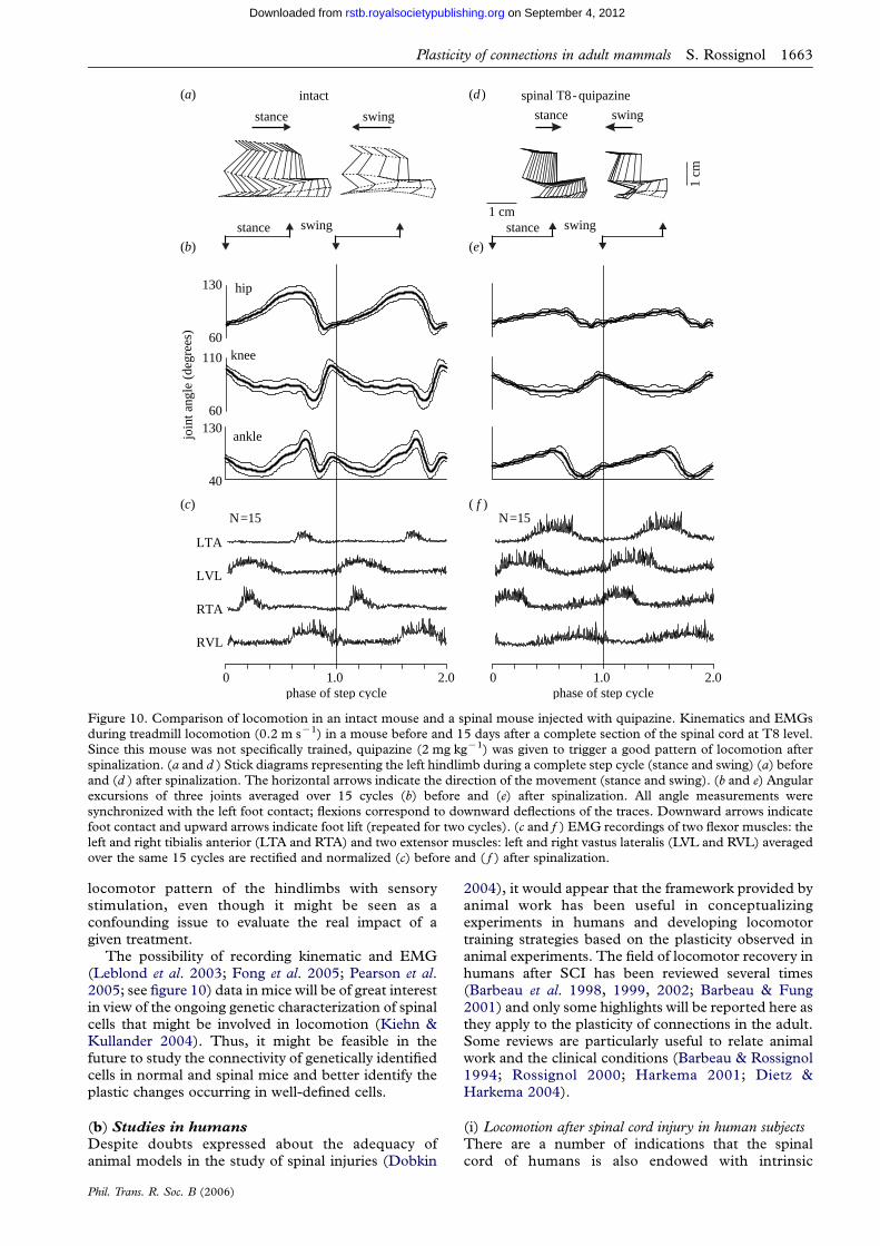
join
t ang
le (
degr
ees)
40
130
60
130
60
110
1 cm
1 cm
LTA
LVL
RTA
RVL
phase of step cycle0 1.0 2.0
phase of step cycle0 1.0 2.0
N=15 N=15
hip
knee
ankle
swingstance swingstance
intact spinal T8-quipazine
swingswing stancestance
(a)
(b)
(c)
(d )
(e)
( f )
Figure 10. Comparison of locomotion in an intact mouse and a spinal mouse injected with quipazine. Kinematics and EMGsduring treadmill locomotion (0.2 m sK1) in a mouse before and 15 days after a complete section of the spinal cord at T8 level.Since this mouse was not specifically trained, quipazine (2 mg kgK1) was given to trigger a good pattern of locomotion afterspinalization. (a and d ) Stick diagrams representing the left hindlimb during a complete step cycle (stance and swing) (a) beforeand (d ) after spinalization. The horizontal arrows indicate the direction of the movement (stance and swing). (b and e) Angularexcursions of three joints averaged over 15 cycles (b) before and (e) after spinalization. All angle measurements weresynchronized with the left foot contact; flexions correspond to downward deflections of the traces. Downward arrows indicatefoot contact and upward arrows indicate foot lift (repeated for two cycles). (c and f ) EMG recordings of two flexor muscles: theleft and right tibialis anterior (LTA and RTA) and two extensor muscles: left and right vastus lateralis (LVL and RVL) averagedover the same 15 cycles are rectified and normalized (c) before and ( f ) after spinalization.
Plasticity of connections in adult mammals S. Rossignol 1663
on September 4, 2012rstb.royalsocietypublishing.orgDownloaded from
locomotor pattern of the hindlimbs with sensorystimulation, even though it might be seen as aconfounding issue to evaluate the real impact of agiven treatment.
The possibility of recording kinematic and EMG(Leblond et al. 2003; Fong et al. 2005; Pearson et al.2005; see figure 10) data in mice will be of great interestin view of the ongoing genetic characterization of spinalcells that might be involved in locomotion (Kiehn &Kullander 2004). Thus, it might be feasible in thefuture to study the connectivity of genetically identifiedcells in normal and spinal mice and better identify theplastic changes occurring in well-defined cells.
(b) Studies in humans
Despite doubts expressed about the adequacy ofanimal models in the study of spinal injuries (Dobkin
Phil. Trans. R. Soc. B (2006)
2004), it would appear that the framework provided byanimal work has been useful in conceptualizingexperiments in humans and developing locomotortraining strategies based on the plasticity observed inanimal experiments. The field of locomotor recovery inhumans after SCI has been reviewed several times(Barbeau et al. 1998, 1999, 2002; Barbeau & Fung2001) and only some highlights will be reported here asthey apply to the plasticity of connections in the adult.Some reviews are particularly useful to relate animalwork and the clinical conditions (Barbeau & Rossignol1994; Rossignol 2000; Harkema 2001; Dietz &Harkema 2004).
(i) Locomotion after spinal cord injury in human subjectsThere are a number of indications that the spinalcord of humans is also endowed with intrinsic
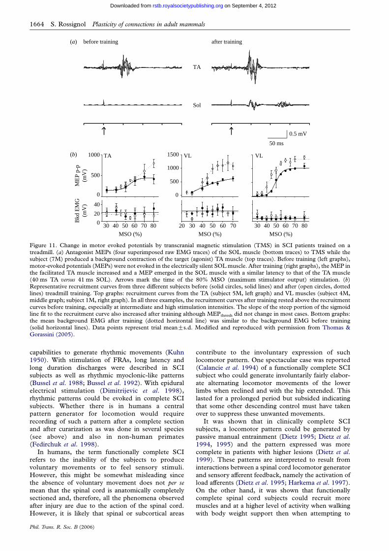
ME
P p-
p(m
V)
MSO (%) MSO (%) MSO (%)
30 40 50 60 70 80
Bkd
EM
G(m
V)
20 30 40 50 60 70
TA VL
40
20
0 30 40 50 60 70 80
VL
0
500
1000
0
500
1000
1500
TA
Sol
before training(a)
(b)
after training
50 ms
0.5 mV
Figure 11. Change in motor evoked potentials by transcranial magnetic stimulation (TMS) in SCI patients trained on atreadmill. (a) Antagonist MEPs (four superimposed raw EMG traces) of the SOL muscle (bottom traces) to TMS while thesubject (7M) produced a background contraction of the target (agonist) TA muscle (top traces). Before training (left graphs),motor-evoked potentials (MEPs) were not evoked in the electrically silent SOL muscle. After training (right graphs), the MEP inthe facilitated TA muscle increased and a MEP emerged in the SOL muscle with a similar latency to that of the TA muscle(40 ms TA versus 41 ms SOL). Arrows mark the time of the 80% MSO (maximum stimulator output) stimulation. (b)Representative recruitment curves from three different subjects before (solid circles, solid lines) and after (open circles, dottedlines) treadmill training. Top graphs: recruitment curves from the TA (subject 5M, left graph) and VL muscles (subject 4M,middle graph; subject 1M, right graph). In all three examples, the recruitment curves after training rested above the recruitmentcurves before training, especially at intermediate and high stimulation intensities. The slope of the steep portion of the sigmoidline fit to the recruitment curve also increased after training although MEPthresh did not change in most cases. Bottom graphs:the mean background EMG after training (dotted horizontal line) was similar to the background EMG before training(solid horizontal lines). Data points represent trial meanGs.d. Modified and reproduced with permission from Thomas &Gorassini (2005).
1664 S. Rossignol Plasticity of connections in adult mammals
on September 4, 2012rstb.royalsocietypublishing.orgDownloaded from
capabilities to generate rhythmic movements (Kuhn
1950). With stimulation of FRAs, long latency and
long duration discharges were described in SCI
subjects as well as rhythmic myoclonic-like patterns
(Bussel et al. 1988; Bussel et al. 1992). With epidural
electrical stimulation (Dimitrijevic et al. 1998),
rhythmic patterns could be evoked in complete SCI
subjects. Whether there is in humans a central
pattern generator for locomotion would require
recording of such a pattern after a complete section
and after curarization as was done in several species
(see above) and also in non-human primates
(Fedirchuk et al. 1998).
In humans, the term functionally complete SCI
refers to the inability of the subjects to produce
voluntary movements or to feel sensory stimuli.
However, this might be somewhat misleading since
the absence of voluntary movement does not per semean that the spinal cord is anatomically completely
sectioned and, therefore, all the phenomena observed
after injury are due to the action of the spinal cord.
However, it is likely that spinal or subcortical areas
Phil. Trans. R. Soc. B (2006)
contribute to the involuntary expression of such
locomotor pattern. One spectacular case was reported
(Calancie et al. 1994) of a functionally complete SCI
subject who could generate involuntarily fairly elabor-
ate alternating locomotor movements of the lower
limbs when reclined and with the hip extended. This
lasted for a prolonged period but subsided indicating
that some other descending control must have taken
over to suppress these unwanted movements.
It was shown that in clinically complete SCI
subjects, a locomotor pattern could be generated by
passive manual entrainment (Dietz 1995; Dietz et al.1994, 1995) and the pattern expressed was more
complete in patients with higher lesions (Dietz et al.1999). These patterns are interpreted to result from
interactions between a spinal cord locomotor generator
and sensory afferent feedback, namely the activation of
load afferents (Dietz et al. 1995; Harkema et al. 1997).
On the other hand, it was shown that functionally
complete spinal cord subjects could recruit more
muscles and at a higher level of activity when walking
with body weight support then when attempting to
Plasticity of connections in adult mammals S. Rossignol 1665
on September 4, 2012rstb.royalsocietypublishing.orgDownloaded from
produce voluntary movements (Maegele et al. 2002).This suggests that remnant voluntary control coupledwith enhanced sensory inputs can combine to shape thelocomotor pattern as is the case in animals.
(ii) Locomotor trainingPatients with partial spinal cord lesions can walk albeitwith some deficits (Pepin et al. 2003a,b). Animal andhuman experiments suggesting a potential for aninvoluntary rhythmogenesis at the spinal cord/brainstemlevel has encouraged the promotion of locomotortraining on a treadmill with body weight support,robotics or muscle stimulation with remarkable results(Barbeau et al. 1987b; Colombo et al. 1998; Werniget al. 1999; Behrman & Harkema 2000; Edgerton et al.2001; Barbeau et al. 2002; Wirz et al. 2005). Thus,patients who were wheel-chair bound could transferthe benefit of their treadmill locomotor training to dailylife situations.
(iii) Changes in ascending and descending pathwaysafter lesionsThis is a very large topic in itself and only very recentwork on aspects relevant to the present theme isdiscussed.
A recent report on patients with a chronic cervicallesion of more than six months describes the emergenceof interlimb reflexes coupling cervical and lumbosacralsegments (Calancie et al. 2005). The relevance in thepresent context is that these interlimb reflexes are notapparent within the first six months but graduallydevelop, suggesting an ongoing plasticity of connec-tions even in the adult after spinal injury.
Another very significant piece of work (Thomas &Gorassini 2005) has shown that functional locomotorimprovement after 3–5 months of locomotor training inhumans with SCI is accompanied by a significant changein motor potentials evoked by transcranial magneticstimulation (TMS; see figure 11). Thus, it can beconcluded that the overall transmission in the cortico-spinal pathway is enhanced by locomotor training andmay, at least in part, be responsible for the functionalamelioration. This could be achieved at different levels asshown for the changes occurring after neurectomies(Bretzner& Drew2005a,b). In the present case, however,a change in intracortical excitability was also observedsuggesting intracortical modifications of excitabilityresulting from locomotor training.
3. CONCLUDING REMARKSThis review has attempted to summarize some aspectsof a very large topic dealing with plasticity ofconnections after spinal injury in adult mammals.The issue of locomotor recovery in animal models wasaddressed and the point was made that recovery ispartly due to plasticity within existing spinal locomotornetworks. Locomotor training appears to change theexcitability of simple reflex pathways as well as morecomplex circuitry. Some work was reported demon-strating the potential intrinsic plasticity of the spinalcord in adapting to lesions (central or peripheral) but,usually, adaptation to lesions entails changes both atspinal and supraspinal levels. Finally, a very brief
Phil. Trans. R. Soc. B (2006)
overview of human work was given to indicate at leastthat the principles regulating the recovery of locomotorfunctions in humans are probably very similar to thoseobserved in animal experiments.
It is comforting to think that the nervous system,even at the adult stage, is plastic enough to optimizeinternal motor sequences after lesion, that this canfurther be optimized by various types of stimulation(pharmacological, electrical) and prepares the terrainfor further success in axonal regeneration to addvoluntary and more refined controls of the expressionof locomotion after spinal lesion.
I wish to acknowledge Daniel Cyr, Janyne Provencher andHugues Leblond for their help with illustrations. I would alsolike to thank former students, postdoctoral fellows orcollaborators whose work is partly illustrated here(C. Langlet, L. Bouyer, D. Orsal and H. Leblond). Specialthanks to Drs J.-P. Gossard, T. Drew and M. Gorassini forproviding modified figures from their published work.A special thanks to Alain Frigon for revising the text. Finally,many thanks are addressed to the Canadian Institute ofHealth Research for continuous support over the yearsthrough individual grants, group grants and a CanadaResearch Chair on the Spinal Cord and a grant from theCanadian Foundation for Innovation.
REFERENCESAfelt, Z. 1974 Functional significance of ventral descending
tracts of the spinal cord in the cat. Acta Neurobiol. Exp. 34,393–407.
Antri, M., Orsal, D. & Barthe, J.-Y. 2002 Locomotor recoveryin the chronic spinal rat: effects of long-term treatmentwith a 5-HT2 agonist. Eur. J. Neurosci. 16, 467–476.(doi:10.1046/j.1460-9568.2002.02088.x)
Antri, M., Mouffle, C., Orsal, D. & Barthe, J. Y. 2003 5-HT1A receptors are involved in short- and long-termprocesses responsible for 5-HT-induced locomotor func-tion recovery in chronic spinal rat. Eur. J. Neurosci. 18,1963–1972. (doi:10.1046/j.1460-9568.2003.02916.x)
Armstrong, D. M. 1986 Supraspinal contributions to theinitiation and control of locomotion in the cat. Prog.Neurobiol. 26, 273–361. (doi:10.1016/0301-0082(86)90021-3)
Armstrong, D. M. 1988 The supraspinal control ofmammalian locomotion. J. Physiol. 405, 1–37.
Barbeau, H. & Fung, J. 2001 The role of rehabilitation in therecovery of walking in the neurological population. Curr.Opin. Neurol. 14, 735–740. (doi:10.1097/00019052-200112000-00009)
Barbeau, H. & Rossignol, S. 1987 Recovery of locomotionafter chronic spinalization in the adult cat. Brain Res. 412,84–95. (doi:10.1016/0006-8993(87)91442-9)
Barbeau, H. & Rossignol, S. 1990 The effects of serotonergicdrugs on the locomotor pattern and on cutaneous reflexesof the adult chronic spinal cat. Brain Res. 514, 55–67.(doi:10.1016/0006-8993(90)90435-E)
Barbeau, H. & Rossignol, S. 1991 Initiation and modulationof the locomotor pattern in the adult chronic spinal cat bynoradrenergic, serotonergic and dopaminergic drugs.Brain Res. 546, 250–260. (doi:10.1016/0006-8993(91)91489-N)
Barbeau, H. & Rossignol, S. 1994 Enhancement oflocomotor recovery following spinal cord injury. Curr.Opin. Neurol. 7, 517–524.
Barbeau, H., Julien, C. & Rossignol, S. 1987a The effects ofclonidine and yohimbine on locomotion and cutaneousreflexes in the adult chronic spinal cat. Brain Res. 437,83–96. (doi:10.1016/0006-8993(87)91529-0)

1666 S. Rossignol Plasticity of connections in adult mammals
on September 4, 2012rstb.royalsocietypublishing.orgDownloaded from
Barbeau, H., Wainberg, M. & Finch, L. 1987b Description
and application of a system for locomotor rehabilitation.
Med. Biol. Eng. Comput. 25, 341–344.
Barbeau, H., Norman, K., Fung, J., Visintin, M. &
Ladouceur, M. 1998 Does neurorehabilitation play a
role in the recovery of walking in neurological populations?
Ann. NY Acad. Sci. 860, 377–392. (doi:10.1111/j.1749-
6632.1998.tb09063.x)
Barbeau, H., Ladouceur, M., Norman, K. E., Pepin, A. &
Leroux, A. 1999 Walking after spinal cord injury:
evaluation, treatment, and functional recovery. Arch.
Phys. Med. Rehabil. 80, 225–235. (doi:10.1016/S0003-
9993(99)90126-0)
Barbeau, H., Fung, J., Leroux, A. & Ladouceur, M. 2002
A review of the adaptability and recovery of locomotion
after spinal cord injury. Prog. Brain Res. 137, 9–25.
Bareyre, F. M., Kerschensteiner, M., Raineteau, O.,
Mettenleiter, T. C., Weinmann, O. & Schwab, M. E.
2004 The injured spinal cord spontaneously forms a new
intraspinal circuit in adult rats. Nat. Neurosci. 7, 269–277.
(doi:10.1038/nn1195)
Barthelemy, D., Leblond, H. & Rossignol, S. 2002 Electrical
stimulation of the lumbar spinal cord and the importance
of mid lumbar segments in eliciting spinal locomotion.
Society for Neuroscience Abstracts 28[65.19].
Barthelemy, D., Leblond H., Mushahwar V. & Rossignol, S.
2005 Use of intraspinal electrical stimulation for inducing
locomotion in spinal cats. 35th Congress of the Inter-
national Union of Physiological Sciences (IUPS) San
Diego, CA. March 31–April 5, 2005.
Basso, D. M. 2004 Behavioral testing after spinal cord
injury: congruities, complexities, and controversies.
J. Neurotrauma 21, 395–404. (doi:10.1089/08977150
4323004548)
Basso, D. M., Murray, M. & Goldberger, M. E. 1994
Differential recovery of bipedal and overground loco-
motion following complete spinal cord hemisection in
cats. Restorative Neurol. Neurosci. 7, 95–110.
Basso, D. M., Beattie, M. S. & Bresnahan, J. C. 1995 A
sensitive and reliable locomotor rating scale for open field
testing in rats. J. Neurotrauma 12, 1–21.
Basso, D. M., Beattie, M. S. & Bresnahan, J. C. 1996 Graded
histological and locomotor outcomes after spinal cord
contusion using the NYU weight-drop device versus
transection. Exp. Neurol. 139, 244–256. (doi:10.1006/
exnr.1996.0098)
Behrman, A. L. & Harkema, S. J. 2000 Locomotor training
after human spinal cord injury: a series of case studies.
Phys. Ther. 80, 688–700.
Belanger, M., Drew, T., Provencher, J. & Rossignol, S. 1996
A comparison of treadmill locomotion in adult cats before
and after spinal transection. J. Neurophysiol. 76, 471–491.
Bem, T., Gorska, T., Majczynski, H. & Zmyslowski, W. 1995
Different patterns of fore-hindlimb coordination during
overground locomotion in cats with ventral and lateral
spinal lesions. Exp. Brain Res. 104, 70–80. (doi:10.1007/
BF00229856)
Bennett, D. J., Sanelli, L., Cooke, C. L., Harvey, P. J. &
Gorassini, M. A. 2004 Spastic long-lasting reflexes in the
awake rat after sacral spinal cord injury. J. Neurophysiol.91, 2247–2258. (doi:10.1152/jn.00946.2003)
Bertrand, S. & Cazalets, J. R. 2003 The respective
contribution of lumbar segments to the generation of
locomotion in the isolated spinal cord of newborn rat. Eur.
J. Neurosci. 16, 1741–1750. (doi:10.1046/j.1460-9568.
2002.02233.x)
Bouyer, L. & Rossignol, S. 1998 The contribution of
cutaneous inputs to locomotion in the intact and the spinal
cat. In Neuronal mechanisms for generating locomotor activity
Phil. Trans. R. Soc. B (2006)
(ed. O. Kiehn, R. M. Harris-Warrick, L. M. Jordan, H.Hultborn & N. Kudo), pp. 508–512. New York, NY: NewYork Academy of Science.
Bouyer, L. & Rossignol, S. 2001 Spinal cord plasticityassociated with locomotor compensation to peripheralnerve lesions in the cat. In Spinal cord plasticity: alterationsin reflex function (ed. M. M. Patterson & J. W. Grau),pp. 207–224. Boston, MA: Kluwer Academic Publishers.
Bouyer, L. J. G. & Rossignol, S. 2003a Contribution ofcutaneous inputs from the hindpaw to the control oflocomotion: 1. Intact cats. J. Neurophysiol. 90, 3625–3639.(doi:10.1152/jn.00496.2003)
Bouyer, L. J. G. & Rossignol, S. 2003b Contribution ofcutaneous inputs from the hindpaw to the control oflocomotion: 2. Spinal cats. J. Neurophysiol. 90,3640–3653. (doi:10.1152/jn.00497.2003)
Bouyer, L. J. G., Drew, T. & Rossignol, S. 2000 A corticalcontribution to the functional recovery observed followinga cutaneous neurectomy of the hindpaw. Society forNeuroscience Abstracts 26[459.13], 1232.
Bouyer, L. J. G., Whelan, P., Pearson, K. G. & Rossignol, S.2001 Adaptive locomotor plasticity in chronic spinal catsafter ankle extensors neurectomy. J. Neurosci. 21,3531–3541.
Bregman, B. S. 1998 Regeneration in the spinal cord. Curr.Opin. Neurobiol. 8, 800–807. (doi:10.1016/S0959-4388(98)80124-4)
Bretzner, F. & Drew, T. 2005a Changes in corticospinalefficacy contribute to the locomotor plasticity observedfollowing unilateral cutaneous denervation of the hindpawin the cat. J. Neurophysiol. 94, 2911–2927. (doi:10.1152/jn.00254.2005)
Bretzner, F. & Drew, T. 2005b Contribution of the motorcortex to the structure and the timing of hindlimblocomotion in the cat: a microstimulation study.J. Neurophysiol. 94, 657–672. (doi:10.1152/jn.01245.2004)
Brustein, E. & Rossignol, S. 1998 Recovery of locomotionafter ventral and ventrolateral spinal lesions in the cat. I.Deficits and adaptive mechanisms. J. Neurophysiol. 80,1245–1267.
Brustein, E. & Rossignol, S. 1999 Recovery of locomotionafter ventral and ventrolateral spinal lesions in the cat. II.Effects of noradrenergic and serotoninergic drugs.J. Neurophysiol. 81, 1513–1530.
Bussel, B. C., Roby-Brami, A., Yakovleff, A. & Bennis, N.1988 Evidences for the presence of a spinal steppinggenerator in patients with a spinal cord section. InPosture and gait: development, adaptation and modulation(ed. B. Amblard, A. Berthoz & F. Clarac), pp. 273–278.North Holland, NY: Elsevier.
Bussel, B., Roby-Brami, A. & Azouvi, P. 1992 Organizationof reflexes elicited by flexor reflex afferents in paraplegicman: evidence for a stepping generator. In Muscle afferentsand spinal control of movement (ed. L. Jami, E. Pierrot-Deseilligny & D. Zytnicki), pp. 427–432. Oxford, UK:Pergamon Press.
Calancie, B., Needham-Shropshire, B., Jacobs, P., Willer, K.,Zych, G. & Green, B. A. 1994 Involuntary stepping afterchronic spinal cord injury. Evidence for a central rhythmgenerator for locomotion in man. Brain 117, 1143–1159.
Calancie, B., Alexeeva, N., Broton, J. G. & Molano, M. R.2005 Interlimb reflex activity after spinal cord injury inman: strengthening response patterns are consistent withongoing synaptic plasticity. Clin. Neurophysiol. 116, 75–86.(doi:10.1016/j.clinph.2004.07.018)
Carrier, L., Brustein, L. & Rossignol, S. 1997 Locomotion ofthe hindlimbs after neurectomy of ankle flexors in intactand spinal cats: model for the study of locomotorplasticity. J. Neurophysiol. 77, 1979–1993.

Plasticity of connections in adult mammals S. Rossignol 1667
on September 4, 2012rstb.royalsocietypublishing.orgDownloaded from
Cazalets, J. R. 2000 Organization of the spinal locomotornetwork in neonatal rat. In Neurobiology of spinal cord injury(ed. R. G. Kalb & S. M. Strittmater), pp. 89–111. Totowa,NJ: Humana Press.
Cazalets, J. R., Borde, M. & Clarac, F. 1995 Localization andorganization of the central pattern generator for hindlimblocomotion in newborn rat. J. Neurosci. 15, 4943–4951.
Chau, C., Barbeau, H. & Rossignol, S. 1998a Earlylocomotor training with clonidine in spinal cats.J. Neurophysiol. 79, 392–409.
Chau, C., Barbeau, H. & Rossignol, S. 1998b Effects ofintrathecal a1- and a2-noradrenergic agonists and nor-epinephrine on locomotion in chronic spinal cats.J. Neurophysiol. 79, 2941–2963.
Chau, C., Giroux, N., Reader, T. A. & Rossignol, S. 2001Ampa and alpha-2 adrenergic receptors in cat lumbo-sacral spinal cord following complete lesions. Society forNeuroscience Abstracts 27[517.2].
Chau, C., Giroux, N., Barbeau, H., Jordan, L. M. &Rossignol, S. 2002 Effects of intrathecal glutamatergicdrugs on locomotion. I. NMDA in short-term spinal cats.J. Neurophysiol. 88, 3032–3045. (doi:10.1152/jn.00138.2002)
Colombo, G., Wirz, M. & Dietz, V. 1998 Effect of locomotortraining related to clinical and electrophysiological exami-nations in spinal cord injured humans. In Neuronalmechanisms for generating locomotor activity (ed. O. Kiehn,R. M. Harris-Warrick, L. M. Jordan, H. Hultborn &N. Kudo), pp. 536–538. New York, NY: New YorkAcademy of Science.
Contamin, F. 1983 Sections medullaires incompletes etlocomotion chez le chat. Bull. Acad. Nat. Med. 167,727–730.
Cote, M.-P. & Gossard, J.-P. 2003 Task-dependent pre-synaptic inhibition. J. Neurosci. 23, 1886–1893.
Cote, M.-P. & Gossard, J.-P. 2004 Step-training depen-dent plasticity in spinal cutaneous pathways.J. Neurosci. 24, 11 317–11 327. (doi:10.1523/JNEUR-OSCI.1486-04.2004)
Cote, M.-P., Menard, A. & Gossard, J.-P. 2003 Spinal cats onthe treadmill: changes in load pathways. J. Neurosci. 23,2789–2796.
Coumans, J. V., Lin, T. T., Dai, H. N., MacArthur, L.,McAtee, M., Nash, C. & Bregman, B. S. 2001 Axonalregeneration and functional recovery after complete spinalcord transection in rats by delayed treatment withtransplants and neurotrophins. J. Neurosci. 21,9334–9344.
Davies, H. E. & Edgley, S. A. 1994 Inputs to group IIactivated midlumbar interneurones from descendingmotor pathways in the cat. J. Physiol. 479, 463–473.
de Leon, R. D., Hodgson, J. A., Roy, R. R. & Edgerton, V. R.1998a Full weight-bearing hindlimb standing followingstand training in the adult spinal cat. J. Neurophysiol. 80,83–91.
de Leon, R. D., Hodgson, J. A., Roy, R. R. & Edgerton, V. R.1998b Locomotor capacity attributable to step trainingversus spontaneous recovery after spinalization in adultcats. J. Neurophysiol. 79, 1329–1340.
de Leon, R. D., Hodgson, J. A., Roy, R. R. & Edgerton, V. R.1999 Retention of hindlimb stepping ability in adult spinalcats after the cessation of step training. J. Neurophysiol. 81,85–94.
de Leon, R. D., Kubasak, M. D., Phelps, P. E., Timoszyk,W. K., Reinkensmeyer, D. J., Roy, R. R. & Edgerton, V. R.2002 Using robotics to teach the spinal cord to walk. BrainRes. Brain Res. Rev. 40, 267–273. (doi:10.1016/S0165-0173(02)00209-6)
Delcomyn, F. 1980 Neural basis of rhythmic behavior inanimals. Science 210, 492–498.
Phil. Trans. R. Soc. B (2006)
Dietz, V. 1995 Locomotor training in paraplegic patients.
Ann. Neurol. 38, 965. (doi:10.1002/ana.410380621)
Dietz, V. & Colombo, G. 1998 Influence of body load on the
gait pattern in Parkinson’s disease. Mov. Disord. 13,
255–261. (doi:10.1002/mds.870130210)
Dietz, V. & Duysens, J. 2000 Significance of load receptor
input during locomotion: a review. Gait Posture 11,
102–110. (doi:10.1016/S0966-6362(99)00052-1)
Dietz, V. & Harkema, S. J. 2004 Locomotor activity in spinal
cord-injured persons. J. Appl. Physiol. 96, 1954–1960.
(doi:10.1152/japplphysiol.00942.2003)
Dietz, V., Colombo, G. & Jensen, L. 1994 Locomotor activity
in spinal man. Lancet 344, 1260–1263. (doi:10.1016/
S0140-6736(94)90751-X)
Dietz, V., Colombo, G., Jensen, L. & Baumgartner, L. 1995
Locomotor capacity of spinal cord in paraplegic patients.
Ann. Neurol. 37, 574–582. (doi:10.1002/ana.410370506)
Dietz, V., Nakazawa, K., Wirz, M. & Erni, T. 1999 Level of
spinal cord lesion determines locomotor activity in spinal
man. Exp. Brain Res. 128, 405–409. (doi:10.1007/
s002210050861)
Dimitrijevic, M. R., Gerasimenko, Y. & Pinter, M. M. 1998
Evidence for a spinal central pattern generator in humans.
Ann. NY Acad. Sci. 860, 360–376. (doi:10.1111/j.1749-
6632.1998.tb09062.x)
Dobkin, B. H. 2004 Neurobiology of rehabilitation. Ann. NY
Acad. Sci. 1038, 148–170. (doi:10.1196/annals.1315.024)
Donelan, J. M. & Pearson, K. G. 2004a Contribution of force
feedback to ankle extensor activity in decerebrate walking
cats. J. Neurophysiol. 92, 2093–2104. (doi:10.1152/jn.
00325.2004)
Donelan, J. M. & Pearson, K. G. 2004b Contribution of
sensory feedback to ongoing ankle extensor activity during
the stance phase of walking. Can. J. Physiol. Pharmacol. 82,
589–598. (doi:10.1139/y04-043)
Douglas, J. R., Noga, B. R., Dai, X. & Jordan, L. M. 1993
The effects of intrathecal administration of excitatory
amino acid agonists and antagonists on the initiation of
locomotion in the adult cat. J. Neurosci. 13, 990–1000.
Drew, T. 1991 Functional organization within the medullary
reticular formation of the intact unanesthetized cat. III.
Microstimulation during locomotion. J. Neurophysiol. 66,
919–938.
Drew, T. & Rossignol, S. 1990a Functional organisation
within the medullary reticular formation of intact
unanaesthetized cat. II. Electromyographic activity evoked
by microstimulation. J. Neurophysiol. 64, 782–795.
Drew, T. & Rossignol, S. 1990b Functional organisation
within the medullary reticular formation of intact
unanaesthetized cat. I. Movements evoked by microsti-
mulation. J. Neurophysiol. 64, 767–781.
Drew, T., Dubuc, R. & Rossignol, S. 1986 Discharge patterns
of reticulospinal and other reticular neurons in chronic,
unrestrained cats walking on a treadmill. J. Neurophysiol.
55, 375–401.
Drew, T., Jiang, W., Kably, B. & Lavoie, S. 1996 Role of the
motor cortex in the control of visually triggered gait
modifications. Can. J. Physiol. Pharmacol. 74, 426–442.
(doi:10.1139/cjpp-74-4-426)
Drew, T., Prentice, S. & Schepens, B. 2004 Cortical and
brainstem control of locomotion. Prog. Brain Res. 143,
251–261.
Duysens, J. & Stein, R. B. 1978 Reflexes induced by nerve
stimulation in walking cats with implanted cuff electrodes.
Exp. Brain Res. 32, 213–224. (doi:10.1007/BF00239728)
Edgerton, V. R., de Leon, R. D., Tillakaratne, N.,
Recktenwald, M. R., Hodgson, J. A. & Roy, R. R. 1997
Use-dependent plasticity in spinal stepping and standing.
Adv. Neurol. 72, 233–247.
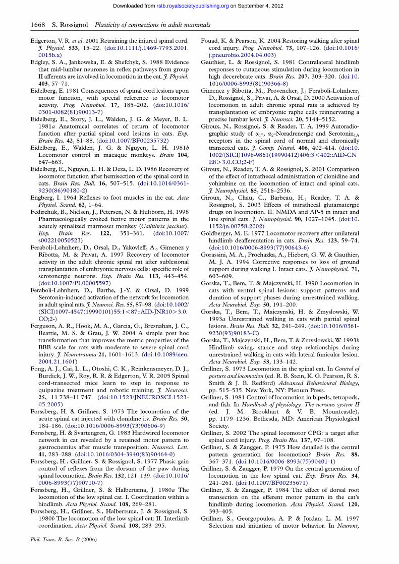
1668 S. Rossignol Plasticity of connections in adult mammals
on September 4, 2012rstb.royalsocietypublishing.orgDownloaded from
Edgerton, V. R. et al. 2001 Retraining the injured spinal cord.
J. Physiol. 533, 15–22. (doi:10.1111/j.1469-7793.2001.
0015b.x)
Edgley, S. A., Jankowska, E. & Shefchyk, S. 1988 Evidence
that mid-lumbar neurones in reflex pathways from group
II afferents are involved in locomotion in the cat. J. Physiol.403, 57–71.
Eidelberg, E. 1981 Consequences of spinal cord lesions upon
motor function, with special reference to locomotor
activity. Prog. Neurobiol. 17, 185–202. (doi:10.1016/
0301-0082(81)90013-7)
Eidelberg, E., Story, J. L., Walden, J. G. & Meyer, B. L.
1981a Anatomical correlates of return of locomotor
function after partial spinal cord lesions in cats. Exp.
Brain Res. 42, 81–88. (doi:10.1007/BF00235732)
Eidelberg, E., Walden, J. G. & Nguyen, L. H. 1981b
Locomotor control in macaque monkeys. Brain 104,
647–663.
Eidelberg, E., Nguyen, L. H. & Deza, L. D. 1986 Recovery of
locomotor function after hemisection of the spinal cord in
cats. Brain Res. Bull. 16, 507–515. (doi:10.1016/0361-
9230(86)90180-2)
Engberg, I. 1964 Reflexes to foot muscles in the cat. Acta
Physiol. Scand. 62, 1–64.
Fedirchuk, B., Nielsen, J., Petersen, N. & Hultborn, H. 1998
Pharmacologically evoked fictive motor patterns in the
acutely spinalized marmoset monkey (Callithrix jacchus).
Exp. Brain Res. 122, 351–361. (doi:10.1007/
s002210050523)
Feraboli-Lohnherr, D., Orsal, D., Yakovleff, A., Gimenez y
Ribotta, M. & Privat, A. 1997 Recovery of locomotor
activity in the adult chronic spinal rat after sublesional
transplantation of embryonic nervous cells: specific role of
serotonergic neurons. Exp. Brain Res. 113, 443–454.
(doi:10.1007/PL00005597)
Feraboli-Lohnherr, D., Barthe, J.-Y. & Orsal, D. 1999
Serotonin-induced activation of the network for locomotion
in adult spinal rats. J. Neurosci. Res. 55, 87–98. (doi:10.1002/
(SICI)1097-4547(19990101)55:1!87::AID-JNR10O3.0.
CO;2-)
Ferguson, A. R., Hook, M. A., Garcia, G., Bresnahan, J. C.,
Beattie, M. S. & Grau, J. W. 2004 A simple post hoc
transformation that improves the metric properties of the
BBB scale for rats with moderate to severe spinal cord
injury. J. Neurotrauma 21, 1601–1613. (doi:10.1089/neu.
2004.21.1601)
Fong, A. J., Cai, L. L., Otoshi, C. K., Reinkensmeyer, D. J.,
Burdick, J. W., Roy, R. R. & Edgerton, V. R. 2005 Spinal
cord-transected mice learn to step in response to
quipazine treatment and robotic training. J. Neurosci.
25, 11 738–11 747. (doi:10.1523/JNEUROSCI.1523-
05.2005)
Forssberg, H. & Grillner, S. 1973 The locomotion of the
acute spinal cat injected with clonidine i.v. Brain Res. 50,
184–186. (doi:10.1016/0006-8993(73)90606-9)
Forssberg, H. & Svartengren, G. 1983 Hardwired locomotor
network in cat revealed by a retained motor pattern to
gastrocnemius after muscle transposition. Neurosci. Lett.
41, 283–288. (doi:10.1016/0304-3940(83)90464-0)
Forssberg, H., Grillner, S. & Rossignol, S. 1977 Phasic gain
control of reflexes from the dorsum of the paw during
spinal locomotion. Brain Res. 132, 121–139. (doi:10.1016/
0006-8993(77)90710-7)
Forssberg, H., Grillner, S. & Halbertsma, J. 1980a The
locomotion of the low spinal cat. I. Coordination within a
hindlimb. Acta Physiol. Scand. 108, 269–281.
Forssberg, H., Grillner, S., Halbertsma, J. & Rossignol, S.
1980b The locomotion of the low spinal cat: II. Interlimb
coordination. Acta Physiol. Scand. 108, 283–295.
Phil. Trans. R. Soc. B (2006)
Fouad, K. & Pearson, K. 2004 Restoring walking after spinal
cord injury. Prog. Neurobiol. 73, 107–126. (doi:10.1016/
j.pneurobio.2004.04.003)
Gauthier, L. & Rossignol, S. 1981 Contralateral hindlimb
responses to cutaneous stimulation during locomotion in
high decerebrate cats. Brain Res. 207, 303–320. (doi:10.
1016/0006-8993(81)90366-8)
Gimenez y Ribotta, M., Provencher, J., Feraboli-Lohnherr,
D., Rossignol, S., Privat, A. & Orsal, D. 2000 Activation of
locomotion in adult chronic spinal rats is achieved by
transplantation of embryonic raphe cells reinnervating a
precise lumbar level. J. Neurosci. 20, 5144–5152.
Giroux, N., Rossignol, S. & Reader, T. A. 1999 Autoradio-
graphic study of a1-, a2-Noradrenergic and Serotonin1A
receptors in the spinal cord of normal and chronically
transected cats. J. Comp. Neurol. 406, 402–414. (doi:10.
1002/(SICI)1096-9861(19990412)406:3!402::AID-CN
E8O3.0.CO;2-F)
Giroux, N., Reader, T. A. & Rossignol, S. 2001 Comparison
of the effect of intrathecal administration of clonidine and
yohimbine on the locomotion of intact and spinal cats.
J. Neurophysiol. 85, 2516–2536.
Giroux, N., Chau, C., Barbeau, H., Reader, T. A. &
Rossignol, S. 2003 Effects of intrathecal glutamatergic
drugs on locomotion. II. NMDA and AP-5 in intact and
late spinal cats. J. Neurophysiol. 90, 1027–1045. (doi:10.
1152/jn.00758.2002)
Goldberger, M. E. 1977 Locomotor recovery after unilateral
hindlimb deafferentation in cats. Brain Res. 123, 59–74.
(doi:10.1016/0006-8993(77)90643-6)
Gorassini, M. A., Prochazka, A., Hiebert, G. W. & Gauthier,
M. J. A. 1994 Corrective responses to loss of ground
support during walking I. Intact cats. J. Neurophysiol. 71,
603–609.
Gorska, T., Bem, T. & Majczynski, H. 1990 Locomotion in
cats with ventral spinal lesions: support patterns and
duration of support phases during unrestrained walking.
Acta Neurobiol. Exp. 50, 191–200.
Gorska, T., Bem, T., Majczynski, H. & Zmyslowski, W.
1993a Unrestrained walking in cats with partial spinal
lesions. Brain Res. Bull. 32, 241–249. (doi:10.1016/0361-
9230(93)90183-C)
Gorska, T., Majczynski, H., Bem, T. & Zmyslowski, W. 1993b
Hindlimb swing, stance and step relationships during
unrestrained walking in cats with lateral funicular lesion.
Acta Neurobiol. Exp. 53, 133–142.
Grillner, S. 1973 Locomotion in the spinal cat. In Control of
posture and locomotion (ed. R. B. Stein, K. G. Pearson, R. S.
Smith & J. B. Redford) Advanced Behavioural Biology,
pp. 515–535. New York, NY: Plenum Press.
Grillner, S. 1981 Control of locomotion in bipeds, tetrapods,
and fish. In Handbook of physiology. The nervous system II(ed. J. M. Brookhart & V. B. Mountcastle),
pp. 1179–1236. Bethesda, MD: American Physiological
Society.
Grillner, S. 2002 The spinal locomotor CPG: a target after
spinal cord injury. Prog. Brain Res. 137, 97–108.
Grillner, S. & Zangger, P. 1975 How detailed is the central
pattern generation for locomotion? Brain Res. 88,
367–371. (doi:10.1016/0006-8993(75)90401-1)
Grillner, S. & Zangger, P. 1979 On the central generation of
locomotion in the low spinal cat. Exp. Brain Res. 34,
241–261. (doi:10.1007/BF00235671)
Grillner, S. & Zangger, P. 1984 The effect of dorsal root
transection on the efferent motor pattern in the cat’s
hindlimb during locomotion. Acta Physiol. Scand. 120,
393–405.
Grillner, S., Georgopoulos, A. P. & Jordan, L. M. 1997
Selection and initiation of motor behavior. In Neurons,
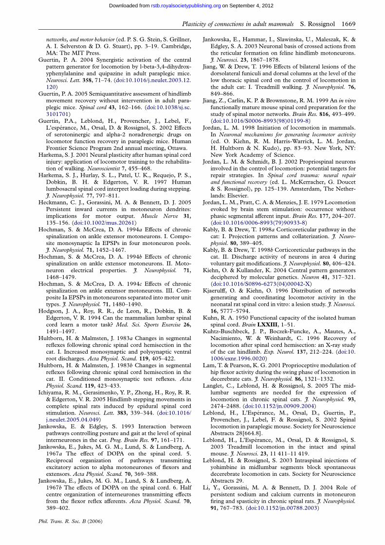
Plasticity of connections in adult mammals S. Rossignol 1669
on September 4, 2012rstb.royalsocietypublishing.orgDownloaded from
networks, and motor behavior (ed. P. S. G. Stein, S. Grillner,A. I. Selverston & D. G. Stuart), pp. 3–19. Cambridge,MA: The MIT Press.
Guertin, P. A. 2004 Synergistic activation of the centralpattern generator for locomotion by l-beta-3,4-dihydrox-yphenylalanine and quipazine in adult paraplegic mice.Neurosci. Lett. 358, 71–74. (doi:10.1016/j.neulet.2003.12.120)
Guertin, P. A. 2005 Semiquantitative assessment of hindlimbmovement recovery without intervention in adult para-plegic mice. Spinal cord 43, 162–166. (doi:10.1038/sj.sc.3101701)
Guertin, P.A., Leblond, H., Provencher, J., Lebel, F.,L’esperance, M., Orsal, D. & Rossignol, S. 2002 Effectsof serotoninergic and alpha-2 noradrenergic drugs onlocomotor function recovery in paraplegic mice. HumanFrontier Science Program 2nd annual meeting, Ottawa.
Harkema, S. J. 2001 Neural plasticity after human spinal cordinjury: application of locomotor training to the rehabilita-tion of walking. Neuroscientist 7, 455–468.
Harkema, S. J., Hurlay, S. L., Patel, U. K., Requejo, P. S.,Dobkin, B. H. & Edgerton, V. R. 1997 Humanlumbosacral spinal cord interpret loading during stepping.J. Neurophysiol. 77, 797–811.
Heckmann, C. J., Gorassini, M. A. & Bennett, D. J. 2005Persistent inward currents in motoneuron dendrites:implications for motor output. Muscle Nerve 31,135–156. (doi:10.1002/mus.20261)
Hochman, S. & McCrea, D. A. 1994a Effects of chronicspinalization on ankle extensor motoneurons. I. Compo-site monosynaptic Ia EPSPs in four motoneuron pools.J. Neurophysiol. 71, 1452–1467.
Hochman, S. & McCrea, D. A. 1994b Effects of chronicspinalization on ankle extensor motoneurons. II. Moto-neuron electrical properties. J. Neurophysiol. 71,1468–1479.
Hochman, S. & McCrea, D. A. 1994c Effects of chronicspinalization on ankle extensor motoneurons. III. Com-posite Ia EPSPs in motoneurons separated into motor unittypes. J. Neurophysiol. 71, 1480–1490.
Hodgson, J. A., Roy, R. R., de Leon, R., Dobkin, B. &Edgerton, V. R. 1994 Can the mammalian lumbar spinalcord learn a motor task? Med. Sci. Sports Exercise 26,1491–1497.
Hultborn, H. & Malmsten, J. 1983a Changes in segmentalreflexes following chronic spinal cord hemisection in thecat. I. Increased monosynaptic and polysynaptic ventralroot discharges. Acta Physiol. Scand. 119, 405–422.
Hultborn, H. & Malmsten, J. 1983b Changes in segmentalreflexes following chronic spinal cord hemisection in thecat. II. Conditioned monosynaptic test reflexes. ActaPhysiol. Scand. 119, 423–433.
Ichiyama, R. M., Gerasimenko, Y. P., Zhong, H., Roy, R. R.& Edgerton, V. R. 2005 Hindlimb stepping movements incomplete spinal rats induced by epidural spinal cordstimulation. Neurosci. Lett. 383, 339–344. (doi:10.1016/j.neulet.2005.04.049)
Jankowska, E. & Edgley, S. 1993 Interaction betweenpathways controlling posture and gait at the level of spinalinterneurones in the cat. Prog. Brain Res. 97, 161–171.
Jankowska, E., Jukes, M. G. M., Lund, S. & Lundberg, A.1967a The effect of DOPA on the spinal cord. 5.Reciprocal organization of pathways transmittingexcitatory action to alpha motoneurones of flexors andextensors. Acta Physiol. Scand. 70, 369–388.
Jankowska, E., Jukes, M. G. M., Lund, S. & Lundberg, A.1967b The effects of DOPA on the spinal cord. 6. Halfcentre organization of interneurones transmitting effectsfrom the flexor reflex afferents. Acta Physiol. Scand. 70,389–402.
Phil. Trans. R. Soc. B (2006)
Jankowska, E., Hammar, I., Slawinska, U., Maleszak, K. &Edgley, S. A. 2003 Neuronal basis of crossed actions fromthe reticular formation on feline hindlimb motoneurons.J. Neurosci. 23, 1867–1878.
Jiang, W. & Drew, T. 1996 Effects of bilateral lesions of thedorsolateral funiculi and dorsal columns at the level of thelow thoracic spinal cord on the control of locomotion inthe adult cat: I. Treadmill walking. J. Neurophysiol. 76,849–866.
Jiang, Z., Carlin, K. P. & Brownstone, R. M. 1999 An in vitrofunctionally mature mouse spinal cord preparation for thestudy of spinal motor networks. Brain Res. 816, 493–499.(doi:10.1016/S0006-8993(98)01199-8)
Jordan, L. M. 1998 Initiation of locomotion in mammals.In Neuronal mechanisms for generating locomotor activity(ed. O. Kiehn, R. M. Harris-Warrick, L. M. Jordan,H. Hultborn & N. Kudo), pp. 83–93. New York, NY:New York Academy of Science.
Jordan, L. M. & Schmidt, B. J. 2002 Propriospinal neuronsinvolved in the control of locomotion: potential targets forrepair strategies. In Spinal cord trauma: neural repairand functional recovery (ed. L. McKerracher, G. Doucet& S. Rossignol), pp. 125–139. Amsterdam, The Nether-lands: Elsevier.
Jordan, L. M., Pratt, C. A. & Menzies, J. E. 1979 Locomotionevoked by brain stem stimulation: occurrence withoutphasic segmental afferent input. Brain Res. 177, 204–207.(doi:10.1016/0006-8993(79)90933-8)
Kably, B. & Drew, T. 1998a Corticoreticular pathway in thecat: I. Projection patterns and collaterization. J. Neuro-physiol. 80, 389–405.
Kably, B. & Drew, T. 1998b Corticoreticular pathways in thecat. II. Discharge activity of neurons in area 4 duringvoluntary gait modifications. J. Neurophysiol. 80, 406–424.
Kiehn, O. & Kullander, K. 2004 Central pattern generatorsdeciphered by molecular genetics. Neuron 41, 317–321.(doi:10.1016/S0896-6273(04)00042-X)
Kjaerulff, O. & Kiehn, O. 1996 Distribution of networksgenerating and coordinating locomotor activity in theneonatal rat spinal cord in vitro: a lesion study. J. Neurosci.16, 5777–5794.
Kuhn, R. A. 1950 Functional capacity of the isolated humanspinal cord. Brain LXXIII, 1–51.
Kuhtz-Buschbeck, J. P., Boczek-Funcke, A., Mautes, A.,Nacimiento, W. & Weinhardt, C. 1996 Recovery oflocomotion after spinal cord hemisection: an X-ray studyof the cat hindlimb. Exp. Neurol. 137, 212–224. (doi:10.1006/exnr.1996.0020)
Lam, T. & Pearson, K. G. 2001 Proprioceptive modulation ofhip flexor activity during the swing phase of locomotion indecerebrate cats. J. Neurophysiol. 86, 1321–1332.
Langlet, C., Leblond, H. & Rossignol, S. 2005 The mid-lumbar segments are needed for the expression oflocomotion in chronic spinal cats. J. Neurophysiol. 93,2474–2488. (doi:10.1152/jn.00909.2004)
Leblond, H., L’Esperance, M., Orsal, D., Guertin, P.,Provencher, J., Lebel, F. & Rossignol, S. 2002 Spinallocomotion in paraplegic mouse. Society for NeuroscienceAbstracts 28[664.8].
Leblond, H., L’Esperance, M., Orsal, D. & Rossignol, S.2003 Treadmill locomotion in the intact and spinalmouse. J. Neurosci. 23, 11 411–11 419.
Leblond, H. & Rossignol, S. 2003 Intraspinal injections ofyohimbine in midlumbar segments block spontaneousdecerebrate locomotion in cats. Society for NeuroscienceAbstracts 29.
Li, Y., Gorassini, M. A. & Bennett, D. J. 2004 Role ofpersistent sodium and calcium currents in motoneuronfiring and spasticity in chronic spinal rats. J. Neurophysiol.91, 767–783. (doi:10.1152/jn.00788.2003)

1670 S. Rossignol Plasticity of connections in adult mammals
on September 4, 2012rstb.royalsocietypublishing.orgDownloaded from
Maegele, M., Muller, S., Wernig, A., Edgerton, V. R. &Harkema, S. J. 2002 Recruitment of spinal motor poolsduring voluntary movements versus stepping after humanspinal cord injury. J. Neurotrauma 19, 1217–1229. (doi:10.1089/08977150260338010)
Marcoux, J. & Rossignol, S. 2000 Initiating or blockinglocomotion in spinal cats by applying noradrenergic drugsto restricted lumbar spinal segments. J. Neurosci. 20,8577–8585.
Matsuyama, K. & Drew, T. 2000a Vestibulospinal andreticulospinal neuronal activity during locomotion in theintact cat I. Walking on a level surface. J. Neurophysiol. 84,2237–2256.
Matsuyama, K. & Drew, T. 2000b Vestibulospinal andreticulospinal neuronal activity during locomotion in theintact cat II. Walking on an inclined plane. J. Neurophysiol.84, 2257–2276.
Matsuyama, K., Mori, F., Nakajima, K., Drew, T., Aoki, M.& Mori, S. 2004a Locomotor role of the corticoreticular-reticulospinal-spinal interneuronal system. Prog. BrainRes. 143, 239–249.
Matsuyama, K., Nakajima, K., Mori, F., Aoki, M. & Mori, S.2004b Lumbar commissural interneurons with reticulosp-inal inputs in the cat: morphology and discharge patternsduring fictive locomotion. J. Comp. Neurol. 474, 546–561.(doi:10.1002/cne.20131)
McDearmid, J. R., Scrymgeour-Wedderburn, J. F. & Sillar,K. T. 1997 Aminergic modulation of glycine release in aspinal network controlling swimming in Xenopus laevis.J. Physiol. 503, 111–117. (doi:10.1111/j.1469-7793.1997.111bi.x) (Pt 1)
McVea, D. A., Donelan, J. M., Tachibana, A. & Pearson,K. G. 2005 A role for hip position in initiating the swing tostance transition in walking cats. J. Neurophysiol. 94,3497–3508. (doi:10.1152/jn.00511.2005)
Merkler, D., Metz, G. A. S., Raineteau, O., Dietz, V.,Schwab, M. E. & Fouad, K. 2001 Locomotor recovery inspinal cord-injured rats treated with an antibody neu-tralizing the myelin-associated neurite growth inhibitorNogo-A. J. Neurosci. 21, 3665–3673.
Mori, S. 1987 Integration of posture and locomotion in acutedecebrate cats and in awake, freely moving cats. Prog.Neurobiol. 28, 161–195. (doi:10.1016/0301-0082(87)90010-4)
Nacimiento, W., Mautes, A., Topper, R., Oestreicher, A. B.,Gispen, W. H., Nacimiento, A. C., Noth, J. & Kreutzberg,G. W. 1993 B-50 (GAP-43) in the spinal cord caudal tohemisection: indication for lack of intraspinal sprouting indorsal root axons. J. Neurosci. Res. 35, 603–617. (doi:10.1002/jnr.490350604)
Nathan, P. W. 1994 Effects on movement of surgical incisionsinto the human spinal cord. Brain 117, 337–346.
Orlovsky, G. N. & Shik, M. L. 1976 Control of locomotion: aneurophysiological analysis of the cat locomotor system.In International review of physiology. Neurophysiology II (ed.R. Portez), pp. 281–309. Baltimore, MD: University ParkPress.
Orsal, D., Barthe, J. Y., Antri, M., Feraboli-Lohnherr, D.,Yakovleff, A., Ribotta, M., Privat, A., Provencher, J. &Rossignol, S. 2002 Locomotor recovery in chronic spinalrat: long-term pharmacological treatment or trans-plantation of embryonic neurons? In Spinal cord trauma:regeneration, neural repair and functional recovery (ed. L. McKerracher, G. Doucet & S. Rossignol), pp. 213–230.New York, NY: Elsevier.
Pearson, K. G. 2000 Neural adaptation in the generation ofrhythmic behavior. Annu. Rev. Physiol. 62, 723–753.(doi:10.1146/annurev.physiol.62.1.723)
Pearson, K. G. & Rossignol, S. 1991 Fictive motor patterns inchronic spinal cats. J. Neurophysiol. 66, 1874–1887.
Phil. Trans. R. Soc. B (2006)
Pearson, K. G., Fouad, K. & Misiaszek, J. E. 1999 Adaptivechanges in motor activity associated with functionalrecovery following muscle denervation in walking cats.J. Neurophysiol. 82, 370–381.
Pearson, K. G., Misiaszek, J. E. & Hulliger, M. 2003Chemical ablation of sensory afferents in the walkingsystem of the cat abolishes the capacity for functionalrecovery after peripheral nerve lesions. Exp. Brain Res.150, 50–60.
Pearson, K. G., Acharya, H. & Fouad, K. 2005 A newelectrode configuration for recording electromyographicactivity in behaving mice. J. Neurosci. Methods 148, 36–42.(doi:10.1016/j.jneumeth.2005.04.006)
Pepin, A., Ladouceur, M. & Barbeau, H. 2003a Treadmillwalking in incomplete spinal-cord-injured subjects: 2.Factors limiting the maximal speed. Spinal Cord 41,271–279. (doi:10.1038/sj.sc.3101453)
Pepin, A., Norman, K. E. & Barbeau, H. 2003b Treadmillwalking in incomplete spinal-cord-injured subjects: 1.Adaptation to changes in speed. Spinal Cord 41, 257–270.(doi:10.1038/sj.sc.3101452)
Perreault, M.-C., Drew, T. & Rossignol, S. 1993 Activity ofmedullary reticulospinal neurons during fictive loco-motion. J. Neurophysiol. 69, 2232–2247.
Perreault, M.-C., Rossignol, S. & Drew, T. 1994 Micro-stimulation of the medullary reticular formation duringfictive locomotion. J. Neurophysiol. 71, 229–245.
Philippson, M. 1905 L’autonomie et la centralisation dans lesysteme nerveux des animaux. Trav. Lab. Physiol. Inst.Solvay (Bruxelles) 7, 1–208.
Prentice, S. D. & Drew, T. 2001 Contributions of thereticulospinal system to the postural adjustments occur-ring during voluntary gait modifications. J. Neurophysiol.85, 679–698.
Prochazka, A., Sontag, K. H. & Wand, P. 1978 Motorreactions to perturbations of gait: proprioceptive andsomesthetic involvement. Neurosci. Lett. 7, 35–39. (doi:10.1016/0304-3940(78)90109-X)
Raineteau, O., Fouad, K., Bareyre, F. M. & Schwab, M. E.2002 Reorganization of descending motor tracts in the ratspinal cord. Eur. J. Neurosci. 16, 1761–1771. (doi:10.1046/j.1460-9568.2002.02243.x)
Reader, T. A., Chau, C., Ghali, Z., Giroux, N., Senecal, J. &Rossignol, S. 2001 Characterization of alpha-2 adrenergicreceptor coupling in rat and cat spinal cords. Society forNeuroscience Abstracts 27[517.1].
Remy-Neris, O., Barbeau, H., Daniel, O., Boiteau, F. &Bussel, B. 1999 Effects of intrathecal clonidine injectionon spinal reflexes and human locomotion in incompleteparaplegic subjects. Exp. Brain Res. 129, 433–440. (doi:10.1007/s002210050910)
Ribotta, M. G., Provencher, J., Feraboli-Lohnherr, D.,Rossignol, S., Privat, A. & Orsal, D. 2000 Activation oflocomotion in adult chronic spinal rats is achieved bytransplantation of embryonic raphe cells reinnervating aprecise lumbar level. J. Neurosci. 20, 5144–5152.
Rossignol, S. 1996 Neural control of stereotypic limbmovements. In Handbook of physiology, section 12. Exercise:regulation and integration of multiple systems (ed. L. B.Rowell & J. T. Sheperd), pp. 173–216. New York, NY:Oxford University Press.
Rossignol, S. 2000 Locomotion and its recovery after spinalinjury. Curr. Opin. Neurobiol. 10, 708–716. (doi:10.1016/S0959-4388(00)00151-3)
Rossignol, S. & Gauthier, L. 1980 An analysis of mechanismscontrolling the reversal of crossed spinal reflexes. BrainRes. 182, 31–45. (doi:10.1016/0006-8993(80)90828-8)
Rossignol, S., Lund, J. P. & Drew, T. 1988 The role of sensoryinputs in regulating patterns of rhythmical movements inhigher vertebrates. A comparison between locomotion,

Plasticity of connections in adult mammals S. Rossignol 1671
on September 4, 2012rstb.royalsocietypublishing.orgDownloaded from
respiration and mastication. In Neural control of rhythmic
movements in vertebrates (ed. A. Cohen, S. Rossignol & S.
Grillner), pp. 201–283. New York, NY: Wiley.
Rossignol, S., Saltiel, P., Perreault, M.-C., Drew, T., Pearson,
K. G. & Belanger, M. 1993 Intralimb and interlimb
coordination in the cat during real and fictive rhythmic
motor programs. Neurosciences 5, 67–75.
Rossignol, S., Chau, C., Brustein, E., Belanger, M., Barbeau,
H. & Drew, T. 1996 Locomotor capacities after complete
and partial lesions of the spinal cord. Acta Neurobiol. Exp.
56, 449–463.
Rossignol, S., Chau, C., Brustein, E., Giroux, N., Bouyer, L.,
Barbeau, H. & Reader, T. 1998 Pharmacological activation
and modulation of the central pattern generator for
locomotion in the cat. In Neuronal mechanisms for generating
locomotor activity (ed. O. Kiehn, R. M. Harris-Warrick,
L. M. Jordan, H. Hulborn & N. Kudo), pp. 346–359.
New York, NY: New York Academy of Science.
Rossignol, S., Drew, T., Brustein, E. & Jiang, W. 1999
Locomotor performance and adaptation after partial or
complete spinal cord lesions in the cat. In Peripheral and
spinal mechanisms in the neural control of movement (ed.
M. D. Binder), pp. 349–365. Amsterdam, The Nether-
lands: Elsevier.
Rossignol, S. et al. 2000 The spinal cat. In Neurobiology ofspinal cord injury (ed. R. G. Kalb & S. M. Strittmatter),
pp. 57–87. Totowa, NJ: Humana Press.
Rossignol, S., Giroux, N., Chau, C., Marcoux, J., Brustein, E.
& Reader, T. A. 2001 Pharmacological aids to locomotor
training after spinal injury in the cat. J. Physiol. 533, 65–74.
(doi:10.1111/j.1469-7793.2001.0065b.x)
Rossignol, S. et al. 2002 The cat model of spinal injury. In
Spinal cord trauma: regeneration, neural repair and functionalrecovery (ed. L. McKerracher, G. Doucet & S. Rossignol),
pp. 151–168. New York, NY: Elsevier.
Rossignol, S., Dubuc, R. & Gossard, J. P. 2006 Dynamic
sensorimotor interactions in locomotion. Physiol. Rev. 86,
89–154. (doi:10.1152/physrev.00028.2005)
Safyants, V. I. 1970 On the mechanism of inhibition following
the reflex discharge in interneurons of the spinal cord.
XXXth. Int. Cong. Physiol. Sci. Vancouver. 2, 3–10.
Saltiel, P. & Rossignol, S. 2004a Critical points in the
forelimb fictive locomotor cycle and motor coordination:
evidence from the effects of tonic proprioceptive pertur-
bations in the cat. J. Neurophysiol. 92, 1329–1341. (doi:10.
1152/jn.00563.2003)
Saltiel, P. & Rossignol, S. 2004b Critical points in the
forelimb fictive locomotor cycle of the cat and motor
coordination: effects of phasic retractions and protractions
of the shoulder in the cat. J. Neurophysiol. 92, 1342–1356.
(doi:10.1152/jn.00564.2003)
Schmidt, B. J. & Jordan, L. M. 2000 The role of serotonin in
reflex modulation and locomotor rhythm production in
the mammalian spinal cord. Brain Res. Bull. 53, 689–710.
(doi:10.1016/S0361-9230(00)00402-0)
Schouenborg, J. 2003 Modular organisation and spinal
somatosensory imprinting. Brain Res. Rev. 40, 80–91.
(doi:10.1016/S0165-0173(02)00191-1)
Shefchyk, S., McCrea, D. A., Kriellaars, D., Fortier, P. &
Jordan, L. 1990 Activity of interneurons within the L4
spinal segment of the cat during brainstem-evoked fictive
locomotion. Exp. Brain Res. 80, 290–295. (doi:10.1007/
BF00228156)
Shefner, J. M., Berman, S. A., Sarkarati, M. & Young, R. R.
1992 Recurrent inhibition is increased in patients with
spinal cord injury. Neurology 42, 2162–2168.
Phil. Trans. R. Soc. B (2006)
Sherrington, C. S. 1910 Flexion-reflex of the limb, crossedextension-reflex, and reflex stepping and standing.J. Physiol. 40, 28–121.
Shik, M. L. 1997 Recognizing propriospinal and reticulo-spinal systems of initiation of stepping. Motor Control 1,310–313.
Shik, M. L., Severin, F. V. & Orlovsky, G. N. 1966 Control ofwalking and running by means of electrical stimulation ofthe mid-brain. Biophysics 11, 756–765.
Shimamura, M., Kogure, I. & Wada, S. I. 1982 Reticularneuron activities associated with locomotion in thalamiccats. Brain Res. 231, 51–62. (doi:10.1016/0006-8993(82)90006-3)
Sinkjaer, T., Andersen, J. B., Ladouceur, M., Christensen,L. O. D. & Nielsen, J. 2000 Major role for sensoryfeedback in soleus EMG activity in the stance phase ofwalking in man. J. Physiol. 523, 817–827. (doi:10.1111/j.1469-7793.2000.00817.x)
Steward, O., Zheng, B. & Tessier-Lavigne, M. 2003 Falseresurrections: distinguishing regenerated from sparedaxons in the injured central nervous system. J. Comp.Neurol. 459, 1–8. (doi:10.1002/cne.10593)
Thomas, S. L. & Gorassini, M. A. 2005 Increases incorticospinal tract function by treadmill training afterincomplete spinal cord injury. J. Neurophysiol. 94,2844–2855. (doi:10.1152/jn.00532.2005)
Vanderhorst, V. G. J. M. & Holstege, G. 1997 Organization oflumbosacral motoneuronal cell groups innervatinghindlimb, pelvic floor, and axial muscles in the cat.J. Comp. Neurol. 382, 46–76. (doi:10.1002/(SICI)1096-9861(19970526)382:1!46::AID-CNE4O3.0.CO;2-K)
Vilensky, J. A., Moore, A. M., Eidelberg, E. & Walden, J. G.1992 Recovery of locomotion in monkeys with spinal cordlesions. J. Motor Behav. 24, 288–296.
Wernig, A., Nanassy, A. & Muller, S. 1999 Laufband(treadmill) therapy in incomplete paraplegia and tetra-plegia. J. Neurotrauma 16, 719–726.
Wetzel, M. C., Atwater, A. E., Wait, J. V. & Stuart, D. G.1976 Kinematics of locomotion by cats with a singlehindlimb deafferented. J. Neurophysiol. 39, 667–678.
Whelan, P. J., Hiebert, G. W. & Pearson, K. G. 1995 Plasticityof the extensor group I pathway controlling the stance toswing transition in the cat. J. Neurophysiol. 74, 2782–2787.
Wirz, M., Zemon, D. H., Rupp, R., Scheel, A., Colombo, G.,Dietz, V. & Hornby, T. G. 2005 Effectiveness ofautomated locomotor training in patients with chronicincomplete spinal cord injury: a multicenter trial. Arch.Phys. Med. Rehabil. 86, 672–680. (doi:10.1016/j.apmr.2004.08.004)
Xia, R. & Rymer, W. Z. 2005 Reflex reciprocal facilitation ofantagonist muscles in spinal cord injury. Spinal Cord 43,14–21. (doi:10.1038/sj.sc.3101656)
Young, W. 2002 Spinal cord contusion models. Prog. BrainRes. 137, 231–255.
Zehr, E. P. & Stein, R. B. 1999 What functions do reflexesserve during human locomotion? Prog. Neurobiol. 58,185–205. (doi:10.1016/S0301-0082(98)00081-1)
Zehr, E. P., Fujita, K. & Stein, R. B. 1998a Reflexes from thesuperficial peroneal nerve during walking in strokesubjects. J. Neurophysiol. 79, 848–858.
Zehr, E. P., Stein, R. B. & Komiyama, T. 1998b Function ofsural nerve reflexes during human walking. J. Physiol. 507,305–314. (doi:10.1111/j.1469-7793.1998.305bu.x)
Zmyslowski, W., Gorska, T., Majczynski, H. & Bem, T. 1993Hindlimb muscle activity during unrestrained walking incats with lesions of the lateral funiculi. Acta Neurobiol. Exp.53, 143–153.