plasmonic ge-doped zno nanocrystals · cite this chem. commun., 2015, 51 ,12369 plasmonic ge-doped...
TRANSCRIPT
This journal is©The Royal Society of Chemistry 2015 Chem. Commun., 2015, 51, 12369--12372 | 12369
Cite this:Chem. Commun., 2015,
51, 12369
Plasmonic Ge-doped ZnO nanocrystals†
Enrico Della Gaspera,*a Noel W. Duffy,b Joel van Embden,a Lynne Waddington,a
Laure Bourgeois,c Jacek J. Jasieniak‡*a and Anthony S. R. Chesman*a
We present the first colloidal synthesis of Ge-doped ZnO nanocrystals,
which are produced by a scalable method that uses only air and
moisture stable precursors. The incorporation of tetravalent Ge ions
within ZnO nanocrystals generates a surface plasmon resonance in the
near-mid infrared, and induces a change in morphology, from isotropic
spheroidal nanocrystals to rod-like, elongated structures with a dis-
tinctive c-axis orientation.
The ability to precisely incorporate dopants into colloidal ZnOnanocrystals (NCs) has created a reliable pathway to tailoringthe optical and electronic properties of ZnO. As a consequence,doped ZnO has application in electrochemistry, photocatalysis,optoelectronics, magnetism and sensing.1–5 In particular,doped ZnO NCs displaying a localized surface plasmon resonance(LSPR) have recently become an attractive alternative to expensiveindium tin oxide (ITO).6–10 Plasmonic ZnO NCs have typically beendoped with trivalent cations via substitution at the zinc site, with thedopant providing free charge carriers that give rise to a plasmonicresonance centered in the near or mid infrared. Due to the similarityin valence and cation size between trivalent substituents (typically Al,Ga or In) and zinc ions, such dopants can be incorporated withinZnO at relatively high concentrations.5–7,11 In contrast, investigationsinto doping ZnO with tetravalent p-block elements by solution-basedroutes have been limited,12 presumably owing to the challenges
associated with the disparity in the physical and electronicproperties between the dopant and host cations.
In light of this, germanium is the most suitable candidateamong the Group IV elements to act as a 4+ dopant in ZnO, as itis closer in size to Zn than Si, and has a more stable tetravalentoxidation state than Sn. Ge-doped ZnO (ZnO:Ge) has beenexamined for its potential use as a photoluminescent material anda transparent conducting oxide (TCO). To date, its synthesis hasbeen limited to the use of expensive vacuum-based depositiontechniques, such as sputtering,13 evaporation,14 and ALD,15 or hightemperature liquid phase epitaxy,16 with no solution-based methodsamenable to cheap, large scale synthesis available.
In this work, we overcome this barrier by presenting the firstsynthesis of Ge-doped ZnO colloidal NCs. The synthetic methoddeveloped here utilizes a scalable heat-up technique instead ofthe commonly adopted hot-injection protocol.17 It employs onlyair and moisture stable precursors, and provides ready access tolarge quantities of ZnO:Ge NCs across a range of doping levels.We examine in detail the effect of the dopant concentration onthe optical properties and the morphology of the NCs. Further-more, we demonstrate the general applicability of this methodby synthesizing ZnO NCs doped with Si and Sn.
ZnO:Ge NCs were synthesized by an adapted colloidal heat-upmethod previously developed for the formation of Al, Ga, andIn-doped ZnO NCs.11 Briefly, an air stable Ge precursor solutionwas first formed by heating [Ge(O,O0-glycolate)2(H2O)2] in oleyl-amine at 120 1C under vacuum.18 Upon cooling, the Ge precursorwas added to zinc stearate suspended in oleic acid, 1-dodecanoland octadecene. The solution was then heated at 230 1C for 2 hoursto give ZnO:Ge NCs (see ESI† for experimental procedures). Thesamples have been labeled ZGeX where X is the nominal Ge/Znratio in atomic %. The reaction proceeds via esterification betweenthe carboxylate groups coordinating the metals and the hydroxylmoieties of the alcohol, which results in the formation of metalhydroxide species that ultimately condense to give doped ZnONCs.11
Optical characterization of the as-prepared colloids is pre-sented in Fig. 1. A distinctive IR feature was observed in the
a CSIRO Manufacturing Flagship Ian Wark Laboratories, Bayview Ave, Clayton,
Victoria 3168, Australia. E-mail: [email protected],
[email protected], [email protected] CSIRO Energy Flagship, Ian Wark Laboratories, Bayview Ave, Clayton,
Victoria 3168, Australiac Monash Centre for Electron Microscopy and Department of Materials Science and
Engineering, Monash University, Wellington Road, Clayton, Victoria 3800,
Australia
† Electronic supplementary information (ESI) available: Experimental details,FTIR spectra, additional UV-Vis-NIR, XRD and TEM with size distributions andEDS data, SEM of thin films, full characterization of ZnO:Si and ZnO:Snnanocrystals. See DOI: 10.1039/c5cc02429c‡ Present address: Materials Science and Engineering, Monash University, WellingtonRd, Clayton, Victoria 3800, Australia.
Received 24th March 2015,Accepted 12th June 2015
DOI: 10.1039/c5cc02429c
www.rsc.org/chemcomm
ChemComm
COMMUNICATION
Ope
n A
cces
s A
rtic
le. P
ublis
hed
on 3
0 Ju
ne 2
015.
Dow
nloa
ded
on 1
4/08
/201
5 00
:46:
21.
Thi
s ar
ticle
is li
cens
ed u
nder
a C
reat
ive
Com
mon
s A
ttrib
utio
n 3.
0 U
npor
ted
Lic
ence
.
View Article OnlineView Journal | View Issue

12370 | Chem. Commun., 2015, 51, 12369--12372 This journal is©The Royal Society of Chemistry 2015
optical absorption spectra of all Ge-doped NC colloidal solutions(Fig. 1a and Fig. S1, ESI†). Examination of Fig. 1a reveals that themagnitude of the absorption in this region increased concomitantlywith Ge doping levels. We ascribe this feature to a LSPR peakarising from dopant-induced generation of free electrons.6,11,19
Consequently, the doped solutions have a pale green color, incomparison to the colorless undoped ZnO (inset of Fig. 1a).This distinctive coloring has also been observed previously forAl-, Ga- and In-doped ZnO NCs, and it is ascribed to thepresence of aliovalent dopants.5,11 Concurrent with higher Gedoping levels, the optical band gap of ZnO increased aspredicted by the Burstein–Moss effect (Fig. 1b and c). Fig. 1cshows that as the nominal Ge doping level is raised from 0 to20% the band gap increases from B3.31 eV to 3.4 eV.
Confirmation of the extent of doping was achieved by X-rayfluorescence (XRF) analysis, which was used to evaluate the realGe/Zn ratio (Fig. 1c and Table S1, ESI†). The doping efficiency,evaluated as the ratio of Ge incorporated within the NCs withrespect to the amount of precursor used, was approximately75% for all Ge precursor concentrations examined in this study,excluding the highest, which gave a doping efficiency of B50%.The decrease in doping efficiency at higher dopant precursorloadings has been observed previously.11,20
X-ray diffraction (XRD) analyses confirmed that all of theprepared materials were in the wurtzite crystalline phase (ICDDNo. 34-1451) with no secondary or contaminating phases (e.g.,GeO2, ZnGeO3, Zn2GeO4) observed (Fig. 2a). A slight shift in
diffraction peaks towards higher angles was observed withincreasing Ge doping levels, consistent with a reduction inthe crystal lattice parameters following the substitution of Zn2+
with smaller Ge4+ ions (Fig. S2, ESI†).15 The broadening of thediffraction peaks is consistent with the crystalline domainsbeing in the nanometer size regime (Fig. S3, ESI†). The crystallitesize evaluated with the Scherrer relationship was B15 nm for theundoped NCs, and slightly smaller (10–12 nm) for the doped NCs,regardless of the level of doping. This is possibly due to the stress-induced reduction in crystal growth caused by the incorporation ofdopants within the ZnO lattice.6,21 In addition to the dopant-induceddecrease in crystallite size, a progressive increase in intensity andreduction in width of the (002) diffraction peak with increasing Gedoping levels was observed (Fig. 2b and c). This phenomenon isusually ascribed to the formation of elongated structures orientedalong the c-axis of the hexagonal wurtzite cell, which is the mostenergetically favourable growth direction.22,23 This is in agreementwith the morphology of the as-prepared ZnO:Ge NCs synthesisedhere (vide infra).
Collectively, these analyses confirmed the successful incorpora-tion of Ge ions within the ZnO NCs, proving the efficacy of ouroptimized synthetic protocol. While Ge dopants were responsiblefor the LSPR in the infrared spectrum, their presence inside the
Fig. 1 (a) Optical absorption spectra of equimolar colloidal solutions ofZnO:Ge in tetrachloroethylene (TCE). The inset shows a digital picture ofthe prepared colloidal solutions. (b) Tauc plot (exponent = 2, direct bandgap) of the ZnO:Ge colloidal solutions. The dashed lines are the linear fitsused to identify the band gap. (c) Optical band gap and real Ge/Zn amountevaluated from XRF as a function of the nominal Ge/Zn ratio.
Fig. 2 (a) XRD patterns of ZnO:Ge powders. The diffraction patterns areoffset vertically for clarity and the peaks are indexed according to the ICDDNo. 34-1451 reference. (b) Zoomed view of the (100), (002) and (101)diffraction peaks. (c) Crystallite size of the prepared NCs as a function ofGe doping. The error bars are plus or minus one standard deviation. Byexcluding the (002) diffraction peak in the estimation of the crystallite size,smaller sizes with lower standard deviations are observed.
Communication ChemComm
Ope
n A
cces
s A
rtic
le. P
ublis
hed
on 3
0 Ju
ne 2
015.
Dow
nloa
ded
on 1
4/08
/201
5 00
:46:
21.
Thi
s ar
ticle
is li
cens
ed u
nder
a C
reat
ive
Com
mon
s A
ttrib
utio
n 3.
0 U
npor
ted
Lic
ence
.View Article Online
This journal is©The Royal Society of Chemistry 2015 Chem. Commun., 2015, 51, 12369--12372 | 12371
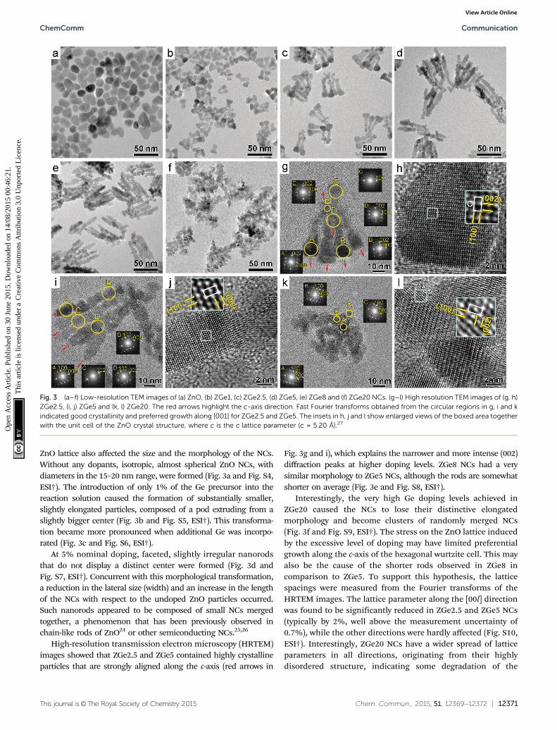
ZnO lattice also affected the size and the morphology of the NCs.Without any dopants, isotropic, almost spherical ZnO NCs, withdiameters in the 15–20 nm range, were formed (Fig. 3a and Fig. S4,ESI†). The introduction of only 1% of the Ge precursor into thereaction solution caused the formation of substantially smaller,slightly elongated particles, composed of a pod extruding from aslightly bigger center (Fig. 3b and Fig. S5, ESI†). This transforma-tion became more pronounced when additional Ge was incorpo-rated (Fig. 3c and Fig. S6, ESI†).
At 5% nominal doping, faceted, slightly irregular nanorodsthat do not display a distinct center were formed (Fig. 3d andFig. S7, ESI†). Concurrent with this morphological transformation,a reduction in the lateral size (width) and an increase in the lengthof the NCs with respect to the undoped ZnO particles occurred.Such nanorods appeared to be composed of small NCs mergedtogether, a phenomenon that has been previously observed inchain-like rods of ZnO24 or other semiconducting NCs.25,26
High-resolution transmission electron microscopy (HRTEM)images showed that ZGe2.5 and ZGe5 contained highly crystallineparticles that are strongly aligned along the c-axis (red arrows in
Fig. 3g and i), which explains the narrower and more intense (002)diffraction peaks at higher doping levels. ZGe8 NCs had a verysimilar morphology to ZGe5 NCs, although the rods are somewhatshorter on average (Fig. 3e and Fig. S8, ESI†).
Interestingly, the very high Ge doping levels achieved inZGe20 caused the NCs to lose their distinctive elongatedmorphology and become clusters of randomly merged NCs(Fig. 3f and Fig. S9, ESI†). The stress on the ZnO lattice inducedby the excessive level of doping may have limited preferentialgrowth along the c-axis of the hexagonal wurtzite cell. This mayalso be the cause of the shorter rods observed in ZGe8 incomparison to ZGe5. To support this hypothesis, the latticespacings were measured from the Fourier transforms of theHRTEM images. The lattice parameter along the [00l] directionwas found to be significantly reduced in ZGe2.5 and ZGe5 NCs(typically by 2%, well above the measurement uncertainty of0.7%), while the other directions were hardly affected (Fig. S10,ESI†). Interestingly, ZGe20 NCs have a wider spread of latticeparameters in all directions, originating from their highlydisordered structure, indicating some degradation of the
Fig. 3 (a–f) Low-resolution TEM images of (a) ZnO, (b) ZGe1, (c) ZGe2.5, (d) ZGe5, (e) ZGe8 and (f) ZGe20 NCs. (g–l) High resolution TEM images of (g, h)ZGe2.5, (i, j) ZGe5 and (k, l) ZGe20. The red arrows highlight the c-axis direction. Fast Fourier transforms obtained from the circular regions in g, i and kindicated good crystallinity and preferred growth along [001] for ZGe2.5 and ZGe5. The insets in h, j and l show enlarged views of the boxed area togetherwith the unit cell of the ZnO crystal structure, where c is the c lattice parameter (c = 5.20 Å).27
ChemComm Communication
Ope
n A
cces
s A
rtic
le. P
ublis
hed
on 3
0 Ju
ne 2
015.
Dow
nloa
ded
on 1
4/08
/201
5 00
:46:
21.
Thi
s ar
ticle
is li
cens
ed u
nder
a C
reat
ive
Com
mon
s A
ttrib
utio
n 3.
0 U
npor
ted
Lic
ence
.View Article Online

12372 | Chem. Commun., 2015, 51, 12369--12372 This journal is©The Royal Society of Chemistry 2015
crystal lattice due to excessive doping. From HRTEM it can beconcluded that lattice shrinkage started along the [00l] direction,which corresponds to the family of lattice planes with the highestsurface energy,28 and then spread to the other [uv0] directions asdoping increases, eventually compromising the structural integrityof the NCs. The preferential shrinkage along the c-axis at lowdoping levels has been previously observed in Al-doped ZnO NCs,and it has been ascribed to a possible preferential site occupationby Al3+ ions.6
The evolution of the NCs as a function of the reaction timewas investigated using optical absorption spectroscopy andTEM; Ge dopants were found to be continuously incorporatedwithin the ZnO NCs over the duration of the 2 h reaction, withthe rate of incorporation decreasing with time (Fig. S12, ESI†).Such a decrease is more evident when the initial amount of Geprecursor was low (ZGe5), because the dopant source is rapidlydepleted from the solution during the initial stages of thereaction. Conversely, at high Ge loadings (ZGe20), there wassufficient Ge precursor available to promote a higher rate ofdoping during the later stages of the 2 h reaction. The heavilydoped ZnO NCs required more time to nucleate, with no ZGe20NCs isolatable 5 minutes after the reaction solution reached230 1C, while ZGe5 NCs were already fully formed in that periodof time, which is additional evidence that the Ge precursorplays a key role in the nucleation, growth and consequentmorphology of the Ge-doped ZnO NCs. Interestingly, the NCsdo not change morphology during the course of the reaction(see Fig. S13 and S14, ESI†), while as discussed before, thedoping is found to constantly increase over time.
As a proof-of-concept study, the versatile reaction methodpresented here was also applied to the synthesis of ZnO NCsdoped with other p-block elements, namely Si and Sn (Fig. S15–S18 and Table S1, see ESI† for details), confirming its widerapplicability towards the preparation of a variety of doped NCs.
Ge-doped ZnO NCs can be dispersed in various organicsolvents (e.g. octane, toluene) at concentrations of up to200 mg mL�1. These could be used to prepare high qualitythin films through several deposition methods, such as spincoating, spray coating, drop casting, and printing (see Fig. S19for SEM and optical characterization of spin coated films, ESI†).The ability to fabricate functional thin films from NC inksmakes doped ZnO colloids attractive for a variety of applica-tions in optoelectronics, sensing and catalysis.
In conclusion, we have developed the first synthesis of Ge-dopedZnO colloidal NCs with plasmonic resonances in the infraredspectrum. The synthesis is based on a scalable, heat-up protocolwhich utilizes only air and moisture stable reagents. Ge dopantswere found to strongly affect not just the functional properties ofthe ZnO NCs, but also their morphology, with a progressivetransformation from isotropic nanoparticles to elongatednanorods, and eventually at very high concentrations (410%atomic) to clusters of merged NCs. This work constitutes asignificant step forward towards further investigations on ZnOdoped with Group IV elements.
This work was funded through the Manufacturing Flagshipof CSIRO as part of Office of the Chief Executive Postdoctoral
Fellowships (E.D.G., J.v.E.). J.J.J. and A.S.R.C. acknowledge theAustralian Research Council for funding through GrantDP110105341 and for a Discovery Early Career Research Award,respectively. Steve Peacock and Thomas R. Gengenbach arethanked for XRF and XPS measurements, respectively. Theauthors acknowledge use of facilities within the Monash Centrefor Electron Microscopy and the support of the State of Victoriaand Monash University for instrumentation.
Notes and references1 P. V. Radovanovic, N. S. Norberg, K. E. McNally and D. R. Gamelin,
J. Am. Chem. Soc., 2002, 124, 15192–15193.2 V. Etacheri, R. Roshan and V. Kumar, ACS Appl. Mater. Interfaces,
2012, 4, 2717–2725.3 E. Della Gaspera, M. Guglielmi, G. Perotto, S. Agnoli, G. Granozzi,
M. L. Post and A. Martucci, Sens. Actuators, B, 2012, 161, 675–683.4 G. Garcia, R. Buonsanti, A. Llordes, E. L. Runnerstrom, A. Bergerud
and D. J. Milliron, Adv. Opt. Mater., 2013, 1, 215–220.5 J. Song, S. A. Kulinich, J. Li, Y. Liu and H. Zeng, Angew. Chem., Int.
Ed., 2015, 54, 462–466.6 R. Buonsanti, A. Llordes, S. Aloni, B. A. Helms and D. J. Milliron,
Nano Lett., 2011, 11, 4706–4710.7 S. Ghosh, M. Saha and S. K. De, Nanoscale, 2014, 6, 7039–7051.8 X. Liang, Y. Ren, S. Bai, N. Zhang, X. Dai, X. Wang, H. He, C. Jin,
Z. Ye, Q. Chen, L. Chen, J. Wang and Y. Jin, Chem. Mater., 2014, 26,5169–5178.
9 E. Della Gaspera, M. Bersani, M. Cittadini, M. Guglielmi, D. Pagani,R. Noriega, S. Mehra, A. Salleo and A. Martucci, J. Am. Chem. Soc.,2013, 135, 3439–3448.
10 S. Cimitan, S. Albonetti, L. Forni, F. Peri and D. Lazzari, J. ColloidInterface Sci., 2009, 329, 73–80.
11 E. Della Gaspera, A. S. R. Chesman, J. van Embden andJ. J. Jasieniak, ACS Nano, 2014, 8, 9154–9163.
12 S. Ghosh, M. Saha, V. Dev Ashok, B. Dalal and S. K. De, J. Phys. Chem. C,2015, 119, 1180–1187.
13 D. H. Fan, Z. Y. Ning and M. F. Jiang, Appl. Surf. Sci., 2005, 245,414–419.
14 Y. Su, X. Meng, Y. Chen, S. Li, Q. Zhou, X. Liang and Y. Feng, Mater.Res. Bull., 2008, 43, 1865–1871.
15 P. R. Chalker, P. A. Marshall, P. J. King, K. Dawson, S. Romani,P. A. Williams, J. Ridealgh and M. J. Rosseinsky, J. Mater. Chem.,2012, 22, 12824–12829.
16 D. Ehrentraut, H. Sato, Y. Kagamitani, A. Yoshikawa, T. Fukuda, J. Pejchal,K. Polak, M. Nikl, H. Odaka, K. Hatanaka and H. Fukumura, J. Mater.Chem., 2006, 16, 3369–3374.
17 J. Van Embden, A. S. R. Chesman and J. Jasieniak, Chem. Mater.,2015, 27, 2246–2285.
18 A. S. R. Chesman, J. van Embden, E. Della Gaspera, N. W. Duffy,N. A. S. Webster and J. J. Jasieniak, Chem. Mater., 2014, 26, 5482–5491.
19 A. Comin and L. Manna, Chem. Soc. Rev., 2014, 43, 3957–3975.20 L. De Trizio, R. Buonsanti, A. M. Schimpf, A. Llordes, D. R. Gamelin,
R. Simonutti and D. J. Milliron, Chem. Mater., 2013, 25, 3383–3390.21 Z. Lu, J. Zhou, A. Wang, N. Wang and X. Yang, J. Mater. Chem., 2011,
21, 4161–4167.22 Y. Yang, Y. Jin, H. He, Q. Wang, Y. Tu, H. Lu and Z. Ye, J. Am. Chem.
Soc., 2010, 132, 13381–13394.23 E. Della Gaspera, J. van Embden, A. S. R. Chesman, N. W. Duffy and
J. J. Jasieniak, ACS Appl. Mater. Interfaces, 2014, 6, 22519–22526.24 C. Pacholski, A. Kornowski and H. Weller, Angew. Chem., Int. Ed.,
2002, 41, 1188–1191.25 K.-S. Cho, D. V. Talapin, W. Gaschler and C. B. Murray, J. Am. Chem.
Soc., 2005, 127, 7140–7147.26 Z. Tang, N. A. Kotov and M. Giersig, Science, 2002, 297, 237–240.27 Attempts to map the distribution of the Ge-dopant in the ZnO lattice
by X-ray energy dispersive spectroscopy (XEDS) was not successful, asalthough Ge is clearly detected, it cannot be mapped with confidencedue to the small amount of Ge, overlap between Ge and Zn signals, andbeam damage to the NCs after prolonged electron beam exposure(Fig. S11, ESI†).
28 C. Woll, Prog. Surf. Sci., 2007, 82, 55–120.
Communication ChemComm
Ope
n A
cces
s A
rtic
le. P
ublis
hed
on 3
0 Ju
ne 2
015.
Dow
nloa
ded
on 1
4/08
/201
5 00
:46:
21.
Thi
s ar
ticle
is li
cens
ed u
nder
a C
reat
ive
Com
mon
s A
ttrib
utio
n 3.
0 U
npor
ted
Lic
ence
.View Article Online