physics of radiography - pusanbml.pusan.ac.kr/resources/lecture/indst/physicsradiography.pdf ·...
TRANSCRIPT

2013-09-24
1
Physics of Radiography
Ho Kyung [email protected]
Pusan National University
Medical Physics
• Ionizing radiation
– Capable of ejecting electrons from atoms
• X-ray
• Gamma-ray
• Particulate radiation
• Radiographic imaging
– Transmission vs. emission imaging
2

2013-09-24
2
• Transmission imaging
– Making use of the transmission of ionizing radiation thru the body
– X-ray tube + imaging detector
– e.g., projection radiography & computed tomography (CT)
– Various tissues & organs attenuate the intensity of the x-ray beam as it passes thru the body
– The attenuation characteristics are determined by the effective atomic number Z & density of the tissues or organs
• Depicting structures within the body; hence anatomical imaging
– CT shows much higher “contrast” than the projection radiography because of the lack of superposition of out-of-plane tissues
3
Ionization
• The ejection of an electron from an atom, creating a free electron & an ion
• Atomic structure
– Atom = nucleus + electrons
– Nucleus (consisting of “nucleons”) = protons + neutrons
– Atomic number, Z
= # of protons & defining the element
• Representing # of orbiting electrons
– Mass number, A
= # of nucleons (= protons + neutrons)
– Nuclide
• Referring to any unique combination of protons & neutrons which forms a nucleus
• Denoted by 𝑍𝐴𝑋 or 𝑋 − 𝐴 (e.g., 6
12𝐶 or 𝐶 − 12)
– Radionuclides
• Unstable nuclides & their atoms are radioactive
• Statistically likely to undergo radioactive decay causing a rearrangement of the nucleus, which in turn gives off energy & results in a more stable nucleus
• e.g., 614𝐶 → 7
14𝑁 + −1𝛽
4

2013-09-24
3
– Electron orbits or shells• K, L, M, …
• Max. number of electrons = 2𝑛2, where 𝑛 = the shell number
5
• Electron binding energy
– E[atom] > E[nucleus] + E[electrons] energy difference binding energy
– BE as 𝑛
– BE of e– in H = 13.6 eV
– BE of e–‘s in Hg = 7.8 eV
• Average binding energy
– Avg. BE for air = ~34 eV
– Avg. BE for Pb = ~1 keV
– Avg. BE for W = ~4 keV
• Ionization & excitation
– Ionization ion + electrons (ion pairs)
– Excitation
• Transferring some energy to a bound electron but less than the electron’s binding energy
e– is raised to a higher energy state (e.g., a more outer orbit) but is
– Characteristic radiation
• Produced when “holes” or vacancies of e–‘s made due to ionization or excitation are filled w/ e–‘s from higher shells
6
2013-09-24
4
Forms of Ionizing Radiation
• Ionizing radiation: particulate vs. electromagnetic
• Particulate radiation
– Subatomic particles (proton, neutron & electron) w/ an enough kinetic energy to ionize an atom
– From Einstein’s theory of relativity
• 𝑚 =𝑚0
1− 𝑣2 𝑐2& 𝐸 = 𝑚𝑐2
– The K.E. of a particle
• 𝐾𝐸 = 𝐸 − 𝐸0 = 𝑚𝑐2 −𝑚0𝑐2
𝐾𝐸 =1
2𝑚𝑣2, 𝑣 ≪ 𝑐
7
• Electromagnetic radiation
– Radio waves, microwaves, infrared light, visible light, ultraviolet light, x rays, rays
– No rest mass, no charge
– Acting like either a particle or wave
– Photons: “packets” of energy
• 𝐸 = ℎν
– ℎ = 6.626 10-34 Js = Plank’s constant
– 𝜈 = frequency (𝜆 = 𝑐 𝜈)
– 𝑐 = 3.0 108 m/s = the speed of light
– X rays from the electron cloud of atoms & gamma rays from the nuclei of atoms
• The same behavior in their propagation properties & interaction w/ matter
8

2013-09-24
5
Nature and Properties of Ionizing Radiation
• Particulate & EM ionizing radiations interact w/ the materials:
– Imparting energy to the material
– Losing energy from & redirecting their own radiation
– Generating new types of particles & radaition
9
Imaging Dose
Particulate
BremsstrahlungCharacteristic radiationPositron annihilationRange
Linear energy transferSpecific ionization
Electromagnetic
AttenuationPhotoelectric effectCompton scatterCharacteristic radiationPolyenergetic
Air kermaDoseDose equivalentEffective dosef-factor
10

2013-09-24
6
Primary energetic electron interaction
• Electron & positron
– Only particles of direct consequences to the formation of medical images
– Positrons: discussed in nuclear medicine (PET)
• Electron interactions
– Electrons continue many (collisional & radiative) interactions successively until the incident e–‘s KE is exhausted
– Collisional energy transfer
• Transferring a typically “small” fraction of the e–‘s KE to another e–w/ which it collides
• Deexcitation process of the affected atom produces heat thru infrared light generation
• Occasionally, transferring a “large” amount of energy to a struck e– creating a new energetic e–‘s delta ray
11
– Radiative energy transfer
• Producing x rays: characteristic vs. bremsstrahlung x rays
• Characteristic radiation
– Loss of energy in EM photons as e– fills the vacancy in the shell
– Energy of characteristic radiation = the difference in e– BE’s btwn two shells
– Discrete (monoenergetic) spectrum
• Bremsstrahlung radiation
– Caused by the interaction of an energetic e– w/ the nucleus of an atom
– Loss of energy in EM photon as e– decelerates (“braking radiation”)
– Intensity ~ E0 of e– & Z of the target
– Primary source of x rays from an x-ray tube
– Continuous (polyenergetic) spectrum
– Max. energy when the rare direct collisions btwn energetic e–’s & nuclei
12

2013-09-24
7
13
Primary EM radiation interactions
14

2013-09-24
8
• Pair production (PP)
– Occurring when E 1.02 MeV
– Negligible in medical imaging because of the typ. photon energies of 25–500 keV
• Photoelectric effect (PE)
– Interactions w/ (tightly-bound) electrons
– Complete energy absorption by an atom
– Ejecting a photoelectron & leaving a vacancy
• 𝐸𝑒− = ℎ𝜈 − 𝐸𝐵
– Filled the vacancy by electron transition, producing “characteristic” radiation
• Transfer of the characteristic radiation E to an outer-orbit electron Auger electron
– Energetic photoelectrons & Auger e–’s further interacts w/ matter (collisional/radiative), contributing to the detrimental biological effects of ionizing EM
– “Primary mechanism providing contrast btwn different types of tissues”
15
• Compton scattering (CS)
– Interactions w/ (loosely-bound or free) electrons
– Ejecting a valence (outer-shell) e–, yielding a new energetic e– Compton electron
• 𝐸𝑒− = ℎ𝜈 − ℎ𝜈′
– Loss of the incident photon E & change in direction Compton photon
• ℎ𝜈′ =ℎ𝜈
1+ℎ𝜈
𝑚0𝑐2(1−cos 𝜃)
– Energy of Compton (or scattered) photons ~ -1
Max. E loss of primary photons when backscatter
– “Primary mechanism limiting the resolution of x-ray images”
16

2013-09-24
9
Probability of EM interactions
• Important for
– Differential attenuation in imaging
– Blocking or shielding from the source of ionizing EM radiation
• Photoelectric effect
– Occurring w/ the coulomb field of the nucleus of an atom (more likely more protons)
– Prob[PE] ∝𝑍𝑒𝑓𝑓4
(ℎ𝜈)3
• For high-Z materials: 𝑍𝑒𝑓𝑓4 → 𝑍𝑒𝑓𝑓
3
• Increasing “abruptly” when the energy rises above BE of L- or K-shell electrons
contrast agent
17
• Compton scattering
– Occurring w/ very loosely bound (or “free”) electrons in the outer shells
– Dependent upon # of e–’s per kilogram of material (electron density, ED)
• ED =𝑁𝐴𝑍
𝑊𝑚[e–’s/g or e–’s/kg]
– 𝑁𝐴 = Avogadro’s number (atoms/mole)
– 𝑍 = e–’s/atom
– 𝑊𝑚 = the molecular weight of the atom (grams/mole)
• ED for various biological materials is nearly the same as 3 1026 e–’s/kg
Prob. of CS is nearly independent of (actual or effective) Z
18
Material Density (kg/m3) Zeff ED (e–’s/kg)
HydrogenCarbon
AirWaterMuscle
FatBone
0.08992250.0000
1.29301000.00001040.0000916.0000
1650.0000
1.06.07.87.57.66.5
12.3
5.97 1026
3.01 1026
3.01 1026
3.34 1026
3.31 1026
3.34 1026
3.19 1026

2013-09-24
10
– Energy dependence
• Klein-Nishina formula: the prob. of CS generally decreases w/ increasing ℎ𝜈
• Prob. of CS is reasonably constant in diagnostic imaging
• Prob[CS] ∝ ED
19
Photon energy (keV)% of Compton
interactions% of deposited E due to Compton interactions
1015203040506080
100150
3.211.826.458.377.988.090.097.098.499.5
0.10.41.36.8
19.337.255.078.889.697.4
Attenuation of EM Radiation
• Attenuation
– Process describing the loss of strength of a beam of EM radiation
– Tissue-dependent attenuation (primarily) creating “contrast” in radiography
• Measures of x-ray beam strength
To characterize the inherent noise in the system;
To adjust the dynamic range of the detection system;
To estimate the (adverse) biological effects of ionizing radiation;
– Photon fluence Φ =𝑁
𝐴[#/mm2]
– Photon fluence rate 𝜙 =𝑁
𝐴Δ𝑡[#/mm2s]
– Energy fluence Ψ =𝑁ℎ𝜈
𝐴[keV/mm2] assuming monoenergetic photons
– Energy fluence rate 𝜓 =𝑁ℎ𝜈
𝐴Δ𝑡[keV/mm2s] assuming monoenergetic photons
• Also known as intensity of an x-ray beam 𝐼 = 𝐸𝜙 where 𝐸 = ℎ𝜈
– For polyenergetic photons or spectrum 𝑆(𝐸)
• 𝜙 = 0∞𝑆 𝐸′ d𝐸′ & 𝐼 = 0
∞𝐸′𝑆 𝐸′ d𝐸′
20
2013-09-24
11
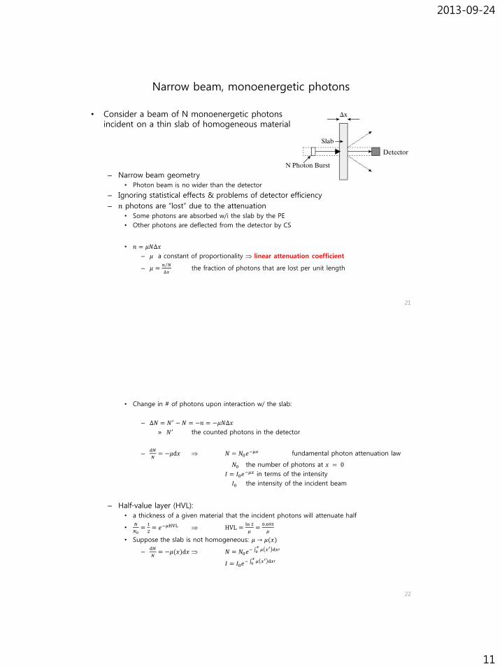
Narrow beam, monoenergetic photons
21
– Narrow beam geometry
• Photon beam is no wider than the detector
– Ignoring statistical effects & problems of detector efficiency
– 𝑛 photons are “lost” due to the attenuation
• Some photons are absorbed w/i the slab by the PE
• Other photons are deflected from the detector by CS
• 𝑛 = 𝜇𝑁∆𝑥
– 𝜇 a constant of proportionality linear attenuation coefficient
– 𝜇 = 𝑛 𝑁
∆𝑥the fraction of photons that are lost per unit length
• Consider a beam of N monoenergetic photons incident on a thin slab of homogeneous material
• Change in # of photons upon interaction w/ the slab:
– ∆𝑁 = 𝑁′ −𝑁 = −𝑛 = −𝜇𝑁∆𝑥
» 𝑁′ the counted photons in the detector
–d𝑁
𝑁= −𝜇d𝑥 𝑁 = 𝑁0𝑒
−𝜇𝑥 fundamental photon attenuation law
𝑁0 the number of photons at 𝑥 = 0
𝐼 = 𝐼0𝑒−𝜇𝑥 in terms of the intensity
𝐼0 the intensity of the incident beam
– Half-value layer (HVL):
• a thickness of a given material that the incident photons will attenuate half
•𝑁
𝑁0=
1
2= 𝑒−𝜇HVL HVL =
ln 2
𝜇=
0.693
𝜇
• Suppose the slab is not homogeneous: 𝜇 → 𝜇(𝑥)
–d𝑁
𝑁= −𝜇(𝑥)d𝑥 𝑁 = 𝑁0𝑒
− 0𝑥𝜇 𝑥′ d𝑥′
𝐼 = 𝐼0𝑒− 0
𝑥𝜇 𝑥′ d𝑥′
22

2013-09-24
12
Narrow beam, polyenergetic photons
• Replace 𝜇 by 𝜇(𝐸)!!!
– For an incident x-ray beam having spectrum 𝑆0(𝐸);
• 𝑆 𝐸 = 𝑆0(𝐸)𝑒−𝜇(𝐸)𝑥
– In addition, for a heterogeneous slab;
• 𝑆(𝑥; 𝐸) = 𝑆0(𝐸)𝑒− 0
𝑥𝜇 𝑥′;𝐸 d𝑥′
– For the overall intensity of the beam;
• 𝐼 = 0∞𝑆0 𝐸′ 𝐸′𝑒−𝜇 𝐸′ ∆𝑥d𝐸′
• 𝐼(𝑥) = 0∞𝑆0 𝐸′ 𝐸′𝑒− 0
𝑥𝜇 𝑥′;𝐸′ d𝑥′d𝐸′
23
24

2013-09-24
13
Broad beam case
• Broad beam geometry
– An additional possibility that photons from outside the detector’s line-of-sight geometry might get scattered toward the detector by Compton interactions
– More photons are generally detected than predicted by a monoenergetic, narrow beam analysis
– Even for the monoenergetci photon incidence, no longer monoenergetic in detected photons due to the CS which reduces photon energy beam softening
25
• In practice, detector collimation makes the “narrow beam geometry” assumption possible
– Narrow-beam geometry for imaging (due to collimation)
– Broad-beam geometry for dose (due to no collimation)
Radiation Dosimetry
• Exposure 𝑋
– # of ion pairs produced in a specific volume of air by EM radiation
• C/kg of air in SI unit
• R (roentgen) in classic unit
• 1 R = 2.58 10-4 C/kg
• 1 C/kg = 3876 R
• Ionization chamber
– Measuring the current produced btwn two plates held at a fixed potential due to radiation producing ions in the air btwn the two plates
26

2013-09-24
14
• Dose 𝐷 & kerma 𝐾
– Absorbed dose
• rad 1 rad = 100 ergs/g in classic unit
» Note: 1 eV = 1.6 10-12 ergs = 1.6 10-19 J
• gray (Gy) 1 Gy = 1 J/kg = 100 rads in SI unit
• In soft tissue, 1 R of exposure 1 rad of absorbed dose
– Kerma
• The amount of energy per unit mass imparted directly to the electrons in a given material
• Measured in unit of “Gy”
• Essentially equivalent to “dose” at diagnostic x-ray energies
• Air kerma 𝐾𝑎𝑖𝑟– Used in air for calibration purposes
27
• Linear energy transfer (LET)
– A measure of the energy transferred by radiation to the material through which it is passing per unit length
– Higher LET radiation producing greater adverse biological consequences
– Specific ionization (SI)
• # of ion pairs formed per unit length
– W-value
• Average amount of energy required to form one ion pair
• The f-factor
– A relationship between exposure in air & dose in air: 1 R = 0.87 rad
– To compute the dose to a material other than air;
• 𝐷 = 𝑓𝑋
– 𝑓 = 0.87( 𝜇 𝜌)𝑚𝑎𝑡𝑒𝑟𝑖𝑎𝑙
( 𝜇 𝜌)𝑎𝑖𝑟
– 𝜇 𝜌 = the mass attenuation coefficient
28

2013-09-24
15
• Dose equivalent
– To consider the fact that different types of radiation can actually have different effects on the body even when delivering the same dose
– 𝐻 = 𝐷𝑄
• 𝑄 quality factor
a property of the types of radiation used
𝑄 ≈ 1 for x rays, gamma rays, electrons & beta particles
𝑄 ≈ 10 for neutrons & protons
𝑄 ≈ 20 for alpha particles
• 𝐻 in rems (rem) for 𝐷 in rads in classic units
• 𝐻 in siverts (Sv) for 𝐷 in grays in SI units
29
• Effective dose
– For the purpose of relating “dose” of ionizing radiation to “risk”
– Comparing risks for different radiations & different target tissues
– An extension of the “dose equivalent” as the “dose equivalent” which would have been received if the whole body had been irradiated uniformly
– The sum of dose equivalents to different organs or body tissues weighted in a such fashion as to provide a value proportional to radiation-induced somatic & genetic risk even when the body is not uniformly irradiated
• 𝐷𝑒𝑓𝑓𝑒𝑐𝑡𝑖𝑣𝑒 = 𝑜𝑟𝑔𝑎𝑛𝑠 𝐻𝑗𝑤𝑗
– 𝐻𝑗 dose equivalent for organ j
– 𝑤𝑗 weighting factor for organ j
– 𝑜𝑟𝑔𝑎𝑛𝑠𝑤𝑗 = 1
– Average annual effective dose ~300 mrems
– Typical chest x-ray 10 mrems
– Fluoroscopy several rem
– Radiogenic carcinogenesis: cancer production
– Note that the physicians & patient together should make the decision that the medical benefits of the imaging procedure outweigh any potential risks
30