radiography safety fda 2012 recommendationsdentallearning.org/course/radiationsafety/radiography...
TRANSCRIPT
Academy - Dental Learning & OSHA Training is an ADA CERP Recognized provider.
ADA CERP is a service of the American Dental Association to assist dental professionals in identifying quality providers of continuing dental education. ADA CERP does not approve or endorse individual courses or instructors, nor does it imply acceptance of credit hours by boards of dentistry.
Concerns or complaints about a CE provider may be directed to the provider or to ADA CERP at www.ada.org/cerp.
Provider Disclosure: Academy of Dental Learning and OSHA Training nor its authors have no commercial interest in the content of this educational activity. Cancellation/Refund Policy: Any participant not 100% satisfied with this course may request a full refund by contacting: The Academy of Dental Learning and OSHA Training at 1.800.522.1307
Radiography Safety FDA 2012 Recommendations
Updated 2012
U.S. DEPARTMENT OF HEALTH AND HUMAN SERVICES
Public Health Service Food and Drug Administration
Revised: 2012
3 credit hours (3 CEs)
Edited by:
MaryLou Austin, RDH, MS
Dental Health Science Education
Publication Date: December 2012 Expiration Date: December 2016

COURSE AND EXAMINATION INSTRUCTIONS
1. Review the Objectives
Objectives provide an overview of the entire course and each chapter. Read the Course Description and focus on the Learning Objectives listed.
2. Study the Chapters in Order
Each chapter contains information essential to understanding subsequent sections. Keep your learning ‘programmed’ by reviewing the materials in order.
3. Complete the Post-Examination Online or by Fax
After studying the course take the test. You can access the test by returning to our website, http://www.dentallearning.org/ and clicking on the course title. Take the test then register (or login if you have already registered) and pay for the course. Your certificate will display for you to print for your records. Answer each question by clicking the button next to the answer you believe to be correct. Each question has only one correct answer. All questions must be answered before the test will be graded. There is no time limit on the test. You may also choose to print the exam and complete it manually. If you choose this option, please Fax (800-886-3009) or mail (ADL, POB 14585, Albany, NY 12212) your answer sheet to us. If you have not already paid for the course online you MUST include payment with your answer sheet. Answer sheets received without payment will not be processed. 4. Complete the Evaluation Form
Some courses require you to complete a course evaluation. If an evaluation appears after you pass the online test please answer the questions, enter the amount of time spent completing the entire course and CE exam, and submit the form.
5. CE Certificate
Your CE Certificate will be displayed on the screen for you to print.
THANK YOU FOR CHOOSING THE ACADEMY OF
DENTAL LEARNING AND OSHA TRAINING!
If you have any questions, please email us at [email protected]
or call our friendly customer service department at 1-800-522-1207
ANSWER SHEET
Radiography Safety FDA 2012 Recommendations
To quickly complete your course and instantly receive your certificate of completion return to our website, http://www.dentallearning.org/ . If you do not have access to a computer please complete and fax this form to 800-886-3009. Faxed answer sheets will be processed within two business days. Name: ___________________________________ Profession: ___________________ License State: ______ License #: _________________ Expiration Date: _____________ Address: ______________________________________________________________________ Address: ______________________________________________________________________ City: _________________________________ State: _______ Zip Code: __________ Phone: _______________________ Fax: ____________________ Email:________________________ If you have not already done so online, enter your payment information below. (To pay by check or money order make it payable to ADL and mail to POB 14585, Albany, NY 12212) Card Type: _______ Card Number: _______________________________________ Exp. Date: _______ Name as it appears on card: ____________________________
Please print the corresponding letter for each answer below:
1._____ 6. _____ 11. _____ 16. _____
2. _____ 7. _____ 12. _____ 17. _____
3. _____ 8. _____ 13. _____ 18. _____
4. _____ 9. _____ 14. _____ 19. _____
5. _____ 10. _____ 15. _____ 20. _____
Table of Contents COURSE LEARNING OBJECTIVES ................................................................... 1
BACKGROUND .................................................................................................... 1
INTRODUCTION .................................................................................................. 2
PATIENT SELECTION CRITERIA ....................................................................... 2
RECOMMENDATIONS FOR PRESCRIBING DENTAL RADIOGRAPHS ................................................. 5
EXPLANATION OF RECOMMENDATIONS FOR PRESCRIBING DENTAL RADIOGRAPHS ................. 8
LIMITING RADIATION EXPOSURE .................................................................. 16
RECEPTOR SELECTION .........................................................................................................................17
RECEPTOR HOLDERS ............................................................................................................................18
COLLIMATION ..........................................................................................................................................18
OPERATING POTENTIAL AND EXPOSURE TIME .................................................................................18
PATIENT SHIELDING AND POSITIONING ..............................................................................................19
OPERATOR PROTECTION ......................................................................................................................19
HAND-HELD X-RAY UNITS ......................................................................................................................20
FILM EXPOSURE AND PROCESSING ....................................................................................................21
QUALITY ASSURANCE............................................................................................................................21
TECHNIQUE CHARTS/PROTOCOLS ......................................................................................................22
RADIATION RISK COMMUNICATION .....................................................................................................22
TRAINING AND EDUCATION ..................................................................................................................24
CONCLUSION .................................................................................................... 24
REFERENCES ................................................................................................... 25
TEST ......................................................................... Error! Bookmark not defined.
1
COURSE LEARNING OBJECTIVES
Upon completion of this course, the dental professional will:
• Understand the FDA’s role in the regulation and promotion of radiation safety in
clinical dentistry.
• Know updated FDA recommendations for patient selection criteria in prescribing
dental radiographs.
• Understand basic radiography technique and rationale for clinical choices in types of
dental radiography.
• Review criteria for radiographic screening with regard to patient medical history,
clinical presentation, and risk assessment.
• Know clinical methods to limit radiation exposure for operator and patient.
• Know the recommendations from the FDA for particular clinical situations
BACKGROUND
The dental profession is committed to delivering the highest quality of care to
each of its individual patients and applying advancements in technology and
science to continually improve the oral health status of the U.S. population.
These guidelines were developed to serve as an adjunct to the dentist’s
professional judgment of how to best use diagnostic imaging for each patient.
Radiographs can help the dental practitioner evaluate and definitively diagnose
many oral diseases and conditions. However, the dentist must weigh the benefits
of taking dental radiographs against the risk of exposing a patient to x-rays, the
effects of which accumulate from multiple sources over time.
The dentist, knowing the patient’s health history and vulnerability to oral disease,
is in the best position to make this judgment in the interest of each patient. For
this reason, the guidelines are intended to serve as a resource for the practitioner
and are not intended as standards of care, requirements or regulations.
The guidelines are not substitutes for clinical examinations and health histories.
The dentist is advised to conduct a clinical examination, consider the patient’s
signs, symptoms and oral and medical histories, as well as consider the patient’s
vulnerability to environmental factors that may affect oral health. This diagnostic
and evaluative information may determine the type of imaging to be used or the
2
frequency of its use. Dentists should only order radiographs when they expect
that the additional diagnostic information will affect patient care.
Based on this premise, the guidelines can be used by the dentist to optimize
patient care, minimize radiation exposure and responsibly allocate health care
resources.
This course deals only with standard dental imaging techniques of intraoral and
common extra-oral examinations, excluding cone-beam computed tomography
(CBCT). At this time the indications for CBCT examinations are not well
developed—and still under consideration by the FDA and ADA.
INTRODUCTION
The guidelines titled, “The Selection of Patients for X-Ray Examination” were first
developed in 1987 by a panel of dental experts convened by the Center for
Devices and Radiological Health of the U.S. Food and Drug Administration (FDA).
The development of the guidelines at that time was spurred by concern about the
U.S. population’s total exposure to radiation from all sources. Thus, the
guidelines were developed to promote the appropriate use of x-rays. In 2002,
the American Dental Association, recognizing that dental technology and science
continually advance, recommended to the FDA that the guidelines be reviewed
for possible updating. The FDA welcomed organized dentistry’s interest in
maintaining the guidelines, and so the American Dental Association, in
collaboration with a number of dental specialty organizations and the FDA,
published updated guidelines in 2004. This report updates the 2004 guidelines
and includes recommendations for limiting exposure to radiation. These
materials issued by the FDA are in the public domain and dental professionals
are encouraged to use the information to make better clinical choices for patient
and operator safety.
PATIENT SELECTION CRITERIA
Radiographs and other imaging modalities are used to diagnose and monitor oral
diseases, as well as to monitor dento-facial development and the progress or
prognosis of therapy. Radiographic examinations can be performed using digital
imaging or conventional film. The available evidence suggests that either is a
suitable diagnostic method. Digital imaging may offer reduced radiation exposure
and the advantage of image analysis that may enhance sensitivity and reduce
error introduced by subjective analysis.
3
A study of 490 patients found that basing selection criteria on clinical evaluations
for asymptomatic patients, combined with selected periapical radiographs for
symptomatic patients, can result in a 43 percent reduction in the number of
radiographs taken without a clinically consequential increase in the rate of
undiagnosed disease. The development and progress of many oral conditions
are associated with a patient’s age, stage of dental development, and
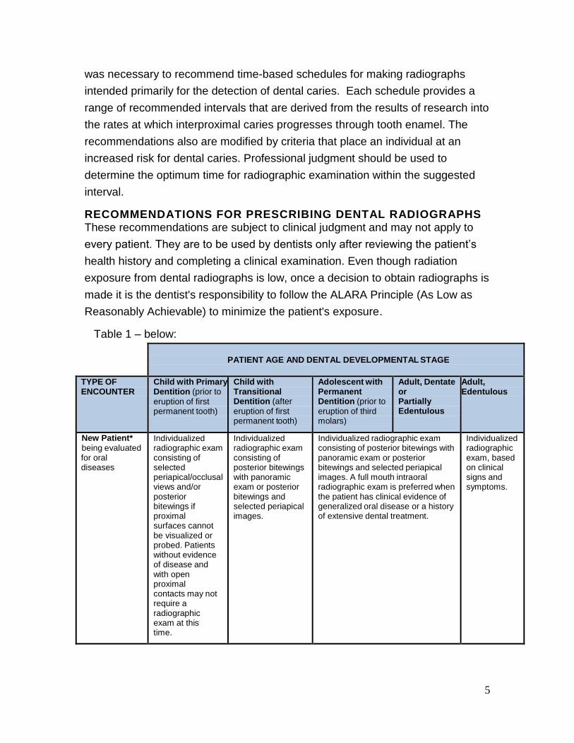
vulnerability to known risk factors. Therefore, the guidelines in Table 1 are
presented within a matrix of common clinical and patient factors, which may
determine the type (s) of radiographs that is commonly needed. The guidelines
assume that diagnostically adequate radiographs can be obtained. If not,
appropriate management techniques should be used after consideration of the
relative risks and benefits for the patient.
Along the horizontal axis of the matrix, patient age categories are described,
each with its usual dental developmental stage: child with primary dentition (prior
to eruption of the first permanent tooth); child with transitional dentition (after
eruption of the first permanent tooth); adolescent with permanent dentition (prior
to eruption of third molars); adult who is dentate or partially edentulous; and adult
who is edentulous.
Along the vertical axis, the type of encounter with the dental system is
categorized (as “New Patient” or “Recall Patient”) along with the clinical
circumstances and oral diseases that may be present during such an encounter.
The “New Patient” category refers to patients who are new to the dentist, and
thus are being evaluated by the dentist for oral disease and for the status of
dental development. Typically, such a patient receives a comprehensive
evaluation or, in some cases, a limited evaluation for a specific problem. The
“Recall Patient” categories describe patients who have had a recent
comprehensive evaluation by the dentist and, typically, have returned as a
patient of record for a periodic evaluation or for treatment. However, a “Recall
Patient” may also return for a limited evaluation of a specific problem, a detailed
and extensive evaluation for a specific problem(s), or a comprehensive
evaluation.
Both categories are marked with a single asterisk that corresponds to a footnote
that appears below the matrix; the footnote lists “Positive Historical Findings” and
“Positive Clinical Signs/Symptoms” for which radiographs may be indicated. The
4
lists are not intended to be all-inclusive, rather they offer the clinician further
guidance on clarifying his or her specific judgment on a case.
The clinical circumstances and oral diseases that are presented with the types of
encounters include: clinical caries or increased risk for caries; no clinical caries or
no increased risk for caries; periodontal disease or a history of periodontal
treatment; growth and development assessment; and other circumstances. A
few examples of “Other Circumstances” proposed are: existing implants, other
dental and craniofacial pathoses, endodontic/restorative needs and
remineralization of dental caries. These examples are not intended to be an
exhaustive list of circumstances for which radiographs or other imaging may be
appropriate.
The categories, “Clinical Caries or Increased Risk for Caries” and “No Clinical
Caries and No Increased Risk for Caries” are marked with a double asterisk that
corresponds to a footnote that appears below the matrix; the footnote contains
links to the ADA Caries Risk Assessment Forms (0 – 6 years of age and over 6
years of age). It should be noted that a patient’s risk status can change over time
and should be periodically reassessed.
The panel also has made the following recommendations that are applicable to
all categories:
1. Intraoral radiography is useful for the evaluation of dento-alveolar trauma. If
the area of interest extends beyond the dento-alveolar complex, extra-oral
imaging may be indicated.
2. Care should be taken to examine all radiographs for any evidence of caries,
bone loss from periodontal disease, developmental anomalies and occult
disease.
3. Radiographic screening for the purpose of detecting disease before clinical
examination should not be performed. A thorough clinical examination,
consideration of the patient history, review of any prior radiographs, caries
risk assessment and consideration of both the dental and the general health
needs of the patient should precede radiographic examination.
In the practice of dentistry, patients often seek care on a routine basis in part
because oral disease may develop in the absence of clinical symptoms. Since
attempts to identify specific criteria that will accurately predict a high probability of
finding interproximal carious lesions have not been successful for individuals, it
5
was necessary to recommend time-based schedules for making radiographs
intended primarily for the detection of dental caries. Each schedule provides a
range of recommended intervals that are derived from the results of research into
the rates at which interproximal caries progresses through tooth enamel. The
recommendations also are modified by criteria that place an individual at an
increased risk for dental caries. Professional judgment should be used to
determine the optimum time for radiographic examination within the suggested
interval.
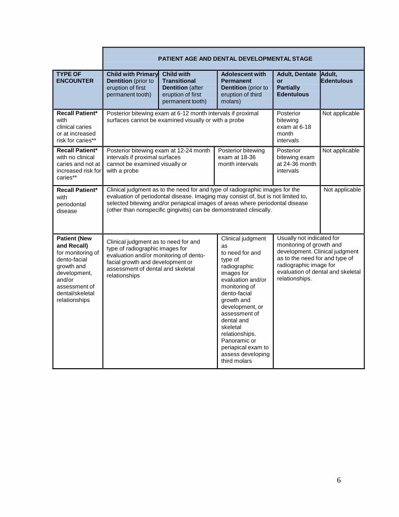
RECOMMENDATIONS FOR PRESCRIBING DENTAL RADIOGRAPHS These recommendations are subject to clinical judgment and may not apply to
every patient. They are to be used by dentists only after reviewing the patient’s
health history and completing a clinical examination. Even though radiation
exposure from dental radiographs is low, once a decision to obtain radiographs is
made it is the dentist's responsibility to follow the ALARA Principle (As Low as
Reasonably Achievable) to minimize the patient's exposure.
Table 1 – below:
PATIENT AGE AND DENTAL DEVELOPMENTAL STAGE
TYPE OF ENCOUNTER
Child with Primary Dentition (prior to
eruption of first permanent tooth)
Child with Transitional Dentition (after
eruption of first permanent tooth)
Adolescent with Permanent Dentition (prior to
eruption of third molars)
Adult, Dentate or Partially Edentulous
Adult, Edentulous
New Patient*
being evaluated for oral diseases
Individualized radiographic exam consisting of selected periapical/occlusal views and/or posterior bitewings if proximal surfaces cannot be visualized or probed. Patients without evidence of disease and with open proximal contacts may not require a radiographic exam at this time.
Individualized radiographic exam consisting of posterior bitewings with panoramic exam or posterior bitewings and selected periapical images.
Individualized radiographic exam consisting of posterior bitewings with panoramic exam or posterior bitewings and selected periapical images. A full mouth intraoral radiographic exam is preferred when the patient has clinical evidence of generalized oral disease or a history of extensive dental treatment.
Individualized radiographic exam, based on clinical signs and symptoms.
6
PATIENT AGE AND DENTAL DEVELOPMENTAL STAGE
TYPE OF ENCOUNTER
Child with Primary Dentition (prior to
eruption of first permanent tooth)
Child with Transitional Dentition (after
eruption of first permanent tooth)
Adolescent with Permanent Dentition (prior to
eruption of third molars)
Adult, Dentate or Partially Edentulous
Adult, Edentulous
Recall Patient*
with clinical caries or at increased risk for caries**
Posterior bitewing exam at 6-12 month intervals if proximal surfaces cannot be examined visually or with a probe
Posterior bitewing exam at 6-18 month intervals
Not applicable
Recall Patient*
with no clinical caries and not at increased risk for caries**
Posterior bitewing exam at 12-24 month intervals if proximal surfaces cannot be examined visually or with a probe
Posterior bitewing exam at 18-36 month intervals
Posterior bitewing exam at 24-36 month intervals
Not applicable
Recall Patient*
with
periodontal disease
Clinical judgment as to the need for and type of radiographic images for the evaluation of periodontal disease. Imaging may consist of, but is not limited to, selected bitewing and/or periapical images of areas where periodontal disease (other than nonspecific gingivitis) can be demonstrated clinically.
Not applicable
Patient (New
and Recall)
for monitoring of dento-facial growth and development, and/or assessment of dental/skeletal relationships
Clinical judgment as to need for and type of radiographic images for evaluation and/or monitoring of dento-facial growth and development or assessment of dental and skeletal relationships
Clinical judgment
as
to need for and type of radiographic images for evaluation and/or monitoring of dento-facial growth and development, or assessment of dental and skeletal relationships. Panoramic or periapical exam to assess developing third molars
Usually not indicated for monitoring of growth and development. Clinical judgment as to the need for and type of radiographic image for evaluation of dental and skeletal relationships.
7
PATIENT AGE AND DENTAL DEVELOPMENTAL STAGE
TYPE OF ENCOUNTER
Child with Primary Dentition (prior to
eruption of first permanent tooth)
Child with Transitional Dentition (after
eruption of first permanent tooth)
Adolescent with Permanent Dentition (prior to
eruption of third molars)
Adult, Dentate or Partially Edentulous
Adult, Edentulous
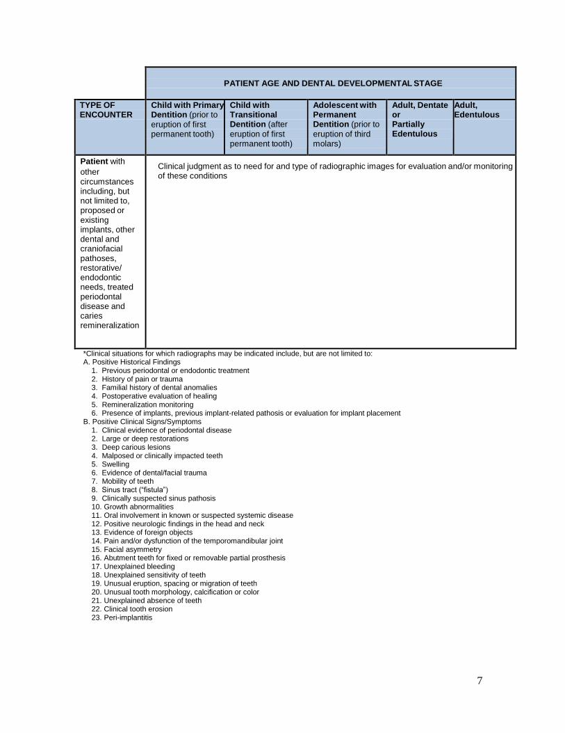
Patient with
other
circumstances including, but not limited to, proposed or existing implants, other dental and craniofacial pathoses, restorative/ endodontic needs, treated periodontal disease and caries remineralization
Clinical judgment as to need for and type of radiographic images for evaluation and/or monitoring of these conditions
*Clinical situations for which radiographs may be indicated include, but are not limited to: A. Positive Historical Findings
1. Previous periodontal or endodontic treatment 2. History of pain or trauma 3. Familial history of dental anomalies 4. Postoperative evaluation of healing 5. Remineralization monitoring 6. Presence of implants, previous implant-related pathosis or evaluation for implant placement
B. Positive Clinical Signs/Symptoms 1. Clinical evidence of periodontal disease 2. Large or deep restorations 3. Deep carious lesions 4. Malposed or clinically impacted teeth 5. Swelling 6. Evidence of dental/facial trauma 7. Mobility of teeth 8. Sinus tract (“fistula”) 9. Clinically suspected sinus pathosis 10. Growth abnormalities 11. Oral involvement in known or suspected systemic disease 12. Positive neurologic findings in the head and neck 13. Evidence of foreign objects 14. Pain and/or dysfunction of the temporomandibular joint 15. Facial asymmetry 16. Abutment teeth for fixed or removable partial prosthesis 17. Unexplained bleeding 18. Unexplained sensitivity of teeth 19. Unusual eruption, spacing or migration of teeth 20. Unusual tooth morphology, calcification or color 21. Unexplained absence of teeth 22. Clinical tooth erosion 23. Peri-implantitis
8
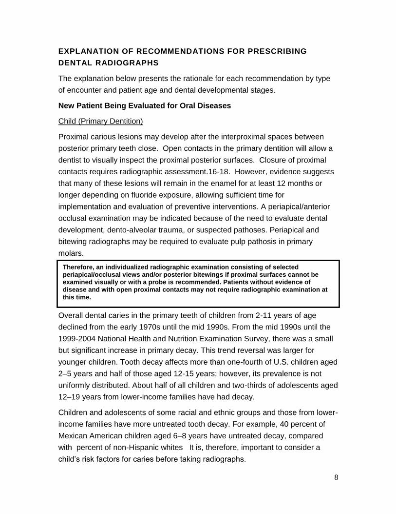
EXPLANATION OF RECOMMENDATIONS FOR PRESCRIBING
DENTAL RADIOGRAPHS
The explanation below presents the rationale for each recommendation by type
of encounter and patient age and dental developmental stages.
New Patient Being Evaluated for Oral Diseases
Child (Primary Dentition)
Proximal carious lesions may develop after the interproximal spaces between
posterior primary teeth close. Open contacts in the primary dentition will allow a
dentist to visually inspect the proximal posterior surfaces. Closure of proximal
contacts requires radiographic assessment.16-18. However, evidence suggests
that many of these lesions will remain in the enamel for at least 12 months or
longer depending on fluoride exposure, allowing sufficient time for
implementation and evaluation of preventive interventions. A periapical/anterior
occlusal examination may be indicated because of the need to evaluate dental
development, dento-alveolar trauma, or suspected pathoses. Periapical and
bitewing radiographs may be required to evaluate pulp pathosis in primary
molars.
Child (Transitional Dentition)
Overall dental caries in the primary teeth of children from 2-11 years of age
declined from the early 1970s until the mid 1990s. From the mid 1990s until the
1999-2004 National Health and Nutrition Examination Survey, there was a small
but significant increase in primary decay. This trend reversal was larger for
younger children. Tooth decay affects more than one-fourth of U.S. children aged
2–5 years and half of those aged 12-15 years; however, its prevalence is not
uniformly distributed. About half of all children and two-thirds of adolescents aged
12–19 years from lower-income families have had decay.
Children and adolescents of some racial and ethnic groups and those from lower-
income families have more untreated tooth decay. For example, 40 percent of
Mexican American children aged 6–8 years have untreated decay, compared
with percent of non-Hispanic whites It is, therefore, important to consider a
child’s risk factors for caries before taking radiographs.
Therefore, an individualized radiographic examination consisting of selected periapical/occlusal views and/or posterior bitewings if proximal surfaces cannot be examined visually or with a probe is recommended. Patients without evidence of disease and with open proximal contacts may not require radiographic examination at
this time.
9
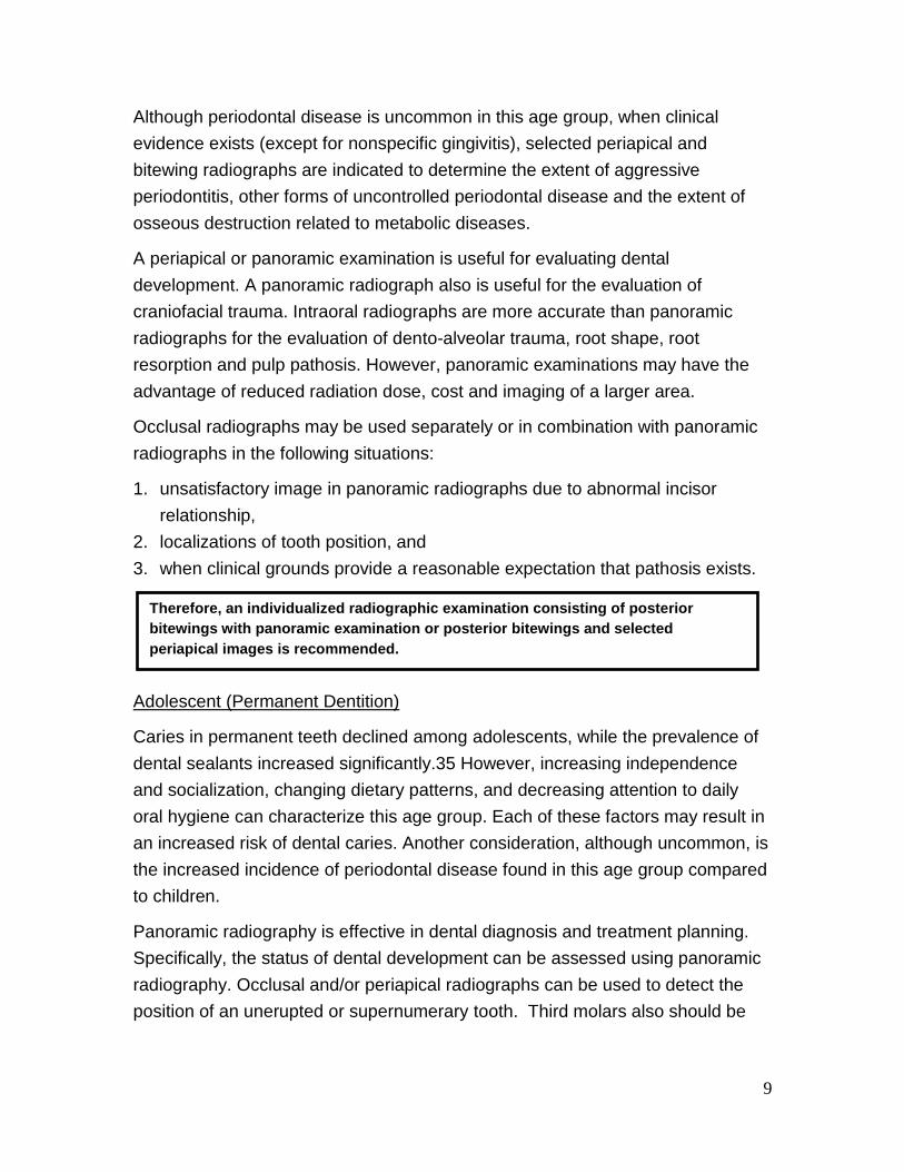
Although periodontal disease is uncommon in this age group, when clinical
evidence exists (except for nonspecific gingivitis), selected periapical and
bitewing radiographs are indicated to determine the extent of aggressive
periodontitis, other forms of uncontrolled periodontal disease and the extent of
osseous destruction related to metabolic diseases.
A periapical or panoramic examination is useful for evaluating dental
development. A panoramic radiograph also is useful for the evaluation of
craniofacial trauma. Intraoral radiographs are more accurate than panoramic
radiographs for the evaluation of dento-alveolar trauma, root shape, root
resorption and pulp pathosis. However, panoramic examinations may have the
advantage of reduced radiation dose, cost and imaging of a larger area.
Occlusal radiographs may be used separately or in combination with panoramic
radiographs in the following situations:
1. unsatisfactory image in panoramic radiographs due to abnormal incisor
relationship,
2. localizations of tooth position, and
3. when clinical grounds provide a reasonable expectation that pathosis exists.
Adolescent (Permanent Dentition)
Caries in permanent teeth declined among adolescents, while the prevalence of
dental sealants increased significantly.35 However, increasing independence
and socialization, changing dietary patterns, and decreasing attention to daily
oral hygiene can characterize this age group. Each of these factors may result in
an increased risk of dental caries. Another consideration, although uncommon, is
the increased incidence of periodontal disease found in this age group compared
to children.
Panoramic radiography is effective in dental diagnosis and treatment planning.
Specifically, the status of dental development can be assessed using panoramic
radiography. Occlusal and/or periapical radiographs can be used to detect the
position of an unerupted or supernumerary tooth. Third molars also should be
Therefore, an individualized radiographic examination consisting of posterior
bitewings with panoramic examination or posterior bitewings and selected
periapical images is recommended.
10
evaluated in this age group for their presence, position, and stage of
development.
Adult (Dentate or Partially Edentulous)
The overall dental caries experience of the adult population has declined from
the early 1970s until the most recent (1999-2004) National Health and Nutrition
Examination Survey. However, risk for dental caries exists on a continuum and
changes over time as risk factors change. Therefore, it is important to evaluate
proximal surfaces in the new adult patient for carious lesions. In addition, it is
important to examine patients for recurrent dental caries.
The incidence of root surface caries increases with age. Although bitewing
radiographs can assist in detecting root surface caries in proximal areas, the
usual method of detecting root surface caries is by clinical examination.
The incidence of periodontal disease increases with age. Although new adult
patients may not have symptoms of active periodontal disease, it is important to
evaluate previous experience with periodontal disease and/or treatment.
Therefore, a high percentage of adults may require selected intraoral radiographs
to determine the current status of the disease.
Taking posterior bitewing radiographs of new adult patients was found to reduce
the number of radiological findings and the diagnostic yield of panoramic
radiography. In addition, the following clinical indicators for panoramic
radiography were identified as the best predictors for useful diagnostic yield:
suspicion of teeth with periapical pathologic conditions, presence of partially
erupted teeth, caries lesions, swelling, and suspected unerupted teeth.
Therefore, an individualized radiographic examination consisting of posterior bitewings with panoramic examination or posterior bitewings and selected periapical images is recommended. A full mouth intraoral radiographic examination is preferred when the patient has clinical evidence of generalized oral disease or a history of extensive dental
treatment.
Therefore, an individualized radiographic examination, consisting of posterior bitewings with selected periapical images or panoramic examination when indicated is recommended. A full mouth intraoral radiographic examination is preferred when the patient has clinical evidence of generalized oral disease or a history of extensive dental
treatment.
11
Adult (Edentulous)
The clinical and radiographic examinations of edentulous patients generally
occur during an assessment of the need for prostheses. The most common
pathological conditions detected are impacted teeth and retained roots with and
without associated disease. Other less common conditions also may be
detected: bony spicules along the alveolar ridge, residual cysts or infections,
developmental abnormalities of the jaws, intraosseous tumors, and systemic
conditions affecting bone metabolism.
The original recommendations for this group called for a full-mouth intraoral
radiographic examination or a panoramic examination for the new, edentulous
adult patient. Firstly, this recommendation was made because examinations of
edentulous patients generally occur during an assessment of the need for
prostheses. Secondly, the original recommendation considered edentulous
patients to be at increased risk for oral disease.
Studies have found that from 30 to 50 percent of edentulous patients exhibited
abnormalities in panoramic radiographs. In addition, the radiographic
examination revealed anatomic considerations that could influence prosthetic
treatment, such as the location of the mandibular canal, the position of the
mental foramen and maxillary sinus, and relative thickness of the soft tissue
covering the edentulous ridge. However, in studies that considered treatment
outcomes, there was little evidence to support screening radiography for new
edentulous patients. For example, one study reported that less than 4 percent of
such findings resulted in treatment modification before denture fabrication, and
another showed no difference in post- denture delivery complaints in patients
who did not receive screening pretreatment radiographs.
This panel concluded that prescription of radiographs is appropriate as part of the
initial assessment of edentulous areas for possible prosthetic treatment. A full
mouth series of periapical radiographs or a combination of panoramic, occlusal
or other extra-oral radiographs may be used to achieve diagnostic and
therapeutic goals. Particularly with the option of dental implant therapy for
edentulous patients, radiographs can be an important aid in diagnosis, prognosis,
and the determination of treatment complexity.
Therefore, an individualized radiographic examination, based on clinical signs,
symptoms, and treatment plan is recommended.
12
Recall Patient with Clinical Caries or Increased Risk for Caries
Child (Primary and Transitional Dentition) and Adolescent (Permanent Dentition)
Clinically detectable dental caries may suggest the presence of proximal carious
lesions that can only be detected with a radiographic examination. In addition,
patients who are at increased risk for developing dental caries because of such
factors as poor oral hygiene, high frequency of exposure to sucrose-containing
foods, and deficient fluoride intake (see caries risk assessment forms, 0 – 6
years of age and over 6 years of age) are more likely to have proximal carious
lesions.
The bitewing examination is the most efficient method for detecting proximal
lesions.
The frequency of radiographic recall should be determined on the basis of caries
risk assessment. It should be noted that a patient’s caries risk status may change
over time and that an individual’s radiographic recall interval may need to be
changed accordingly.
Adult (Dentate and Partially Edentulous)
Adults who exhibit clinical dental caries or who have other increased risk factors
should be monitored carefully for any new or recurrent lesions that are detectable
only by radiographic examination. The frequency of radiographic recall should be
determined on the basis of caries risk assessment. It should be noted that a
patient’s risk status can change over time and that an individual’s radiographic
recall interval may need to be changed accordingly.
Recall Patient (Edentulous Adult)
A study that assessed radiographs of edentulous recall patients showed that
previously detected incidental findings did not progress and that no intervention
was indicated. The data suggest that patients who receive continuous dental
care do not exhibit new findings that require treatment.
Therefore, a posterior bitewing examination is recommended at 6 to 12 month intervals if
proximal surfaces cannot be examined visually or with a probe.
Therefore, a posterior bitewing examination is recommended at 6 to 18 month intervals.
13
An examination for occult disease in this group cannot be justified on the basis of
prevalence, morbidity, mortality, radiation dose, and cost.
Recall Patient with No Clinical Caries and No Increased Risk for Caries
Child (Primary and Transitional Dentition)
Despite the general decline in dental caries activity, recent data show that
subgroups of children have a higher caries experience than the overall
population. The identification of patients in these subgroups may be difficult on
an individual basis. For children who present for recall examination without
evidence of clinical caries and who are not considered at increased risk for the
development of caries, it remains important to evaluate proximal surfaces by
radiographic examination. In primary teeth the caries process can take
approximately one year to progress through the outer half of the enamel and
about another year through the inner half. Considering this rate of progression of
carious lesions through primary teeth, a time- based interval of radiographic
examinations from one to two years for this group appears appropriate. The
prevalence of carious lesions has been shown to increase during the stage of
transitional dentition. Children under routine professional care would be expected
to be at a lower risk for caries. Nevertheless, newly erupted teeth are at risk for
the development of dental caries.
Adolescent (Permanent Dentition)
Adolescents with permanent dentition, who are free of clinical dental caries and
factors that would place them at increased risk for developing dental caries,
should be monitored carefully for development of proximal carious lesions, which
may only be detected by radiographic examination. The caries process, on
average, takes more than three years to progress through the enamel. However,
evidence suggests that the enamel of permanent teeth undergoes post-eruptive
maturation and that young permanent teeth are susceptible to faster progression
of carious lesions.
Therefore, no radiographic examination is recommended without evidence of disease.
Therefore, a radiographic examination consisting of posterior bitewings is recommended at intervals of 12 to 24 months if proximal surfaces cannot be examined visually or with a
probe.
14
Adult (Dentate and Partially Edentulous)
Adult dentate patients, who receive regularly scheduled professional care and
are free of signs and symptoms of oral disease, are at a low risk for dental caries.
Nevertheless, consideration should be given to the fact that caries risk can vary
over time as risk factors change.
Advancing age and changes in diet, medical history and periodontal status may
increase the risk for dental caries.
Recall Patient with Periodontal Disease
Child (Primary and Transitional Dentition), Adolescent (Permanent Dentition),
and Adult (Dentate and Partially Edentulous)
The decision to obtain radiographs for patients who have clinical evidence or a
history of periodontal disease/treatment should be determined on the basis of the
anticipation that important diagnostic and prognostic information will result.
Structures or conditions to be assessed should include the level of supporting
alveolar bone, condition of the interproximal bony crest, length and shape of
roots, bone loss in furcations, and calculus deposits. The frequency and type of
radiographic examinations for these patients should be determined on the basis
of a clinical examination of the periodontium and documented signs and
symptoms of periodontal disease. The procedure for prescribing radiographs for
the follow-up/recall periodontal patient would be to use selected intraoral
radiographs to verify clinical findings on a patient-by-patient basis.
Therefore, a radiographic examination consisting of posterior bitewings is
recommended at intervals of 18 to 36 months.
Therefore, a radiographic examination consisting of posterior bitewings is
recommended at intervals of 24 to 36 months.
Therefore, it is recommended that clinical judgment be used in determining the need
for, and type of radiographic images necessary for, evaluation of periodontal disease.
Imaging may consist of, but is not limited to, selected bitewing and/or periapical
images of areas where periodontal disease (other than nonspecific gingivitis) can be
identified clinically.
15
Patient (New and Recall) for Monitoring of Dento-facial Growth and
Development, and/or Assessment of Dental/Skeletal Relationships
Child (Primary and Transitional Dentition)
For children with primary dentition, before the eruption of the first permanent
tooth, radiographic examination to assess growth and development in the
absence of clinical signs or symptoms is unlikely to yield productive information.
Any abnormality of growth and development suggested by clinical findings should
be evaluated radiographically on an individual basis. After eruption of the first
permanent tooth, the child may have a radiographic examination to assess
growth and development. This examination need not be repeated unless dictated
by clinical signs or symptoms. Cephalometric radiographs may be useful for
assessing growth, and/or dental and skeletal relationships.
Adolescent (Permanent Dentition)
During adolescence there is often a need to assess the growth status and/or the
dental and skeletal relationships of patients in order to diagnose and treat their
malocclusion. Appropriate radiographic assessment of the malocclusion should
be determined on an individual basis.
An additional concern relating to growth and development for patients in this age
group is to determine the presence, position and development of third molars.
This determination can best be made by the use of selected periapical images or
a panoramic examination, once the patient is in late adolescence (16 to 19 years
of age).
Therefore, it is recommended that clinical judgment be used in determining the need for, and type of radiographic images necessary for, evaluation and/or monitoring of dento-facial growth and development, or assessment of dental and skeletal
relationships.
Therefore, it is recommended that clinical judgment be used in determining the need for, and type of radiographic images necessary for, evaluation and/or monitoring of dento-facial growth and development, or assessment of dental and skeletal relationships. Panoramic or periapical examination may be used to assess
developing third molars.
16
Adult (Dentate, Partially Edentulous and Edentulous)
In the absence of any clinical signs or symptoms suggesting abnormalities of
growth and development in adults, no radiographic examinations are indicated
for this purpose.
Patients with Other Circumstances
(Including, but not limited to, proposed or existing implants, other dental and
craniofacial pathoses, restorative/endodontic needs, treated periodontal disease
and caries remineralization)
All Patient Categories
The use of imaging, as a diagnostic and evaluative tool, has progressed beyond
the longstanding need to diagnose caries and evaluate the status of periodontal
disease. The expanded technology in imaging is now used to diagnose other
orofacial clinical conditions and evaluate treatment options. A few examples of
other clinical circumstances are the use of imaging for dental implant treatment
planning, placement, or evaluation; the monitoring of dental caries and
remineralization; the assessment of restorative and endodontic needs; and the
diagnosis of soft and hard tissue pathoses.
LIMITING RADIATION EXPOSURE
Dental radiographs account for approximately 2.5 percent of the effective dose
received from medical radiographs and fluoroscopies. Even though radiation
exposure from dental radiographs is low, once a decision to obtain radiographs is
made it is the dentist's responsibility to follow the ALARA Principle (As Low as
Reasonably Achievable) to minimize the patient's exposure. Examples of good
radiologic practice include
Therefore, in the absence of clinical signs and symptoms, no radiographic
examination is recommended.
Therefore it is recommended that clinical judgment be used in determining the need
for, and type of radiographic images necessary for, evaluation and/or monitoring in
these circumstances.
17
• use of the fastest image receptor compatible with the diagnostic task (F-speed film or
digital);
• collimation of the beam to the size of the receptor whenever feasible;
• proper film exposure and processing techniques;
• use of protective aprons and thyroid collars, when appropriate; and
• limiting the number of images obtained to the minimum necessary to obtain essential
diagnostic information.
RECEPTOR SELECTION The American National Standards Institute and the International Organization for
Standardization have established standards for film speed. Film speeds available
for dental radiography are D-speed, E-speed and F-speed, with D-speed being
the slowest and F-speed the fastest. According to the U.S. Food and Drug
Administration, switching from D to E speed can produce a 30 to 40 percent
reduction in radiation exposure. The use of F-speed film can reduce exposure 20
to 50 percent compared to use of E-speed film, without compromising image
quality.
Exposure of extra-oral films such as panoramic radiographs requires intensifying
screens to minimize radiation exposure to patients. The intensifying screen
consists of layers of phosphor crystals that fluoresce when exposed to radiation.
In addition to the radiation incident on the film, the film is exposed primarily to the
light emitted from the intensifying screen. Previous generations of intensifying
screens were composed of phosphors such as calcium tungstate. However, rare-
earth intensifying screens are recommended because they reduce a patient’s
radiation exposure by 50 percent compared with calcium tungstate-intensifying
screens. Rare-earth film systems, combined with a high-speed film of 400 or
greater, can be used for panoramic radiographs. Older panoramic equipment can
be retrofitted to reduce the radiation exposure to accommodate the use of rare-
earth, high-speed systems.
Digital imaging provides an opportunity to further reduce the radiation dose by 40
to 60 percent. In digital radiography, there are three types of receptors that take
the place of conventional film: charge-coupled device (CCD), complementary-
metal-oxide-semiconductor (CMOS), and photo-stimulable phosphor (PSP)
plates. Systems that use CCD and CMOS- based, solid-state detectors are
called “direct.” When these sensors receive energy from the x- ray beam, the
CCD or CMOS chip sends a signal to the computer and an image appears on the
monitor within seconds. Systems that use PSP plates are called “indirect.” When
18
these plates are irradiated, a latent image is stored on them. The plate is then
scanned and the scanner transmits the image to the computer.
RECEPTOR HOLDERS Holders that align the receptor precisely with the collimated beam are
recommended for periapical and bitewing radiographs. Heat-sterilizable or
disposable intraoral radiograph receptor-holding devices are recommended for
optimal infection control. Dental professionals should not hold the receptor holder
during exposure. Under extraordinary circumstances in which members of the
patient’s family (or other caregiver) must provide restraint or hold a receptor
holder in place during exposure, such a person should wear appropriate
shielding.
COLLIMATION Collimation limits the amount of radiation, both primary and scattered, to which
the patient is exposed. An added benefit of rectangular collimation is an
improvement in contrast as a result of a reduction in fogging caused by
secondary and scattered radiation. The x-ray beam should not exceed the
minimum coverage necessary, and each dimension of the beam should be
collimated so that the beam does not exceed the receptor by more than 2
percent of the source-to-image receptor distance. Since a rectangular collimator
decreases the radiation dose by up to fivefold as compared with a circular one,
radiographic equipment should provide rectangular collimation for exposure of
periapical and bitewing radiographs. Use of a receptor-holding device minimizes
the risk of cone-cutting (non-exposure of part of the image receptor due to
malalignment of the x-ray beam). The position-indicating device should be open
ended and have a metallic lining to restrict the primary beam and reduce the
tissue volume exposed to radiation. Use of long source-to-skin distances of 40
cm, rather than short distances of 20 cm, decreases exposure by 10 to 25
percent. Distances between 20 and 40 cm are appropriate, but the longer
distances are optimal.
OPERATING POTENTIAL AND EXPOSURE TIME The operating potential of dental x-ray units affects the radiation dose and
backscatter radiation. Lower voltages produce higher-contrast images and higher
entrance skin doses, and lower deep-tissue doses and levels of backscatter
radiation. However, higher voltages produce lower contrast images that enable
better separation of objects with differing densities. Thus, the diagnostic
purposes of the radiograph should be used to determine the selection of kilovolt
19
setting. A setting above 90 kV(p) will increase the patient dose and should not be
used. The optimal operating potential of dental x-ray units is between 60 and 70
kVp.
Filmless technology is much more forgiving to overexposure often resulting in
unnecessary radiation exposure. Facilities should strive to set the x-ray unit
exposure timer to the lowest setting providing an image of diagnostic quality. If
available, the operator should always confirm that the dose delivered falls within
the manufacturer’s exposure index. Imaging plates should be evaluated at least
monthly and cleaned as necessary.
PATIENT SHIELDING AND POSITIONING The amount of scattered radiation striking the patient’s abdomen during a
properly conducted radiographic examination is negligible The thyroid gland is
more susceptible to radiation exposure during dental radiographic exams given
its anatomic position, particularly in children. Protective thyroid collars and
collimation substantially reduce radiation exposure to the thyroid during dental
radiographic procedures. Because every precaution should be taken to minimize
radiation exposure, protective thyroid collars should be used whenever possible.
If all the recommendations for limiting radiation exposure are put into practice,
the gonadal radiation dose will not be significantly affected by use of abdominal
shielding. Therefore, use of abdominal shielding may not be necessary.
Protective aprons and thyroid shields should be hung or laid flat and never folded,
and manufacturer’s instructions should be followed. All protective shields should
be evaluated for damage (e.g. tears, folds, and cracks) monthly using visual and
manual inspection.
Proper education and training in patient positioning is necessary to ensure that
panoramic radiographs are of diagnostic quality.
OPERATOR PROTECTION Although dental professionals receive less exposure to ionizing radiation than do
other occupationally exposed health care workers, operator protection measures
are essential to minimize exposure. Operator protection measures include
education, the implementation of a radiation protection program, occupational
radiation exposure limits, recommendations for personal dosimeters and the use
of barrier shielding. The maximum permissible annual dose of ionizing radiation
for health care workers is 50 millisieverts (mSv) and the maximum permissible
lifetime dose is 10 mSv multiplied by a person’s age in years. Personal
20
dosimeters should be used by workers who may receive an annual dose greater
than 1 mSv to monitor their exposure levels. Pregnant dental personnel operating
x-ray equipment should use personal dosimeters, regardless of anticipated
exposure levels.
Operators of radiographic equipment should use barrier protection when possible,
and barriers should ideally contain a leaded glass window to enable the operator
to view the patient during exposure. When shielding is not possible, the operator
should stand at least two meters from the tube head and out of the path of the
primary beam. The National Council on Radiation Protection & Measurements
report “Radiation Protection in Dentistry” offers detailed information on shielding
and office design. State radiation control agencies can help assess whether
barriers meet minimum standards.
HAND-HELD X-RAY UNITS Hand-held, battery-powered x-ray systems are available for intra-oral
radiographic imaging.
The hand-held exposure device is activated by a trigger on the handle of the
device. However, dosimetry studies indicate that these hand-held devices
present no greater radiation risk than standard dental radiographic units to the
patient or the operator. No additional radiation protection precautions are needed
when the device is used according to the manufacturer’s instructions. These
include: 1. holding the device at mid-torso height, 2. orienting the shielding ring
properly with respect to the operator, and 3. keeping the cone as close to the
patient’s face as practical. If the hand-held device is operated without the ring
shield in place, it is recommended that the operator wear a lead apron.
All operators of hand-held units should be instructed on their proper storage.
Due to the portable nature of these devices, they should be secured properly
when not in use to prevent accidental damage, theft, or operation by an
unauthorized user. Hand-held units should be stored in locked cabinets, locked
storage rooms, or locked work areas when not under the direct supervision of an
individual authorized to use them. Units with user-removable batteries should be
stored with the batteries removed. Records listing the names of approved
individuals who are granted access and use privileges should be prepared and
kept current.
21
FILM EXPOSURE AND PROCESSING All film should be processed following the film and processer manufacturer
recommendations. Once this is achieved, the x-ray operator can adjust the tube
current and time and establish a technique that will provide consistent dental
radiographs of diagnostic quality. Poor processing technique, including sight-
developing, most often results in underdeveloped films, forcing the x-ray operator
to increase the dose to compensate, resulting in patient and personnel being
exposed to unnecessary radiation.
A safelight does not provide completely safe exposure for an indefinite period of
time. Extra-oral film is much more sensitive to fogging. The length of time for
which a film can be exposed to the safelight should be determined for the specific
safelight/film combination in use.
QUALITY ASSURANCE Quality assurance protocols for the x-ray unit, imaging receptor, film processing,
dark room, and patient shielding should be developed and implemented for each
dental health care setting. All quality assurance procedures, including date,
procedure, results, and corrective action, should be logged for documentation
purposes. A qualified expert should survey all x- ray units on their placement and
should resurvey the equipment every four years or after any changes that may
affect the radiation exposure of the operator and others. Surveys typically are
performed by state agencies, and individual state regulations should be
consulted regarding specific survey intervals. The film processor should be
evaluated at its initial installation and on a monthly basis afterward. The
processing chemistry should be evaluated daily, and each type of film should be
evaluated monthly or when a new box or batch of film is opened. Abdominal
shielding and thyroid collars should be inspected visually for creases or clumping
that may indicate voids in their integrity on a monthly basis. Damaged abdominal
shielding and collars should be replaced. Table 2 (below) lists specific methods
of quality assurance procedures, covering not only inspection of the x-ray unit
itself but also of the film processor, the image receptor devices, and the
darkroom and abdominal shielding and collars.
It is imperative that the operator’s manual for all imaging acquisition hardware is
readily available to the user, and that the equipment is operated and maintained
following the manufacturer’s instructions, including any appropriate adjustments
for optimizing dose and image quality.
22
TECHNIQUE CHARTS/PROTOCOLS Size-based technique charts/protocols with suggested parameter settings are
important for ensuring that radiation exposure is optimized for all patients.
Technique charts should be used for all systems with adjustable settings, such
as tube potential, tube current, and time or pulses. The purpose of using the
charts is to control the amount of radiation to the patient and receptor. Technique
charts are tables that indicate appropriate settings on the x-ray unit for a specific
anatomical area and will ensure the least amount of radiation exposure to
produce a consistently good-quality radiograph.
Technique charts for intraoral and extra-oral radiography should list the type of
exam, the patient size (small, medium, large) for adults and a pediatric setting.
The speed of film used, or use of a digital receptor, should also be listed on the
technique chart. The chart should be posted near the control panel where the
technique is adjusted for each x-ray unit. A technique chart that is regularly
updated should be developed for each x-ray unit. The charts will also need to be
updated when a different film or sensor, new unit, or new screens are used.
RADIATION RISK COMMUNICATION Dentists should be prepared to discuss with their patients the benefits and risks
of the x-ray exam. To help answer patient and parent questions about dental
radiology radiation safety, the American Academy of Oral and Maxillofacial
Radiology and the Alliance for Radiation Safety in Pediatric Imaging partnered to
create a brochure targeted at parents and patients.
Table 2
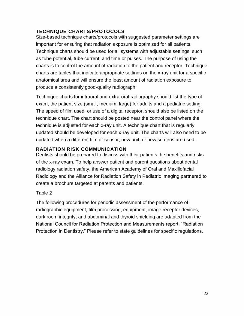
The following procedures for periodic assessment of the performance of
radiographic equipment, film processing, equipment, image receptor devices,
dark room integrity, and abdominal and thyroid shielding are adapted from the
National Council for Radiation Protection and Measurements report, “Radiation
Protection in Dentistry.” Please refer to state guidelines for specific regulations.
23
Quality Assurance Procedures for Assessment of Radiographic Equipment
Equipment
Frequency
Method
X-ray Machine On installation At regular intervals as recommended by state regulations Whenever there are any changes in installation workload or operating conditions
Inspection by qualified expert (as specified by government regulations and manufacturers recommendations).
Film Processor On installation Daily
Method 1: Sensitometry and Densitometry
A sensitometer is used to expose a film, followed by standard processing of the film. The processed film will have a defined pattern of optical densities. The densities are measured with a densitometer. The densitometer measurements are compared to the densities of films exposed and processed under ideal conditions. A change in densitometer values indicates a problem with either the development time, temperature or the developer solutions. Advantages Accuracy Speed Disadvantage Expense of additional equipment Method 2: Reference Film
A film exposed and processed under ideal conditions is attached to the corner of a view box as a reference film. Subsequent films are compared with the reference film. Advantage Cost effectiveness Disadvantage
Less sensitive
Image Receptor Devices Intensifying Screen and
Monthly With each new batch of film Every six months
Method 1: Sensitometry and Densitometry
(as described above) Method 2: Reference Image (as
described above) Visual inspection of cassette integrity Examination of intensifying screen for scratches
Extra-oral Cassettes Development of an unexposed film that has been in the cassette exposed to normal lighting for one hour or more
24
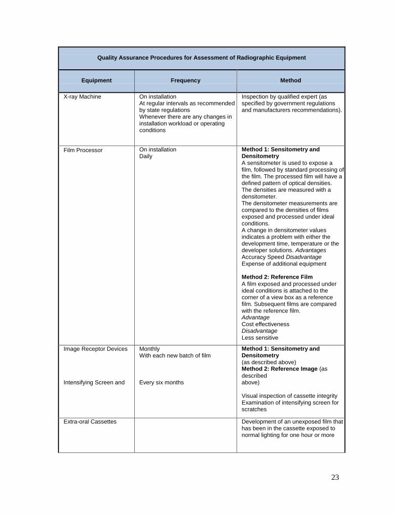
Quality Assurance Procedures for Assessment of Radiographic Equipment
Equipment
Frequency
Method
Darkroom Integrity On installation Monthly After a change in the lighting filter or lamp
While in a darkroom with the safelight on, place metal object (such as a coin) on unwrapped film for a period that is equivalent to the time required for a typical darkroom procedure Develop film Detection of the object indicates a problem with the safelight or light leaks in the darkroom
Abdominal and Thyroid Shielding
Monthly (visual and manual inspection)
All protective shields should be evaluated for damage (e.g., tears, folds, and cracks) monthly using visual and manual inspection. If a defect in the attenuating material is suspected, radiographic or fluoroscopic inspection may be performed as an alternative to immediately removing the item from service. Consideration should be given to minimizing the radiation exposure of inspectors by minimizing unnecessary fluoroscopy.
TRAINING AND EDUCATION Where permitted by law, auxiliary dental personnel can perform intraoral and
extra-oral imaging.103 Personnel certified to take dental radiographs should
receive appropriate education. Practitioners should remain informed about safety
updates and the availability of new equipment, supplies and techniques that
could further improve the diagnostic quality of radiographs and decrease
radiation exposure. Free training materials are available for limiting radiation
exposure in dental imaging through the International Atomic Energy Agency.
CONCLUSION
Dentists should conduct a clinical examination, consider the patient’s oral and
medical histories, as well as consider the patient’s vulnerability to environmental
factors that may affect oral health before conducting a radiographic examination.
This information should guide the dentist in the determination of the type of
imaging to be used, the frequency of its use, and the number of images to obtain.
Radiographs should be taken only when there is an expectation that the
diagnostic yield will affect patient care.
25
Dentists should develop and implement a radiation protection program in their
offices. In addition, practitioners should remain informed on safety updates and
the availability of new equipment, supplies, and techniques that could further
improve the diagnostic ability of radiographs and decrease exposure.
REFERENCES *Original FDA reference list to support evidence based material as presented in the course.
1. The American Dental Association Council on Scientific Affairs. The use of cone-beam
computed tomography in dentistry. J Am Dent Assoc 2012;143(8):899-202.
2. Anbiaee N, Mohassel AR, Imanimoghaddam M, Moazzami SM. A comparison of the
accuracy of digital and conventional radiography in the diagnosis of recurrent caries.
JContemp Dent Pract 2010;11(6):E025-032.
3. Senel B, Kamburoglu K, Ucok O, et al. Diagnostic accuracy of different imaging modalities in
detection of proximal caries. Dentomaxillofac Radiol 2010;39(8):501-11.
4. Ulusu T, Bodur H, Odabas ME. In vitro comparison of digital and conventional bitewing
radiographs for the detection of approximal caries in primary teeth exposed and viewed by a
new wireless handheld unit. Dentomaxillofac Radiol 2010;39(2):91-4.
5. Tracy KD, Dykstra BA, Gakenheimer DC, et al. Utility and effectiveness of computer- aided
diagnosis of dental caries. Gen Dent 2011;59(2):136-44.
6. Atchison KA, White SC, Flack VF, Hewlett ER. Assessing the FDA guidelines for ordering
dental radiographs. J Am Dent Assoc 1995;126(10):1372-83.
7. Atchison KA, White SC, Flack VF, Hewlett ER, Kinder SA. Efficacy of the FDA selection
criteria for radiographic assessment of the periodontium. J Dent Res 1995;74(7):1424-32.
8. Pitts NB, Kidd EA. The prescription and timing of bitewing radiography in the diagnosis and
management of dental caries: contemporary recommendations. Br Dent J1992;172(6):225-
7.
9. Smith NJ. Selection criteria for dental radiography. Br Dent J 1992;173(4):120-1.
10. Hintze H. Screening with conventional and digital bite-wing radiography compared to clinical
examination alone for caries detection in low-risk children. Caries Res 1993;27(6):499-504.
11. Hintze H, Wenzel A. Clinically undetected dental caries assessed by bitewing screening in
children with little caries experience. Dentomaxillofac Radiol 1994;23(1):19-23.
12. Ferguson F, Festa SA. Radiography for children and adolescents. N Y State Dent
J1993;59(2):25-9.
13. Henderson NJ, Crawford PJ. Guidelines for taking radiographs of children. Dent
Update1995;22(4):158-61.
14. Wenzel A. Current trends in radiographic caries imaging. Oral Surg Oral Med Oral Pathol
1995;80(5):527-39.
15. White SC, Heslop EW, Hollender LG, et al. Parameters of radiologic care: An official report
of the American Academy of Oral and Maxillofacial Radiology. Oral Surg Oral Med Oral
Pathol Oral Radiol Endod 2001;91(5):498-511.
16. Newman B, Seow WK, Kazoullis S, Ford D, Holcombe T. Clinical detection of caries in the
primary dentition with and without bitewing radiography. Aust Dent J 2009;54(1):23-30.
17. Clark HC, Curzon ME. A prospective comparison between findings from a clinical
examination and results of bitewing and panoramic radiographs for dental caries diagnosis
in children. Eur J Paediatr Dent 2004;5(4):203-9.
26
18. Hopcraft MS, Morgan MV. Comparison of radiographic and clinical diagnosis of approximal
and occlusal dental caries in a young adult population. Community Dent Oral Epidemiol
2005; 33(3):212-8.
19. Tinanoff N, Douglass JM. Clinical decision-making for caries management in primary teeth.
J Dent Educ 2001;65(10):1133
20. Arrow P. Incidence and progression of approximal carious lesions among school children in
Western Australia. Aust Dent J 2007;52(3):216-26.
21. Lith A. Frequency of radiographic caries examinations and development of dental caries.
Swed Dent J Suppl 2001(147):1-72.
22. National Institute of Dental Research. The prevalence of dental caries in United States
children, 1979-1980. Department of Health and Human Services - National Institutes of
Health. 1981;NIH publication no. 82-2245.
23. National Institute of Dental Research. The national survey of dental caries in U.S. School
Children: 1986-1987. Department of Health and Human Services – National Institutes of
Health. 1989;NIH publication no. 89-2247.
24. Kaste LM, Selwitz RH, Oldakowski RJ, et al. Coronal caries in the primary and permanent
dentition of children and adolescents 1-17 years of age: United States, 1988-1991. J Dent
Res 1996;75 Spec No:631-41.
25. National Center for Chronic Disease Prevention and Health Promotion. Oral Health:
Preventing Cavities, Gum Disease, Tooth Loss, and Oral Cancers at a Glance 2011.
www.cdc.gov/chronicdisease/resources/publications/AAG/doh.htm (accessed November 14,
2011).
26. Research Science and Therapy Committee American Academy of Periodontology. Position
paper: Periodontal diseases of children and adolescents. J Periodontol 2003;74(11):1696-
704.
27. Oh TJ, Eber R, Wang HL. Periodontal diseases in the child and adolescent. J Clin
Periodontol 2002;29(5):400-10.
28. Corbet EF, Ho DK, Lai SM. Radiographs in periodontal disease diagnosis and management.
Aust Dent J 2009
29. Hollier LH, Jr., Sharabi SE, Koshy JC, Stal S. Facial trauma: general principles of
management. J Craniofac Surg 2010;21(4):1051-3.
30. Alcala-Galiano A, Arribas-Garcia IJ, Martin-Perez MA, et al. Pediatric facial fractures:
children are not just small adults. Radiographics 2008;28(2):441-61; quiz 618.
31. Sameshima GT, Asgarifar KO. Assessment of root resorption and root shape: periapical vs
panoramic films. Angle Orthod 2001;71(3):185-9.
32. Witcher TP, Brand S, Gwilliam JR, McDonald F. Assessment of the anterior maxilla in
orthodontic patients using upper anterior occlusal radiographs and dental panoramic
tomography: a comparison. Oral Surg Oral Med Oral Pathol Oral Radiol Endod
2010;109(5):765-74.
33. Tai CC, Miller PA, Packota GV, Wood RE. The occlusal radiograph revisited. Oral Health
1994;84(11):47-50, 53.
34. Taylor NG, Jones AG. Are anterior occlusal radiographs indicated to supplement panoramic
radiography during an orthodontic assessment? Br Dent J 1995;179(10):377-81.
35. Tomar SL, Reeves AF. Changes in the oral health of U.S. children and adolescents and
dental public health infrastructure since the release of the Healthy People 2010 Objectives.
Acad Pediatr 2009;9(6):388-95.
27
36. Albandar JM, Tinoco EM. Global epidemiology of periodontal diseases in children and young
persons. Periodontol 2000 2002;29:153-76.
37. Atieh MA. Diagnostic accuracy of panoramic radiography in determining relationship
between inferior alveolar nerve and mandibular third molar. J Oral Maxillofac Surg
2010;68(1):74-82.
38. Le T, Nassery K, Kahlert B, Heithersay G. A comparative diagnostic assessment of anterior
tooth and bone status using panoramic and periapical radiography. Aust Orthod J
2011;27(2):162-8.
39. Nohadani N, Ruf S. Assessment of vertical facial and dento-alveolar changes using
panoramic radiography. EurJ Orthod 2008;30(3):262-8.
40. Garvey MT, Barry HJ, Blake M. Supernumerary teeth--an overview of classification,
diagnosis and management. J Can Dent Assoc 1999;65(11):612-6.
41. Tsai HH. Panoramic radiographic findings of the mandibular growth from deciduous dentition
to early permanent dentition. J Clin Pediatr Dent 2002;26(3):279-84.
42. Anthonappa RP, King NM, Rabie AB, Mallineni SK. Reliability of panoramic radiographs for
identifying supernumerary teeth in children. Inter J Paediatr Dent 2012;22(1):37-43.
43. National Institute for Dental and Craniofacial Research. Dental Caries (Tooth Decay) in
Adults (Age 20 to 64).
http://www.nidcr.nih.gov/DataStatistics/FindDataByTopic/DentalCaries/DentalCariesAdu
lts20to64 (accessed March 21, 2012).
44. Centers for Disease Control and Prevention. Recommendations for using fluoride to prevent
and control dental caries in the United States. MMWR 2001;50(No. RR-14).
45. Ritter AV, Shugars DA, Bader JD. Root caries risk indicators: a systematic review of risk
models. Community Dent Oral Epidemiol 2010;38(5):383-97.
46. Topping GVA, Pitts NB. Clinical Visual Caries Detection. Monogr Oral Sci 2009;21:15-
47. Hugoson A, Sjodin B, Norderyd O. Trends over 30 years, 1973-2003, in the prevalence and
severity of periodontal disease. J Clin Periodontol 2008;35:405-14.
48. Rushton VE, Horner K, Worthington HV. Screening panoramic radiography of new adult
patients: diagnostic yield when combined with bitewing radiography and identification of
selection criteria. Br Dent J 2002;192(5):275-9.
49. Rushton MN, Rushton VE. A study to determine the added value of 740 screening
panoramic radiographs compared to intraoral radiography in the management of adult (>18
years) dentate patients in a primary care setting. J Dent 2012;40(8)661-9.
50. Rushton VE, Horner K, Worthington HV. Routine panoramic radiography of new adult
patients in general dental practice: relevance of diagnostic yield to treatment and
identification of radiographic selection criteria. Oral Surg Oral Med Oral Pathol Oral Radiol
Endod 2002; 93(4):488-95.
51. Jindal SK, Sheikh S, Kulkarni S, Singla A. Significance of pre-treatment panoramic
radiographic assessment of edentulous patients--a survey. Med Oral Patol Oral Cir Bucal.
Jul 2011;16(4):e600-6.
52. Edgerton M, Clark P. Location of abnormalities in panoramic radiographs of edentulous
patients. Oral Surg Oral Med Oral Pathol 1991;71(1):106-9.
53. Sumer AP, Sumer M, Guler AU, Bicer I. Panoramic radiographic examination of edentulous
mouths. Quintessence Int 2007;38(7):e399-403.
54. Masood F, Robinson W, Beavers KS, Haney KL. Findings from panoramic radiographs of
the edentulous population and review of the literature. Quintessence Int 2007;38(6):e298-
305.
28
55. Awad EA, Al-Dharrab A. Panoramic radiographic examination: a survey of 271 edentulous
patients. Int J Prosthodont 2011;24(1):55-7.
56. Bohay RN, Stephens RG, Kogon SL. A study of the impact of screening or selective
radiography on the treatment and postdelivery outcome for edentulous patients. Oral Surg
Oral Med Oral Pathol Oral Radiol Endod 1998;86(3):353-9.
57. Feine JS, Carlsson GE, Awad MA, et al. The McGill consensus statement on overdentures.
Mandibular two-implant overdentures as first choice standard of care for edentulous
patients. Montreal, Quebec, May 24-25, 2002. Int J Oral Maxillofac Implants 2002;17(4):601-
2.
58. da Silva RP, Assaf AV, Pereira SM, et al. Validity of caries-detection methods under
epidemiological setting. Am J Dent 2011;24(6):363-6.
59. Steiner M, Buhlmann S, Menghini G, Imfeld C, Imfeld T. Caries risks and appropriate
intervals between bitewing x-ray examinations in schoolchildren. Schweiz Monatsschr
Zahnmed 2011;121(1):12-24.
60. Patel S, Bay RC, Glick M. A systematic review of dental recall intervals and incidence of
dental caries. J Am Dent Assoc 2010;141(5):527-39.
61. Pitts NB. The use of bitewing radiographs in the management of dental caries: scientific and
practical considerations. Dentomaxillofac Radiol 1996;25(1):5-16.
62. Garcia RI, Valachovic RW, Chauncey HH. Longitudinal study of the diagnostic yield of
panoramic radiographs in aging edentulous men. Oral Surg Oral Med Oral Pathol
1987;63(4):494-7.
63. Dye BA, Thornton-Evans G. Trends in oral health by poverty status as measured by Healthy
People 2010 objectives. Public Health Rep 2010;125(6):817-30.
64. Dye BA, Arevalo O, Vargas CM. Trends in paediatric dental caries by poverty status in the
United States, 1988-1994 and 1999-2004. Int J Paediatr Dent 2010;20(2):132-43.
65. Shwartz M, Grondahl HG, Pliskin JS, Boffa J. A longitudinal analysis from bite-wing
radiographs of the rate of progression of approximal carious lesions through humandental
enamel. Arch Oral Biol 1984;29(7):529-36.
66. Berkey CS, Douglass CW, Valachovic RW, Chauncey HH. Longitudinal radiographic
analysis of carious lesion progression. Community Dent Oral Epidemiol 1988;16(2):83-90.
67. Mejare I, Kallest l C, Stenlund H. Incidence and progression of approximal caries from 11 to
22 years of age in Sweden: A prospective radiographic study. Caries Res 1999;33(2):93-
100.
68. Sheiham A, Sabbah W. Using universal patterns of caries for planning and evaluating dental
care. Caries Research 2010;44(2):141-50.
69. Chankanka O, Marshall TA, Levy SM, et al. Mixed dentition cavitated caries incidence and
dietary intake frequencies. Pediatr Dent 2011;33(3):233-40.
70. Gruythuysen RJ, van der Linden LW, Woltgens JH, Geraets WG. Differences between
primary and permanent teeth in posteruptive age dependency of radiological changes in
enamel during the development of approximal caries. J Biol Buccale 1992;20(1):59-62.
71. Kotsanos N, Darling AI. Influence of posteruptive age of enamel on its susceptibility to
artificial caries. Caries Res 1991;25(4):241-50.
72. Woltgens JH, Etty EJ, Geraets WG. Posteruptive age dependency of cariogenic changes in
enamel of permanent teeth of children. J Biol Buccale 1990;18(1):49-53.
73. Cardoso CA, Magalhaes AC, Rios D, Lima JE. Cross-sectional hardness of enamel from
human teeth at different posteruptive ages. Caries Res 2009;43(6):491-4.
29
74. Gutteridge DL. The use of radiographic techniques in the diagnosis and management of
periodontal diseases. Dentomaxillofac Radiol 1995;24(2):107-13.
75. National Council on Radiation Protection and Measurements, ed NCRP Report No. 160 -
Ionizing Radiation Exposure of the Population of the United States. Bethesda: National
Council on Radiation Protection and Measurements; 2009.
76. American National Standards Institute. Photography - Intra-oral dental radiographic film -
Specification. New York: American National Standards Institute. 1997;ANSI/NAPM IT2.49-
1997. ANSI/ISO 3665:1996.
77. American National Standards Institute. Photography - Direct-exposing medical and dental
radiographic film/process systems - Determination of ISO speed and ISO average gradient.
New York: American National Standards Institute. 1983;ISO 5799:1991. ANSI PH2.50-1983.
78. U.S. Food and Drug Administration. Dental Radiography: Doses and Film Speed.
http://www.fda.gov/Radiation- EmittingProducts/RadiationSafety/NationwideEvaluationofX-
RayTrendsNEXT/ucm116524.htm. (accessed August 2011).
79. Farman TT, Farman AG. Evaluation of a new F speed dental X-ray film. The effect of
processing solutions and a comparison with D and E speed films. Dentomaxillofac Radiol
2000;29(1):41-5.
80. Hadley DL, Replogle KJ, Kirkam JC, Best AM. A comparison of five radiographic systems to
D-speed film in the detection of artificial bone lesions. J Endod 2008;34(9):1111-4.
81. Alkurt MT, Peker I, Bala O, Altunkaynak B. In vitro comparison of four different dental X- ray
films and direct digital radiography for proximal caries detection. Oper Dent 2007;32(5):504-
9.
82. Schulze RK, Nackat D, D'Hoedt B. In vitro carious lesion detection on D-, E-, and F- speed
radiographic films. Oral Surg Oral Med Oral Pathol Oral Radiol Endod 2004;97(4):529-34.
83. Sheaffer JC, Eleazer PD, Scheetz JP, Clark SJ, Farman AG. Endodontic measurement
accuracy and perceived radiograph quality: effects of film speed and density. Oral Surg Oral
Med Oral Pathol Oral Radiol Endod 2003;96(4):441-8.
84. Syriopoulos K, Velders XL, Sanderink GC, van Der Stelt PF. Sensitometric and clinical
evaluation of a new F-speed dental X-ray film. Dentomaxillofac Radiol 2001;30(1):40-4.
85. Woolhiser GA, Brand JW, Hoen MM, et al. Accuracy of film-based, digital, and enhanced
digital images for endodontic length determination. Oral Surg Oral Med Oral Pathol Oral
Radiol Endod 2005;99(4):499-504.
86. National Council for Radiation Protection & Measurements, ed. NCRP Report No. 145 -
Radiation Protection in Dentistry. Bethesda: National Council on Radiation Protection and
Measurement; 2003.
87. Gratt BM, White SC, Packard FL, Petersson AR. An evaluation of rare-earth imaging
systems in panoramic radiography. Oral Surg Oral Med Oral Pathol 1984;58(4):475-82.
88. Kaugars GE, Fatouros P. Clinical comparison of conventional and rare earth screen-film
systems for cephalometric radiographs. Oral Surg Oral Med Oral Pathol1982;53(3):322-5.
89. Goren AD, Lundeen RC, Deahl II ST, et al. Updated quality assurance self-assessment
exercise in intraoral and panoramic radiography. Oral Surg Oral Med Oral Pathol Oral Radiol
Endod 2000;89(3):369-74.
90. Gavala S, Donta C, Tsiklakis K, et al. Radiation dose reduction in direct digital panoramic
radiography. Eur J Radiol 2009;71(1):42-8.
91. Alcaraz M, Parra C, Martinez Beneyto Y, Velasco E, Canteras M. Is it true that the radiation
dose to which patients are exposed has decreased with modern radiographic films?
Dentomaxillofac Radiol 2009;38(2):92-7.
30
92. Dannewitz B, Hassfeld S, Eickholz P, Muhling J. Effect of dose reduction in digital dental
panoramic radiography on image quality. Dentomaxillofac Radiol 2002;31(1):50-5.
93. Kaeppler G, Dietz K, Herz K, Reinert S. Factors influencing the absorbed dose in intraoral
radiography. Dentomaxillofac Radiol 2007;36(8):506-13.
94. Centers for Disease Control and Prevention. Guidelines for Infection Control in Dental
Health-Care Settings - 2003. MMWR 2003;52(No. RR-17):31.
95. Freeman JP, Brand JW. Radiation doses of commonly used dental radiographic surveys.
Oral Surg Oral Med Oral Pathol 1994;77(3):285-9.
96. Gibbs SJ. Effective dose equivalent and effective dose: comparison for common projections
in oral and maxillofacial radiology. Oral Surgery Oral Med Oral Pathol OralRadiolEendod
2000;90(4):538-45.
97. Gibbs SJ, Pujol A, Jr., Chen TS, James A, Jr. Patient risk from intraoral dental radiography.
Dentomaxillofac Radiol 1988;17(1):15-23.
98. Gibbs SJ. Biological effects of radiation from dental radiography. Council on Dental
Materials, Instruments, and Equipment. J Am Dent Assoc 1982;105(2):275-81.
99. Hujoel P, Hollender L, Bollen AM, et al. Head-and-neck organ doses from an episode of
orthodontic care. Am J Orthod Dento-facial Orthop 2008;133(2):210-7.
100. Ohman A, Kull L, Andersson J, Flygare L. Radiation doses in examination of lower third
molars with computed tomography and conventional radiography. Dentomaxillofac Radiol
2008;37(8):445-52.
101. Kircos LT, Angin LL, Lorton L. Order of magnitude dose reduction in intraoral radiography. J
Am Dent Assoc 1987;114(3):344-7.
102. Sinnott B, Ron E, Schneider AB. Exposing the thyroid to radiation: a review of its current
extent, risks, and implications. Endocr Rev 2010;31(5):756-73.
103. Lambert K, McKeon T. Inspection of lead aprons: criteria for rejection. Health Phys
2001;80(5 Suppl):S67-9.
104. Michel R, Zorn MJ. Implementation of an X-ray radiation protective equipment inspection
program. Health Phys 2002;82(2 Suppl):S51-3.
105. U.S. Food and Drug Administration. Pediatric X-ray Imaging. 2012
http://www.fda.gov/Radiation-
EmittingProducts/RadiationEmittingProductsandProcedures/MedicalImaging/ucm298899.ht
m. (accessed August 2012).
106. Alliance for Radiation Safety in Pediatric Imaging in partnership with the American Academy
of Oral and Maxillofacial Radiology. What Parents Should Know about the Safety of Dental
Radiology. 2011
http://www.pedrad.org/associations/5364/files/What%20Parents%20Should%20Know%20ab
outthe%20Safety%20of%20Dental%20Radiology.pdf. (accessed August 2012).
107. International Atomic Energy Agency. Diagnostic and lnterventional Radiology. 2012
https:llrpop.iaea.org/RPOPIRPoP!Content!Additiona/Resources/Training/1_TrainingMateriai!
Radiology.htm. (accessed August 2012).
31
Test Radiography Safety: FDA 2012 Recommendations
1. Digital imaging may offer reduced radiation exposure and the advantage of image
analysis that may enhance sensitivity and reduce error introduced by subjective
analysis.
a. True
b. False
2. Even though radiation exposure from dental radiographs is low, once a decision to
obtain radiographs is made it is the dentist's responsibility to follow the ALARA
Principle (As Low as Reasonably Achievable) to minimize the patient's exposure.
a. True
b. False
3. Intraoral radiographs are less accurate than panoramic radiographs for the
evaluation of dento-alveolar trauma, root shape, root resorption and pulp pathosis.
a. True
b. False
4. In pediatric patients, occlusal radiographs may be used separately or in combination
with panoramic radiographs in the following situations:
a. Unsatisfactory image in panoramic radiographs due to abnormal incisor
relationship,
b. Localizations of tooth position
c. When clinical grounds provide a reasonable expectation that pathosis exists.
d. All of the above
5. Although bitewing radiographs can assist in detecting root surface caries in proximal
areas, the usual method of detecting root surface caries is by clinical examination.
a. True
b. False
32
6. Studies have found that from 10 to 30 percent of edentulous patients exhibited
abnormalities in panoramic radiographs.
a. True
b. False
7. The bitewing examination is the most efficient method for detecting proximal lesions.
a. True
b. False
8. In adolescents a radiographic examination consisting of posterior bitewings is
recommended at intervals of 18 to 36 months.
a. True
b. False
9. Dental radiographs account for approximately 10 percent of the effective dose
received from medical radiographs and fluoroscopies.
a. True
b. False
10. The ALARA Principle stands for “As Low as Reasonably Achievable”.
a. True
b. False
11. Digital imaging provides an opportunity to further reduce the radiation dose by 40 to
60 percent.
a. True
b. False
12. Collimation does not appear to limit the amount of radiation, both primary and
scattered, to which the patient is exposed.
a. True
b. False
33
13. Use of long source-to-skin distances of 40 cm, rather than short distances of 20 cm,
decreases exposure by 10 to 25 percent.
a. True
b. False
14. A setting above 90 kV(p) will increase the patient dose and should not be used.
a. True
b. False
15. The optimal operating potential of dental x-ray units is between 60 and 70 kVp.
a. True
b. False
16. The most vulnerable organ for patients in radiographic exposure is:
a. parotid gland
b. thyroid gland
c. parathyroid gland
d. submandibular gland
17. All protective shields should be evaluated for damage (e.g. tears, folds, and cracks)
annually using visual and manual inspection.
a. True
b. False
18. Operator protection measures include:
a. Education
b. The implementation of a radiation protection program
c. Occupational radiation exposure limits, recommendations for personal
dosimeters and the use of barrier shielding.
d. All of the above

34
19. Dentists should develop and implement a radiation protection program in their
offices.
a. True
b. False
20. Technique charts for intraoral and extra-oral radiography should list the type of
exam, the patient size (small, medium, large) for adults and a pediatric setting.
a. True
b. False