photons concordia college. the ultraviolet catastrophe a blackbody is an idealised object which...
TRANSCRIPT
Photons
Concordia College
The Ultraviolet Catastrophe
A blackbody is an idealised object which absorbs and emits all frequencies.
Classical physics can be used to derive an equation which describes the intensity of blackbody radiation as a function of frequency for a fixed temperature--the result is known as the Rayleigh-Jeans law.
The Ultraviolet Catastrophe
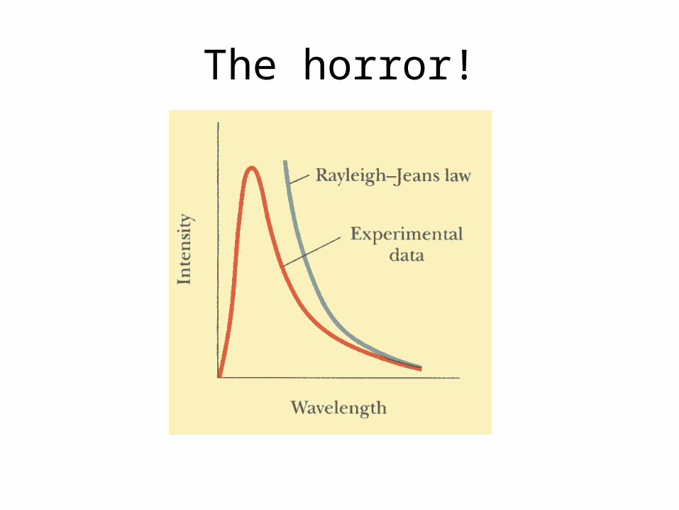
Although this law worked well for low frequencies (high wavelength) it did not for high frequencies (ultraviolet region.)
So shocking was this find, that it was labelled the Ultraviolet Catastrophe
The horror!
Where did it all go wrong?
The error involved in these shocking events was the assumption that the energy levels of the radiation was continuous.
i.e. any energy level could be obtained.
Max Planck, however, thought differently.

Max Planck

Photons
Max Planck theorised that energy was not continuous, but came in discrete packets. For light, we call these photons.
He developed his theory and mathematics that matched experimental data.
This managed to solve all of our problems……..

Problem Solved!
………………………….or is it?

New Problems
This created problems more difficult than the so-called catastrophe. The implication is that light must be a particle, if it comes in discrete packets.
But how can we explain all these wave-related phenomena with particles?
Scientists are still working on it…..

Young’s Double Slit
We can arrange Young’s Double slit experiment so that instead of having a strong light source, we have a source which emits just one photon at a time………….
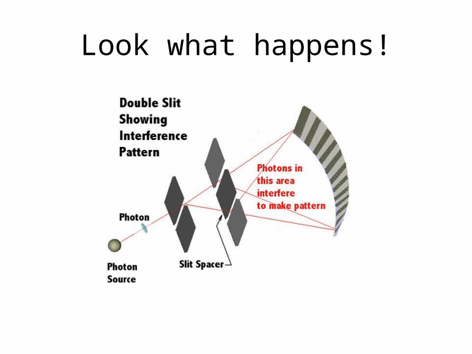
Look what happens!

Young’s Double Slit
We still get an interference pattern!
Our previous explanation involved one ray interfering with another.
How can one photon interfere with itself?
Welcome to the strange world of quantum mechanics……..

Energy of a photon
The energy of a photon is given by:
hfE
Jsh 341063.6 h is known as Planck’s Constant, named after Max Planck

Photon Energy
This can be re-arranged……
hc
hfE

More properties
Another natural consequence of light being a particle is that it should have similar properties to particles as well.
One of these is momentum.
It was found that when photons collided with electrons, they moved in such a way that it looked like conservation of momentum.

Photon Momentum
Momentum of Photons
The question of giving momentum to photons, which are massless, seems difficult.
Einstein came up with the solution.
He said that energy and mass are different versions of the same thing.

EinsteinI knew I shouldn’t have worn my wife’s
shoes E = mc2

Photon Momentum
We can substitute Einstein’s equation into our expression for the energy of a photon to get the ‘mass’ of a photon:
Equating: and
Give:
Therefore:
2mcE hfE
hc
hfmc 2
c
hm

Photon Momentum
We know momentum, p =mv
For photons, v = c, so….
cc
hp
mcp
mvp
h
p

Example
Calculate the energy (in joules and electron volts) and momentum of each of the following photons.a) Orange light of wavelength 600nm
b) An X-ray of wavelength 0.1nm.
The Photoelectric Effect
The photoelectric effect is the ejection of electrons from the surface of a material when light of sufficiently high frequency is incident upon it.
Usually the electrons are emitted from a metal, and are called photoelectrons.
Example
A laser emitting monochromatic light of wavelength 780nm, is rated at a power of 0.1mW. How many photons per second is it emitting?

Photoelectric Effect Setup
Here, a clean metal surface—the cathode—is illuminated with light from an external source.
If the light causes photoelectrons to be emitted, they will be detected at the anode. This flow of electrons is called the photoelectric current, and is registered by a sensitive ammeter.
Photoelectric Effect
The circuit includes a variable voltage supply which can be used to make the cathode negative (and the anode positive). When this is done, the photoelectrons will be helped by the resulting electric field to the anode, anda maximum possible current will be measured.
Alternatively, the voltage may be adjusted to make the cathode positive and the anode negative—a reverse potential. This arrangement is used to investigate the kinetic energy carried by the emitted photoelectrons.
Measuring the Kinetic Energy
- Fix the frequency above the threshold frequency for electrons to be emitted
- Apply an increasing reverse potential difference. As the reverse voltage increases, the current decreases
- A point will arrive when there is no measured current. This occurs at the stopping potential, Vs.

Measuring the Kinetic Energy
The kinetic energy of the electrons can now be measured using: E = eΔV
i.e. Kmax = eVs
Kinetic Energy of Emitted Electrons
The maximum kinetic energy can be found to depend on two factors:
- the frequency of the radiation (E=hf)
- the surface from which electrons are emitted
Free electrons
Conductors have free (de-localised) electrons.
These are not bound to a specific atom, but to the conductor as a whole.
It is these electrons which make electric current possible.
Photoelectric Effect explained
ln order to explain the photoelectric effect, Einstein used the photon concept that Planck had developed.
He considered that, within the metal, each electron was bound to the metal by a different amount of energy. (At that time he knew nothing of electron shells.)
Some electrons required substantial amounts of energy to become free, while others required less energy.
Einstein was able to represent this situation using a ‘potential well’.

Photoelectric Effect explained
If the y-axis represents the total energy of the electrons, the electron will be bound to the metal where the energy is negative, will be freed where the energy is positive, and this energy is kinetic energy. An electron with zero energy would be free, but have no speed.
Photoelectric Effect Explained
- If the frequency is below the threshold frequency, even the least bound electron will not be emitted. This is independent of the intensity. E.g. if the minimum energy is 4.0eV, you can’t get two photons of energy 2.1eV to emit the electron.
- When the frequency is above the threshold frequency, the photon can be absorbed, giving the electron more energy, and it becomes free. E.g. An electron which requires 3.2eV can absorb a 7.0eV photon, and be emitted with an energy of 7.0eV – 3.2 eV = 3.8eV

Photoelectric Effect Explained
- Since the photon is absorbed, the electrons are emitted instantaneously (almost). (This is a strong argument for the particle theory of light)
- We call the minimum energy required to free the electron the ‘Work Function’

Work Function Values
Metal Work Function (eV)
Metal Work Function (ev)
Na 2.46 Pt 6.35
Al 4.08 Pb 4.14
Cu 4.70 Fe 4.50
Zn 4.31 Cs 1.90
Ag 4.73 K 2.24
W 4.58 Mo 4.2
Photoelectric Effect Equation
- Radiation of sufficiently high frequency, f, is incident on a metal surface of work function W.
- This photon, of energy E, is absorbed by the electron
- This electron thus leaves with kinetic energy Kmax = E - W

Photoelectric Effect Equation
WhfK max
)()(max eVWfe
heVK
Or if you like electron volts……

Photoelectric Effect Graph
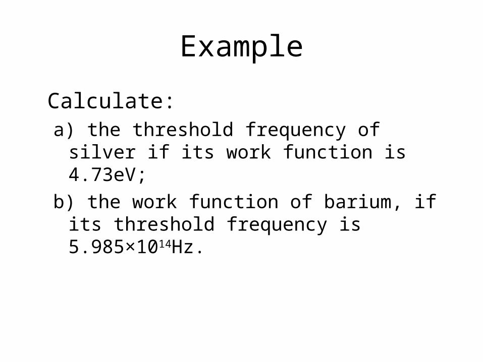
Example
Calculate:a) the threshold frequency of silver if its work
function is 4.73eV;
b) the work function of barium, if its threshold frequency is 5.985×10¹⁴Hz.

Example
The work function of aluminium is 4.08eV. Determinea) whether light of frequency 9×10¹⁴Hz will
cause electrons to be emitted;
b) the maximum kinetic energy of the emitted electrons if the metal is irradiated with ultra-violet light of wavelength 2.0×10⁻⁷m.

Example
The work function W of zinc is 4.3l eV. A zinc surface is irradiated with lights of different frequency. Sketch a graph of the maximum kinetic energy of emitted electrons against the frequency of light.

Example
The stopping potentials for electrons emitted from a metal by light of four different
wavelengths are given in the table below
Draw a graph of the maximum kinetic energy of the emitted electrons against frequency.
From your graph determine the following:
a) Planck’s constant
b) The work function of the metal
c) The threshold frequency of the metal
d) The maximum kinetic energy of electrons emitted by light of frequency 8.0 ×10¹⁴Hz.

X-Rays
If electromagnetic radiation can give electrons kinetic energy, the converse should also be true.
That is, a loss of kinetic energy should produce electromagnetic radiation.
German Physicist Wilhelm Roentgen discovered X-rays
using this principle.
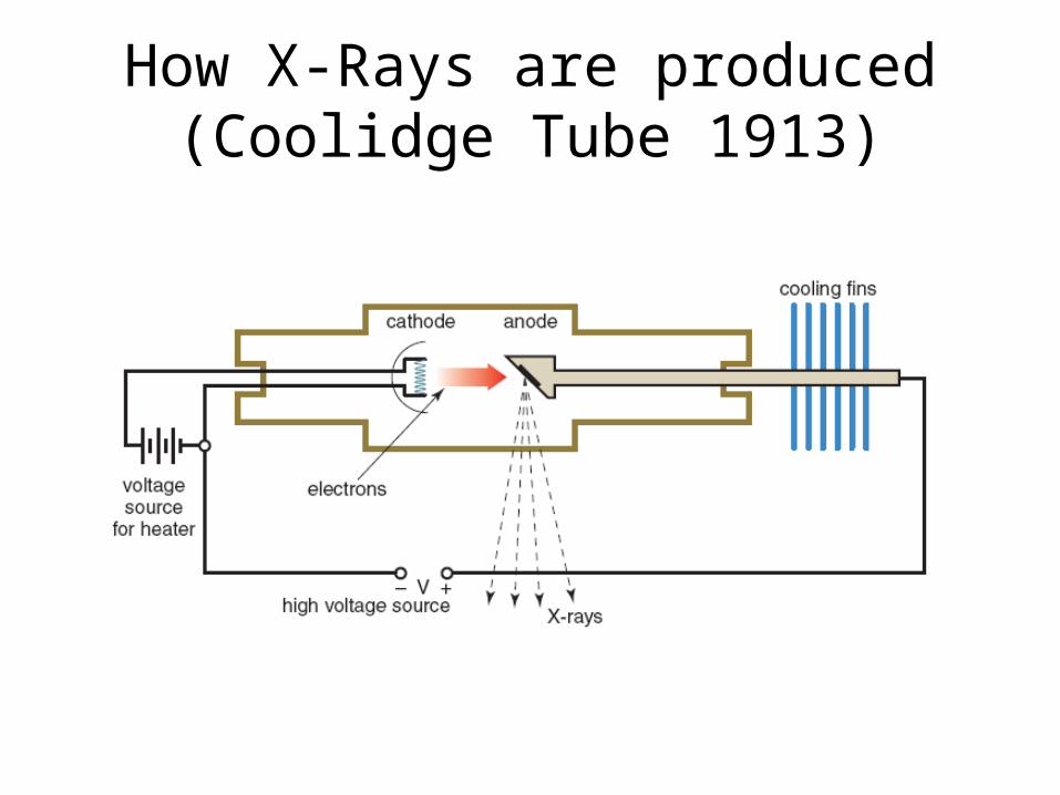
How X-Rays are produced (Coolidge Tube 1913)
What’s happening in the Coolidge Tube?
- A high potential difference is applied over the cathode and anode
- The voltage source for heater heats the cathode, providing energy to the electrons, allowing them to leave more easily
- These electrons are torn off of the cathode towards the anode
What’s happening in the Coolidge Tube?
- The anode is a good conductor of heat, and this heat is conducted to the cooling fins- The face of the anode has a hard metal of tungsten. This is where the electrons strike.- As the electrons enter the anode, the electrons encounter the positive nuclei of the metal
What’s happening in the Coolidge Tube?
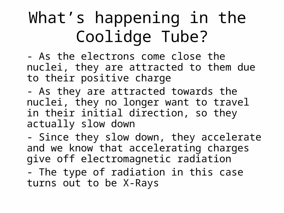
- As the electrons come close the nuclei, they are attracted to them due to their positive charge- As they are attracted towards the nuclei, they no longer want to travel in their initial direction, so they actually slow down- Since they slow down, they accelerate and we know that accelerating charges give off electromagnetic radiation- The type of radiation in this case turns out to be X-Rays

X-Ray Spectrum(Bremsstrahlung Radiation)
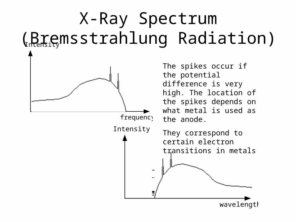
-The spectrum shows a continuous range of frequencies (this graph shows wavelength) up to a maximum
- Each voltage has a cut off frequency
- As the voltage increases the maximum frequency (minimum wavelength) increases
- As voltage increases, so does the intensity of the emitted X-Rays
X-Ray Spectrum(Bremsstrahlung Radiation)
frequency
Intensity
wavelength
Intensity
The spikes occur if the potential difference is very high. The location of the spikes depends on what metal is used as the anode.
They correspond to certain electron transitions in metals
Why continuous?
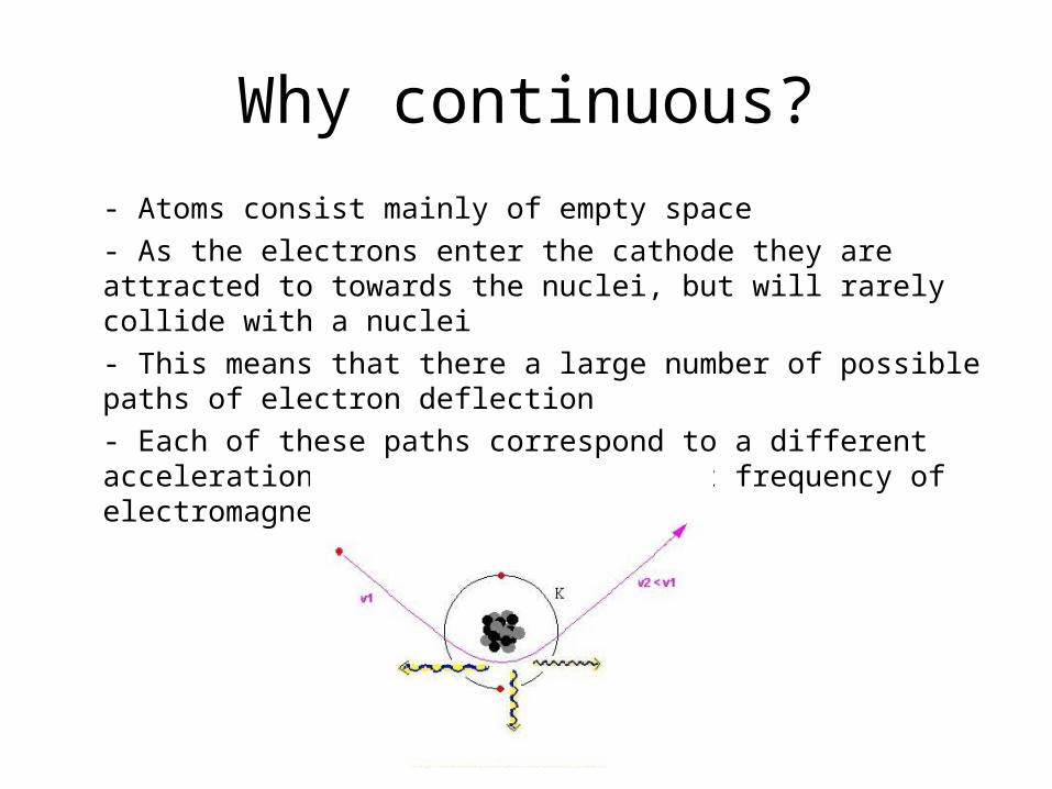
- Atoms consist mainly of empty space
- As the electrons enter the cathode they are attracted to towards the nuclei, but will rarely collide with a nuclei
- This means that there a large number of possible paths of electron deflection
- Each of these paths correspond to a different acceleration, and hence a different frequency of electromagnetic radiation

Maximum Energy of Photons
The energy of the electrons is given by:
The maximum energy of photons is therefore:
So….
VeK
VeE max
Vehf max
h
Vef
max
X-Rays in Medicine

Example
X-rays are generated in an X-ray tube with an operating voltage of 50kV.a) The maximum energy of the emitted photons;
b) The maximum frequency of the emitted photons.
Example
An X-ray tube emits radiation with a minimum wavelength of 0.03nm. What is the operating voltage of the tube?

Uses of X-Rays in Medicine
- Since X-rays have such high energy, they are able to penetrate matter quite well- When X-rays are incident on a photographic plate, they can expose the film and create an image- This is what happens when we take X-rays when bones break etc.- As the X-rays pass through the body, some are absorbed. Therefore the intensity varies, forming an image

What affects the exposure?
- The degree of absorption (attenuation) of the X-rays
- The penetrating power of the X-rays
- The exposure time

Absorption (Attenuation)
The denser the material, the greater the attenuation. Here the bone has the greater attenuation, and shows up best.
A similar effect happens with thick materials.
Elements with high atomic numbers tend to have a greater attenuation

Penetrating Power
- Highly penetrating X-rays are described by ‘hardness’
- Hard X-rays are typically 100-150kV
- Soft X-rays are typically around 50kV
- Hard X-rays are used to take images of bones and similar materials
- Soft X-rays are used for images of tissues and organs

Exposure Time
- Unfortunately, X-rays are harmful to living tissue.- Lead shielding is used to protect you and the radiographer- It is best to keep exposure to a minimum- The exposure depends on :
- the hardness. (harder = more dangerous)
- the intensity (high intensity = more dangerous) - exposure time

Determining Exposure Time
- Depends on medical condition- Shorter exposure time means less chance of injury, but- Shorter exposure time may not result in the appropriate clarity of the image
X-rays are set to an appropriate voltage (to get hardness) and an appropriate current (to get intensity)