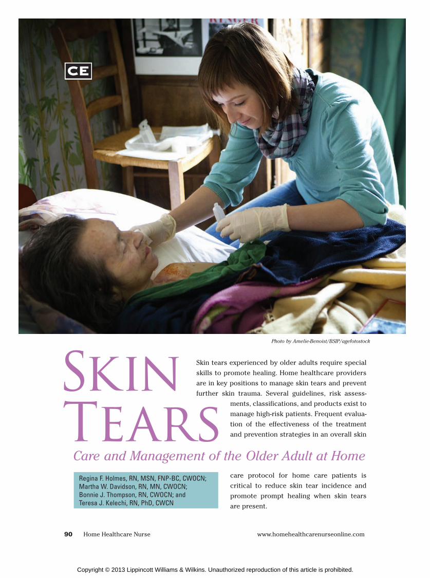
photo by amelie-benoist/bsip/agefotostock skin...
TRANSCRIPT
Skin tears experienced by older adults require special
skills to promote healing. Home healthcare providers
are in key positions to manage skin tears and prevent
further skin trauma. Several guidelines, risk assess-
ments, classifications, and products exist to
manage high-risk patients. Frequent evalua-
tion of the effectiveness of the treatment
and prevention strategies in an overall skin
care protocol for home care patients is
critical to reduce skin tear incidence and
promote prompt healing when skin tears
are present.
Skin Tears
Care and Management of the Older Adult at Home
Regina F. Holmes, RN, MSN, FNP-BC, CWOCN; Martha W. Davidson, RN, MN, CWOCN; Bonnie J. Thompson, RN, CWOCN; and Teresa J. Kelechi, RN, PhD, CWCN
Photo by Amelie-Benoist/BSIP/agefotostock
90 Home Healthcare Nurse www.homehealthcarenurseonline.com
Copyright © 2013 Lippincott Williams & Wilkins. Unauthorized reproduction of this article is prohibited.

vol. 31 • no. 2 • February 2013 Home Healthcare Nurse 91
Skin tears, one of the most dreaded conse-
quences of trauma to aging skin, require a
skilled assessment approach and special
wound care practices to prevent complications
and promote healing. Although skin tears can
occur anywhere on the body including the but-
tocks and legs, the hands and arms are most
vulnerable. Although recent figures are unavailable,
it is estimated that skin tears affect 1.5 million
hospitalized and long-term care older patients
with prevalence rates thought to be between 14%
and 24% (Bank & Nix, 2006; Malone et al., 1991).
Approximately 5% of older adults residing in
residential community settings have skin tears
(Carville et al., 2007); however, the prevalence of
skin tears of older adults living at home and/or
receiving homecare is unknown and could be
much higher than in residential settings.
With the increase in the aging population,
especially in the old–old age group, and older
adults living at home with chronic conditions,
the care of the skin becomes a critical health
issue. Home healthcare providers are in a key
position to prevent skin tears. They are able to
assess the home environment and can teach
caregivers, families, and the older adults how to
make simple modifications. The care of older
adults with skin tears should focus on controlling
bleeding, preventing infection, controlling pain,
restoring skin integrity, and promoting a healing
environment (LeBlanc & Baranoski, 2009). Risk
assessment, skin tear classification, best-practice
prevention, and treatment guidelines presented
in this article should assist the home healthcare
provider to manage skin tears and identify those
at risk for these wounds.
Current definitions of skin tears suggest
there are two types: a partial-thickness wound
where the epidermis is separated from dermis
secondary to friction or shearing force trauma;
or, full thickness where both the epidermis and
the dermis separate from the underlying tissue
(Carville et al., 2007; Payne & Martin, 1993).
Although skin tears do not usually result in
serious problems, they can predispose persons
to infection, cause major discomfort, and be
costly to manage if not treated quickly and
appropriately (Payne & Martin, 1993). Because
skin tears are often mismanaged and misdiag-
nosed, methods to prevent, predict, assess,
and treat skin tears have recently been the
focus of an international consensus panel to
address the need for a validated, comprehen-
sive program for managing skin tears (LeBlanc
& Baranoski, 2011).
Susceptibility of Aging Skin and Risk FactorsAlthough skin tears can occur at any age, those
65 years old and older are at high risk and those
80 years old and above at greatest risk because of
more pronounced physiological changes in the
skin. Older individuals have also endured a life-
time of environmental exposure to the sun and
other hazards, and have more chronic conditions
that impact the skin.
Physiological ChangesPhysiological changes play a pivotal role in ini-
tiating a cascade of events that predispose the
skin to tearing, beginning with a 20% atrophy or
“thinning” of the dermis and the epidermis
(Ratliff & Fletcher, 2007). Thinning causes a
weakening of the skin structure between its lay-
ers and produces a paper-thin appearance. The
structures that keep the skin layers attached to
each other flatten causing a decrease in the
cohesiveness of the skin layers. For example,
epidermal papillae flatten and the rete pegs or
ridges—extensions in the epithelium that
project down into the underlying connective
tissue holding the epidermis and dermis to-
gether loosen, while the subcutaneous fatty
layer becomes lessened. Effects of aging also
result in a reduced blood supply. Fewer nerve
endings decrease sensation, particularly in re-
sponse to pain, tactile sensitivity, and tempera-
ture (Sibbald & Krasner, 2009). Many older
adults are unaware that a skin tear has occurred
until they see droplets of blood on their cloth-
ing or are told by another person that their skin
is bleeding.
Decreased cell growth and turnover rate as
well as decreased production of sebum (oil)
glands and sweat glands result in dry and itchy
patches of skin. The immune system is weaker
due to decreased production of T-lymphocytes
and mast cells with subsequent reduction in
antigen response, thus increasing the risk of in-
fection and cancer. The skin becomes less elastic
and the tensile strength weakens, causing the
skin to be less resilient or quick to recover from
abrupt changes. Vascular changes caused by
capillary fragility and pathological change from
Copyright © 2013 Lippincott Williams & Wilkins. Unauthorized reproduction of this article is prohibited.

92 Home Healthcare Nurse www.homehealthcarenurseonline.com
atherosclerosis cause rupture with subsequent
bruising (senile pupura) or ecchymosis beneath
the skin. Approximately 40% of skin tears are
associated with senile purpura, which tend to
occur on the back of the hands (Ratliff & Fletcher,
2007). Medications such as corticosteroids com-
promise skin integrity. Corticosteroids inhibit
collagen synthesis and reduce the strength and
elasticity of the skin. The combination of aging,
pathological changes in the vascular system,
comorbid conditions such as cancer, as well as
medications lead to a higher risk of tears
(Xu et al., 2009).
AppearanceFor some older adults, changes in the physical
appearance of the skin can lead to negative psy-
chological consequences. Many express dismay
at paper-thin skin and bruising, especially the
back of their hands, the second most visible part
of the body after the face. This perceived un-
sightly appearance of the skin may lead to social
isolation and depression. Unfortunately there
are limited “cosmetic” approaches to conceal
frail skin.
Review of the LiteratureThere is a limited body of knowledge that in-
cludes studies that contribute to best prac-
tices, prevalence, and an understanding of the
financial burden to older adults living at
home. There are few randomized clinical trials
or systematic reviews; however, there are a
growing number of guidelines and consensus
statements published in the last 5 years on skin
tear prevention and management. A recent in-
ternational consensus statement, published in
2011 (LeBlanc & Baranoski, 2011) was based in
part on the data from a large international sur-
vey (LeBlanc & Christensen, 2010) conducted
by the lead author of the consensus statement.
The survey focused on exploring current prac-
tices on the assessment, prediction, preven-
tion, and treatment of skin tears of healthcare
professionals from 16 countries (LeBlanc &
Christensen, 2010). Of the 1,127 respondents
surveyed, 69.6% reported a problem with cur-
rent assessment and documentation of skin
tears in their practice settings. In particular,
approximately 90% admitted to not using any
instrument or classification system for assess-
ing and documenting skin tears. They indicated
they would use a simplified method for docu-
menting and assessing skin tears if available.
The findings from this survey continue to sup-
port the need for more research aimed at the
prevention, prediction, assessment, and treat-
ment of skin tears. Fortunately, the interna-
tional consensus statement (LeBlanc &
Baranoski, 2011) on skin tears serves as a guide
for clinicians in home care settings.
Practice GuidelinesThere are a number of clinical practice guide-
lines and best practice recommendations avail-
able for skin tears (Box 1). For example, the
State of Pennsylvania developed a statewide
skin tear initiative, Preventing Pressure Ulcers and Skin Tears, a toolkit available for down-
loading at http://www.guideline.gov through
the National Guidelines Clearinghouse (Penn-
sylvania Safety Authority Skin Tear Initiative,
2006). The toolkit focuses on preventing skin
tears, identifying those at risk, and fostering
healing of skin tears, targeting those who are
immobile, undernourished or malnourished,
incontinent, have friable skin, and impaired
cognition. As part of this guideline, the Payne–
Martin Classification System (LeBlanc &
Baranoski, 2011; Payne & Martin, 1993) is
recommended to categorize skin tears. The
guideline also provides recommendations for
Box 1. Resources • Preventing Pressure Ulcers and Skin Tears
Toolkit: http://www.guideline.gov
• The Best Practice Recommendations for
the Prevention and Treatment of Skin
Tears (LeBlanc & Baranoski, 2009)
• Payne-Martin Classification System
(Payne & Martin, 1993)
• International Consensus Statement
(LeBlanc & Baranoski, 2011)
• Skin Tear Audit Research (STAR) Classifi-
cation System (Carville et al., 2007)
• Skin Integrity Risk Assessment Tool
(White et al., 1994)
• Say Goodbye to Wet-to-Dry Wound care
Dressings (Dale & Wright, 2011)
Copyright © 2013 Lippincott Williams & Wilkins. Unauthorized reproduction of this article is prohibited.
vol. 31 • no. 2 • February 2013 Home Healthcare Nurse 93
promoting a safe environment by educating
staff and caregivers, protecting the older adult
from self-injury or injury during routine care,
and managing skin tears if they occur. The Best Practice Recommendations for the Prevention and Treatment of Skin Tears (LeBlanc & Baranoski,
2009) and the International Consensus State-ment (LeBlanc & Baranoski, 2011) both pro-
mote recommendations consistent with the
toolkit but suggest the Skin Tear Audit Research
(STAR) Classification System (Carville et al.,
2007) for classifying skin tears, touting the fact
that it is simpler to use. Regardless of which
classification system is used, it is imperative to
classify skin tears for the purpose of guiding
management strategies and documenting heal-
ing outcomes.
Classification Systems for Skin TearsThe first classification system for skin tears was
developed by Payne and Martin in 1993 and
groups characteristics and degrees of skin dam-
age into three main categories:
• Category I: Skin tears without skin loss
(which takes about average 10 days to heal),
• Category II: Skin tears with partial-thickness
skin loss (healing takes and average of
14 days), and
• Category III: Skin tears with complete tissue
loss (average 21 days to heal).
This skin tear classification system was fur-
ther developed for the STAR project, which
resulted in the validated STAR Skin Tear Clas-
sification System (Supplemental Digital Con-
tent 1, http://links.lww.com/HHN/A23) (Carville
et al., 2007). Nurses in home settings should
consider using STAR as part of the overall skin
assessment when skin tears are present. How-
ever, to prevent the occurrence of skin tears,
the steps of an overall skin tear prevention ap-
proach begin with predicting which patients
are at high risk.
Step 1: Predicting RiskIt is critical to predict and identify those at
high risk for skin tears so that an appropriate
prevention program can be implemented be-
fore injury occurs (Bank & Nix, 2006; Carville et
al., 2007; LeBlanc et al., 2008). Older adults are
at high risk for the development of skin tears in
home care settings, and thus, a comprehensive
risk assessment for skin tears should be
performed on all older patients. Guidelines
recommend a risk assessment be performed
that includes a head-to-toe assessment on ad-
mission to the home health agency, with a
change in the individual’s condition, or per
agency/ facility policies (National Pressure Ulcer
Advisory Panel and European Pressure Ulcer
Advisory Panel, 2009).
Unlike well-validated risk assessment in-
struments used to determine pressure ulcer
risk such as the Braden Scale (Bergstrom et
al., 1987), there is a lack of validated instru-
ments to predict skin tear risk, and the use of
existing instruments (STAR) to classify skin
tears are generally underused. Although
developed almost 20 years ago, the Skin
Integrity Risk Assessment Tool (LeBlanc &
Baranoski, 2011; White et al., 1994) is relevant
for home care patients as it places patients
into groups and recommends implementing a
skin tear risk prevention plan for those pa-
tients who meet:
• any criteria in Group 1: history of skin
tears within last 90 days; has open skin
tear;
• four or more criteria in Group 2: decision-
making skills impaired; vision impairment;
extensive assistance/total dependence for
activities of daily living (ADLs); wheel-
chair-assistance required; loss of balance;
confined to bed or chair; unsteady gait;
bruises;
• five or more criteria in Group 3: physically
abusive, resists ADL care; agitation; hearing
impaired; decreased tactile stimulation;
wheels self; manually/mechanically lifted;
contracture of arms, legs, shoulders, hands;
hemiplegia/hemiparesis; trunk—partial or
total inability to balance or turn body;
pitting edema of legs; open lesions on ex-
tremities; three to four senile purpura on
extremities; dry, scaly skin; or
• three criteria in Group 2 and three or more
criteria in Group 3.
Risk Factors
Although physiological changes in the skin
contribute to the predisposition of skin tears
in older adults, older adults who return home
Copyright © 2013 Lippincott Williams & Wilkins. Unauthorized reproduction of this article is prohibited.
94 Home Healthcare Nurse www.homehealthcarenurseonline.com
from the hospital after a critical illness such
as a myocardial infarction or stroke, those
who are medically compromised (diabetes,
thyroid disorders), and those who require as-
sistance with ADLs or have altered mobility
are particularly vulnerable to skin tears with
even the most minimal of friction or shear
force trauma (Carville et al., 2007; LeBlanc et
al., 2008). In addition to medical and func-
tional comorbidities, cognitive impairment,
dehydration, poor nutrition, medications (im-
munosuppressives, anti-inflammatories, anti-
coagulants), alkaline soaps and antibacterial
skins cleansers (strip protective acid man-
tel; see Box 2) and smoking place older adults
at high risk for skin tears (Bank & Nix, 2006;
Sibbald et al., 2006). Multiple factors should
be included in the overall skin assessment to
determine skin tear risk (LeBlanc & Baranoski,
2011; LeBlanc et al., 2008; Stephen-Haynes
et al., 2011). Risk factors for skin tears in older
patients residing at home are extensive and
include five major areas: skin problems, co-
morbid conditions, functional impairments,
cognitive dysfunction, and environmental
hazards (White et al., 1994). High risk factors
for skin include a history of tears within
90 days, one or more open tears, skin that
bruises easily, presence of purpura, long nails
that can traumatize intact skin, and skin that
is macerated or moist. Several comorbid
conditions also place older patients at risk
including diabetes, thyroid disorders, stoke,
chronic lung disease, malnutrition, dehydra-
tion, and taking medications such as cortico-
steroids and anticoagulants. Impaired function
in vision and hearing, as well as neuropathy
that causes loss of protective sensation, im-
paired balance, or unsteady gait, increases
one’s risk. Those using assistive devices and
having edema of the legs can suffer skin tear
of the lower extremities. Cognitive problems
related to agitation and combativeness are
related to skin tears caused by trauma from
flailing limbs.
Step 2: Prevention StrategiesMost skin tears occur accidentally during rou-
tine patient care activities. Education and in-
volvement of family and caregivers in the pre-
vention of skin tear development is imperative.
Many of the basic strategies for prevention of a
skin tear, while appearing to be commonsense
approaches, should be included in the preven-
tion plan. It is also important to remind caregiv-
ers that despite their best efforts, not all skin
tears are 100% preventable (Roberts, 2007).
However, every effort should be made to pre-
vent skin tears whenever possible. The follow-
ing prevention steps can be taken to minimize
the potential for occurrences or reoccurrences
to reduce the opportunity for future skin injury.
Several recommendations focus on environmen-
tal management, the maintenance of skin
integrity and factors such as nutrition and
hygiene (Hampton, 2010; Krasner, 2010; LeBlanc
& Baranoski, 2011; Sussman & Golding, 2011).
Create a Safe Environment
A safe environment is a critical component of an
overall prevention plan. Several actions should
be implemented as follows:
1. Assess the home environment for routine
household items that cause an accidental
skin tear such as exposure of sharp corners
of countertops, open drawers, or other pro-
truding objects. Provide adequate lighting to
aid in avoiding unnecessary bumps or
knocks to the skin against firm objects. Limit
items with protruding legs such as tables
and footstools that can be accidentally
Box 2. Soaps Avoid alkaline, antibacterial, or heavily per-
fumed soaps that can be drying for aging
skin:
• Dial
• Irish Spring
• Ivory
• Zest
Instead, recommend patients use
pH- balanced products such as:
• Basis
• Aveeno
• Neutrogena
Source: Barbara Dale, personal communication, March 30,
2012.
Copyright © 2013 Lippincott Williams & Wilkins. Unauthorized reproduction of this article is prohibited.

vol. 31 • no. 2 • February 2013 Home Healthcare Nurse 95
bumped as older adults are moving about
their homes. Remove small throw rugs or
shoes that could be easily tripped over.
2. Move or transfer older adults with altered
mobility correctly across a bed or into a
chair to prevent shear and friction that
could cause tears on the buttocks or arms.
The adaptation of good manual handling
techniques with the use of lift devices,
draw sheets, or slide sheets can prevent or
decrease shear or friction injury. Special
belts can aid in assisting older adults to
stand from a sitting position. Care must be
taken when using any adaptive equipment
and requires instructions to caregivers on
proper use.
3. Pad bed rails, chairs, wheelchairs, or walkers
to prevent accidental skin tears when arms
or legs bump against these firm surfaces.
Commercialized arm and shin pads can be
used on the extremities for protection
against rubbing or hitting an extremity on a
hard surface.
Maintain Skin Integrity
One of the most crucial aspects of prevention is
to keep the skin in the best possible condition.
Steps to foster skin integrity include:
1. Ensure optimal nutritional to improve basic
skin health, assist with the healing of a cur-
rent skin tear, and aid in the prevention of
future skin tears. Dietitians can provide in-
formation on the amount and types of foods
that are important for wound healing such as
diary products, meats, beans/legumes, nuts,
eggs, and soy products. Recommendations
often include adding additional protein in
the daily diet or supplemental nutritional
drinks. It is important that a tailored plan be
developed as many older adults have food
sensitivities and conditions that affect diges-
tion and absorption of nutrients. Overall,
older adults are encouraged to consume
four to six small meals per day and should
con sider taking a daily multivitamin after
discussion with their healthcare provider.
2. Encourage hydration by providing addi-
tional fluids between meals as well as a
variety of fluids throughout the day, un-
less there is a fluid restriction. Hydrated
skin is at lower risk of tearing. It is recom-
mended people drink at least 2 to 3 quarts
of fluid daily but this can vary based on
the size of the individual. Fluids can in-
clude any noncaffeinated beverages, water,
juice, sports drinks, or milk. Any food that
turns to liquid in the mouth is considered
a fluid such as yogurt, ice cream, jello, and
popsicles. Handy water bottles also pro-
vide an easy option for older adults to use,
especially those with tremors or arthritic
hands.
3. Use hypoallergenic moisturizers twice each
day especially on the hands and arms to
help hydrate dry aged skin. It is best to
apply moisturizers after showering when
the skin is still damp to aid in maintaining
skin elasticity and resilience.
4. Routine bathing and shower should be lim-
ited. The natural decrease in lubrication
from diminished sebaceous and sweat
gland activity places older adults at a
higher risk for skin tears as the skin be-
comes more susceptible to dryness. Bath-
ing removes the body’s natural oils from
the skin surface and can be naturally dehy-
drating. A shower with warm-tepid water,
approximately 94°F, is preferred over tub
baths. Cleansing with mild pH-balanced
soaps is recommended. Unfortunately most
soap is alkaline, which tends to increase
the pH of the skin and reduces the skin’s
protective acid mantle. Healthy skin is
meant to have a pH in the range of 5.4 to
5.9. For many older adults, the use of soap
is not indicated, especially for arms and
legs. With the decrease in the skin’s natural
lubrication, application of moisturizers to
damp skin after bathing will aid in skin hy-
dration. Consider nonalkaline and glycer-
ine-based products and washes, many of
which are available over-the-counter.
5. Protect arms and legs with long sleeves and
pants. Tubular stockinet on arms or thick
athletic socks with the foot cut out can be
placed on the arms for additional protec-
tion. Avoid tight and restrictive clothing
that can interfere with ease of movement.
Also encourage older adults to wear pro-
tective footwear with hard soles to prevent
them from tripping when walking.
6. Avoid adhesives on fragile skin. Use skin
sealants applied to the skin under tapes to
Copyright © 2013 Lippincott Williams & Wilkins. Unauthorized reproduction of this article is prohibited.
96 Home Healthcare Nurse www.homehealthcarenurseonline.com
reduce skin damage during tape removal. It
is advised to use adhesive removers to fa-
cilitate tape removal while applying coun-
ter pressure to the skin in the opposition
when the tape is slowly rolled off. If tape is
used on the skin, do so minimally if possi-
ble. Silicone, paper, and cloth hypoaller-
genic tapes are gentler for removal than
other tapes.
7. Assess the length of fingernails and toenails
to determine the need for trimming or filing.
Long fingernails of caregivers, family, or
older adults themselves can cause skin dam-
age from scratching or accidental pinches.
Many care providers are discouraged from
wearing artificial nails as they can harbor
organisms that, if passed onto open skin,
can cause infection.
Step 3: TreatmentAlthough prevention of skin tears should remain
the primary focus, evidence-based wound care
principles should be used when a skin tear devel-
ops. The same principles used for other wounds
should be employed when treating skin tears. A
wound treatment plan (see Box 3) should also
consider nutritional support, pain management,
local wound conditions, and optimal dressing
selection (Sibbald et al., 2006). Wound assess-
ment should be performed, with the documenta-
tion of local wound conditions (location, size,
flap status, exudate, pain) and skin tear category.
A multifactorial plan should include:
1. Cleanse the wound to remove surface bacte-
ria and necrotic debris as this prepares the
wound for the dressing. This can be achieved
by irrigating with noncytotoxic solutions
such as normal saline or nonionic surfactant
cleansers using safe pressures of less than 10
to 15 pounds per square inch (psi). A 19-gauge
angiocatheter and a 35-mL piston syringe
can also be used. Skin tears without debris
should be gently cleansed at lower pressures
(less than 8 psi to protect granulating tissue,
using similar cleansers (Gardner & Frantz,
2008; Krasner, 2010; Sibbald et al., 2006).
Most cleansers come with specially designed
spray bottles to deliver the appropriate force
of pressure to cleanse the wounds. Tepid
showers are acceptable to cleanse and rinse
the wound as well. Rubbing or wiping over
the wound is contraindicated as it can disrupt
fragile tissue.
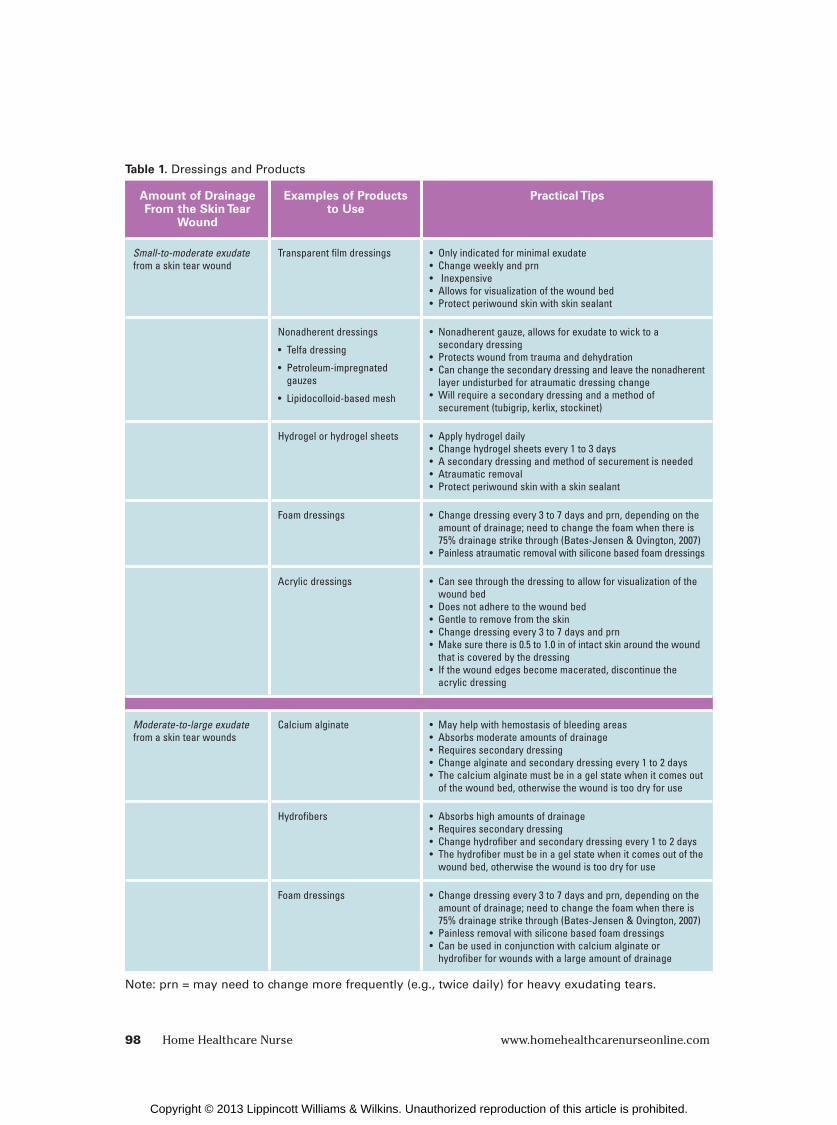
2. Apply dressings to cover, protect, and aid in
healing the skin tear. There are several dress-
ing options (Table 1). Selection is based on
products available to the home healthcare
agency, clinical expertise, and the degree
of skin damage from the skin tear. Ideally,
dressings for skin tears should absorb the
exudate (drainage), maintain a moist wound
environment, allow for pain-free removal,
and stay in place for several days, no longer
than 7 days. Moist wound healing remains the
method of choice when selecting a dressing
and has a high level of evidence to support
the use for skin tears. Moist wound healing
provides an environment that supports cell
growth and healing (Baranoski, 2008).
Several recommendations (Ovington & Pierce,
2001; Stephen-Haynes & Carville, 2010) guide
the selection of dressings that will:
• Maintain constant moisture balance,
• Suit the local wound environment,
• Protect the periwound skin,
• Control or manage exudate,
• Control or manage infection, and
• Optimize caregiver time.
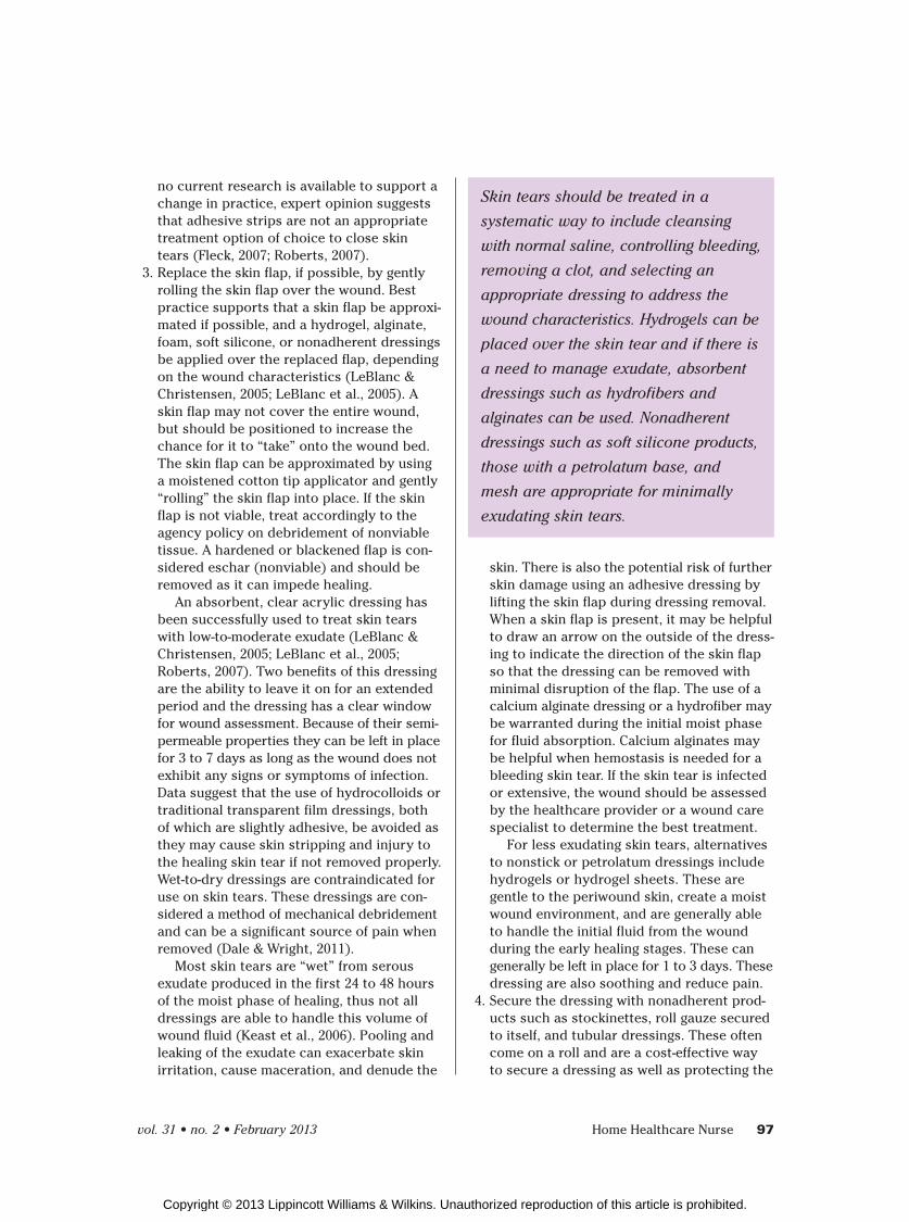
Skin tears should be treated in a systematic
way to include cleansing with normal saline,
controlling bleeding, removing a clot, and
selecting an appropriate dressing to address
the wound characteristics. Hydrogels can be
placed over the skin tear and if there is a
need to manage exudate, absorbent dress-
ings such as hydrofibers and alginates can
be used. Nonadherent dressings such as soft
silicone products, those with a petrolatum
base, and mesh are appropriate for mini-
mally exudating skin tears.
Closure of the skin tear, based on wound
characteristics, should be considered. Unlike
chronic wounds, skin tears are acute wounds
with the potential to be closed by primary
intention. Wounds closed by primary inten-
tion are traditionally secured with suture or
staples; however, given the fragility of aged
skin, sutures, and staples are not viable
options. The practice of using adhesive
strips for skin tears is dated, and although
Copyright © 2013 Lippincott Williams & Wilkins. Unauthorized reproduction of this article is prohibited.
vol. 31 • no. 2 • February 2013 Home Healthcare Nurse 97
no current research is available to support a
change in practice, expert opinion suggests
that adhesive strips are not an appropriate
treatment option of choice to close skin
tears (Fleck, 2007; Roberts, 2007).
3. Replace the skin flap, if possible, by gently
rolling the skin flap over the wound. Best
practice supports that a skin flap be approxi-
mated if possible, and a hydrogel, alginate,
foam, soft silicone, or nonadherent dressings
be applied over the replaced flap, depending
on the wound characteristics (LeBlanc &
Christensen, 2005; LeBlanc et al., 2005). A
skin flap may not cover the entire wound,
but should be positioned to increase the
chance for it to “take” onto the wound bed.
The skin flap can be approximated by using
a moistened cotton tip applicator and gently
“rolling” the skin flap into place. If the skin
flap is not viable, treat accordingly to the
agency policy on debridement of nonviable
tissue. A hardened or blackened flap is con-
sidered eschar (nonviable) and should be
removed as it can impede healing.
An absorbent, clear acrylic dressing has
been successfully used to treat skin tears
with low- to-moderate exudate (LeBlanc &
Christensen, 2005; LeBlanc et al., 2005;
Roberts, 2007). Two benefits of this dressing
are the ability to leave it on for an extended
period and the dressing has a clear window
for wound assessment. Because of their semi-
permeable properties they can be left in place
for 3 to 7 days as long as the wound does not
exhibit any signs or symptoms of infection.
Data suggest that the use of hydrocolloids or
traditional transparent film dressings, both
of which are slightly adhesive, be avoided as
they may cause skin stripping and injury to
the healing skin tear if not removed properly.
Wet-to-dry dressings are contraindicated for
use on skin tears. These dressings are con-
sidered a method of mechanical debridement
and can be a significant source of pain when
removed (Dale & Wright, 2011).
Most skin tears are “wet” from serous
exudate produced in the first 24 to 48 hours
of the moist phase of healing, thus not all
dressings are able to handle this volume of
wound fluid (Keast et al., 2006). Pooling and
leaking of the exudate can exacerbate skin
irritation, cause maceration, and denude the
skin. There is also the potential risk of further
skin damage using an adhesive dressing by
lifting the skin flap during dressing removal.
When a skin flap is present, it may be helpful
to draw an arrow on the outside of the dress-
ing to indicate the direction of the skin flap
so that the dressing can be removed with
minimal disruption of the flap. The use of a
calcium alginate dressing or a hydrofiber may
be warranted during the initial moist phase
for fluid absorption. Calcium alginates may
be helpful when hemostasis is needed for a
bleeding skin tear. If the skin tear is infected
or extensive, the wound should be assessed
by the healthcare provider or a wound care
specialist to determine the best treatment.
For less exudating skin tears, alternatives
to nonstick or petrolatum dressings include
hydrogels or hydrogel sheets. These are
gentle to the periwound skin, create a moist
wound environment, and are generally able
to handle the initial fluid from the wound
during the early healing stages. These can
generally be left in place for 1 to 3 days. These
dressing are also soothing and reduce pain.
4. Secure the dressing with nonadherent prod-
ucts such as stockinettes, roll gauze secured
to itself, and tubular dressings. These often
come on a roll and are a cost-effective way
to secure a dressing as well as protecting the
Skin tears should be treated in a
systematic way to include cleansing
with normal saline, controlling bleeding,
removing a clot, and selecting an
appropriate dressing to address the
wound characteristics. Hydrogels can be
placed over the skin tear and if there is
a need to manage exudate, absorbent
dressings such as hydrofibers and
alginates can be used. Nonadherent
dressings such as soft silicone products,
those with a petrolatum base, and
mesh are appropriate for minimally
exudating skin tears.
Copyright © 2013 Lippincott Williams & Wilkins. Unauthorized reproduction of this article is prohibited.
98 Home Healthcare Nurse www.homehealthcarenurseonline.com
Amount of Drainage From the Skin Tear
Wound
Examples of Products to Use
Practical Tips
Small-to-moderate exudate from a skin tear wound
Transparent film dressings • Only indicated for minimal exudate• Change weekly and prn• Inexpensive• Allows for visualization of the wound bed• Protect periwound skin with skin sealant
Nonadherent dressings
• Telfa dressing
• Petroleum-impregnated gauzes
• Lipidocolloid-based mesh
• Nonadherent gauze, allows for exudate to wick to a secondary dressing
• Protects wound from trauma and dehydration• Can change the secondary dressing and leave the nonadherent
layer undisturbed for atraumatic dressing change• Will require a secondary dressing and a method of
securement (tubigrip, kerlix, stockinet)
Hydrogel or hydrogel sheets • Apply hydrogel daily• Change hydrogel sheets every 1 to 3 days• A secondary dressing and method of securement is needed• Atraumatic removal• Protect periwound skin with a skin sealant
Foam dressings • Change dressing every 3 to 7 days and prn, depending on the amount of drainage; need to change the foam when there is 75% drainage strike through (Bates-Jensen & Ovington, 2007)
• Painless atraumatic removal with silicone based foam dressings
Acrylic dressings • Can see through the dressing to allow for visualization of the wound bed
• Does not adhere to the wound bed• Gentle to remove from the skin• Change dressing every 3 to 7 days and prn• Make sure there is 0.5 to 1.0 in of intact skin around the wound
that is covered by the dressing• If the wound edges become macerated, discontinue the
acrylic dressing
Moderate-to-large exudate from a skin tear wounds
Calcium alginate • May help with hemostasis of bleeding areas• Absorbs moderate amounts of drainage• Requires secondary dressing• Change alginate and secondary dressing every 1 to 2 days• The calcium alginate must be in a gel state when it comes out
of the wound bed, otherwise the wound is too dry for use
Hydrofibers • Absorbs high amounts of drainage• Requires secondary dressing• Change hydrofiber and secondary dressing every 1 to 2 days• The hydrofiber must be in a gel state when it comes out of the
wound bed, otherwise the wound is too dry for use
Foam dressings • Change dressing every 3 to 7 days and prn, depending on the amount of drainage; need to change the foam when there is 75% drainage strike through (Bates-Jensen & Ovington, 2007)
• Painless removal with silicone based foam dressings• Can be used in conjunction with calcium alginate or
hydrofiber for wounds with a large amount of drainage
Note: prn = may need to change more frequently (e.g., twice daily) for heavy exudating tears.
Table 1. Dressings and Products
Copyright © 2013 Lippincott Williams & Wilkins. Unauthorized reproduction of this article is prohibited.
vol. 31 • no. 2 • February 2013 Home Healthcare Nurse 99
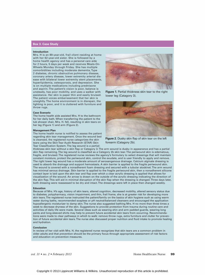
Introduction
Mrs. H is an 89-year-old, frail client residing at home
with her 62-year-old sister. She is followed by a
home health agency and has a personal care aide
for 2 hours, 5 days per week and receives Meals-On-
Wheels Monday through Friday. She has multiple
comorbidities including moderate dementia, Type
2 diabetes, chronic obstructive pulmonary disease,
coronary artery disease, lower extremity arterial dis-
ease with bilateral lower extremity stent placements,
hyperlipidemia, osteoporosis, and depression. She
is on multiple medications including prednisone
and aspirin. The patient’s vision is poor, balance is
unsteady, has poor mobility, and uses a walker with
assistance. Her skin is paper thin and easily bruised.
The patient voices embarrassment that her skin is
unsightly. The home environment is in disrepair, the
lighting is poor, and it is cluttered with furniture and
throw rugs.
Case Scenario
The home health aide assisted Mrs. H to the bathroom
for her daily bath. When transferring the patient to the
tub shower chair, Mrs. H. fell, resulting in skin tears on
her leg (Figure 1) and arm (Figure 2).
Management Plan
The home health nurse is notified to assess the patient
regarding skin tear management. Once the wound bed
is cleansed, the registered nurse categorizes the skin
tears using the Skin Tear Audit Research (STAR) Skin
Tear Classification System. The leg wound is a partial-
thickness skin tear without a skin flap or Category 3. The arm wound is dusky in appearance and has a partial
skin flap remaining. The leg wound is classified as a Category 2b skin tear. The periwound skin is edematous,
fragile, and bruised. The registered nurse reviews the agency’s formulary to select dressings that will maintain
constant moisture, protect the periwound skin, control the exudate, and is user friendly to apply and remove.
The right lower leg wound has a moderate amount of serosangenous drainage. Calcium alginate dressing is
used to absorb the drainage and support hemostasis. A skin barrier is applied to the fragile periwound skin.
The wound is covered with a nonadherent foam dressing and secured with a tubular stockinet. The arm wound
has minimal serous drainage. Skin barrier is applied to the fragile periwound skin, then a nonadherent silicone
contact layer is laid upon the skin tear and flap over which a clear acrylic dressing is applied that allows for
visualization of the wound. An arrow is drawn on the outside of the acrylic dressing indicating the direction of
the skin flap. This will aid in minimal disruption of the skin flap when the dressing is changed. Three days later,
both dressing were reassessed to be dry and intact. The dressings were left in place then changed weekly.
Education
Because of Mrs. H’s age, history of skin tears, altered cognition, decreased mobility, altered sensory status due
to diabetes, polypharmacy, vision impairment, and thin, frail frame, she is at greater risk for developing more
skin tears. The registered nurse instructed the patient/family on the basics of skin hygiene such as using warm
water during baths, recommended soapless or pH neutral/balanced cleansers and encouraged the application
hypoallergenic moisturizer to damp skin. The nurse also suggested bathing Mrs. H no more than three times a
week to decrease dryness of the skin. Suggestions to provide protection from trauma during routine care and
activities of daily life were made. Several ideas such as wearing shin and arm padded guards, wearing long
pants and long-sleeved shirts may help to prevent future accidental skin tears from occurring. Recommenda-
tions were made to clear pathways in which to walk: remove throw rugs, extra furniture and clutter for preven-
tion of future accidental skin tears. The nurse also discussed proper nutrition and fluid intake to promote healing
and hydration.
Conclusion
In review of her visit with Mrs. H, the registered nurse recognizes that skin tears are a common problem in
older adults and that prevention should be the primary focus through appropriate assessment of risk factors
and education on prevention measures.
Box 3. Case Study
Figure 1. Partial thickness skin tear to the right
lower leg (Catagory 3).
Figure 2. Dusky skin flap of skin tear on the left
forearm (Catagory 2b).
Copyright © 2013 Lippincott Williams & Wilkins. Unauthorized reproduction of this article is prohibited.
100 Home Healthcare Nurse www.homehealthcarenurseonline.com
limb from further trauma. If tapes and adhe-
sives are the only option to secure the dress-
ing, nonaggressive, hypoallergenic paper or
silicone tapes are preferred. The use of skin
sealants applied to the periwound skin
around the wound edges as well as under
the dressing or tape is helpful to avoid fur-
ther tissue damage from moisture and trauma.
Care should be taken when removing any
dressing or tape. These should be removed
by gently lifting off the dressing, starting at
one edge, while pressing and gently applying
pressure and drawing the skin taut. It is im-
portant to premoisten the dressing with sa-
line if it has adhered to the skin to prevent
further damage to the healing wound.
Step 4: EvaluationFrequent evaluation of the effectiveness of the
treatment strategies is imperative. If older adults
express pain, particularly during dressing removal,
consider analgesic options. Pain can disrupt
quality of life and can impede healing. The ease
of application and costs of dressings should be
major considerations as caregiver responsibility
can be overwhelming when meeting multiple
needs of their older loved one.
ConclusionOlder adults living at home, especially those at
high risk of developing skin tears, require a
thorough risk assessment of factors that are
amenable to prevention. Armed with an overall
risk assessment approach and a thorough
understanding of risk factors, home care
providers are in keys positions to reduce skin
tear incidence and can initiate prompt treatment
when skin tears are present. Clinical guidelines
and best practices are available, such as those
proposed in this article, to aid in the develop-
ment of a skin tear prevention and management
approach for use in home care settings as part of
the overall skin care protocol for your agency.
Regina F. Holmes, RN, MSN, FNP-BC, CWOCN, is a Wound, Ostomy, and Continence Nurse/ Emergency Room Nurse Practitioner at McLeod Loris Hospital, Loris, South Carolina.
Martha W. Davidson, RN, MN, CWOCN, is a Wound, Ostomy, and Continence Nurse at Vander-bilt University Medical Center, Nashville, Tennessee.
Bonnie J. Thompson, RN, CWOCN, is a Wound, Ostomy, and Continence Nurse at Vander-bilt University Medical Center, Nashville, Tennessee.
Teresa J. Kelechi, RN, PhD, CWCN, is a Professor at Medical University of South Carolina, Charleston, South Carolina.
Supplemental digital content is available for this article. Direct URL citations appear in the printed text and are provided in the HTML and PDF ver-sions of this article on the journal’s Web site (http://journals.lww.com/homehealthcarenurseonline).
The authors and nurse planners have disclosed that they have no financial relationships related to this article.
Address for correspondence: Teresa J. Kelechi, RN, PhD, CWCN, Medical University of South Carolina, 99 Jonathan Lucas St., MSC 160, Charleston, SC 29425-1600 ([email protected]).
DOI:10.1097/NHH.0b013e31827f458a
REFERENCES
Bank, D., & Nix, D. (2006). Preventing skin tears in a
nursing and rehabilitation center: An interdisci-
plinary effort. Ostomy Wound Management, 52(9),
38-46.
Bates-Jensen, B. M., & Ovington, L. G. (2007). Manage-
ment of exudate and infection. In C. Sussman & B.
Bates-Jensen (Eds.). Wound care: A collaborative practice manual for physical therapists and nurses
(3rd ed., pp. 215-233). Philadelphia, PA: Lippincott,
Williams & Wilkins.
Frequent evaluation of the effectiveness
of the treatment strategies is imperative.
If older adults express pain, particularly
during dressing removal, consider
analgesic options. Pain can disrupt
quality of life and can impede healing.
The ease of application and costs of
dressings should be major considerations
as caregiver responsibility can be
overwhelming when meeting multiple
needs of their older loved one.
Copyright © 2013 Lippincott Williams & Wilkins. Unauthorized reproduction of this article is prohibited.

vol. 31 • no. 2 • February 2013 Home Healthcare Nurse 101
Baranoski, S. (2008). Choosing a dressing: Part 2.
Advances in Skin & Wound Care, 38(2), 14-15.
Bergstrom, N., Braden, B., Laguzza, A., & Holman, V.
(1987). The Braden Scale for predicting pressure
sore risk. Nursing Research, 36(4), 205-210.
Carville, K., Newall, G., Haslehurt, P., Sanatmaria, R.,
& Roberts, N. (2007). STAR: A consensus for skin
tear classification. Primary Intention, 15(1),
18-28.
Dale, B. A., & Wright, H. D. (2011). Say goodbye to
wet-to-dry wound dressing. Home Healthcare Nurse, 29(7), 429-440.
Fleck C. (2007). Preventing and treating skin tears.
Advances for Skin & Wound Care, 20(6), 315-320.
Gardner, S., & Frantz, R. (2008). Wound bioburden.
In: Baranoski, S., & Ayello, E. (Eds.). Wound Care Essentials: Practice Principles (2nd ed., pp.
93-114). Philadelphia, PA: Lippincott Williams &
Wilkins.
Hampton, S. (2010). How to preserve skin integrity and
prevent skin tears. Nursing & Residential Care, 12(6),
284-287.
Keast, D., Parslow, N., Houghton, P, Norton, L., &
Fraser, C. (2006). Best practice recommendations
for the prevention and treatment of pressure ulcers:
Update 2006. Advances for Skin & Wound Care, 20(8), 447-460.
Krasner, D. (2010). Skin tears: Understanding prob-
lem leads to prevention, proper care. Journal of American Geriatric Society, 29(5), 30-33.
LeBlanc, K., & Baranoski, S. (2009). Prevention and
management of skin tears. Advances for Skin & Wound Care, 22(7), 325-334.
LeBlanc, K., & Baranoski, S. (2011). Skin tears: State
of the science: consensus statements for the pre-
vention, prediction, assessment, and treatment of
skin tears. Advances for Skin & Wound Care, 24(9),
2-15.
LeBlanc, K., & Christensen, D. (2005, November 12–
15). An approach to managing skin tears in the el-derly population: A case series. Poster presented at
the Canadian Association of Wound Care Annual
Conference, Montreal, Quebec. Retrieved from
http://www.caet.ca/link/caet-link-2010-03-kim-
leblanc.pdf
LeBlanc, K., & Christensen, D. (2010). Demystifying skin
tears, Part 1. Nursing2010, 7(1), 62-67.
LeBlanc, K., Christensen, D., & Cuillier, B. (2005, November
12–15). Managing skin tears in long-term care. Poster
presented at the Canadian Association of Wound Care
Annual Conference, Montreal, Quebec. Retrieved from
http://www.caet.ca/link/caet-link-2010-03-kim-leblanc.pdf
LeBlanc, K., Christensen, D., Orsted, H., & Keast, D.
(2008). Prevention and treatment of skin tears.
Wound Care Canada, 6(1), 14-30.
Malone, M., Rozario, N., Gavinski, M., & Goodwin, J.
(1991). The epidemiology of skin tears in the
institutionalized elderly. Journal of the American Geriatrics Society, 39(6), 591-595.
National Pressure Ulcer Advisory Panel and European
Pressure Ulcer Advisory Panel. (2009). Prevention and Treatment of Pressure Ulcers: Clinical Practice Guideline. Washington, DC: National Pressure Ulcer
Advisory Panel.
Ovington, L., & Pierce, B. (2001). Wound dressings:
Form, function, feasibility, and facts. In D. Krasner,
G. Rodeheaver, & G. Sibbald (Eds.). Chronic wound care: A Clinical Source Book for Healthcare Profes-sionals (3rd ed., pp. 311-320). Wayne, PA: HMP
Communications.
Payne, R., & Martin, M. (1993). Defining and classifying
skin tears: need for a common language. Ostomy Wound Management, 39(5), 16-26.
Pennsylvania Safety Authority Skin Tear Initiative.
(2006). PA-PSRS Patient Safety Advisory, 3(3). Retrieved
from http://www.patientsafetyauthority.org.
Ratliff, C., & Fletcher, K. (2007). Skin tears: a review of
the evidence to support prevention and treatment.
Ostomy Wound Management, 53(3), 32-42.
Roberts, J. (2007). Preventing and managing skin tears:
A review. Journal of the Wound Ostomy Continence Nurses Society, 34(3), 256-259.
Sibbald, R.G., & Krasner, D. (2009). Skin changes at the
end of life consensus statements. Advances for Skin & Wound Care, 23(5), 237-239.
Sibbald, R. G., Orstead, H., Coutts, P., & Keast, D.
(2006). Best practice recommendations for prepar-
ing the wound bed: Update 2006. Wound Care Canada, 4(1), 19-29.
Stephen-Haynes, J., Callaghan, R., Bethell, E., & Green-
wood, M. (2011). The assessment and management
of skin tears in care homes. British Journal of Nursing, 20(11), 12-32.
Stephen-Haynes, J., & Carville, K. (2010). Skin tears
made easy. Wounds International, 2(4). Retrieved from
http://www.woundsinternational.com/made-easys/
skin-tears-made-easy&print.
Sussman, G., & Golding, M. (2011). Skin tears: Should
the emphasis be only their management. Wound Practice and Research, 19(2), 66-77.
Xu, X., Lau, K., Taira, B., & Singer, A. (2009). The current
management of skin tears. American Journal of Emergency Medicine, 27(6), 729-733.
White, M. W., Karam, S., & Cowell, B. (1994). Skin tears
in frail elders: A practical approach to prevention.
Geriatric Nursing, 15(95), 95-99.
For more than 70 additional continuing nursing education articles on skin and wound care, go to nursingcenter.com/ce.
Copyright © 2013 Lippincott Williams & Wilkins. Unauthorized reproduction of this article is prohibited.