pharmacological characterization of human incretin ... characterization of human incretin receptor...
TRANSCRIPT
JPET #160531 1.
Pharmacological Characterization of Human Incretin
Receptor Missense Variants
Jean-Philippe Fortin, Jonathan C. Schroeder, Yuantee Zhu,
Martin Beinborn and Alan S. Kopin
Molecular Pharmacology Research Center, Molecular Cardiology Research Institute,
Tufts Medical Center, Tufts University School of Medicine, Boston, MA 02111.
J.-P.F., J.C.S., Y.Z., M.B., A.S.K.
JPET Fast Forward. Published on October 19, 2009 as DOI:10.1124/jpet.109.160531
Copyright 2009 by the American Society for Pharmacology and Experimental Therapeutics.
This article has not been copyedited and formatted. The final version may differ from this version.JPET Fast Forward. Published on October 19, 2009 as DOI: 10.1124/jpet.109.160531
at ASPE
T Journals on M
ay 1, 2017jpet.aspetjournals.org
Dow
nloaded from
JPET #160531 2.
Running Title: Pharmacology of incretin receptor missense variants
Corresponding author: Dr. Alan S. Kopin, 750 Washington Street,
P.O. Box 7703, Boston, MA 02111; Tel.: 617-636-4834; Fax: 617-636-8692;
Email: [email protected]
Number of text pages: 28
Number of tables: 2
Number of figures: 5
Number of references: 36
Number of words in Abstract: 239
Number of words in Introduction: 518
Number of words in Discussion: 1453
Abbreviations: Glucagon-like peptide 1 (GLP-1), Glucose-dependent insulinotropic
peptide (GIP), Glucagon-like peptide 1 receptor (GLP-1R), Glucose-dependent
insulinotropic peptide receptor (GIP-R), G protein-coupled receptor (GPCR),
transmembrane domain (TMD), extracellular domain (ECD), hemagglutinin (HA),
horseradish peroxidase (HRP), cAMP-responsive element (CRE), Type 2 diabetes (T2D).
Recommended section assignement: Metabolism, Transport, and Pharmacogenomics
This article has not been copyedited and formatted. The final version may differ from this version.JPET Fast Forward. Published on October 19, 2009 as DOI: 10.1124/jpet.109.160531
at ASPE
T Journals on M
ay 1, 2017jpet.aspetjournals.org
Dow
nloaded from
JPET #160531 3.
Abstract
Glucagon-like peptide 1 (GLP-1) and glucose-dependent insulinotropic polypeptide
(GIP) are gut-derived incretin hormones that regulate blood glucose levels. In addition to
their widely accepted insulinotropic role, there is evidence that GLP-1 modulates feeding
behavior and GIP regulates lipid metabolism thereby promoting postprandial fat
deposition. In this study, we investigated whether naturally-occurring polymorphisms in
the GLP-1 receptor (GLP-1R) and the GIP receptor (GIP-R) affect the pharmacological
properties of these proteins. After transient expression of the receptors in HEK293 cells,
basal as well as ligand-induced cAMP production were assessed using luciferase reporter
gene assays. Our data reveal that the wild-type GIP-R displays a considerable degree of
ligand-independent activity. In comparison, the GIP-R variants C46S, G198C, R316L
and E354Q show a marked decrease in basal signaling that may, at least in part, be
explained by reduced cell surface expression. When stimulated with GIP, the C46S and
R316L mutants display significantly reduced potency (>1000 and 25- fold, respectively)
compared to wild type. Complementary competition binding assays further demonstrate
that the C46S variant fails to bind radio-iodinated GIP whereas all other GIP-R mutants
maintain normal ligand affinity. In contrast to the GIP-R, the wild-type GLP-1R lacks
constitutive activity. Furthermore, none of the ten GLP-1R missense mutations showed
an alteration in pharmacological properties versus wild type. The extent to which
abnormalities in GIP-R function may lead to physiological changes or affect drug
sensitivity in selected populations (e.g. obese, diabetic individuals) remains to be further
investigated.
This article has not been copyedited and formatted. The final version may differ from this version.JPET Fast Forward. Published on October 19, 2009 as DOI: 10.1124/jpet.109.160531
at ASPE
T Journals on M
ay 1, 2017jpet.aspetjournals.org
Dow
nloaded from

JPET #160531 4.
Introduction
The incretin hormones glucose-dependent insulinotropic polypeptide (GIP) and
glucagon-like peptide 1 (GLP-1) are homologous peptides released from intestinal
enteroendocrine cells in response to food intake. Both hormones are important
modulators of metabolic function. In the pancreas, GLP-1 and GIP potentiate nutrient-
stimulated insulin secretion, as well as promote the expansion of pancreatic islet mass via
induction of β-cell proliferation and survival (Kim et al., 2005; Kim and Egan, 2008). In
light of these insulinotropic actions, drugs that mimic or prolong the biological functions
of GIP and GLP-1 have attracted considerable attention as treatment options for type 2
diabetes (T2D) (Lovshin and Drucker, 2009). Exendin-4 (Exenatide), a potent long-
acting agonist of the GLP-1 receptor (GLP-1R), represents the first incretin-based
pharmaceutical to reach the market for the treatment of T2D. Inhibitors of the enzyme
dipeptidyl dipeptidase IV, which plays a major in inactivating both incretin hormones,
have also recently been approved as therapeutics for T2D.
Considerable efforts have focused on unraveling additional metabolic functions
triggered by the incretins (Kim and Egan, 2008). Accumulating evidence supports that
GIP modulates adipocyte metabolism, triggering fat deposition following feeding.
Highlighting the physiological relevance of this function, previous studies have shown
that targeted disruption of the GIP receptor (GIP-R) in mice results in protection from
both diet-induced obesity and insulin resistance (Miyawaki et al., 2002). Consistent with
these observations, inhibition of GIP-R signaling using a selective antagonist, or passive
immunization against GIP, were both shown to decrease body weight and to protect
This article has not been copyedited and formatted. The final version may differ from this version.JPET Fast Forward. Published on October 19, 2009 as DOI: 10.1124/jpet.109.160531
at ASPE
T Journals on M
ay 1, 2017jpet.aspetjournals.org
Dow
nloaded from

JPET #160531 5.
against glucose intolerance in animals that were fed a high fat diet (Gault et al., 2007;
Fulurija et al., 2008).
GLP-1 also modulates metabolic function, in part by acting on GLP-1Rs in
extrapancreatic tissues (Kim and Egan, 2008). This peptide triggers delayed gastric
emptying which in turn slows the absorption of food thus delaying the rise in blood
glucose levels. In addition, GLP-1 has been shown to inhibit feeding behavior by
stimulation of cognate receptors in the brain. Taken together, the incretin hormones and
their receptors contribute at multiple levels to maintaining normal glucose homeostasis
and regulating body weight.
Both the GIP-R and the GLP-1R belong to the glucagon subfamily of class B1 G
protein-coupled receptors (GPCRs). These seven transmembrane domain (TMD)
proteins, when stimulated with ligand, undergo a conformational change from putative
inactive to active conformations, thereby triggering a Gαs-mediated increase in cAMP
production (Hoare, 2005). It has been observed with other wild-type and mutant GPCRs
that partially active receptor conformations may occur even in the absence of agonist,
leading to constitutive, ligand-independent signaling (Kenakin, 2004). Although
engineered constitutively active incretin receptors have been generated (Tseng and Lin,
1997; M.B. unpublished data); the extent to which detectable basal signaling is
influenced by naturally-occurring polymorphic/mutant incretin receptors has not been
investigated.
This article has not been copyedited and formatted. The final version may differ from this version.JPET Fast Forward. Published on October 19, 2009 as DOI: 10.1124/jpet.109.160531
at ASPE
T Journals on M
ay 1, 2017jpet.aspetjournals.org
Dow
nloaded from
JPET #160531 6.
It is well established that missense mutations in GPCRs can result in a variety of
pharmacologic abnormalities (e.g. alterations in basal and ligand-dependent activity,
receptor affinity, expression) which predispose to physiologic changes or disease (Seifert
and Wenzel-Seifert, 2002). In the current study, we examined the molecular
pharmacologic consequences of naturally-occurring mutations/polymorphisms in the
GIP-R and GLP-1R using a series of in vitro assays.
This article has not been copyedited and formatted. The final version may differ from this version.JPET Fast Forward. Published on October 19, 2009 as DOI: 10.1124/jpet.109.160531
at ASPE
T Journals on M
ay 1, 2017jpet.aspetjournals.org
Dow
nloaded from
JPET #160531 7.
Methods
Generation of incretin receptor variants. The complementary DNA encoding the GIP-R
was obtained from the Missouri S&T cDNA Resource Center (www.cdna.org) and
subcloned into pcDNA1.1. The human GLP-1R cDNA was previously reported
(Tibaduiza et al., 2001). Single amino acid substitutions, as well as a hemagglutinin (HA)
tag, were introduced into the receptor sequence using oligonucleotide-directed site-
specific mutagenesis as previously described (Fortin et al., 2009). The nucleotide
sequences of all receptor coding regions were confirmed by automated DNA sequencing.
Cell Culture and Transfection. Human embryonic kidney (HEK) 293 cells were grown
in Dulbecco’s modified Eagle’s medium (Gibco, Carlsbad, CA) supplemented with 10%
fetal bovine serum, 100 U/ml penicillin G and 100 μg/ml streptomycin. The cells were
maintained at 37ºC in a humidified environment containing 5 % CO2.
Luciferase Reporter Gene Assay. Receptor-mediated signaling was assessed using a
previously described luciferase assay (Fortin et al., 2009). In brief, HEK293 cells were
plated at a density of 2000-3000 cells per well onto clear-bottom, white 96-well plates
and grown for 2 days to ~ 80% confluency. Cells were then transiently transfected using
LipofectamineR reagent (Invitrogen, Carlsbad, CA) with cDNAs encoding (i) a GPCR (or
empty expression vector), (ii) a cAMP responsive element-luciferase reporter gene
(CRE6X-luc) and (iii) β-galactosidase, to enable correction for interwell variability in
transfection efficiency and cell survival. Forty eight hours after transfection, cells were
This article has not been copyedited and formatted. The final version may differ from this version.JPET Fast Forward. Published on October 19, 2009 as DOI: 10.1124/jpet.109.160531
at ASPE
T Journals on M
ay 1, 2017jpet.aspetjournals.org
Dow
nloaded from
JPET #160531 8.
incubated for 6 hours with or without the appropriate peptide ligand (American Peptide
Company Inc., Sunnyvale, CA) in serum-free medium. Following agonist treatment, the
medium was gently aspirated, the cells were lysed and luciferase activity was measured
using SteadyliteR reagent (PerkinElmer, Boston, MA). A β-galactosidase assay was then
performed after adding the enzyme substrate, 2-Nitrophenyl β-D-galactopyranoside.
Following incubation at 37 ºC for 30-60 minutes, substrate cleavage was quantified by
measurement of optical density at 420 nm using a SpectraMaxR microplate reader
(Molecular Devices, Sunnyvale, CA). Corresponding values were used to normalize the
luciferase data.
Assessment of Receptor Expression Using ELISA. The surface expression levels of the
HA-tagged GIPRs were assessed using a previously-described approach (Shinyama et al.,
2003). In agreement with previous reports (Lee et al., 1994; Qi et al., 1997), two
independent predictor tools (http://bmbpcu36.leeds.ac.uk/prot_analysis/Signal.html and
http://www.cbs.dtu.dk/services/) supported the presence of a signal sequence in the GIP-
R extracellular domain (ECD) that is cleaved during receptor maturation. An HA tag was
thus inserted immediately downstream of the putative 24 amino acids GIP-R signal
peptide (shown in Figure 1B). HEK293 cells grown in 96-well clear Primaria plates (BD
Biosciences, Bedford, MA) were transiently transfected with increasing amounts of either
pcDNA1.1 or a cDNA encoding the HA-tagged GIPR. Forty-eight hours post-
transfection, the cells were washed once with phosphate buffered saline (PBS) (pH 7.4)
and fixed with 4% paraformaldehyde in PBS for 10 min at room temperature. After
washing with 100 mM glycine in PBS, the cells were incubated for 30 min in blocking
This article has not been copyedited and formatted. The final version may differ from this version.JPET Fast Forward. Published on October 19, 2009 as DOI: 10.1124/jpet.109.160531
at ASPE
T Journals on M
ay 1, 2017jpet.aspetjournals.org
Dow
nloaded from

JPET #160531 9.
solution (PBS containing 20% bovine serum). A horseradish peroxidase (HRP)-
conjugated antibody directed against the HA epitope tag (Roche; clone 3F10,
monoclonal, 1:500 in blocking buffer) was then added to the cells. After 1 hour, the cells
were washed five times with PBS. Fifty μl per well of a solution containing the
peroxidase substrate BM-blue (3.3’-5, 5’-tetramethylbenzidine, Roche Applied Science,
Indianapolis, IN) was then added. After incubation for 30 min at room temperature,
conversion of this substrate by antibody-linked HRP was terminated by adding 2.0 M
sulfuric acid (50 μl per well). Results were quantified by measuring light absorbance at
450 nm.
Radioligand Binding Studies. HEK293 cells were plated at a density of 30,000 cells per
well onto 24-well plates coated with poly-L-lysine, and grown for 18-24 hours to ~80%
confluency. Cells were then transiently transfected using lipofectamine with receptor
cDNA (100ng/well) and grown for an additional 18-24 hours. Whole cell binding studies
were initiated by washing cells twice with cold (4 oC) assay buffer (DMEM with 0.1%
BSA and 15mM HEPES), followed by addition of the same media with 20,000 counts per
minute of 125I-GIP (Perkin Elmer) and varying concentrations of unlabeled GIP.
Following an 8 hour incubation period at 4oC, the cells were washed twice with cold
assay buffer and solubilized in 0.1N NaOH. The lysates were then counted using a
Packard Cobra Quantum gamma counter to determine cell-associated radioactivity.
Data and statistical analysis. GraphPad Prism software version 5.0 (GraphPad, San
Diego, CA) was used for sigmoidal curve fitting. Half maximal effective concentrations
This article has not been copyedited and formatted. The final version may differ from this version.JPET Fast Forward. Published on October 19, 2009 as DOI: 10.1124/jpet.109.160531
at ASPE
T Journals on M
ay 1, 2017jpet.aspetjournals.org
Dow
nloaded from
JPET #160531 10.
(EC50 values) were calculated as an index of ligand potency and half maximal inhibitory
concentrations (IC50 values) as an index of receptor binding affinity. pEC50, basal activity
and surface expression values for each of the mutants were compared to the
corresponding control value at the WT receptor using one-way ANOVA followed by
Dunnett’s post test (GraphPad INSTAT software).
This article has not been copyedited and formatted. The final version may differ from this version.JPET Fast Forward. Published on October 19, 2009 as DOI: 10.1124/jpet.109.160531
at ASPE
T Journals on M
ay 1, 2017jpet.aspetjournals.org
Dow
nloaded from
JPET #160531 11.
Results
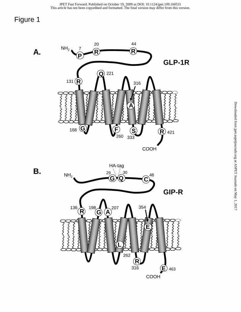
Human incretin receptor variants. Receptor constructs containing naturally-
occuring missense mutations of the human GLP-1R (P7L, R20K, R44H, R131Q, F260L,
A316G, A316T, S333C, R421C) and human GIP-R (C46S, R136W, G198C, A207V,
L262V, R316L, E354Q) were generated for investigation. The position of each amino
acid substitution is illustrated in cartoons of the GLP-1R and GIP-R (Figs 1A and B,
respectively). Each incretin receptor variant appeared in the NaVa (Natural Variants)
database, which catalogs known human GPCR polymorphisms (frequency >1%), as well
as rarer mutations (Kazius et al., 2008). As outlined in the discussion, three GIP-R
variants have been described previously in the literature (Kubota et al., 1996; Almind et
al., 1998). Site-directed mutagenesis was utilized to introduce amino acid substitutions
corresponding to the receptor variants. Each of the mutant receptor constructs or
corresponding wild type proteins were expressed in HEK293 cells and pharmacologically
characterized.
Missense variants of the GLP-1R exhibit normal basal and agonist-induced
signaling. Basal signaling in cells expressing the wild type GLP-1R (assessed using a
cAMP-responsive luciferase construct) was indistinguishable from that observed in cells
transfected with the empty expression vector, pcDNA1.1 (data not shown). This
observation confirms that the GLP-1R lacks constitutive activity. In addition, none of the
10 GLP-1R variants showed a significant level of basal signaling (Table 1).
This article has not been copyedited and formatted. The final version may differ from this version.JPET Fast Forward. Published on October 19, 2009 as DOI: 10.1124/jpet.109.160531
at ASPE
T Journals on M
ay 1, 2017jpet.aspetjournals.org
Dow
nloaded from
JPET #160531 12.
Agonist-induced GLP-1R function was assessed using two structurally-related agonists,
GLP-1 and exendin-4 (illustrated for the wild type receptor and representative variants in
Figure 2). At each mutant receptor, both peptides demonstrate potency and efficacy
values that are comparable to wild type (Table 1).
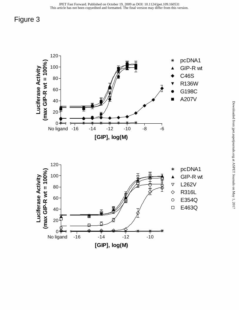
Selected GIP-R variants show altered basal and/or GIP-mediated activity.
Basal as well as GIP-induced signaling was examined at each GIP-R isoform (Figure 3).
In contrast to the GLP-1R, the wild type GIP-R showed constitutive activity (~25.0 ±
4.8% of the GIP-induced maximum) that markedly exceeded control values (determined
using vector-transfected cells). Four GIP-R variants, C46S, G198C, R316L and E354Q
showed a significant reduction in basal activity (Table 2). Of these functionally abnormal
receptors, two also showed a marked decrease in GIP potency. The C46S variant showed
a greater than 1000-fold reduction whereas R316L had a ~25 fold decrease in GIP
potency compared to that at the wild type GIP-R. In contrast, the EC50s for GIP at the
R136W, G198C, A207V, L262V, E354Q and E463 mutants were comparable to the
reference value at the wild type receptor (Table 2).
Impaired binding affinity of GIP at the C46S GIP-R variant. To complement
the functional studies of GIP-R mutants, we evaluated the affinity of GIP at each receptor
variant by radioligand competition binding assays (Figure 4, Table 2). In agreement with
previous work using other cell lines (Manhart et al., 2003), equilibrium binding of the
radioligand to HEK293 cells expressing the recombinant GIP-R was reached within a
seven hour incubation period at 4oC (data not shown). Consistent with the marked
This article has not been copyedited and formatted. The final version may differ from this version.JPET Fast Forward. Published on October 19, 2009 as DOI: 10.1124/jpet.109.160531
at ASPE
T Journals on M
ay 1, 2017jpet.aspetjournals.org
Dow
nloaded from
JPET #160531 13.
reduction in agonist potency at the C46S isoform (Figure 3), no specific binding of [125I]
GIP was detectable for this variant (not shown). In contrast, competition assays revealed
that each of the other variants, R136W, G198C, A207V, L262V, R316L, E354Q and
E463Q, had an affinity for GIP comparable to that observed at the wild type receptor.
GIP-R variants with decreased basal activity also show reduced cell surface
expression. Reduced basal activity of several GIP-R variants (C46S, G198C, R316L and
E354Q; Figure 3) was confirmed in experiments where ligand-independent signaling was
measured after transfecting cells with increasing concentrations of respective receptor
cDNAs (Figure 5A). In a parallel experiment using the same transfection protocol,
receptor expression levels at the cell surface were determined by ELISA (Figure 5B).
These studies revealed that at each cDNA level, receptors with reduced basal signaling
were also expressed at a significantly decreased density relative to the wild type protein
(100%). In contrast, the GIP-R variants where basal activity was not altered (R136W,
A207V, L262V, E463Q) showed normal expression levels (Figure 5 and Table 2). The
reduced cell expression of the C46S, G198C, R316L and E354Q variants was also
confirmed by confocal microscopy (Supplemental Figure 1).
Figure 5C illustrates the linear relationship that exists between expression level
and basal activity of corresponding GIP-R isoforms. The slope of the regresssion line of
the C46S mutant is similar to wild type, suggesting that the decreased basal activity
observed with this receptor variant is largely attributable to its diminished expression. In
contrast, the lower slope value of the G198C, R316L and E354Q variants suggests that
basal activity in these cases is disproportionately reduced relative to corresponding
This article has not been copyedited and formatted. The final version may differ from this version.JPET Fast Forward. Published on October 19, 2009 as DOI: 10.1124/jpet.109.160531
at ASPE
T Journals on M
ay 1, 2017jpet.aspetjournals.org
Dow
nloaded from
JPET #160531 14.
expression level (Table 2). It therefore appears that with the latter three receptors
additional mutation-induced changes (e.g. diminished G protein affinity) contribute to the
loss of ligand-independent signaling.
This article has not been copyedited and formatted. The final version may differ from this version.JPET Fast Forward. Published on October 19, 2009 as DOI: 10.1124/jpet.109.160531
at ASPE
T Journals on M
ay 1, 2017jpet.aspetjournals.org
Dow
nloaded from
JPET #160531 15.
Discussion
The related peptides, GIP and GLP-1, play important physiological roles in maintaining
blood glucose homeostasis, most notably by potentiating glucose-stimulated insulin
secretion by pancreatic β-cells (Kim and Egan, 2008; Lovshin and Drucker, 2009). These
peptides have additional peripheral and central functions, including the regulation of fat
metabolism in adipocytes (GIP) and the induction of satiety (GLP-1). To investigate the
effect of naturally-occurring polymorphisms on the function of cognate incretin
receptors, we compared the pharmacological properties of known human GIP-R and
GLP-1R missense variants with those of corresponding wild type GPCRs.
Introduction of ten naturally-occurring mutations in the human GLP-1R sequence did
not interfere with the ability of GLP-1 or Exendin-4 to trigger receptor-mediated activity.
This is the case despite the occurrence of variants in domains which are susceptible to
mutation-induced pharmacologic alteration. These include the N-terminus, a well-
established site of GLP-1 binding (Runge et al., 2008), and multiple intracellular domains
(loops 1, 2, and 3 and the C terminus), implicated in G protein coupling (Mathi et al.,
1997). These new findings complement an earlier report by our laboratory describing a
GLP-1R variant, T149M, which decreases endogenous agonist (GLP-1) as well as
exendin-4 affinity and potency (Beinborn et al., 2005).
Studies of the GIP-R revealed that two variants (C46S and R316L) while largely
preserving GIP efficacy, result in >10,000-fold and 25-fold reduced agonist potency
This article has not been copyedited and formatted. The final version may differ from this version.JPET Fast Forward. Published on October 19, 2009 as DOI: 10.1124/jpet.109.160531
at ASPE
T Journals on M
ay 1, 2017jpet.aspetjournals.org
Dow
nloaded from
JPET #160531 16.
compared to wild type values. Mutation-induced abnormalities in agonist potency may
be triggered by two distinct mechanisms: (1) alteration of the hormone binding site
and/or (2) defective GPCR transitioning from the inactive to the active receptor state (the
conformation triggering G protein activation) (Beinborn et al., 2004).
The C46 substitution is found in the N terminus of the GIP-R, a domain which plays
an important role in ligand binding (Parthier et al., 2007). It is thus likely that the
observed decrease in potency as well as the absence of radioligand binding to the C46S
variant (Table 2) is due to a mutation-induced alteration of the hormone binding domain.
This conclusion is supported by analysis of the recently obtained crystal structure of the
GIP-R extracellular domain bound to GIP. Experimental evidence from this study
suggests that three conserved disulfide bridges, including a link between C46 and C70,
stabilize the secondary structure of the extracellular domain (Parthier et al., 2007).
Furthermore, mutation of homologous cysteine residues in other class B1 GPCRs have
been shown to disrupt ligand affinity (Lee et al., 1994; Gaudin et al., 1995; Qi et al.,
1997; Lisenbee et al., 2005).
In contrast to C46S, the other GIP-R polymorphism that decreases agonist potency
(R316L) is found in the third intracellular loop. Since this receptor region is far removed
from the ligand binding domains, it is unlikely that the reduced GIP potency observed at
the R316L variant results from a direct change in the hormone docking site. Consistent
with this conclusion, the R316L mutant maintains normal affinity for radioiodinated GIP
(which reflects the initial step of ligand-receptor interaction) despite reduced agonist
potency (a measure of subsequent ligand-induced receptor activation). Of note, previous
structure-function studies on the related GLP-1R and PTH-R revealed that important G-
This article has not been copyedited and formatted. The final version may differ from this version.JPET Fast Forward. Published on October 19, 2009 as DOI: 10.1124/jpet.109.160531
at ASPE
T Journals on M
ay 1, 2017jpet.aspetjournals.org
Dow
nloaded from

JPET #160531 17.
protein coupling determinants localize in the N-terminal section of the third intracellular
loop of these receptors (i.e. the region where R316L is found in the GIP-R) (Huang et al.,
1996; Mathi et al., 1997). It is thus probable that the reduction in GIP potency at the
R316L isoform reflects an altered ability of this variant to couple and/or activate
stimulatory G proteins.
Our studies revealed normal potency and affinity for the G198C mutant (Table 2). It
is of note that this result contrasts with an earlier study that reported lower potency for
this GIP-R variant relative to wild type (Kubota et al., 1996). The basis for this
discrepancy is not clear, however it is possible that the divergent findings are at least in
part explained by differences in methodologies used for receptor characterization
(including the choice of cells for cDNA expression and the type of signaling assay).
While in vitro findings provide valuable insight into the potential of mutations to affect
receptor function (Seifert and Wenzel-Seifert, 2002), they do not necessarily cover the
full range of possible mutation-induced changes. Some alterations in receptor-mediated
function may only be detectable when utilizing a particular experimental setup and/or
with specific functional readouts.
Illustrating this limitation, the current study is the first to clearly demonstrate that the
human wild type GIP-R is constitutively active (Figures 3 and 5). There is only one prior
report in the literature suggesting that the GIP-R has a low degree of constitutive activity
(Almind et al., 1998). Our ability in the current study to readily detect a pronounced
elevation in GIP-R basal activity is likely explained by the sensitivity of the luciferase-
based system that was utilized to assess receptor-mediated signaling. For the broader
This article has not been copyedited and formatted. The final version may differ from this version.JPET Fast Forward. Published on October 19, 2009 as DOI: 10.1124/jpet.109.160531
at ASPE
T Journals on M
ay 1, 2017jpet.aspetjournals.org
Dow
nloaded from
JPET #160531 18.
group of class B1 GPCRs (i.e. the secretin-glucagon family), there are few reports of
significant ligand-independent signaling of unmodified wild type receptors (Seifert and
Wenzel-Seifert, 2002; Hoare et al., 2008). Our demonstration of GIP-R constitutive
activity provided the basis on which to define the effects of specific missense mutations
on this receptor property.
Four GIP-R variants (C46S, G198C, R316L and E354Q) are characterized by a
significant reduction in ligand-independent signaling relative to wild type (Figure 5,
Table 2). For one of these mutants (C46S), this functional change appears to be largely
acounted for by reduced cell surface expression whereas additional factors may underlie
the decreased basal activity of the G198C, R316L and E354Q variants. As a contributing
mechanism, mutation-induced structural changes may shift the putative equilibrium
between active and inactive receptor conformations (Lefkowitz et al., 1993) and/or may
alter G protein-receptor interaction (as discussed above for the R316L mutant). Given
that the G198C and E354Q substitutions are localized outside the intracellular receptor
portion (in EC loop I and TMD VI), it is possible that these mutations induce structural
changes which primarily shift the receptor equilibrium and thereby indirectly
compromise G protein interaction.
Loss of function in the GIP-R could provide a potential mechanism for altered
glucose homeostasis or fat deposition (Miyawaki et al., 2002; Gault et al., 2007; Fulurija
et al., 2008; Kim and Egan, 2008). It is of note that a previous study reported that glucose
tolerant subjects homozygous for the E354Q polymorphism (a variant which in our hands
This article has not been copyedited and formatted. The final version may differ from this version.JPET Fast Forward. Published on October 19, 2009 as DOI: 10.1124/jpet.109.160531
at ASPE
T Journals on M
ay 1, 2017jpet.aspetjournals.org
Dow
nloaded from
JPET #160531 19.
showed reduced basal activity) had a decreased serum C-peptide concentration (an index
of insulin secretion) (Almind et al., 1998). This abnormality was observed under fasting
conditions and after an oral glucose load, relative to subjects with the wild-type GIP-R.
Future efforts will explore whether functional abnormalities of GIP-R variants, including
the E354Q polymorphism, contribute to metabolic phenotypes.
The relatively high rate of mutation-induced functional changes in the GIP-R (four of
the eight known variants were pharmacologically distinct from wild type) contrasts with
our parallel analysis of the GLP-1R where none of the 10 variants that were investigated
showed detectable abnormalities. On this background, it is of note that the GIP-R is
constitutively active whereas the GLP-1R is not, raising the possibility that constitutively
active receptors are more sensitive to polymorphism-induced alterations in
pharmacology. In fact, such a parallel is also suggested by our recent study of the
dopamine D1R and D2R (Al-Fulaij et al., 2008). Reminiscent of our current findings, this
prior study revealed that several variants of the constitutively active D1R were associated
with decreased basal activity and/or expression, whereas all missense mutants of the D2R,
which displayed no ligand-independent signaling, appeared pharmacologically normal.
We also recently reported that a majority of missense variants found in the constitutively
active ghrelin receptor lead to alterations in ligand-independent signaling, potency and/or
expression (Liu et al., 2007). The human melanocortin-4 receptor (MC4R) provides an
additional well known example of a constitutively active GPCR for which a high number
of naturally-occurring missense mutants with altered function have been identified
This article has not been copyedited and formatted. The final version may differ from this version.JPET Fast Forward. Published on October 19, 2009 as DOI: 10.1124/jpet.109.160531
at ASPE
T Journals on M
ay 1, 2017jpet.aspetjournals.org
Dow
nloaded from
JPET #160531 20.
(Vaisse et al., 2000). As a group, loss of function MC4R missense mutations comprises
the most frequent monogenic cause of obesity.
It is of note that the low expression level of selected wild-type and mutant GPCRs
displaying high level of basal signaling has been explained by structural instability of
corresponding proteins (Gether et al., 1997; Samama et al., 1997; Alewijnse et al., 1998).
It is therefore possible that the sensitivity of constitutively active GPCRs, like the GIP-R,
to mutation-induced functional alterations in part reflects higher structural fragility
compared to receptors that lack agonist-independent signaling.
Taken together, our current findings and earlier observations suggest an emerging
trend that constitutive receptor activation may increase the likelihood of mutation-
induced functional abnormalities. This apparent link could have important implications
for predicting a subset of receptors that are likely to show missense variant-induced
changes in signaling with consequent alterations in physiologic response.
This article has not been copyedited and formatted. The final version may differ from this version.JPET Fast Forward. Published on October 19, 2009 as DOI: 10.1124/jpet.109.160531
at ASPE
T Journals on M
ay 1, 2017jpet.aspetjournals.org
Dow
nloaded from
JPET #160531 21.
References
Al-Fulaij MA, Ren Y, Beinborn M and Kopin AS (2008) Pharmacological analysis of
human D1 AND D2 dopamine receptor missense variants. J Mol Neurosci
34:211-223.
Alewijnse AE, Smit MJ, Hoffmann M, Verzijl D, Timmerman H and Leurs R (1998)
Constitutive activity and structural instability of the wild-type human H2 receptor.
J Neurochem 71:799-807.
Almind K, Ambye L, Urhammer SA, Hansen T, Echwald SM, Holst JJ, Gromada J,
Thorens B and Pedersen O (1998) Discovery of amino acid variants in the human
glucose-dependent insulinotropic polypeptide (GIP) receptor: the impact on the
pancreatic beta cell responses and functional expression studies in Chinese
hamster fibroblast cells. Diabetologia 41:1194-1198.
Beinborn M, Ren Y, Blaker M, Chen C and Kopin AS (2004) Ligand function at
constitutively active receptor mutants is affected by two distinct yet interacting
mechanisms. Mol Pharmacol 65:753-760.
Beinborn M, Worrall CI, McBride EW and Kopin AS (2005) A human glucagon-like
peptide-1 receptor polymorphism results in reduced agonist responsiveness. Regul
Pept 130:1-6.
Fortin JP, Zhu Y, Choi C, Beinborn M, Nitabach MN and Kopin AS (2009) Membrane-
tethered ligands are effective probes for exploring class B1 G protein-coupled
receptor function. Proc Natl Acad Sci U S A 106:8049-8054.
This article has not been copyedited and formatted. The final version may differ from this version.JPET Fast Forward. Published on October 19, 2009 as DOI: 10.1124/jpet.109.160531
at ASPE
T Journals on M
ay 1, 2017jpet.aspetjournals.org
Dow
nloaded from
JPET #160531 22.
Fulurija A, Lutz TA, Sladko K, Osto M, Wielinga PY, Bachmann MF and Saudan P
(2008) Vaccination against GIP for the treatment of obesity. PLoS One 3:e3163.
Gaudin P, Couvineau A, Maoret JJ, Rouyer-Fessard C and Laburthe M (1995) Mutational
analysis of cysteine residues within the extracellular domains of the human
vasoactive intestinal peptide (VIP) 1 receptor identifies seven mutants that are
defective in VIP binding. Biochem Biophys Res Commun 211:901-908.
Gault VA, McClean PL, Cassidy RS, Irwin N and Flatt PR (2007) Chemical gastric
inhibitory polypeptide receptor antagonism protects against obesity, insulin
resistance, glucose intolerance and associated disturbances in mice fed high-fat
and cafeteria diets. Diabetologia 50:1752-1762.
Gether U, Ballesteros JA, Seifert R, Sanders-Bush E, Weinstein H and Kobilka BK
(1997) Structural instability of a constitutively active G protein-coupled receptor.
Agonist-independent activation due to conformational flexibility. J Biol Chem
272:2587-2590.
Hoare SR (2005) Mechanisms of peptide and nonpeptide ligand binding to Class B G-
protein-coupled receptors. Drug Discov Today 10:417-427.
Hoare SR, Fleck BA, Gross RS, Crowe PD, Williams JP and Grigoriadis DE (2008)
Allosteric ligands for the corticotropin releasing factor type 1 receptor modulate
conformational states involved in receptor activation. Mol Pharmacol 73:1371-
1380.
Huang Z, Chen Y, Pratt S, Chen TH, Bambino T, Nissenson RA and Shoback DM (1996)
The N-terminal region of the third intracellular loop of the parathyroid hormone
This article has not been copyedited and formatted. The final version may differ from this version.JPET Fast Forward. Published on October 19, 2009 as DOI: 10.1124/jpet.109.160531
at ASPE
T Journals on M
ay 1, 2017jpet.aspetjournals.org
Dow
nloaded from
JPET #160531 23.
(PTH)/PTH-related peptide receptor is critical for coupling to cAMP and inositol
phosphate/Ca2+ signal transduction pathways. J Biol Chem 271:33382-33389.
Kazius J, Wurdinger K, van Iterson M, Kok J, Back T and Ijzerman AP (2008) GPCR
NaVa database: natural variants in human G protein-coupled receptors. Hum
Mutat 29:39-44.
Kenakin T (2004) Principles: receptor theory in pharmacology. Trends Pharmacol Sci
25:186-192.
Kim SJ, Winter K, Nian C, Tsuneoka M, Koda Y and McIntosh CH (2005) Glucose-
dependent insulinotropic polypeptide (GIP) stimulation of pancreatic beta-cell
survival is dependent upon phosphatidylinositol 3-kinase (PI3K)/protein kinase B
(PKB) signaling, inactivation of the forkhead transcription factor Foxo1, and
down-regulation of bax expression. J Biol Chem 280:22297-22307.
Kim W and Egan JM (2008) The role of incretins in glucose homeostasis and diabetes
treatment. Pharmacol Rev 60:470-512.
Kubota A, Yamada Y, Hayami T, Yasuda K, Someya Y, Ihara Y, Kagimoto S, Watanabe
R, Taminato T, Tsuda K and Seino Y (1996) Identification of two missense
mutations in the GIP receptor gene: a functional study and association analysis
with NIDDM: no evidence of association with Japanese NIDDM subjects.
Diabetes 45:1701-1705.
Lee C, Gardella TJ, Abou-Samra AB, Nussbaum SR, Segre GV, Potts JT, Jr.,
Kronenberg HM and Juppner H (1994) Role of the extracellular regions of the
parathyroid hormone (PTH)/PTH-related peptide receptor in hormone binding.
Endocrinology 135:1488-1495.
This article has not been copyedited and formatted. The final version may differ from this version.JPET Fast Forward. Published on October 19, 2009 as DOI: 10.1124/jpet.109.160531
at ASPE
T Journals on M
ay 1, 2017jpet.aspetjournals.org
Dow
nloaded from
JPET #160531 24.
Lisenbee CS, Dong M and Miller LJ (2005) Paired cysteine mutagenesis to establish the
pattern of disulfide bonds in the functional intact secretin receptor. J Biol Chem
280:12330-12338.
Liu G, Fortin JP, Beinborn M and Kopin AS (2007) Four missense mutations in the
ghrelin receptor result in distinct pharmacological abnormalities. J Pharmacol
Exp Ther 322:1036-1043.
Lovshin JA and Drucker DJ (2009) Incretin-based therapies for type 2 diabetes mellitus.
Nat Rev Endocrinol 5:262-269.
Manhart S, Hinke SA, McIntosh CH, Pederson RA and Demuth HU (2003) Structure-
function analysis of a series of novel GIP analogues containing different helical
length linkers. Biochemistry 42:3081-3088.
Mathi SK, Chan Y, Li X and Wheeler MB (1997) Scanning of the glucagon-like peptide-
1 receptor localizes G protein-activating determinants primarily to the N terminus
of the third intracellular loop. Mol Endocrinol 11:424-432.
Miyawaki K, Yamada Y, Ban N, Ihara Y, Tsukiyama K, Zhou H, Fujimoto S, Oku A,
Tsuda K, Toyokuni S, Hiai H, Mizunoya W, Fushiki T, Holst JJ, Makino M,
Tashita A, Kobara Y, Tsubamoto Y, Jinnouchi T, Jomori T and Seino Y (2002)
Inhibition of gastric inhibitory polypeptide signaling prevents obesity. Nat Med
8:738-742.
Parthier C, Kleinschmidt M, Neumann P, Rudolph R, Manhart S, Schlenzig D, Fanghanel
J, Rahfeld JU, Demuth HU and Stubbs MT (2007) Crystal structure of the
incretin-bound extracellular domain of a G protein-coupled receptor. Proc Natl
Acad Sci U S A 104:13942-13947.
This article has not been copyedited and formatted. The final version may differ from this version.JPET Fast Forward. Published on October 19, 2009 as DOI: 10.1124/jpet.109.160531
at ASPE
T Journals on M
ay 1, 2017jpet.aspetjournals.org
Dow
nloaded from
JPET #160531 25.
Qi LJ, Leung AT, Xiong Y, Marx KA and Abou-Samra AB (1997) Extracellular
cysteines of the corticotropin-releasing factor receptor are critical for ligand
interaction. Biochemistry 36:12442-12448.
Runge S, Thogersen H, Madsen K, Lau J and Rudolph R (2008) Crystal structure of the
ligand-bound glucagon-like peptide-1 receptor extracellular domain. J Biol Chem
283:11340-11347.
Samama P, Bond RA, Rockman HA, Milano CA and Lefkowitz RJ (1997) Ligand-
induced overexpression of a constitutively active beta2-adrenergic receptor:
pharmacological creation of a phenotype in transgenic mice. Proc Natl Acad Sci
U S A 94:137-141.
Seifert R and Wenzel-Seifert K (2002) Constitutive activity of G-protein-coupled
receptors: cause of disease and common property of wild-type receptors. Naunyn
Schmiedebergs Arch Pharmacol 366:381-416.
Shinyama H, Masuzaki H, Fang H and Flier JS (2003) Regulation of melanocortin-4
receptor signaling: agonist-mediated desensitization and internalization.
Endocrinology 144:1301-1314.
Tibaduiza EC, Chen C and Beinborn M (2001) A small molecule ligand of the glucagon-
like peptide 1 receptor targets its amino-terminal hormone binding domain. J Biol
Chem 276:37787-37793.
Tseng CC and Lin L (1997) A point mutation in the glucose-dependent insulinotropic
peptide receptor confers constitutive activity. Biochem Biophys Res Commun
232:96-100.
This article has not been copyedited and formatted. The final version may differ from this version.JPET Fast Forward. Published on October 19, 2009 as DOI: 10.1124/jpet.109.160531
at ASPE
T Journals on M
ay 1, 2017jpet.aspetjournals.org
Dow
nloaded from

JPET #160531 26.
Vaisse C, Clement K, Durand E, Hercberg S, Guy-Grand B and Froguel P (2000)
Melanocortin-4 receptor mutations are a frequent and heterogeneous cause of
morbid obesity. J Clin Invest 106:253-262.
This article has not been copyedited and formatted. The final version may differ from this version.JPET Fast Forward. Published on October 19, 2009 as DOI: 10.1124/jpet.109.160531
at ASPE
T Journals on M
ay 1, 2017jpet.aspetjournals.org
Dow
nloaded from

JPET #160531 27.
Footnotes
This work was supported by the Fonds de la Recherche en Santé du Québec and the
Canadian Institutes of Health Research (Fellowship Awards to J.-P.F.), the National
Institutes of Health National Institute of Diabetes and Digestive and Kidney Diseases
[Grant R01DK072497] and the American Diabetes Association [Grant 7-05-RA-08].
Send reprint requests to Dr. Alan S. Kopin, 750 Washington Street, Box 7703, Boston,
MA 02111; Tel.: 617-636-4834; Fax: 617-636-8692;
Email: [email protected].
This article has not been copyedited and formatted. The final version may differ from this version.JPET Fast Forward. Published on October 19, 2009 as DOI: 10.1124/jpet.109.160531
at ASPE
T Journals on M
ay 1, 2017jpet.aspetjournals.org
Dow
nloaded from
JPET #160531 28.
Legends for Figures
Figure 1. Localization of the GLP-1R and GIP-R missense mutations within the
receptor protein. Cartoon illustrating the location of amino acid substitutions within the
7-transmembrane domain structure of the human GLP-1R (A) and GIP-R (B). Respective
residues in the wild type proteins are indicated by the single letter code.
Figure 2. GLP-1R variants show a pharmacological response to GLP-1 and
Exendin-4 that is similar to wild type. HEK 293 cells were transiently transfected with
a receptor-encoding cDNA and a CRE-Luc reporter gene construct. Forty-eight hours
post-transfection, cells were stimulated for 4 hrs with media containing either no peptide
(basal) or increasing concentrations of GLP-1 (A) or Exendin-4 (B). Following
stimulation, luciferase activity was quantified as described in Methods. All activity
values were normalized relative to the GLP-1 or Exendin-4 induced maximal stimulation
(A or B) at the wild type GLP-1R. Average values for basal and GLP-1/Exendin-4
induced maximum luciferase activity were 3.51 ± 0.62 x 10⁴ and 2.38 ± 0.34 x 10⁶/ 2.34
± 0.21 x 10⁶counts per seconds, respectively. Data represent the mean ± SEM from at
least 3 independent experiments, each performed in triplicate.
Figure 3. Selected GIP-R mutations alter GIP-induced signaling. HEK 293 cells were
transiently transfected with the empty vector pcDNA1.1 or a receptor-encoding cDNA,
together with a CRE-Luc reporter gene construct. Forty-eight hours post-transfection,
cells were stimulated for 6 hrs with media containing either no peptide (basal) or
This article has not been copyedited and formatted. The final version may differ from this version.JPET Fast Forward. Published on October 19, 2009 as DOI: 10.1124/jpet.109.160531
at ASPE
T Journals on M
ay 1, 2017jpet.aspetjournals.org
Dow
nloaded from

JPET #160531 29.
increasing concentrations of GIP. Following stimulation, luciferase activity was
quantified as described in Methods. All activity values were normalized relative to the
GIP-stimulated maximum at the wild type GIP-R. Average values for basal and GIP-
induced maximum luciferase activity were 1.50 ± 0.22 x 10⁶ and 5.62 ± 0.70 x 10⁶
counts per seconds, respectively. Data represent the mean ± SEM from at least 3
independent experiments, each performed in quadruplicate.
Figure 4. Effect of GIP-R mutations on GIP binding affinity. 125I GIP radioligand
binding with increasing concentrations of unlabeled GIP was evaluated in HEK293 cells
transiently expressing either the wild-type or a mutant GIP-R. The cells were incubated in
the presence of radioligand with indicated concentrations of unlabeled GIP for 8 hours at
4oC. Data represent the mean ± S.E.M. from at least three independent experiments, each
performed in quadruplicate.
Figure 5. Selected GIP-R missense mutations alter basal signaling and cell surface
expression. (A) Basal activity of multiple GIP-Rs increases as a function of cDNA
concentration. HEK 293 cells were transfected with increasing amounts of plasmid
encoding either the wild type or a mutant GIPR, together with a CRE-Luc reporter gene
construct. After 48 hours, ligand-independent luciferase activity was measured as
described in Methods. (B) Cell surface expression of HA-tagged GIP-Rs increases as a
function of cDNA concentration. HEK 293 cells were transfected with increasing
amounts of plasmid encoding either the wild type or a mutant HA-tagged GIP-R. After
48 hours, surface expression was measured by ELISA as described in Methods. (C)
This article has not been copyedited and formatted. The final version may differ from this version.JPET Fast Forward. Published on October 19, 2009 as DOI: 10.1124/jpet.109.160531
at ASPE
T Journals on M
ay 1, 2017jpet.aspetjournals.org
Dow
nloaded from

JPET #160531 30.
Surface expression and basal activity of wild type and mutant GIPRs show a linear
correlation. The slope of the correlation lines for most mutants approximates the wild
type value, with the exception of the G198C, R316L and E354Q variants (see Table 2).
Basal signaling and expression data are shown as a percentage of the maximal value
observed at the wild type GIP-R (transfection of 2 ng cDNA/well). Each data point
represents the mean ± SEM from at least 3 independent experiments, each performed in
triplicate.
This article has not been copyedited and formatted. The final version may differ from this version.JPET Fast Forward. Published on October 19, 2009 as DOI: 10.1124/jpet.109.160531
at ASPE
T Journals on M
ay 1, 2017jpet.aspetjournals.org
Dow
nloaded from
JPET #160531 31.
Tables
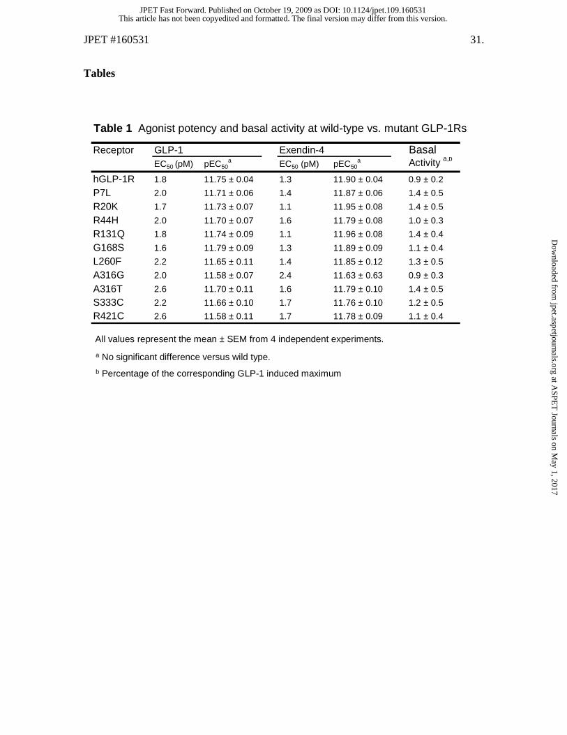
Table 1 Agonist potency and basal activity at wild-type vs. mutant GLP-1Rs
All values represent the mean ± SEM from 4 independent experiments.
a No significant difference versus wild type.
b Percentage of the corresponding GLP-1 induced maximum
Receptor GLP-1 Exendin-4 BasalEC50 (pM) pEC50
a EC50 (pM) pEC50a Activity a,b
hGLP-1R 1.8 11.75 ± 0.04 1.3 11.90 ± 0.04 0.9 ± 0.2
P7L 2.0 11.71 ± 0.06 1.4 11.87 ± 0.06 1.4 ± 0.5
R20K 1.7 11.73 ± 0.07 1.1 11.95 ± 0.08 1.4 ± 0.5
R44H 2.0 11.70 ± 0.07 1.6 11.79 ± 0.08 1.0 ± 0.3
R131Q 1.8 11.74 ± 0.09 1.1 11.96 ± 0.08 1.4 ± 0.4
G168S 1.6 11.79 ± 0.09 1.3 11.89 ± 0.09 1.1 ± 0.4
L260F 2.2 11.65 ± 0.11 1.4 11.85 ± 0.12 1.3 ± 0.5
A316G 2.0 11.58 ± 0.07 2.4 11.63 ± 0.63 0.9 ± 0.3
A316T 2.6 11.70 ± 0.11 1.6 11.79 ± 0.10 1.4 ± 0.5
S333C 2.2 11.66 ± 0.10 1.7 11.76 ± 0.10 1.2 ± 0.5
R421C 2.6 11.58 ± 0.11 1.7 11.78 ± 0.09 1.1 ± 0.4
This article has not been copyedited and formatted. The final version may differ from this version.JPET Fast Forward. Published on October 19, 2009 as DOI: 10.1124/jpet.109.160531
at ASPE
T Journals on M
ay 1, 2017jpet.aspetjournals.org
Dow
nloaded from
JPET #160531 32.
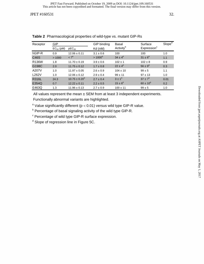
Table 2 Pharmacological properties of wild-type vs. mutant GIP-Rs
All values represent the mean ± SEM from at least 3 independent experiments.
a Value significantly different (p < 0.01) versus wild type GIP-R value. b Percentage of basal signaling activity of the wild type GIP-R. c Percentage of wild type GIP-R surface expression. d Slope of regression line in Figure 5C.
Functionally abnormal variants are highlighted.
Receptor GIP GIP binding Basal Surface Sloped
EC50 (pM) pEC50 Kd (nM) Activityb Expressionc
hGIP-R 0.9 12.06 ± 0.11 3.1 ± 0.6 100 100 1.0
C46S > 1000 < 7a > 1000a 34 ± 4a 31 ± 6a1.1
R136W 1.8 11.70 ± 0.19 3.9 ± 0.6 102 ± 1 102 ± 8 0.9
G198C 2.0 11.75 ± 0.12 1.7 ± 0.8 22 ± 4a 56 ± 9a0.3
A207V 1.0 11.97 ± 0.05 2.6 ± 0.9 104 ± 10 99 ± 5 1.1
L262V 1.0 12.06 ± 0.12 2.9 ± 0.4 99 ± 11 97 ± 13 1.0
R316L 24.3 10.75 ± 0.20a2.7 ± 0.4 3 ± 1a 37 ± 7a
0.01
E354Q 0.7 12.22 ± 0.11 2.2 ± 0.5 15 ± 6a 60 ± 10a0.2
E463Q 1.3 11.96 ± 0.13 2.7 ± 0.9 100 ± 11 99 ± 5 1.0
This article has not been copyedited and formatted. The final version may differ from this version.JPET Fast Forward. Published on October 19, 2009 as DOI: 10.1124/jpet.109.160531
at ASPE
T Journals on M
ay 1, 2017jpet.aspetjournals.org
Dow
nloaded from
AR
E
E
C
G
L
R
46
136 198 207
316
463
N-
C-
354
262
Figure 1
A.
GIP-R
B.
C-
PN- R R
R
G
Q
F S
A
R
720 44
131
168
221
260
316
333421
GLP-1R
29 30
HA-tag
E 463
NH2
G
COOH
NH2
COOH
Q
This article has not been copyedited and formatted. The final version may differ from this version.JPET Fast Forward. Published on October 19, 2009 as DOI: 10.1124/jpet.109.160531
at ASPE
T Journals on M
ay 1, 2017jpet.aspetjournals.org
Dow
nloaded from
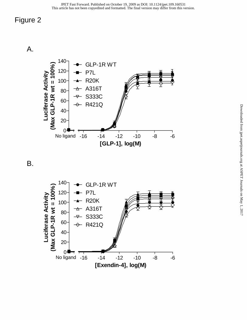
Figure 2
A.
B.
No ligand
No ligand
0
20
40
60
80
100
120
140
-16 -14 -12 -10 -8 -6
GLP-1R WTP7LR20KA316TS333CR421Q
[GLP-1], log(M)
Lu
cife
rase
Act
ivit
y(M
ax G
LP
-1R
wt
= 1
00%
)
0
20
40
60
80
100
120
140
-16 -14 -12 -10 -8 -6
GLP-1R WTP7LR20KA316TS333CR421Q
[Exendin-4], log(M)
Lu
cife
rase
Act
ivit
y(M
ax G
LP
-1R
wt
= 1
00%
)
This article has not been copyedited and formatted. The final version may differ from this version.JPET Fast Forward. Published on October 19, 2009 as DOI: 10.1124/jpet.109.160531
at ASPE
T Journals on M
ay 1, 2017jpet.aspetjournals.org
Dow
nloaded from
-170
20
40
60
80
100
120
-16 -14 -12 -10
pcDNA1GIP-R wtL262VR316LE354QE463Q
[GIP], log(M)
Luc
ifera
se A
ctiv
ity(m
ax G
IP-R
wt
= 1
00%
)
-170
20
40
60
80
100
120
-16 -14 -12 -10 -8 -6
pcDNA1GIP-R wtC46SR136WG198CA207V
[GIP], log(M)
Lu
cife
rase
Act
ivit
y(m
ax G
IP-R
wt
= 1
00%
)
Figure 3
No ligand
No ligand
This article has not been copyedited and formatted. The final version may differ from this version.JPET Fast Forward. Published on October 19, 2009 as DOI: 10.1124/jpet.109.160531
at ASPE
T Journals on M
ay 1, 2017jpet.aspetjournals.org
Dow
nloaded from

-110
20
40
60
80
100
120
-10 -9 -8 -7 -6
GIP-R WT
R136WG198CA207V
R316LE354Q
L262V
E463Q
[GIP], log(M)
125 I-
GIP
Bin
ding
(%B
/Bo)
Figure 4
Control
This article has not been copyedited and formatted. The final version may differ from this version.JPET Fast Forward. Published on October 19, 2009 as DOI: 10.1124/jpet.109.160531
at ASPE
T Journals on M
ay 1, 2017jpet.aspetjournals.org
Dow
nloaded from

Figure 5
A.
B.
0.0 0.5 1.0 1.5 2.00
20
40
60
80
100
120GIP-R WTC46SR136WG198CA207VL262V
E354QE463Q
R316L
[Receptor cDNA], (ng/well)
Sur
face
Exp
ress
ion
(Max
wt
= 1
00%
)
0.0 0.5 1.0 1.5 2.00
20
40
60
80
100
120 GIP-R WTC46SR136WG198CA207VL262VR316LE354QE463QpcDNA1
[Receptor cDNA], (ng/well)
Luci
fera
se A
ctiv
ity
(Max
wt
= 10
0%)
C.
0 20 40 60 80 100 1200
20
40
60
80
100
120GIPR-WTC46SR136WG198CA207VL262VR316LE354QE463Q
Receptor Expression(WT = 100%)
Bas
al A
ctiv
ity
(WT
= 1
00%
)
This article has not been copyedited and formatted. The final version may differ from this version.JPET Fast Forward. Published on October 19, 2009 as DOI: 10.1124/jpet.109.160531
at ASPE
T Journals on M
ay 1, 2017jpet.aspetjournals.org
Dow
nloaded from