peroxisomeproliferator-activatedreceptor … this novel pathway offers unique opportunities to...
TRANSCRIPT

Peroxisome Proliferator-activated Receptor � Co-activator1-� as a Critical Co-activator of the Murine Hepatic OxidativeStress Response and Mitochondrial Biogenesis inStaphylococcus aureus Sepsis*
Received for publication, August 21, 2013, and in revised form, November 6, 2013 Published, JBC Papers in Press, November 19, 2013, DOI 10.1074/jbc.M113.512483
Anne D. Cherry1, Hagir B. Suliman, Raquel R. Bartz, and Claude A. PiantadosiFrom the Departments of Anesthesiology, Medicine and Pathology, Duke University Medical Center,Durham, North Carolina 27710
Background: PGC-1� regulates mitochondrial biogenesis, and may participate in antioxidant gene regulation.Results: PGC-1�-deficient mice in sepsis demonstrated increased hepatocellular mitochondrial oxidative stress and impairedantioxidant enzyme induction, reflecting PGC-1� interaction with the ARE-dependent Nfe2l2 transcription factor and Sod2activation.Conclusion: PGC-1� is critical to mitochondrial SOD-2 induction during hepatic inflammation.Significance: This novel pathway offers unique opportunities to mitigate oxidative mitochondrial damage.
A key transcriptional regulator of cell metabolism, the per-oxisome proliferator-activated receptor � co-activator 1-�(PPARGC-1-� or PGC-1�), also regulates mitochondrial bio-genesis, but its role in antioxidant gene regulation is not wellunderstood. Here, we asked whether genetic heterozygosity ofPGC-1� modulates gene expression for the mitochondrial anti-oxidant enzyme SOD-2 during hepatic inflammatory stress.Using Staphylococcus aureus peritonitis in mice, we found sig-nificant Sod2 gene induction in WT mice, whereas PGC-1�heterozygotes (PGC-1��/�) failed to augment Sod2 mRNA andprotein levels. Impaired Sod2 regulation in PGC-1��/� micewas accompanied by oxidative stress shown by elevated mito-chondrial GSSG/GSH and protein carbonyls. In silico analysis ofthe mouse proximal Sod2 promoter region revealed consensusbinding sites for the Nfe2l2 (Nrf2) transcription factor. Chroma-tin immunoprecipitation demonstrated diminished Nfe2l2 pro-tein binding to the antioxidant response element promoter siteproximal to the Sod2 start site in PGC-1� heterozygous mice,implicating PGC-1� in facilitation of Nfe2l2 DNA binding.Nuclear protein co-immunoprecipitation demonstrated aninteraction between hepatic Nfe2l2 and PGC-1� in WT micethat was greatly reduced in PGC-1��/� mice. The data indicatethat PGC-1� promotes mitochondrial antioxidant enzymeexpression through Nfe2l2-mediated SOD-2 expression in sep-sis. The presence of this new PGC-1�-dependent signaling axisindicates that PGC-1� opposes mitochondrial oxidative stressby means of selective induction of one or more antioxidantresponse element-driven genes. By implication, exploitation ofthis axis could lead to new pharmacological interventions toimprove the antioxidant defenses during oxidative stress-in-duced mitochondrial damage.
Aerobic energy conservation within living cells is dependenton the functional density of mitochondria, which in turn is reg-ulated by energy requirements (1). Indeed, the physiologicaldemands of thermogenesis (2), exercise (3), and postnatalgrowth (4), all states of up-regulated metabolism, induce mito-chondrial biogenesis. This induction is responsive to externalcues, indicating the need for increased ATP production, and ismediated by a variety of transcription factors, co-activators,and co-repressors.
The PPARg co-activator 1 (PGC-1)2 family of co-activators iscentrally important to the regulation of mitochondrial biogen-esis. PGC-1�, one of three known co-activators of the family,partners with multiple transcription factors including nuclearrespiratory factor-1 (NRF-1), nuclear respiratory factor-2(NRF-2; GABPA), MEF-2, estrogen-related receptor � (ERR�),and the peroxisome proliferator-activated receptors, all ofwhich regulate respiratory gene expression (5). PGC-1� isoften designated a master regulator of the metabolicresponses to cold exposure, fasting, exercise, and mitochon-drial stress (6 –12).
As the rate of oxidative metabolism increases, so does therate of mitochondrial reactive oxygen species (ROS) produc-tion (13). An imbalance between this higher mitochondrial ROSproduction and ROS scavenging leads to the induction of ROS-detoxifying enzymes such as superoxide dismutase (SOD-2) andothers. Normally, functional nuclear PGC-1� is integral to theexpression of pathways that enhance oxidative metabolism, butthe protein has also been implicated in the up-regulation of ROS-detoxifying enzymes (14–17).
Given the importance of PGC-1� to the maintenance ofmetabolism and energy production for a broad range of condi-
* This work was supported, in whole or in part, by National Institutes of HealthGrants RO1-A1095424 and R01-HL108801 (to C. A. P.).
1 To whom correspondence should be addressed: Dept. of Anesthesiology,Duke University, DUMC Box 3094, Durham, NC 27710. Tel.: 919-668-6208;Fax: 919-613-5264; E-mail: [email protected].
2 The abbreviations used are: PGC-1, PPARg co-activator 1; PPAR, peroxisomeproliferator-activated receptor; NRF-1, nuclear respiratory factor-1; GABPA,GA-binding protein � chain; ROS, reactive oxygen species; SOD, superox-ide dismutase; mtTFA, mitochondrial transcription factor A; ARE, antioxi-dant response element; DNPH, 2,4-dinitrophenylhydrazine; Co-IP, co-immunoprecipitation.
THE JOURNAL OF BIOLOGICAL CHEMISTRY VOL. 289, NO. 1, pp. 41–52, January 3, 2014© 2014 by The American Society for Biochemistry and Molecular Biology, Inc. Published in the U.S.A.
JANUARY 3, 2014 • VOLUME 289 • NUMBER 1 JOURNAL OF BIOLOGICAL CHEMISTRY 41
by guest on March 27, 2019
http://ww
w.jbc.org/
Dow
nloaded from

tions, it is not surprising that PGC-1� knock-out mice display anumber of maladaptive phenotypes including miniature stat-ure, exercise and cold intolerance, fasting hypoglycemia,hepatic steatosis, decreased cardiac reserve and heart failure,hyperactivity, and in the brain, evidence of axonal degeneration(18 –20). However, PGC-1� heterozygous knock-out (PGC-1��/�) mice are viable and healthy, and they are capable ofmaintaining basal metabolism and mitochondrial biogenesis.
During severe inflammatory stress, such as bacterial sepsis,mitochondrial damage can be extensive. Such damage leads toincreased synthesis and activity of PGC-1� and its transcriptionfactor partners, with the induction of mitochondrial biogen-esis and restoration of respiratory function (21, 22). Here,using an established mouse model of Staphylococcus aureusperitonitis and sepsis, we compared the impact of variablePGC-1� induction on the regulation of the mitochondrialantioxidant defenses in wild type (WT) and PGC-1�heterozygote knock-out (PGC-1��/�) mice. We hypothe-sized that a single Ppargc1� (PGC-1�) allele would be insuffi-cient for the transcriptional co-activation and maintenance ofhigh expression levels of critical target antioxidant genes in asentinel organ, the liver, under stress. This could lead todepressed enzyme function and decreased defenses againstincreased hepatic ROS production in sepsis. After a series ofexploratory studies, we further evaluated the role of PGC-1� inthe antioxidant response element (ARE)-dependent expressionof the key mitochondrial antioxidant enzyme SOD-2.
EXPERIMENTAL PROCEDURES
Mouse Studies—The mouse studies were conducted on aprotocol approved by the Duke University (Durham, NC) Insti-tutional Animal Care and Use Committee. Young adultC57Bl6/J mice purchased from The Jackson Laboratory (BarHarbor, ME) served as WT control animals. HeterozygotePGC-1� null mice (PGC-1��/�) on a C57Bl6/J backgroundwere obtained from The Jackson Laboratory and bred at ourinstitution. Healthy mice underwent sterile midline laparotomyafter induction of anesthesia with a single injection of ketamineand xylazine. Using an established sepsis model (21, 22), theperitoneal cavities were implanted with 500-�l fibrin clots con-taining 1 � 106 CFU/ml S. aureus. Closure was achieved usingproline sutures. Mice were killed at 6 and 24 h by aortic tran-section under isoflurane anesthesia. The livers were harvestedimmediately for fresh use or flash-frozen for later use. Controllivers were obtained from WT and PGC-1a heterozygoteknock-out mice without clot implantation.
Preparation of Total, Nuclear, and Mitochondrial ProteinSamples—Total protein extracts were prepared from flash-fro-zen liver using mechanical homogenization followed by sonica-tion in radioimmunoprecipitation assay buffer. High speedcentrifugation (13,200 rpm for 18 min) was used to eliminateremaining heavy cellular components. Fresh liver nuclei wereisolated immediately after organ harvest using Dounce homog-enization (glass/glass) and low speed centrifugation (3000 rpmfor 10 min) followed by resuspension and a 15-min incubationin cell lysis buffer (5 mM PIPES, 85 mM KCl, and 0.5% NonidetP-40 with protease inhibitors/PMSF). After low speed centrif-ugation (5000 rpm for 5 min), the pellet was stored at �80 °C.
Before freezing, nuclear samples for chromatin immunopre-cipitation (ChIP) assays were treated with 1% formaldehydefollowed by quenching with 2.5 M glycine for DNA-proteincross-linking.
Mitochondria were isolated from fresh tissue by glasshomogenization in isolation buffer (0.25 M sucrose, 10 mM Trisbase, 0.5 mM EDTA, and 0.5% BSA at pH 7.4) (23) followed bylow speed centrifugation (4000 rpm for 4 min) to eliminateheavy aggregates. The supernatant was centrifuged at highspeed (10,200 rpm for 8 min) to recover the mitochondrialfraction.
PAGE and Western Blot Analysis—After protein assay, indi-vidual proteins were resolved by electrophoresis on NovexTris-glycine polyacrylamide gels (Invitrogen) followed bytransfer to polyvinylidene difluoride membranes (PVDF, Milli-pore, Billerica, MA). Proteins were probed with primary anti-bodies to polyclonal rabbit anti-PGC-1� (Cayman Chemical,Ann Arbor, MI), rabbit anti-SOD-2 (Abcam, Cambridge, MA),rabbit anti-nuclear factor (erythroid-derived 2)-like 2 (Nfe2l2H300, Santa Cruz Biotechnology, Santa Cruz, CA), rabbit anti-programmed cell death-1 (mPD-1, AnaSpec, Fremont, CA),rabbit anti-mitofusin-2 (Mfn-2, Epitomics, Burlingame, CA),and mouse anti-NAD(P)H dehydrogenase (quinone) 1 (QR1,Santa Cruz Biotechnology). Antibodies to NRF-1, NRF-2(GABPA), and mitochondrial transcription factor A (mtTFA)developed in rabbits were characterized in our laboratory (24).After application of primary antibodies and washing, mem-branes were incubated with the appropriate horseradish perox-idase-conjugated secondary antibodies (Santa Cruz Biotech-nology). Membranes were developed with chemiluminescencereagent (Calbiochem RapidStep, EMD Chemicals; or luminol,Santa Cruz Biotechnology), and proteins were quantified ondigitized images using Quantity One (Bio-Rad). Densitometrymeasurements were normalized to �-actin or tubulin in thesame samples.
Extraction and Quantification of mtDNA and mRNA—Genomic DNA was purified from flash-frozen tissue after tissuedigestion, cell lysis, protein denaturation (proteinase K), andRNA degradation (RNase) using a silica-based membrane(GenElute mammalian genomic DNA miniprep kit, Sigma-Al-drich). The mtDNA copy number was determined using real-time PCR (quantitative PCR) (25).
Total RNA was extracted from liver tissue using TRIzol reagent(Invitrogen). RNA purity was confirmed on 1.2% agarose, and theRNA was converted to cDNA using oligo(dT) (ImProm-II reversetranscription system; A3800). Real-time RT-PCR was performedon an ABI Prism 7000 using gene expression assays (Applied Bio-systems). A �Ct method was used to quantify mRNA levels forPpargc1�, Nrf1, mitochondrial transcription factor A (Tfam),Sod2, Hmox1, mitochondrial thioredoxin (Txn2), tumor necro-sis factor (Tnf), C-C motif chemokine 2 (Ccl2), and interleu-kin-10 (IL10). Amplification efficiency was checked with aninternal 18 S rRNA (AB PN 4332078) over a 0.9 –90-ng range ofRNA, and gene expression was quantified using ABI Prism 7000SDS and MS Excel software. Each sample was assayed intriplicate.
TUNEL Assay—Paraffin-embedded liver thin sections wereused for the TUNEL assay. Samples from healthy control and
PGC-1� Co-activation of the Oxidative Stress Response
42 JOURNAL OF BIOLOGICAL CHEMISTRY VOLUME 289 • NUMBER 1 • JANUARY 3, 2014
by guest on March 27, 2019
http://ww
w.jbc.org/
Dow
nloaded from
24-h post-sepsis livers were heated to 60 °C for 45 min anddeparaffinized by two changes of xylene. Sections were rehy-drated through a series of 5-min soaks in graded ethanol solu-tions. After washing for 10 min, sections were permeabilized for30 min at 37 °C with proteinase K solution. A TUNEL reactionmixture (enzyme and label, Roche Applied Science, Mann-heim, Germany) was then applied to the sections for 1 h at37 °C. Negative and positive control sections were preparedwith label solution only or by preinduction of strand break-age with recombinant DNase I. After incubation, sampleswere analyzed for the proportion of apoptotic cells usingfluorescence microscopy.
GSSG/GSH and Glutathionylation Assays—Mitochondria (1�g of protein/�l) were centrifuged at 700 � g for 10 min. Super-natant was removed, and protein was precipitated from thepelleted mitochondria via sonication in 5% metaphosphoricacid followed by centrifugation at 1000 � g for 10 min at 4 °C.The supernatant, containing mitochondrial GSH, was rapidlyremoved and stored at �80 °C until GSH assay. The total glu-tathione (GSH) and glutathione disulfide (GSSG) content of themitochondria was determined by an enzymatic recycling assaybased on glutathione reductase (26). GSSG levels were deter-mined after derivatization of GSH by 2-vinylpyridine.
Mitochondrial protein S-glutathionylation levels were meas-ured using a bacterial glutaredoxin 1 (GRX1) reduction assaywith S-biotinylation and chemiluminescence detection. Mito-chondrial samples were treated with 20 mM N-ethylmaleimideat room temperature for 30 min to alkylate free thiols. Afterrepeated acetone precipitation, samples were resuspended inTris buffer (pH 7.5), and S-glutathionylated proteins werereduced to free thiols by incubation with Escherichia coli GRX1(Cayman Chemical), GSSG reductase (Sigma), 1 mM GSH, 1mM NADPH, and 1 mM EDTA for 15 min at 37 °C. Reducedsulfhydryl groups were labeled by application of 2 mM biotinN-[6-(Biotinamido)hexyl]-3�-(2�-pyridyldithio)-propionamide(Biotin-HPDP) for 1 h at room temperature. Negative controlswere treated identically but without GRX1, whereas positivecontrols were treated with 2 mM DTT. Samples and controlswere applied to an HSI vacuum slot blotter containing a PVDFmembrane (Hoefer Scientific, San Francisco, CA). StreptavidinHRP (Thermo Fisher Scientific) was applied, and the mem-branes were developed using chemiluminescence detection(Santa Cruz Biotechnology) and quantified on digitized imagesusing Image J (National Institutes of Health, Bethesda, MD).
DNPH-derivatized Protein Carbonyl Quantification—Flash-frozen tissue was homogenized (glass/glass) on ice in Tris/HClcontaining 1 mM butylated hydroxytoluene, desferoxaminemesylate, and commercial protease and phosphatase inhibitormixture. Aliquots of 1.5 mg of protein were reacted with 10 mM
DNPH in 2.5 M HCl. After mixing with cold TCA, the sampleswere centrifuged at high speed, and the pellets were againwashed with TCA and then repeatedly washed (�6) with 1:1ethanol/ethyl acetate to remove free DNPH. The final threewashes were accompanied by sonication. The pellets wereallowed to dry, dissolved in 6 M guanidine HCl, and diluted �0.1with sodium phosphate. After high speed centrifugation to clar-ify the supernatant, the protein content was adjusted to a stand-ard concentration (5 �g/100 �l). Samples, along with a negative
control (treated with NaBH4), were applied to an HSI vacuumslot blotter containing a PVDF membrane (Hoefer Scientific).An immunoassay was performed using a primary antibody(rabbit anti-dinitrophenol, 1:150 dilution, Millipore) followedby a secondary antibody (goat anti-rabbit, 1:300 dilution, Mil-lipore). The membranes were developed using chemilumines-cence detection, and protein carbonyl concentration was quan-tified on digitized images using Quantity One (Bio-Rad).
Myeloperoxidase Assay—A myeloperoxidase activity assaywas used as described to estimate the neutrophil content ofliver tissue (27). Frozen liver tissue was mixed with 1% hexade-cyltrimethylammonium bromide solution, homogenized, andsonicated. After centrifugation, the supernatant was mixedwith o-dianisidine and 0.3% H2O2. Spectrophotometric mea-surement of change in optical density (406 nm) per minute pergram of liver was reported.
Bioinformatics—In silico analysis of the mouse Sod2 pro-moter (Ensembl Gene ID ENSMUSG00000006818) was per-formed for consensus sequences for transcription factorsNRF-1, GABPA, and Nfe2l2 binding sites using DNASIS (Hita-chi Software; Alameda, CA) and confirmed with MatInspector(Genomatix Software; München, Germany).
Chromatin Immunoprecipitation (ChIP)—Nuclei for ChIPassays were sonicated in shearing buffer, and shearing effective-ness was confirmed by electrophoresis on ethidium bromide-stained agarose gels. Samples were then processed for immu-noprecipitation using a kit (ChIP IT assay kit, Active Motif,Carlsbad, CA) and antibodies to NRF-1 (Cayman), GABPA(Cayman), and Nfe2l2 according to the manufacturer’s instruc-tions. After precipitation, cross-linking was reversed, and PCRwas carried out using 1 �l of each sample (input DNA dilution1:10, immunoprecipitated fractions undiluted) in PCR buffer(Qiagen, Valencia, CA) containing dNTP (Invitrogen) and TaqDNA polymerase (Qiagen) with the primers 900CGCTTCGCT-GTGTCCTTGC882 and 776TAATGTTGTGTCGGGCGGC794
to amplify NRF-1 site, 922CCGTCCTCCCCTCCGCTGAT941,and 774ACGCCGCCCGACACAACATT793 to amplify Nfe2l2sites, and 1383GCTGCACCCGGAGTCCGCAA1364 and 1134TGG-CTGTGAGTCGCAAAGCTTCC1156 to amplify GABPA sitesfor the Sod2 promoter. For the Hmox1 promoter, we usedprimers 1350CCTGAAGGGCTACTCCCGTCTTCC1327 and1051TTGCAACATCCAGCCCGGAGGC1072. DNA concen-tration was measured and standardized across samples priorto PCR. PCR products were analyzed by agarose gelelectrophoresis.
Protein Co-immunoprecipitation (Co-IP)—Nuclear protein(500 �g) in Co-IP buffer (120 mM sodium chloride, 50 mM
Hepes [pH 7.5], anti-protease, and anti-phosphatase inhibitors)with 0.25% digitonin was incubated for 3 h with the primaryantibody (10 �l of rabbit polyclonal PGC-1 primary antibody(Cayman Chemical)) for 3 h at 4 °C. A suspension of proteinG-Sepharose beads (Sigma E3403 EZview Red protein G affin-ity gel) was added for 30 min to select for proteins bound toPGC-1�. The captured immune complexes were layered on a1-ml 1 M sucrose cushion (in Co-IP buffer with 0.5% w/w digi-tonin) and centrifuged (10,000 � g for 5 min) followed by wash-ing at least three times with Co-IP buffer (containing 0.25% w/wdigitonin). The beaded complex was washed once with Co-IP
PGC-1� Co-activation of the Oxidative Stress Response
JANUARY 3, 2014 • VOLUME 289 • NUMBER 1 JOURNAL OF BIOLOGICAL CHEMISTRY 43
by guest on March 27, 2019
http://ww
w.jbc.org/
Dow
nloaded from
buffer lacking digitonin. After washing, 1� sample buffer (Bio-Rad) with DTT was added to the samples, which were dena-tured by heating to 95 °C for 10 min. Separate 20-�g nuclearsamples were diluted 1:1 with 2� sample buffer and heated asinput controls. Co-IP samples were magnetized after heating,and supernatant aliquots were analyzed for Nfe2l2 (66 kDa;Santa Cruz Biotechnology) by Western blot analysis using beadand input control samples.
Statistical Analysis—All grouped data are presented asmeans � S.D. Statistical significance was determined using twoway analysis of variance and Student’s t test for post hoc analy-sis with JMP software (version 11, SAS Institute Inc., Cary, NC).p � 0.05 was considered significant.
RESULTS
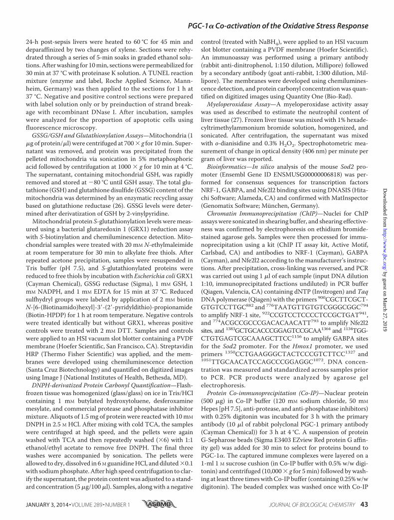
Ppargc1� mRNA and Protein Expression in HeterozygousPGC-1� Mice—Differences in hepatic Ppargc1� mRNA levelswere detectable by real-time RT-PCR in WT and in PGC-1��/� mice following S. aureus clot implantation. There wereno differences between the groups in base-line Ppargc1� tran-script levels, but after peritoneal inoculation, the mRNA levelsincreased by 6 h and returned toward base line by 24 h (Fig. 1A).As expected, the magnitude of the increase in Ppargc1� mRNAwas significantly less in the PGC-1��/� mice than in WT mice.Western blot analysis of total liver PGC-1� protein showed asignificant increase from base line in WT mice at 24 h but nochange in the PGC-1��/� animals (Fig. 1, B and C). Thus,PGC-1� heterozygosity significantly blunts hepatic Ppargc1�mRNA and protein induction in response to S. aureus sepsis.
Induction of Mitochondrial Biogenesis—The redox-depen-dent induction of mitochondrial biogenesis is a key survivalfactor in sepsis (28); hence we evaluated mRNA and proteinlevels in our strains of mice for three key transcription factorsinvolved in mitochondrial biogenesis: NRF-1, GABPA, andmtTFA. NRF-1 and GABPA are necessary for the up-regulationof mtTFA and other nucleus-encoded, mitochondria-targetedproteins involved in mtDNA transcription and replication. Wefound a progressive increase in Nrf1 and Tfam mRNA levels byreal-time RT-PCR at 6 and 24 h in the WT mice, but nonsignif-icant changes in these mRNA levels in PGC-1��/� animals(Fig. 2, A and B). Liver protein levels assessed at 0 and 24 h byWestern blot revealed an increase in NRF-1, GABPA, andmtTFA levels by 24 h in WT and PGC-1��/� mice, but equalinduction of only GABPA in the PGC-1��/� mice (Fig. 2, C–F).The mtDNA copy number was stable at 6 h, but we found anincrease in both strains at 24 h that was significantly greater inthe WT mice (Fig. 2G). These results support an adverse effectof impaired PGC-1� protein synthesis in the PGC-1��/� miceon the induction of mitochondrial biogenesis and the bioener-getic response to sepsis. Moreover, hepatic nuclear protein lev-els for PGC-1� and NRF-1 were impressively elevated in WTmice when compared with PGC-1��/� mice (Fig. 2, B and D).In addition, there were significant between strain differences inthe nuclear levels of two other key redox-sensitive proteinsinvolved in the regulation of mitochondrial biogenesis, Nfe2l2and SIRT2 (Fig. 3, C and E).
Oxidative Stress Assays—We evaluated overall hepatic oxida-tive stress by assaying mitochondrial glutathione levels. Base-
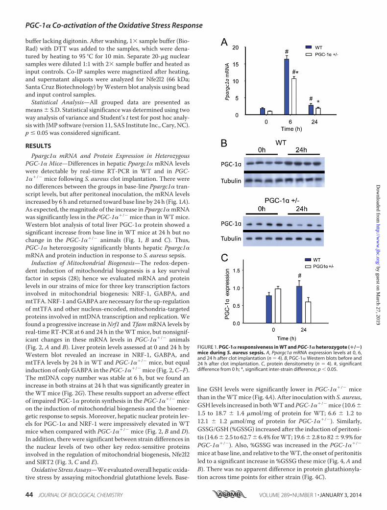
line GSH levels were significantly lower in PGC-1��/� micethan in the WT mice (Fig. 4A). After inoculation with S. aureus,GSH levels increased in both WT and PGC-1��/� mice (10.6 �1.5 to 18.7 � 1.4 �mol/mg of protein for WT; 6.6 � 1.2 to12.1 � 1.2 �mol/mg of protein for PGC-1��/�). Similarly,GSSG/GSH (%GSSG) increased after the induction of peritoni-tis (14.6 � 2.5 to 62.7 � 6.4% for WT; 19.6 � 2.8 to 82 � 9.9% forPGC-1��/�). Also, %GSSG was increased in the PGC-1��/�
mice at base line, and relative to the WT, the onset of peritonitisled to a significant increase in %GSSG these mice (Fig. 4, A andB). There was no apparent difference in protein glutathionyla-tion across time points for either strain (Fig. 4C).
FIGURE 1. PGC-1� responsiveness in WT and PGC-1� heterozygote (�/�)mice during S. aureus sepsis. A, Ppargc1a mRNA expression levels at 0, 6,and 24 h after clot implantation (n � 4). B, PGC-1� Western blots before and24 h after clot implantation. C, protein densitometry (n � 4). #, significantdifference from 0 h; *, significant inter-strain difference; p 0.05.
PGC-1� Co-activation of the Oxidative Stress Response
44 JOURNAL OF BIOLOGICAL CHEMISTRY VOLUME 289 • NUMBER 1 • JANUARY 3, 2014
by guest on March 27, 2019
http://ww
w.jbc.org/
Dow
nloaded from

Differences in oxidative stress levels between WT and PGC-1��/� mice were confirmed by quantification of mitochondrialprotein oxidation using a standardized protein carbonyl assay.Levels of oxidized mitochondrial protein increased in the liversof mice of both strains after induction of peritonitis (Fig. 4D).The PGC-1��/� mice demonstrated higher base-line proteincarbonyl levels than their WT counterparts, and this differencepersisted at 6 h after peritonitis. By 24 h after inoculation, thelevels of oxidized mitochondrial protein had decreased toward
base-line level, particularly in WT mice, perhaps reflecting acti-vation of GSH synthesis, more rapid disposal of oxidized pro-tein, and/or an increase in other antioxidant defenses and anability to limit protein carbonylation (29).
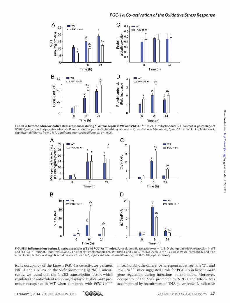
Inflammation and Hepatocellular Damage—To quantify thedegree of hepatocellular damage induced by oxidative stressduring S. aureus sepsis, we measured myeloperoxidase activityas a surrogate for acute inflammation, which typically reflectsthe early neutrophilic inflammatory response to sepsis.
FIGURE 2. Mitochondrial biogenesis transcription factor expression in sepsis. A and B, changes in mRNA levels at 0, 6, and 24 h after clot implantation forNrf1 (A) and Tfam (n � 4) (B). C, representative Western blot analysis. D–F, protein densitometry for NRF-1 (D); GABPA (E); and mtTFA (n � 4) (F). G, mtDNA copynumber in WT and PGC-1��/� mice at 0 h (controls) and 24h (n � 2). #, significant difference from 0 h; *, significant inter-strain difference; p 0.05.
PGC-1� Co-activation of the Oxidative Stress Response
JANUARY 3, 2014 • VOLUME 289 • NUMBER 1 JOURNAL OF BIOLOGICAL CHEMISTRY 45
by guest on March 27, 2019
http://ww
w.jbc.org/
Dow
nloaded from

Myeloperoxidase levels were similarly elevated at 6 and 24 h inboth strains of mice (Fig. 5A). However, the hepatic inductionof early-phase proinflammatory cytokines and chemokines dif-fered in WT and PGC-1��/� mice before and after S. aureusinoculation (Fig. 5, B and C). The mRNA levels for two keypotentially damaging mediators, Tnf and Ccl2, increased signif-icantly more in PGC-1��/� than in WT mice, whereas anti-inflammatory IL10 cytokine levels were lower in PGC-1��/�
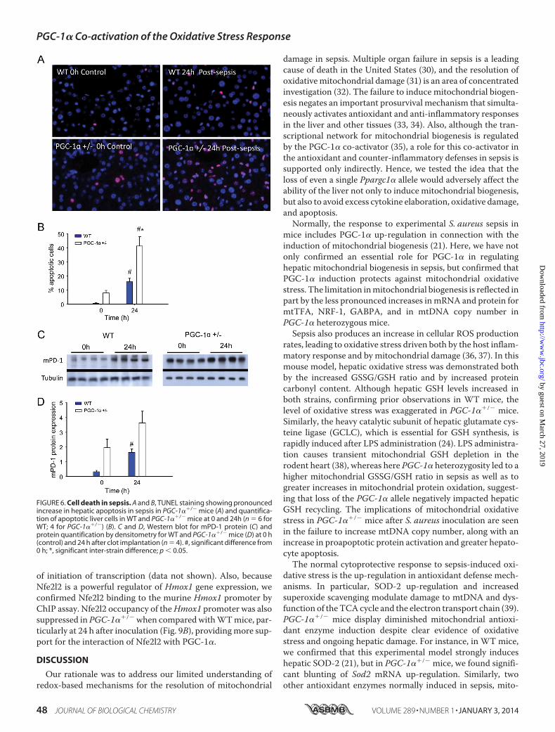
mice than in WT mice (Fig. 5D). The two proinflammatorycytokine levels peaked at 6 h and returned to base line by 24 h inWT mice, but remained elevated in PGC-1��/� mice. Quanti-fication of hepatocellular cell death by TUNEL staining sug-gested a trend toward more base-line apoptosis in PGC-1��/�
mice, but a significant increase in the fraction of apoptotic cellsat 24 h in PGC-1��/� when compared with WT mice (0.01 �0.01 to 0.16 � 0.06 for WT versus 0.08 � 0.04 to 0.41 � 0.02 for
PGC-1��/�) (Fig. 6, A and B). Of further interest, the extent ofhepatic cell death indicated by programmed cell death protein 1(mPD-1) increased from base line in both strains of mice at 24 hafter S. aureus inoculation, but remained significantly higher inthe PGC-1��/� mice (Fig. 6, C and D).
Antioxidant Enzyme Induction in WT and PGC-1��/� Micein Sepsis—The significant oxidative stress in PGC-1��/� dur-ing the infection relative to WT mice raised the possibility ofdifferences in antioxidant enzyme induction between the twostrains. Examination of hepatic mRNA expression for Sod2,Txn2, and Hmox1 mRNA in WT mice by real-time RT-PCRrevealed sustained increases in Txn2 and Hmox1 and anincrease in Sod2 mRNA level at 24 h after inoculation (Fig. 7,A–C). In the PGC-1��/� mice, however, Sod2 and Hmox1mRNA levels had increased only marginally at 24 h, whereasTxn2 increased marginally at 6 h and returned to base line at24 h. Among the three antioxidant enzymes, we also noted pro-nounced differences in mRNA levels in the PGC-1��/� mice insepsis (0 versus 24 h), suggesting that PGC-1� contributes dif-ferentially to the regulation of these genes (Fig. 7D).
PGC-1� and Regulation of Nfe2l2 and NRF-1 DownstreamGenes—To evaluate PGC-1� as a transcriptional co-activator,we next measured protein content for three specific nucleus-encoded downstream genes: the mitochondrial antioxidantenzyme SOD-2, Mfn-2, regulated in part by NRF-1, and theNAD(P)H dehydrogenase QR1, regulated by Nfe2l2. The PGC-1��/� mice displayed less Mfn-2 and QR1 protein inductionduring sepsis when compared with the WT (Fig. 8, A–C). Incontrast to a robust increase in SOD-2 protein in WT mice,SOD-2 did not increase after inoculation in PGC-1��/� mice(Fig. 8D). Thus, the capacity to express PGC-1� was importantfor the activation of genes downstream of both NRF-1 andNfe2l2 during sepsis. This activation enhances steady-stateexpression of QR1, Mfn-2, and SOD-2. The regulatory role ofPGC-1� in NRF-1-dependent genes is known, but the possibil-ity of PGC-1� contributing to redox-regulated gene expressionthrough an interaction with Nfe2l2 is novel. Therefore we eval-uated the association of these two proteins at 0, 6, and 24 h inour strains of mice. A protein-protein interaction for PGC-1�-Nfe2l2 was readily discernible, as was evidence of significantlyless PGC-1� binding to Nfe2l2 in PGC-1��/� mouse livernuclei at each time point (depicted in Fig. 9A, repeated twicewith separate samples for verification).
Active NRF-1, GABPA, and Nfe2l2 Binding Sites in the Sod2Proximal Promoter Region—Because SOD-2 expression was sostrongly induced in WT, but not in PGC-1��/� mice, we spe-cifically evaluated Sod2 gene promoter regulation in the mouseliver. By in silico analysis of mouse Sod2 5�-UTR (DNAsis andGenomatix), we identified potential NRF-1, GABPA, andNfe2l2 binding sites within the conserved Sod2 promotersequence. Putative core ARE (RTGAYnnnGC) canonical bind-ing sites of 100% homology were identified at positions �1701and �910. A canonical binding site (100% homology) for theNRF-1 consensus was found at position 843. We also found twoputative GABPA binding sites of at least 85% homology at posi-tions �1383 and �1100. We performed ChIP assays for Nfe2l2,NRF1, and GABPA occupancy of the Sod2 promoter at 0, 6, and24 h after S. aureus inoculation. The data demonstrated signif-
FIGURE 3. Western analysis for hepatic nuclear proteins. A, immunoblotsfor nuclear Nfe2l2 and NRF-1 transcription factors, PGC-1�, and Sir2 proteins.Tubulin is a reference protein. B–E, protein densitometry of nuclear Nfe2l2 (B);NRF-1 (C); PGC-1� (D); and SIRT2 (E) in WT and PGC-1��/� mice at 0 (control)and 24 h after clot implantation (n � 4). #, significant difference from 0 h.
PGC-1� Co-activation of the Oxidative Stress Response
46 JOURNAL OF BIOLOGICAL CHEMISTRY VOLUME 289 • NUMBER 1 • JANUARY 3, 2014
by guest on March 27, 2019
http://ww
w.jbc.org/
Dow
nloaded from
icant occupancy of the known PGC-1� co-activator partnersNRF-1 and GABPA on the Sod2 promoter (Fig. 9B). Concur-rently, we found that the Nfe2l2 transcription factor, whichregulates the antioxidant response, displayed higher Sod2 pro-moter occupancy in WT when compared with PGC-1��/�
mice. Notably, the difference in responses between the WT andPGC-1��/� mice suggested a role for PGC-1� in hepatic Sod2gene regulation during infectious inflammation. Moreover,occupancy of the Sod2 promoter by NRF-1 and Nfe2l2 wasaccompanied by recruitment of DNA polymerase II, indicative
FIGURE 4. Mitochondrial oxidative stress responses during S. aureus sepsis in WT and PGC-1��/� mice. A, mitochondrial GSH content. B, percentage ofGSSG. C, mitochondrial protein carbonyls. D, mitochondrial protein S-glutathionylation (n � 4). x-axis shows 0 (controls), 6, and 24 h after clot implantation. #,significant difference from 0 h; *, significant inter-strain difference; p 0.05.
FIGURE 5. Inflammation during S. aureus sepsis in WT and PGC-1��/� mice. A, myeloperoxidase activity (n � 4). B–D, changes in mRNA expression in WTand PGC-1��/� mice at 0 (controls), 6, and 24 h after clot implantation: Ccl2 (B), Tnf (C), and IL10 (D) mRNA levels (n � 4). x-axis shows 0 (controls), 6, and 24 hafter clot implantation. #, significant difference from 0 h; *, significant inter-strain difference; p 0.05. OD, optical density.
PGC-1� Co-activation of the Oxidative Stress Response
JANUARY 3, 2014 • VOLUME 289 • NUMBER 1 JOURNAL OF BIOLOGICAL CHEMISTRY 47
by guest on March 27, 2019
http://ww
w.jbc.org/
Dow
nloaded from
of initiation of transcription (data not shown). Also, becauseNfe2l2 is a powerful regulator of Hmox1 gene expression, weconfirmed Nfe2l2 binding to the murine Hmox1 promoter byChIP assay. Nfe2l2 occupancy of the Hmox1 promoter was alsosuppressed in PGC-1��/� when compared with WT mice, par-ticularly at 24 h after inoculation (Fig. 9B), providing more sup-port for the interaction of Nfe2l2 with PGC-1�.
DISCUSSION
Our rationale was to address our limited understanding ofredox-based mechanisms for the resolution of mitochondrial
damage in sepsis. Multiple organ failure in sepsis is a leadingcause of death in the United States (30), and the resolution ofoxidative mitochondrial damage (31) is an area of concentratedinvestigation (32). The failure to induce mitochondrial biogen-esis negates an important prosurvival mechanism that simulta-neously activates antioxidant and anti-inflammatory responsesin the liver and other tissues (33, 34). Also, although the tran-scriptional network for mitochondrial biogenesis is regulatedby the PGC-1� co-activator (35), a role for this co-activator inthe antioxidant and counter-inflammatory defenses in sepsis issupported only indirectly. Hence, we tested the idea that theloss of even a single Ppargc1� allele would adversely affect theability of the liver not only to induce mitochondrial biogenesis,but also to avoid excess cytokine elaboration, oxidative damage,and apoptosis.
Normally, the response to experimental S. aureus sepsis inmice includes PGC-1� up-regulation in connection with theinduction of mitochondrial biogenesis (21). Here, we have notonly confirmed an essential role for PGC-1� in regulatinghepatic mitochondrial biogenesis in sepsis, but confirmed thatPGC-1� induction protects against mitochondrial oxidativestress. The limitation in mitochondrial biogenesis is reflected inpart by the less pronounced increases in mRNA and protein formtTFA, NRF-1, GABPA, and in mtDNA copy number inPGC-1� heterozygous mice.
Sepsis also produces an increase in cellular ROS productionrates, leading to oxidative stress driven both by the host inflam-matory response and by mitochondrial damage (36, 37). In thismouse model, hepatic oxidative stress was demonstrated bothby the increased GSSG/GSH ratio and by increased proteincarbonyl content. Although hepatic GSH levels increased inboth strains, confirming prior observations in WT mice, thelevel of oxidative stress was exaggerated in PGC-1��/� mice.Similarly, the heavy catalytic subunit of hepatic glutamate cys-teine ligase (GCLC), which is essential for GSH synthesis, israpidly induced after LPS administration (24). LPS administra-tion causes transient mitochondrial GSH depletion in therodent heart (38), whereas here PGC-1� heterozygosity led to ahigher mitochondrial GSSG/GSH ratio in sepsis as well as togreater increases in mitochondrial protein oxidation, suggest-ing that loss of the PGC-1� allele negatively impacted hepaticGSH recycling. The implications of mitochondrial oxidativestress in PGC-1��/� mice after S. aureus inoculation are seenin the failure to increase mtDNA copy number, along with anincrease in proapoptotic protein activation and greater hepato-cyte apoptosis.
The normal cytoprotective response to sepsis-induced oxi-dative stress is the up-regulation in antioxidant defense mech-anisms. In particular, SOD-2 up-regulation and increasedsuperoxide scavenging modulate damage to mtDNA and dys-function of the TCA cycle and the electron transport chain (39).PGC-1��/� mice display diminished mitochondrial antioxi-dant enzyme induction despite clear evidence of oxidativestress and ongoing hepatic damage. For instance, in WT mice,we confirmed that this experimental model strongly induceshepatic SOD-2 (21), but in PGC-1��/� mice, we found signifi-cant blunting of Sod2 mRNA up-regulation. Similarly, twoother antioxidant enzymes normally induced in sepsis, mito-
FIGURE 6. Cell death in sepsis. A and B, TUNEL staining showing pronouncedincrease in hepatic apoptosis in sepsis in PGC-1��/� mice (A) and quantifica-tion of apoptotic liver cells in WT and PGC-1��/� mice at 0 and 24h (n � 6 forWT; 4 for PGC-1��/�) (B). C and D, Western blot for mPD-1 protein (C) andprotein quantification by densitometry for WT and PGC-1��/� mice (D) at 0 h(control) and 24 h after clot implantation (n � 4). #, significant difference from0 h; *, significant inter-strain difference; p 0.05.
PGC-1� Co-activation of the Oxidative Stress Response
48 JOURNAL OF BIOLOGICAL CHEMISTRY VOLUME 289 • NUMBER 1 • JANUARY 3, 2014
by guest on March 27, 2019
http://ww
w.jbc.org/
Dow
nloaded from
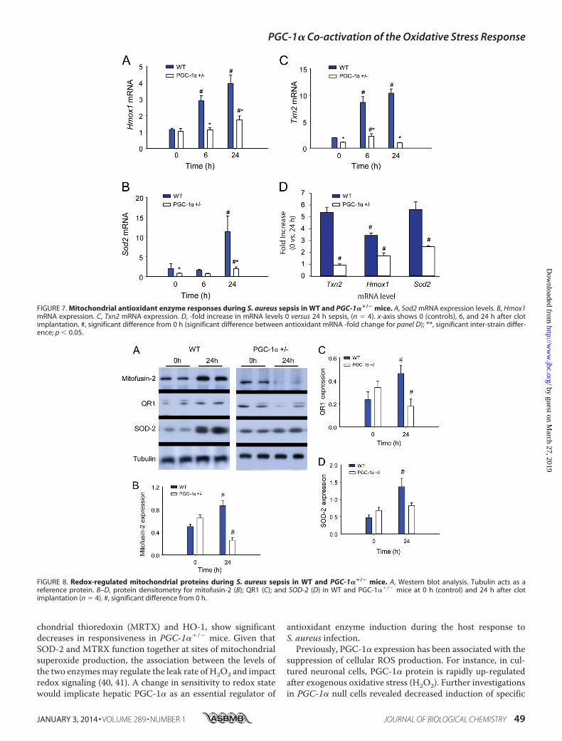
chondrial thioredoxin (MRTX) and HO-1, show significantdecreases in responsiveness in PGC-1��/� mice. Given thatSOD-2 and MTRX function together at sites of mitochondrialsuperoxide production, the association between the levels ofthe two enzymes may regulate the leak rate of H2O2 and impactredox signaling (40, 41). A change in sensitivity to redox statewould implicate hepatic PGC-1� as an essential regulator of
antioxidant enzyme induction during the host response toS. aureus infection.
Previously, PGC-1� expression has been associated with thesuppression of cellular ROS production. For instance, in cul-tured neuronal cells, PGC-1� protein is rapidly up-regulatedafter exogenous oxidative stress (H2O2). Further investigationsin PGC-1� null cells revealed decreased induction of specific
FIGURE 7. Mitochondrial antioxidant enzyme responses during S. aureus sepsis in WT and PGC-1��/� mice. A, Sod2 mRNA expression levels. B, Hmox1mRNA expression. C, Txn2 mRNA expression. D, -fold increase in mRNA levels 0 versus 24 h sepsis, (n � 4). x-axis shows 0 (controls), 6, and 24 h after clotimplantation. #, significant difference from 0 h (significant difference between antioxidant mRNA -fold change for panel D); **, significant inter-strain differ-ence; p 0.05.
FIGURE 8. Redox-regulated mitochondrial proteins during S. aureus sepsis in WT and PGC-1��/� mice. A, Western blot analysis. Tubulin acts as areference protein. B–D, protein densitometry for mitofusin-2 (B); QR1 (C); and SOD-2 (D) in WT and PGC-1��/� mice at 0 h (control) and 24 h after clotimplantation (n � 4). #, significant difference from 0 h.
PGC-1� Co-activation of the Oxidative Stress Response
JANUARY 3, 2014 • VOLUME 289 • NUMBER 1 JOURNAL OF BIOLOGICAL CHEMISTRY 49
by guest on March 27, 2019
http://ww
w.jbc.org/
Dow
nloaded from

ROS-detoxifying enzymes (SOD-1, SOD-2, catalase, and GPx1)during an oxidative stress (14). In vascular endothelial cells,PGC-1� overexpression led to the induction of the mitochon-drial antioxidant defenses and a reduction in oxidative stress(42). In addition, PGC-1��/� mice show low basal cardiacmitochondrial antioxidant enzyme levels that fail to respond tosystolic heart overload (16).
The dampening of PGC-1� induction found in PGC-1��/�
mice in association with decreased antioxidant enzyme induc-tion with the oxidative stress of sepsis also suggests a causativerole for PGC-1� in the antioxidant response, but the molecularpathways involved could be diverse. The literature emphasizesPGC-1� regulation of mitochondrial biogenesis and metabolicpathways (43), but its role in the genetic regulation of mito-chondrial antioxidant enzymes such as SOD-2 has been cir-cumstantial. Although it is well known that the Nfe2l2 tran-scription factor interacts with ARE motifs in the promoterregions of many antioxidant genes, including Sod1, Hmox1, andenzymes of the glutathione cycle (44), PGC-1� has not beenrecognized as an Nfe2l2 binding partner. The diminished levelsof NRF-1 and Nfe2l2 protein and the lack of SOD-2 inductionin PGC-1��/� mice in sepsis raised the prospect of one or moreinteractions between the PGC-1� co-activator and one or bothgenes for these transcription factors or with the nuclear pro-teins at the Sod2 gene promoter.
The in silico identification of putative ARE binding sites forNfe2l2 on the Sod2 promoter led us to evaluate occupancy ofthose sites by ChIP assay. This demonstrated both NRF-1 andNfe2l2 occupancy of binding sites on the Sod2 promoter, whichled us to check the interaction of the PGC-1� co-activator withthe Nfe2l2 hepatic nuclear protein by co-immunoprecipitation.This demonstrated the association of PGC-1� with Nfe2l2 pro-tein (Fig. 9A) and demonstrated by subsequent ChIP assay thatNfe2l2 binding to murine Sod2 was decreased in PGC-1��/�
mice in sepsis when compared with WT mice. These data sup-port PGC-1� co-activation of Nfe2l2 during Sod2 induction.
As a result, although mPD-1 levels increased in sepsis in bothstrains of mice, this response was exaggerated in the PGC-1��/� mice. Activation of mPD-1 was initially reported in apo-ptotic lymphoid cell lines in mice (45), and the protein is wellknown to repress the immune response by inhibiting T cellsand decreasing cytokine synthesis (46). More recently, the pro-
tein was implicated in the immune response to sepsis becausethe knock-out mice exhibit decreased lymphocyte apoptosis insepsis (47) and show improved regional and systemic bacterialclearance after cecal ligation and puncture (48). In humanpatients, differences in mPD-1 levels in sepsis survivors versusnonsurvivors correlate with rates of secondary nosocomialinfections (49). Here in mice, we observed greater mPD-1induction in PGC-1��/� mice corresponding to the observedincrease in hepatocyte apoptosis, yet it remains unclearwhether the mPD-1 up-regulation was the proximate cause ormerely associated with the apoptosis, for example through pos-sible modulation of the immune response. We did evaluate theacute neutrophilic response in the livers by myeloperoxidaseassay and found no significant inter-strain differences in activ-ity. Nonetheless the low PGC-1� expression in the heterozy-gous mice did vary directly with the early proinflammatoryresponse and inversely with counter-inflammatory IL-10production.
In conclusion, we provide new evidence that PGC-1� isrequired not only for the induction of hepatic mitochondrialbiogenesis in sepsis, but also for mitochondrial antioxidantenzyme induction, in particular SOD-2. The partnership ofPGC-1� with both NRF-1 and Nfe2l2 is detectable on mouseSod2 promoter by ChIP. Further evidence for this antioxidantrole of PGC-1� is seen in the dampening of induction of twoother ARE-regulated genes, Txn2 and Hmox1, in sepsis in thePGC-1��/� mice. The significant phenotypic manifestations inthe PGC-1��/� mice during the infections include greatermitochondrial oxidative stress, higher proinflammatory cyto-kine levels, and a higher proportion of apoptotic cells within theliver. Thus, the absence of even a single Ppargc1� allele impairsthe murine host response to S. aureus peritonitis, resulting inincreased levels of oxidative stress and hepatic cell death.
REFERENCES1. Hoppeler, H., Lüthi, P., Claassen, H., Weibel, E. R., and Howald, H. (1973)
The ultrastructure of the normal human skeletal muscle. A morphometricanalysis on untrained men, women and well-trained orienteers. PflugersArch. 344, 217–232
2. Cannon, B., and Nedergaard, J. (2004) Brown adipose tissue: function andphysiological significance. Physiol. Rev. 84, 277–359
3. Holloszy, J. O., and Booth, F. W. (1976) Biochemical adaptations to endur-ance exercise in muscle. Annu. Rev. Physiol. 38, 273–291
FIGURE 9. PGC-1� co-immunoprecipitation with Nfe2l2. A, representative Western blot for Nfe2l2 transcription factor after pulldown with anti-PGC-1� (n �1). B, representative chromatin IP of the Sod2 promoter region demonstrating Nfe2l2, NRF-1, and GABPA transcription factor binding to promoter recognitionsites (n � 1). Binding for all three transcription factors is more pronounced at 6 or 24 h of sepsis in WT mice than in PGC-1��/� mice. The bottom lane showsgreater induction of Nfe2l2 recruitment to the Hmox1 promoter in WT mice than in PGC-1��/� mice in sepsis.
PGC-1� Co-activation of the Oxidative Stress Response
50 JOURNAL OF BIOLOGICAL CHEMISTRY VOLUME 289 • NUMBER 1 • JANUARY 3, 2014
by guest on March 27, 2019
http://ww
w.jbc.org/
Dow
nloaded from
4. Valcarce, C., Navarrete, R. M., Encabo, P., Loeches, E., Satrústegui, J., andCuezva, J. M. (1988) Postnatal development of rat liver mitochondrialfunctions: the roles of protein synthesis and of adenine nucleotides. J. Biol.Chem. 263, 7767–7775
5. Scarpulla, R. C. (2008) Transcriptional paradigms in mammalian mito-chondrial biogenesis and function. Physiol. Rev. 88, 611– 638
6. Baar, K., Wende, A. R., Jones, T. E., Marison, M., Nolte, L. A., Chen, M.,Kelly, D. P., and Holloszy, J. O. (2002) Adaptations of skeletal muscle toexercise: rapid increase in the transcriptional coactivator PGC-1. FASEB J.16, 1879 –1886
7. Goto, M., Terada, S., Kato, M., Katoh, M., Yokozeki, T., Tabata, I., andShimokawa, T. (2000) cDNA cloning and mRNA analysis of PGC-1 inepitrochlearis muscle in swimming-exercised rats. Biochem. Biophys. Res.Commun. 274, 350 –354
8. Lin, J., Wu, H., Tarr, P. T., Zhang, C. Y., Wu, Z., Boss, O., Michael, L. F.,Puigserver, P., Isotani, E., Olson, E. N., Lowell, B. B., Bassel-Duby, R., andSpiegelman, B. M. (2002) Transcriptional co-activator PGC-1� drives theformation of slow-twitch muscle fibres. Nature 418, 797– 801
9. Puigserver, P., Wu, Z., Park, C. W., Graves, R., Wright, M., and Spiegel-man, B. M. (1998) A cold-inducible coactivator of nuclear receptors linkedto adaptive thermogenesis. Cell 92, 829 – 839
10. Russell, A. P., Hesselink, M. K., Lo, S. K., and Schrauwen, P. (2005) Regu-lation of metabolic transcriptional co-activators and transcription factorswith acute exercise. FASEB J. 19, 986 –988
11. Wu, H., Kanatous, S. B., Thurmond, F. A., Gallardo, T., Isotani, E., Bassel-Duby, R., and Williams, R. S. (2002) Regulation of mitochondrial biogen-esis in skeletal muscle by CaMK. Science 296, 349 –352
12. Yoon, J. C., Puigserver, P., Chen, G., Donovan, J., Wu, Z., Rhee, J., Adel-mant, G., Stafford, J., Kahn, C. R., Granner, D. K., Newgard, C. B., andSpiegelman, B. M. (2001) Control of hepatic gluconeogenesis through thetranscriptional coactivator PGC-1. Nature 413, 131–138
13. Nohl, H., and Hegner, D. (1978) Do mitochondria produce oxygen radi-cals in vivo? Eur. J. Biochem. 82, 563–567
14. St-Pierre, J., Drori, S., Uldry, M., Silvaggi, J. M., Rhee, J., Jäger, S., Hand-schin, C., Zheng, K., Lin, J., Yang, W., Simon, D. K., Bachoo, R., andSpiegelman, B. M. (2006) Suppression of reactive oxygen species and neu-rodegeneration by the PGC-1 transcriptional coactivators. Cell 127,397– 408
15. Valle, I., Alvarez-Barrientos, A., Arza, E., Lamas, S., and Monsalve, M.(2005) PGC-1� regulates the mitochondrial antioxidant defense system invascular endothelial cells. Cardiovasc. Res. 66, 562–573
16. Lu, Z., Xu, X., Hu, X., Fassett, J., Zhu, G., Tao, Y., Li, J., Huang, Y., Zhang,P., Zhao, B., and Chen, Y. (2010) PGC-1� regulates expression of myocar-dial mitochondrial antioxidants and myocardial oxidative stress afterchronic systolic overload. Antioxid. Redox Signal. 13, 1011–1022
17. Geng, T., Li, P., Yin, X., and Yan, Z. (2011) PGC-1� promotes nitric oxideantioxidant defenses and inhibits FOXO signaling against cardiac ca-chexia in mice. Am. J. Pathol. 178, 1738 –1748
18. Arany, Z., He, H., Lin, J., Hoyer, K., Handschin, C., Toka, O., Ahmad, F.,Matsui, T., Chin, S., Wu, P. H., Rybkin, I. I., Shelton, J. M., Manieri, M.,Cinti, S., Schoen, F. J., Bassel-Duby, R., Rosenzweig, A., Ingwall, J. S., andSpiegelman, B. M. (2005) Transcriptional coactivator PGC-1� controlsthe energy state and contractile function of cardiac muscle. Cell Metab 1,259 –271
19. Leone, T. C., Lehman, J. J., Finck, B. N., Schaeffer, P. J., Wende, A. R.,Boudina, S., Courtois, M., Wozniak, D. F., Sambandam, N., Bernal-Miz-rachi, C., Chen, Z., Holloszy, J. O., Medeiros, D. M., Schmidt, R. E., Saffitz,J. E., Abel, E. D., Semenkovich, C. F., and Kelly, D. P. (2005) PGC-1�
deficiency causes multi-system energy metabolic derangements: muscledysfunction, abnormal weight control and hepatic steatosis. PLoS Biol. 3,e101
20. Lin, J., Wu, P. H., Tarr, P. T., Lindenberg, K. S., St-Pierre, J., Zhang, C. Y.,Mootha, V. K., Jäger, S., Vianna, C. R., Reznick, R. M., Cui, L., Manieri, M.,Donovan, M. X., Wu, Z., Cooper, M. P., Fan, M. C., Rohas, L. M., Zavacki,A. M., Cinti, S., Shulman, G. I., Lowell, B. B., Krainc, D., and Spiegelman,B. M. (2004) Defects in adaptive energy metabolism with CNS-linkedhyperactivity in PGC-1� null mice. Cell 119, 121–135
21. Haden, D. W., Suliman, H. B., Carraway, M. S., Welty-Wolf, K. E., Ali,
A. S., Shitara, H., Yonekawa, H., and Piantadosi, C. A. (2007) Mitochon-drial biogenesis restores oxidative metabolism during Staphylococcus au-reus sepsis. Am. J. Respir. Crit. Care Med. 176, 768 –777
22. Sweeney, T. E., Suliman, H. B., Hollingsworth, J. W., and Piantadosi, C. A.(2010) Differential regulation of the PGC family of genes in a mouse modelof Staphylococcus aureus sepsis. PLoS One 5, e11606
23. Clark, J. B., and Nicklas, W. J. (1970) The metabolism of rat brain mito-chondria. Preparation and characterization. J. Biol. Chem. 245, 4724 –4731
24. Suliman, H. B., Carraway, M. S., Welty-Wolf, K. E., Whorton, A. R., andPiantadosi, C. A. (2003) Lipopolysaccharide stimulates mitochondrial bio-genesis via activation of nuclear respiratory factor-1. J. Biol. Chem. 278,41510 – 41518
25. Suliman, H. B., Welty-Wolf, K. E., Carraway, M. S., Schwartz, D. A., Hol-lingsworth, J. W., and Piantadosi, C. A. (2005) Toll-like receptor 4 medi-ates mitochondrial DNA damage and biogenic responses after heat-inac-tivated E. coli. FASEB J. 19, 1531–1533
26. Baker, M. A., Cerniglia, G. J., and Zaman, A. (1990) Microtiter plate assayfor the measurement of glutathione and glutathione disulfide in largenumbers of biological samples. Anal. Biochem. 190, 360 –365
27. Clayton, C. E., Carraway, M. S., Suliman, H. B., Thalmann, E. D., Thal-mann, K. N., Schmechel, D. E., and Piantadosi, C. A. (2001) Inhaled carbonmonoxide and hyperoxic lung injury in rats. Am. J. Physiol. Lung Cell. Mol.Physiol. 281, L949 –L957
28. Brealey, D., Brand, M., Hargreaves, I., Heales, S., Land, J., Smolenski, R.,Davies, N. A., Cooper, C. E., and Singer, M. (2002) Association betweenmitochondrial dysfunction and severity and outcome of septic shock. Lan-cet 360, 219 –223
29. Suliman, H. B., Carraway, M. S., and Piantadosi, C. A. (2003) Postlipopo-lysaccharide oxidative damage of mitochondrial DNA. Am. J. Respir. Crit.Care Med. 167, 570 –579
30. Guidet, B., Aegerter, P., Gauzit, R., Meshaka, P., and Dreyfuss, D. (2005)Incidence and impact of organ dysfunctions associated with sepsis. Chest127, 942–951
31. Kantrow, S. P., Taylor, D. E., Carraway, M. S., and Piantadosi, C. A. (1997)Oxidative metabolism in rat hepatocytes and mitochondria during sepsis.Arch. Biochem. Biophys. 345, 278 –288
32. Carré, J. E., Orban, J. C., Re, L., Felsmann, K., Iffert, W., Bauer, M., Suliman,H. B., Piantadosi, C. A., Mayhew, T. M., Breen, P., Stotz, M., and Singer, M.(2010) Survival in critical illness is associated with early activation of mi-tochondrial biogenesis. Am. J. Respir. Crit. Care Med. 182, 745–751
33. MacGarvey, N. C., Suliman, H. B., Bartz, R. R., Fu, P., Withers, C. M.,Welty-Wolf, K. E., and Piantadosi, C. A. (2012) Activation of mitochon-drial biogenesis by heme oxygenase-1-mediated NF-E2-related factor-2induction rescues mice from lethal Staphylococcus aureus sepsis. Am. J.Respir. Crit. Care Med. 185, 851– 861
34. Piantadosi, C. A., Withers, C. M., Bartz, R. R., MacGarvey, N. C., Fu, P.,Sweeney, T. E., Welty-Wolf, K. E., and Suliman, H. B. (2011) Heme oxy-genase-1 couples activation of mitochondrial biogenesis to anti-inflam-matory cytokine expression. J. Biol. Chem. 286, 16374 –16385
35. Wu, Z., Puigserver, P., Andersson, U., Zhang, C., Adelmant, G., Mootha,V., Troy, A., Cinti, S., Lowell, B., Scarpulla, R. C., and Spiegelman, B. M.(1999) Mechanisms controlling mitochondrial biogenesis and respirationthrough the thermogenic coactivator PGC-1. Cell 98, 115–124
36. Schulze-Osthoff, K., Bakker, A. C., Vanhaesebroeck, B., Beyaert, R., Jacob,W. A., and Fiers, W. (1992) Cytotoxic activity of tumor necrosis factor ismediated by early damage of mitochondrial functions: evidence for theinvolvement of mitochondrial radical generation. J. Biol. Chem. 267,5317–5323
37. Taylor, D. E., Ghio, A. J., and Piantadosi, C. A. (1995) Reactive oxygenspecies produced by liver mitochondria of rats in sepsis. Arch. Biochem.Biophys. 316, 70 –76
38. Suliman, H. B., Welty-Wolf, K. E., Carraway, M., Tatro, L., and Piantadosi,C. A. (2004) Lipopolysaccharide induces oxidative cardiac mitochondrialdamage and biogenesis. Cardiovasc. Res. 64, 279 –288
39. Holley, A. K., Bakthavatchalu, V., Velez-Roman, J. M., and St Clair, D. K.(2011) Manganese superoxide dismutase: guardian of the powerhouse.Int. J. Mol. Sci. 12, 7114 –7162
PGC-1� Co-activation of the Oxidative Stress Response
JANUARY 3, 2014 • VOLUME 289 • NUMBER 1 JOURNAL OF BIOLOGICAL CHEMISTRY 51
by guest on March 27, 2019
http://ww
w.jbc.org/
Dow
nloaded from

40. Das, K. C., Lewis-Molock, Y., and White, C. W. (1997) Elevation of man-ganese superoxide dismutase gene expression by thioredoxin. Am. J. Re-spir. Cell Mol. Biol. 17, 713–726
41. Lowes, D. A., and Galley, H. F. (2011) Mitochondrial protection by thethioredoxin-2 and glutathione systems in an in vitro endothelial model ofsepsis. Biochem. J. 436, 123–132
42. Zhang, K., Lu, J., Mori, T., Smith-Powell, L., Synold, T. W., Chen, S., andWen, W. (2011) Baicalin increases VEGF expression and angiogenesis byactivating the ERR�/PGC-1� pathway. Cardiovasc. Res. 89, 426 – 435
43. Scarpulla, R. C. (2011) Metabolic control of mitochondrial biogenesisthrough the PGC-1 family regulatory network. Biochim. Biophys. Acta1813, 1269 –1278
44. Kensler, T. W., Wakabayashi, N., and Biswal, S. (2007) Cell survival re-sponses to environmental stresses via the Keap1-Nrf2-ARE pathway.Annu. Rev. Pharmacol. Toxicol. 47, 89 –116
45. Ishida, Y., Agata, Y., Shibahara, K., and Honjo, T. (1992) Induced expres-sion of PD-1, a novel member of the immunoglobulin gene superfamily,
upon programmed cell death. EMBO J. 11, 3887–389546. Francisco, L. M., Sage, P. T., and Sharpe, A. H. (2010) The PD-1 pathway in
tolerance and autoimmunity. Immunol. Rev. 236, 219 –24247. Brahmamdam, P., Inoue, S., Unsinger, J., Chang, K. C., McDunn, J. E., and
Hotchkiss, R. S. (2010) Delayed administration of anti-PD-1 antibody re-verses immune dysfunction and improves survival during sepsis. J. Leukoc.Biol. 88, 233–240
48. Huang, X., Venet, F., Wang, Y. L., Lepape, A., Yuan, Z., Chen, Y., Swan, R.,Kherouf, H., Monneret, G., Chung, C. S., and Ayala, A. (2009) PD-1 ex-pression by macrophages plays a pathologic role in altering microbialclearance and the innate inflammatory response to sepsis. Proc. Natl.Acad. Sci. U.S.A. 106, 6303– 6308
49. Guignant, C., Lepape, A., Huang, X., Kherouf, H., Denis, L., Poitevin, F.,Malcus, C., Chéron, A., Allaouchiche, B., Gueyffier, F., Ayala, A., Mon-neret, G., and Venet, F. (2011) Programmed death-1 levels correlate withincreased mortality, nosocomial infection and immune dysfunctions inseptic shock patients. Crit. Care 15, R99
PGC-1� Co-activation of the Oxidative Stress Response
52 JOURNAL OF BIOLOGICAL CHEMISTRY VOLUME 289 • NUMBER 1 • JANUARY 3, 2014
by guest on March 27, 2019
http://ww
w.jbc.org/
Dow
nloaded from

Anne D. Cherry, Hagir B. Suliman, Raquel R. Bartz and Claude A. Piantadosi SepsisStaphylococcus aureusBiogenesis in
Co-activator of the Murine Hepatic Oxidative Stress Response and Mitochondrial as a Criticalα Co-activator 1-γPeroxisome Proliferator-activated Receptor
doi: 10.1074/jbc.M113.512483 originally published online November 19, 20132014, 289:41-52.J. Biol. Chem.
10.1074/jbc.M113.512483Access the most updated version of this article at doi:
Alerts:
When a correction for this article is posted•
When this article is cited•
to choose from all of JBC's e-mail alertsClick here
http://www.jbc.org/content/289/1/41.full.html#ref-list-1
This article cites 49 references, 8 of which can be accessed free at
by guest on March 27, 2019
http://ww
w.jbc.org/
Dow
nloaded from