mitochondrial dysfunction and nuclear-mitochondrial shuttling of …mitotool.org/lab/pdf/febs letter...
TRANSCRIPT

FEBS Letters 587 (2013) 1656–1662
journal homepage: www.FEBSLetters .org
Mitochondrial dysfunction and nuclear-mitochondrial shuttling of TERTare involved in cell proliferation arrest induced by G-quadruplex ligands
0014-5793/$36.00 � 2013 Federation of European Biochemical Societies. Published by Elsevier B.V. All rights reserved.http://dx.doi.org/10.1016/j.febslet.2013.04.010
Abbreviations: DODC, 3,30-diethyloxadicarbocyanine iodide; TMPyP4,5,10,15,20-tetra (N-methyl-4-pyridyl) porphine; ROS, reactive oxygen species;MMP, mitochondrial membrane potential; OCR, oxygen consumption rate⇑ Corresponding author. Address: Key Laboratory of Animal Models and Human
Disease Mechanisms, Kunming Institute of Zoology, Chinese Academy of Sciences,32 Jiaochang Donglu, Kunming, Yunnan 650223, China. Fax: +86 871 5180085.
E-mail address: [email protected] (Y.-G. Yao).
Xin-Ying Zhuang, Yong-Gang Yao ⇑Key Laboratory of Animal Models and Human Disease Mechanisms of Chinese Academy of Sciences & Yunnan Province, Kunming Institute of Zoology, Kunming, Yunnan, China
a r t i c l e i n f o
Article history:Received 8 February 2013Revised 8 April 2013Accepted 8 April 2013Available online 18 April 2013
Edited by Varda Rotter
Keywords:TelomeraseG-quadruplex ligandMitochondrial functionCell apoptosis
a b s t r a c t
G-quadruplex ligands DODC and TMPyP4 have different binding modes to quadruplex structure andcause cell proliferation arrest. Here we showed that DODC was more efficient in cell growth inhibi-tion than TMPyP4. Both G-quadruplex ligands induced nuclear-cytoplasmic shuttling and accumu-lation of TERT in mitochondria. This effect was not fully dependent on cellular oxidative stress.DODC induced robust cell apoptosis by perturbing mitochondrial function intensively. Overexpres-sion of TERT could not counteract the effects of DODC on mitochondrial respiratory function. Takentogether, our results suggest that interference of mitochondrial function by DODC is one of maintargets for its anti-tumor ability.� 2013 Federation of European Biochemical Societies. Published by Elsevier B.V. All rights reserved.
1. Introduction
Telomeres are composed of tandem d(TTAGGG) repeats with aprotracted single stranded overhang at the 30 end and protectchromosome ends from being recognized as DNA breakage [1].This guanine-rich region tends to form a 4-stranded DNAs by 4guanines in a planar structure which termed as G-quadruplex[2]. Small molecules that stabilize the G-quadruplex conformationwill interfere with the maintenance of telomere, leading to telo-mere shortening and cell senescence, and ultimately cell apopto-sis [3].
G-quadruplex ligands are recognized as potential drugs foranticancer therapy and their anticancer functions are estimatedin cell-based assays [4,5]. Some unexpected findings showed thatG-quadruplex ligands induced cell senescence within a short timeand even without telomere shortening [6,7]. These results arecontradictive with the initial theory that G-quadruplex ligands in-hibit cell proliferation by interfering telomere lengthening [8].The molecular mechanisms of instant anti-proliferation effect of
G-quadruplex ligands include inducing DNA damage responsedue to double-strand breakage and provoking telomere uncap-ping by competitive displacement of telomere binding protein[9,10].
Cancer cells generally are heterogenic in their telomere lengthand some have extremely short telomeres. It has been suggestedthat these cells with critical short telomere play a crucial role tothe overall cell population [11]. Therefore, one cannot exclude apossibility that the instant effect of telomerase inhibition on criti-cal short telomere was actually caused by G-quadruplex ligands.Recent studies showed that telomerase catalytic subunit TERT istranslocated to mitochondria and plays a protective role under oxi-dative stress [12,13]. Whether G-quadruplex ligands interfere withcellular localization of telomerase has not been studied.
In this study, we showed that two G-quandruplex ligands,TMPyP4 and DODC, have similar effects on telomerase activityinhibition by cell-based assays although they have different bind-ing mode and stabilization ability to G-quadruplex structuresin vitro. G-quandruplex ligands induce the nuclear export of TERTwithin 6 h and nuclear-cytoplasmic shuttling is not dependent oncellular oxidative stress.
Mitochondria played an important role in cell apoptosis in-duced by G-quandruplex ligands. The strategy for testing theanti-tumor efficiency of G-quandruplex ligands should not be re-stricted to only evaluating the G-quadruplex structure stabilizationbinding ability.

X.-Y. Zhuang, Y.-G. Yao / FEBS Letters 587 (2013) 1656–1662 1657
2. Materials and methods
2.1. Cell culture and chemicals
Human cervical cancer cell line HeLa, human hepatoma cell lineHepG2 and human glioma cell lines U251 were cultured in RPMI1640 supplemented with 10% fetal bovine serum (FBS) at 37 �Cin a 5% CO2 incubator with 100% humidity. Human hepatic L02 cell,a TERT-negative cell, was cultured in DMEM supplemented with10% FBS under same condition. 3,30-diethyloxadicarbocyanine io-dide (DODC) and 5,10,15,20-tetra (N-methyl-4-pyridyl) porphine(TMPyP4) were purchased from Sigma–Aldrich. These drugs wereprepared as 1000� stock solution in DMSO and were diluted in cellculture medium immediately before treatment.
2.2. Determination of cell proliferation
Cells were seeded in 96-well plates at 50% confluence and cul-tured for 16 h before the assay. MTT (Promega) was added to cellsand incubated for 4 h at 37 �C. The reduction of MTT was analyzedin an ELISA Reader (BioTek) at 570 nm. Each treatment was deter-mined in triplicate.
2.3. Assay for telomerase enzyme activity
Telomerase activity was measured by the telomerase repeatamplification protocol (TRAP) as described before [14]. Primerextension was carried out in the presence of an internal standard(IS). Cell lysate were diluted using lysis buffer to reach an approx-imate concentration of 1000 cells/lL. PCR products were resolvedusing 10% polyacrylamide gel and were stained with ethidium bro-mide to visualize the 6 bp ladder. The heat-inactivated cell lysatewas used as negative control. Telomerase activity was taken asthe integrated density of all PCR product bands by using the Quan-tity-One software for Bio-Rad Image analysis systems (Bio-RadLaboratories), followed the previously described procedure [14].
2.4. Cell fractionation and immunoblotting
Whole cell lysates were prepared in RIPA-buffer. Mitochondriawere prepared using Mitochondria Isolation Kit for Cultured Cells(Pierce) according to the manufacturer’s protocol. Protein concen-tration was determined using the Bradford method. Equal amountof protein (20 lg) was subjected to 10% SDS–PAGE and transferredonto high-quality polyvinylidene difluoride (PVDF) membrane(Roche). The protein bands were probed with different primaryantibodies including rabbit polyclonal antibody against humanTERT (ab32020, Abcam), mouse monoclonal antibodies againstmitochondria (ab3298, Abcam), tubulin (E12-043, Enogene) andb-actin (E12-041, Enogene), and were visualized by the ImmobilonWestern Chemiluminescent HRP Substrate (Millipore).
2.5. Immunocytochemistry
HeLa cells were grown on coverslips for 24 h. After treatmentwith TMPyP4 or DODC, cells were fixed in 4% parformaldehydeand permeabilized with 0.4% Triton X-100. All procedures weredone at room temperature. Cells were washed twice in phosphatebuffered saline (PBS) and were blocked in 1% BSA for 1 h at 37 �C,followed by an overnight incubation with antibody against TERT(ab32020, Abcam, 1:25). For fluorescence detection, cells wereincubated with FITC-conjugated secondary antibody (KPL, 1:50).Nuclei were counterstained with 1 lg/ml DAPI (Roche). Thestained cells were analyzed by laser scanning confocal microscopy(ZEISS, LSM 510 META).
2.6. Assay of cellular oxidative stress, mitochondrial membranepotential (MMP) and apoptosis
Cellular reactive oxygen species (ROS) was measured by stain-ing with 5 lM DCF-DA (Sigma–Aldrich). To exclude potential arti-facts of this probe, we also quantified the ROS level in HeLa cellstreated with TMPyP4 using 1 lM DHE (Invitrogen). We did notanalyze cells treated with DODC using DHE, simply because DODCis a dye with similar emission wavelength as this probe. We used2 lg/mL JC-1 (Invitrogen) to quantify the MMP in HeLa cells withand without drug treatment. The fluorescence of cells was ana-lyzed by flow cytometry (BD FACScan system, Vantage SE). Apop-tosis of HeLa cells were determined by flow cytometry using anAnnexin V-FITC Kit (Bender Medsystems, eBioscience) accordingto the manufacture.
2.7. Oxygen consumption
Cellular oxygen consumption was measured using intact cellsand a Clark-type oxygen sensor (Hansatech instruments) at 25 �C.Equal number of HeLa cells (3 � 106) with different treatmentswas analyzed in growth culture medium in triplicate.
2.8. Plasmids and cell transfection
The cDNA of human TERT was amplified and cloned into thepCMV-myc vector (Clontech) and pCMV-myc-mito vector (Invitro-gen). All constructs were confirmed by sequencing. HeLa cells weretransiently transfected using FuGENE HD Transfection Reagent(Roche) according to the manufacture’s instruction. Briefly,1 � 106 cells were seeded in a 6-well plate 12 h before transfection.2 lg of vector and 3 lL of transfection reagent were mixed in100 lL Opti-MEM (Gibco) and added to cells drop by drop. Aftertransfection for 48 h, cells were treated by TMPyP4 or DODC for2 h before harvest.
2.9. Statistical analysis
All experiments were repeated for three times unless otherwisestated. Data are presented as mean ± S.D. of three independentexperiments. Differences between cells treated with or withoutdrug were analyzed using unpaired Student’s t-test. A value ofP < 0.05 was considered statistically significant (�).
3. Results
3.1. DODC and TMPyP4 inhibit cell proliferation
The two G-quadruplex ligands, DODC and TMPyP4, can bindand stabilize the G-quadruplex structure which result in inhibitionof telomerase activity in some types of cancer cells [15–17]. In par-ticular, DODC had poor effects on telomerase activity inhibition asrevealed by the cell-free TRAP and exonuclease I hydrolysis assay,whereas TMPyP4 showed good capability of stabilizing G-quadru-plex structure formed by telomere sequences and G-rich region ofthe oncogene c-MYC promoter [18]. Due to the difference betweenDODC and TMPyP4, we suspect that TMPyP4 might be more effi-cient in inhibiting cell proliferation. As shown in Fig. 1A, the viabil-ity of three cancer cell lines (HeLa, HepG2 and U251) presented adosage-dependent change when cells were treated with differentconcentrations of these drugs for 48 h. Unexpectedly, the half max-imal inhibitory concentration (IC50) of DODC (10 lM) was muchlower than that of TMPyP4 (100 lM) for HeLa cells. Treatment ofDODC and TMPyP4 in human hepatic L02 cells had similar effectsas observed in these cancer cell lines (Supplementary Fig. 1).
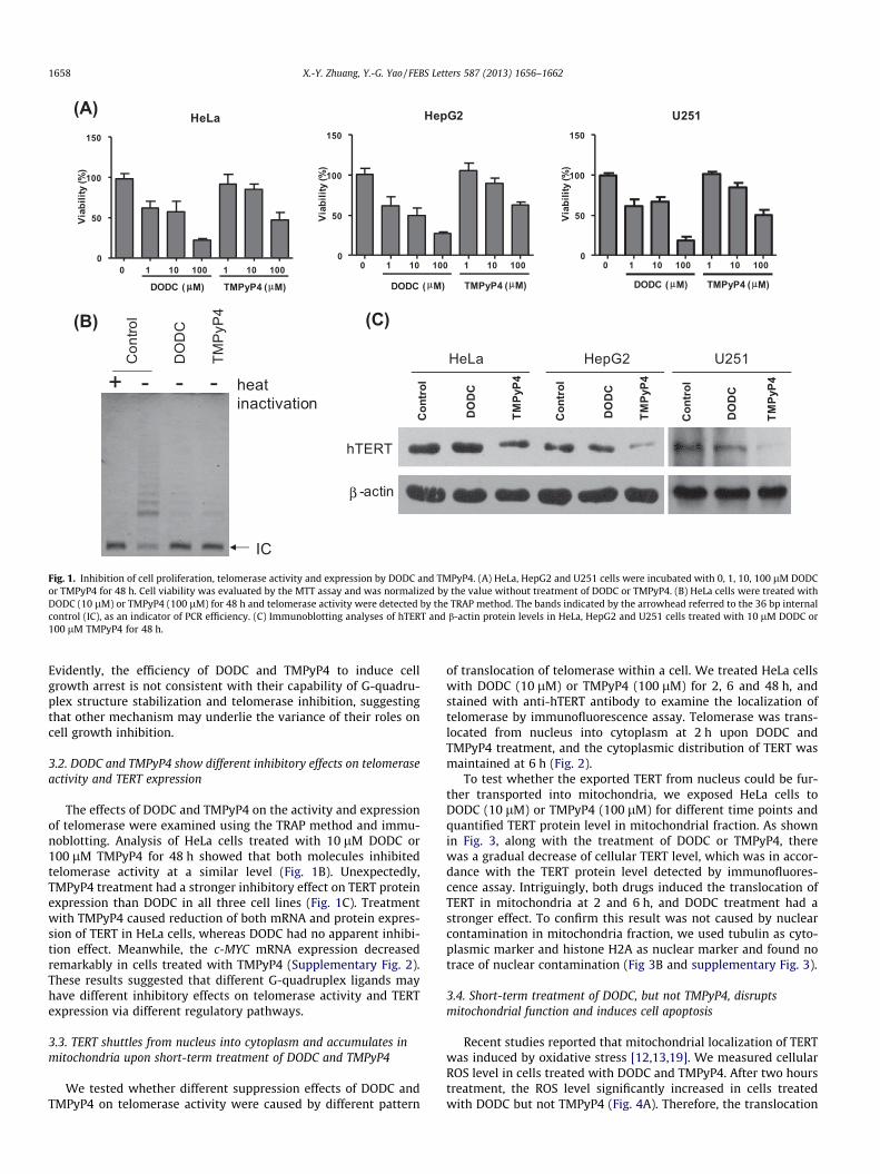
(B)
(A)C
ontro
l
IC
heat inactivation
+ - - -
DO
DC
TMP
yP4
HeLa
0 1 10 100 1 10 1000
50
100
150
DODC ( μμM) TMPyP4 (μM)
Viab
ility
(%)
HepG2
0 1 10 100 1 10 1000
50
100
150
DODC ( μM) TMPyP4 (μM)
Viab
ility
(%)
U251
0 1 10 100 1 10 1000
50
100
150
DODC ( μM) TMPyP4 (μM)
Viab
ility
(%)
(C)
β
hTERT
Con
trol
TMPy
P4
TMPy
P4
TMPy
P4
DO
DC
DO
DC
-actin
DO
DC
Con
trol
Con
trol
HeLa HepG2 U251
Fig. 1. Inhibition of cell proliferation, telomerase activity and expression by DODC and TMPyP4. (A) HeLa, HepG2 and U251 cells were incubated with 0, 1, 10, 100 lM DODCor TMPyP4 for 48 h. Cell viability was evaluated by the MTT assay and was normalized by the value without treatment of DODC or TMPyP4. (B) HeLa cells were treated withDODC (10 lM) or TMPyP4 (100 lM) for 48 h and telomerase activity were detected by the TRAP method. The bands indicated by the arrowhead referred to the 36 bp internalcontrol (IC), as an indicator of PCR efficiency. (C) Immunoblotting analyses of hTERT and b-actin protein levels in HeLa, HepG2 and U251 cells treated with 10 lM DODC or100 lM TMPyP4 for 48 h.
1658 X.-Y. Zhuang, Y.-G. Yao / FEBS Letters 587 (2013) 1656–1662
Evidently, the efficiency of DODC and TMPyP4 to induce cellgrowth arrest is not consistent with their capability of G-quadru-plex structure stabilization and telomerase inhibition, suggestingthat other mechanism may underlie the variance of their roles oncell growth inhibition.
3.2. DODC and TMPyP4 show different inhibitory effects on telomeraseactivity and TERT expression
The effects of DODC and TMPyP4 on the activity and expressionof telomerase were examined using the TRAP method and immu-noblotting. Analysis of HeLa cells treated with 10 lM DODC or100 lM TMPyP4 for 48 h showed that both molecules inhibitedtelomerase activity at a similar level (Fig. 1B). Unexpectedly,TMPyP4 treatment had a stronger inhibitory effect on TERT proteinexpression than DODC in all three cell lines (Fig. 1C). Treatmentwith TMPyP4 caused reduction of both mRNA and protein expres-sion of TERT in HeLa cells, whereas DODC had no apparent inhibi-tion effect. Meanwhile, the c-MYC mRNA expression decreasedremarkably in cells treated with TMPyP4 (Supplementary Fig. 2).These results suggested that different G-quadruplex ligands mayhave different inhibitory effects on telomerase activity and TERTexpression via different regulatory pathways.
3.3. TERT shuttles from nucleus into cytoplasm and accumulates inmitochondria upon short-term treatment of DODC and TMPyP4
We tested whether different suppression effects of DODC andTMPyP4 on telomerase activity were caused by different pattern
of translocation of telomerase within a cell. We treated HeLa cellswith DODC (10 lM) or TMPyP4 (100 lM) for 2, 6 and 48 h, andstained with anti-hTERT antibody to examine the localization oftelomerase by immunofluorescence assay. Telomerase was trans-located from nucleus into cytoplasm at 2 h upon DODC andTMPyP4 treatment, and the cytoplasmic distribution of TERT wasmaintained at 6 h (Fig. 2).
To test whether the exported TERT from nucleus could be fur-ther transported into mitochondria, we exposed HeLa cells toDODC (10 lM) or TMPyP4 (100 lM) for different time points andquantified TERT protein level in mitochondrial fraction. As shownin Fig. 3, along with the treatment of DODC or TMPyP4, therewas a gradual decrease of cellular TERT level, which was in accor-dance with the TERT protein level detected by immunofluores-cence assay. Intriguingly, both drugs induced the translocation ofTERT in mitochondria at 2 and 6 h, and DODC treatment had astronger effect. To confirm this result was not caused by nuclearcontamination in mitochondria fraction, we used tubulin as cyto-plasmic marker and histone H2A as nuclear marker and found notrace of nuclear contamination (Fig 3B and supplementary Fig. 3).
3.4. Short-term treatment of DODC, but not TMPyP4, disruptsmitochondrial function and induces cell apoptosis
Recent studies reported that mitochondrial localization of TERTwas induced by oxidative stress [12,13,19]. We measured cellularROS level in cells treated with DODC and TMPyP4. After two hourstreatment, the ROS level significantly increased in cells treatedwith DODC but not TMPyP4 (Fig. 4A). Therefore, the translocation

DODC
hTERT
hTERT/DAPI
TMPyP4
2h 6h 48hControl 2h 6h 48h
Fig. 2. Distribution of telomerase in cells treated by DODC or TMPyP4. HeLa cells were incubated with 10 lM DODC or 100 lM TMPyP4 for 2, 6 and 48 h. hTERT (green) andnucleus (blue) were stained by the immunofluorescence assay to visualize the cellular distribution of telomerase.
hTERT
Con
trol
2 h
6 h 48 h
2 h
6 h
48 h
β-actin
DODC TMPyP4(A)
hTERT
Con
trol
WC
L co
ntro
lMitochondria
Mitochondrialmarker
(B)
2 h
6 h 48 h
TMPyP4
2 h
6 h
48 h
DODC
Tubulin
Fig. 3. Time course of telomerase expression in HeLa cells treated with DODC orTMPyP4. (A) Decreased level of hTERT was observed in whole cell lysate of HeLacells treated with 10 lM DODC or 100 lM TMPyP4 for 2, 6 and 48 h. (B) Telomeraseprotein in mitochondrial fractions from treated HeLa cells. Equal protein loadingwas demonstrated with antibody against mitochondria (mitochondrial marker) andwhole cell lysate (b-actin). Antibody against tubulin was used as a cytoplasmicmarker to show the purity of mitochondria. WCL, whole cell lysate.
X.-Y. Zhuang, Y.-G. Yao / FEBS Letters 587 (2013) 1656–1662 1659
of telomerase from nucleus into mitochondria induced by thesetwo drugs was not fully dependent on cellular oxidative stress.To validate the pattern as revealed by DCF-DA, we further quanti-fied the ROS level in HeLa cells treated with TMPyP4 using DHE(Invitrogen) and observed no change of ROS (SupplementaryFig. 4), which was consistent with the above result.
In agreement with the rising of ROS, flow cytometric analysisindicated that the proportion of cells with low mitochondrialmembrane potential (MMP) increased in cells treated with DODC(Fig. 4B). To further investigate if treatment of DODC and TMPyP4altered mitochondrial function, we measured the oxygen con-sumption rate (OCR) in cells treated with both drugs using aClark-type oxygen sensor. As shown in Fig. 4C, DODC treatment
significantly reduced the OCR (up to 75% of the untreated cells),whereas TMPyP4 treatment had no apparent effect.
We further evaluated the effects of DODC and TMPyP4 on cellapoptosis within the time frame of mitochondrial localization ofTERT. Fraction of apoptotic cells was significantly elevated in cellstreated with DODC for 2 h but not for cells treated with TMPyP4(Fig. 5).
3.5. Over-expression of TERT counteracts the elevated ROS but fails toimprove the mitochondrial respiratory function decline induced byDODC
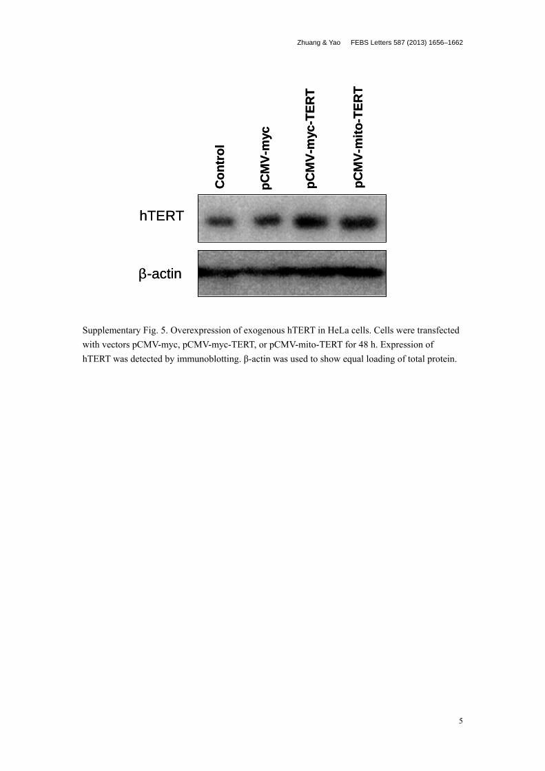
To investigate whether TERT overexpession would counteractthe negative effects of DODC on mitochondria, we measured thechange of cellular oxidative stress in TERT-overexpressing cells,with and without targeted export to mitochondria. We confirmedoverexpression of exogenous TERT in HeLa cells by immunoblot-ting (Supplementary Fig. 5). The ROS level was measured in trans-fected cells after treatment with DODC or TMPyP4 for 2 h. Wefound that ectopic expression of TERT, regardless of its cellularlocalization, reduced the ROS level induced by DODC althoughthe difference was not statistically significant. We observed similarsalvaging effects in all three independent experiments. However,we discerned no alteration of the ROS level in transfected cellstreated with TMPyP4 (Fig. 6A), a result consistent with the aboveobservations (Fig. 4).
Since the decrease of ROS level might be resulted from therecovery of normal mitochondrial respiratory chain activity, wesought to determine whether the respiration rate of cells treatedby DODC could be restored by overexpression of TERT. Our resultsshowed that overexpression TERT could not rescue the decreasedrespiratory rate induced by DODC (Fig. 6B).
4. Discussion
G-quadruplex ligands are potential anticancer agents [4,5,20].Recent studies showed that the instant anti-proliferation effect ofG-quadruplex ligands was more complex than we had thought[6,7,21]. In this study, we aimed to examine whether G-quadruplexligands such as DODC and TMPyP4 could interfere with telomeraseexpression and/or mitochondrial localization.
The two commercially available G-quadruplex ligands DODCand TMPyP4 had different G-quadruplexes binding affinity andspecificity [18]. DODC, but not TMPyP4, was reported to have littleinhibitory effect on telomerase activity as evaluated by exoge-nously adding DODC to cell-free TRAP reaction mixtures [18].

Control
TMPyP4
DODC
*
(A)
(B)
(C)
Control
2.44% 9.58%
5.91%
DODC
TMPyP4
Con
trol
DO
DC
TMPy
P4
0
20
40
60
80 *
DC
F-D
A Fl
uore
scen
ce(a
rtific
ially
gat
ed)
Con
trol
DO
DC
TMPy
P4
0.0
2.5
5.0
7.5
10.0
12.5 *
perc
enta
ge o
f low
MM
P(a
rtific
ially
gat
ed)
Con
trol
DO
DC
TMPy
P4 0.0
0.2
0.4
0.6
0.8
1.0
rela
tvie
resp
iratio
n ra
te
Fig. 4. ROS, MMP and OCR in cells treated by DODC or TMPyP4. HeLa cells wereincubated with 10 lM DODC or 100 lM TMPyP4 for 2 h. Cellular level of ROS wasmeasured by staining with DCF-DA (A) and change of MMP was determined bymitochondrial membrane sensitive dye JC-1 (B). (C) Relative oxygen consumptionrates were measured in intact cells treated by DODC (10 lM) or TMPyP4 (100 lM)for 2 h. Cells without treatment were used for normalization. Bars representmean ± S.D. of three independent experiments. *P < 0.05 relative to control.
1660 X.-Y. Zhuang, Y.-G. Yao / FEBS Letters 587 (2013) 1656–1662
However, whether the antitumor ability of DODC and TMPyP4 istotally dependent on their stabilization with quadruplex structurehas not been sufficiently studied. In our study, we found that bothDODC and TMPyP4 led to reduced cell viability, and DODC had astronger anti-proliferation ability than TMPyP4 in all three cancercell lines (Fig. 1). Treatment with TMPyP4 and DODC decreased cellproliferation and increased cellular ROS level in L02 cells, whichsuggested that the effects of TMPyP4 and DODC on cell prolifera-
tion were TERT-independent (Supplementary Fig. 1). Intriguingly,at their IC50 for cell viability, there was a similar inhibitory effecton telomerase activity in HeLa cells treated with DODC or TMPyP4,although the mRNA and protein levels of TERT were reduced incells treated with TMPyP4 but not in cells treated with DODC(Fig. 1 and Supplementary Fig. 2). The reduction of TERT mRNA le-vel induced by TMPyP4 was probably caused by transcriptionalinhibition through G-quadruplexes structure formed in the pro-moter region of the TERT and c-MYC genes [4,22] (SupplementaryFig. 2).
We further showed that both DODC and TMPyP4 induced thetranslocation of TERT from nucleus into mitochondria within 2 hof treatment and DODC had a stronger effect on the cytoplasmictranslocation of telomerase (Figs. 2 and 3). Inactive type of hTERTthat was localized in cytoplasm would lead to an increased degra-dation of wild-type hTERT [23]. Therefore, it is most likely thatTMPyP4 inhibited telomerase activity mainly at the transcription/translation level whereas DODC mainly at the post-translation le-vel. The short-term response of cells (apoptosis and death) treatedby these ligands could not be merely explained by telomeraseinhibition.
The translocation of telomerase from nucleus to mitochondriaupon treatment of DODC and TMPyP4 is very intriguing. It linksmitochondria with the anti-proliferation effect of G-quadruplex li-gands. Hitherto, several studies characterized mitochondrial telo-merase, albeit its reported role in mitochondria remainscontradictory [12,19]. We found that the ROS level, MMP andOCR of cells treated with DODC presented significant differencewith those of non-treated cells (Fig. 4). These findings suggestedthat the observed effect of DODC was most likely pertinent tomitochondria. In accordance to mitochondrial dysfunction inducedby DODC, percentage of apoptotic cells elevated significantly incells treated by this small molecule (Fig. 5). DODC was used as acarbocyanine membrane dye to probe the microenvironment ofmitochondria and enhanced mitochondrial dysfunction inducedby chemotherapy drugs [24]. Our results were consistent with thisnotion that DODC could disrupt the function of mitochondria [24].In contrast, the basic structure of TMPyP4 is a porphyrin and has alarge aromatic planar geometry, which means that all atoms lie in asingle plane will stack with the plane formed by G-quadruplexDNA. Previous studies had reported that induction of apoptosisby TMPyP4 was associated with activation of DNA damage re-sponse and cell cycle regulatory factors, and TMPyP4 also inhibitedthe expression of crucial components in cell growth and prolifera-tion, such as c-MYC [9]. We speculated that DNA damage andchange of expression profile of key regulators controlling cell sur-vival caused by TMPyP4 might be a driving force for telomerasedelocalization from telomere and exporting from nucleus.
In an effort to confirm the mitochondrial protection role of TERT[16], ectopic TERT were used to test whether it could confer resis-tance to effect of DODC. Increased TERT expression reduced cellu-lar ROS levels but had no effect on mitochondrial respiratoryfunction during the short-term treatment of DODC (Fig. 6). Thesedata were in agreement with previous reports linking TERT expres-sion with reduced ROS in different types of cells [25].
TERT with disability to shuttle between nucleus and cytoplasmnegatively impacted mitochondrial function [26,27]. Mice withoutTERT expression had a compromise of mitochondrial biogenesisand metabolism, suggesting depletion or down-regulation of TERTdid not simply affect telomere maintenance [28]. All these studiesshowed that telomerase plays an important role in mitochondrialbiology. Meanwhile, mitochondrial competence is important forthe survival of cancer cells, and coincidental extinction of mito-chondrial maintenance factor and telomerase activity enhancedantitumor effect [29]. Our results showed that DODC had a dualrole on inhibiting telomerase activity and mitochondrial function.

Control
DODC
TMPyP4
Unstained
PI
FITC
2%
15.68%
1.5%
Stained with Annexin V and PI(A)
(B)
Cont
rol
DODC
TMPy
P4
0
5
10
15
20
25
30
*
perc
enta
ge o
f apo
ptos
is c
ell
(arti
ficia
lly g
ated
)
Fig. 5. Short-term treatment of DODC induced robust apoptosis. HeLa cells were incubated with 10 lMDODC or 100 lM TMPyP4 for 2 h, followed by staining with Annexin Vand PI. Cells without Annexin V and PI staining were used as criteria of gating to exclude the interference of DODC and TMPyP4. (A) Representative images of the flowcytometric analysis. (B) Average of three independent analyses. Bars represent mean ± S.D. *P < 0.05 relative to control.
(A)
0
10
20
30
40
50
60
Control DODC TMPyP4
pCMV-myc-TERT
untransfected
pCMV-myc
pCMV-mito-TERT
DC
F-D
A Fl
uore
scen
ce c
ells
(B)
0.0
0.5
1.0
1.5
2.0
Control DODC TMPyP4
untransfectedpCMV-myc
pCMV-mito-TERT
pCMV-myc-TERT
Rel
ativ
e re
spira
tion
rate
Fig. 6. Effects of ectopic telomerase on mitochondrial function in cells treated by DODC and TMPyP4. HeLa cells were transfected with an empty control vector (pCMV-myc),pCMV-myc-TERT, or pCMV-mito-TERT for 48 h, then treated with 10 lM DODC or 100 lM TMPyP4 for 2 h. Cellular ROS (A) and OCR (B) were determined in transfected cellsas indicated. Data are mean ± S.D. of three independent experiments.
X.-Y. Zhuang, Y.-G. Yao / FEBS Letters 587 (2013) 1656–1662 1661
This is probably the reason why DODC displayed a stronger anti-proliferation ability than TMPyP4.
In short, we showed that treatment of DODC and TMPyP4 incancer cell lines caused mitochondrial localization of TERT, but
the underlying mechanism of this effect might be quite different.Our results showed that cytotoxic mechanism of G-quadruplex li-gands, such as perturbation of mitochondrial function, should beconsidered besides the traditional concept of the G-quadruplex

1662 X.-Y. Zhuang, Y.-G. Yao / FEBS Letters 587 (2013) 1656–1662
stabilization. Further study is necessary to answer the questionwhether nuclear-cytoplasmic shuttling and mitochondrial localiza-tion of TERT induced by G-quadruplex ligands is a cause or conse-quence of drug effect and how this could contribute to a betterunderstanding of TERT function in extra-nuclear compartments.
Acknowledgements
This work was supported by the Ministry of Science and Tech-nology of China (2011CB910900), ‘‘Light in Western China (西部
之光)’’ of the Chinese Academy of Sciences and Yunnan Province(2009CI119).
Appendix A. Supplementary data
Supplementary data associated with this article can be found, inthe online version, at http://dx.doi.org/10.1016/j.febslet.2013.04.010.
References
[1] Makarov, V.L. et al. (1997) Long G tails at both ends of human chromosomessuggest a C strand degradation mechanism for telomere shortening. Cell 88,657–666.
[2] Burge, S. et al. (2006) Quadruplex DNA: sequence, topology and structure.Nucleic Acids Res. 34, 5402–5415.
[3] Neidle, S. et al. (2002) Telomere maintenance as a target for anticancer drugdiscovery. Nat. Rev. Drug Disc. 1, 383–393.
[4] Siddiqui-Jain, A. et al. (2002) Direct evidence for a G-quadruplex in a promoterregion and its targeting with a small molecule to repress c-MYC transcription.Proc. Natl. Acad. Sci. USA 99, 11593–11598.
[5] Riou, J.F. et al. (2002) Cell senescence and telomere shortening induced by anew series of specific G-quadruplex DNA ligands. Proc. Natl. Acad. Sci. USA 99,2672–2677.
[6] Pennarun, G. et al. (2005) Apoptosis related to telomere instability and cellcycle alterations in human glioma cells treated by new highly selective G-quadruplex ligands. Oncogene 24, 2917–2928.
[7] Zhou, W.J. et al. (2009) G-quadruplex ligand SYUIQ-5 induces autophagy bytelomere damage and TRF2 delocalization in cancer cells. Mol. Cancer Ther. 8,3203–3213.
[8] Kelland, L. (2007) Targeting the limitless replicative potential of cancer: thetelomerase/telomere pathway. Clin. Cancer Res. 13, 4960–4963.
[9] Mikami-Terao, Y. et al. (2009) Antitumor activity of TMPyP4 interacting G-quadruplex in retinoblastoma cell lines. Exp. Eye Res. 89, 200–208.
[10] Fu, Y.T. et al. (2009) BRACO19 analog dimers with improved inhibition oftelomerase and hPot 1. Bioorg. Med. Chem. 17, 2030–2037.
[11] Hemann, M.T. et al. (2001) The shortest telomere, not average telomerelength, is critical for cell viability and chromosome stability. Cell 107, 67–77.
[12] Ahmed, S. et al. (2008) Telomerase does not counteract telomere shorteningbut protects mitochondrial function under oxidative stress. J. Cell Sci. 121,1046–1053.
[13] Haendeler, J. et al. (2009) Mitochondrial telomerase reverse transcriptasebinds to and protects mitochondrial DNA and function from damage.Arterioscler. Thromb. Vasc. Biol. 29, 929–935.
[14] Kim, N.W. et al. (1997) Advances in quantification and characterization oftelomerase activity by the telomeric repeat amplification protocol (TRAP).Nucleic Acids Res. 25, 2595–2597.
[15] Chen, Q. et al. (1996) Spectroscopic recognition of guanine dimeric hairpinquadruplexes by a carbocyanine dye. Proc. Natl. Acad. Sci. USA 93, 2635–2639.
[16] Li, C.P. et al. (2004) A G-quadruplex ligand 3,30-diethyloxadicarbocyanineiodide induces mitochondrion-mediated apoptosis but not decrease oftelomerase activity in nasopharyngeal carcinoma NPC-TW01 cells. Pharm.Res. 21, 93–100.
[17] Mikami-Terao, Y. et al. (2008) Antitumor activity of G-quadruplex-interactiveagent TMPyP4 in K562 leukemic cells. Cancer Lett. 261, 226–234.
[18] Yao, Y. et al. (2007) An exonuclease I hydrolysis assay for evaluating G-quadruplex stabilization by small molecules. Nucleic Acids Res. 35, e68.
[19] Santos, J.H. et al. (2006) Mitochondrial localization of telomerase as adeterminant for hydrogen peroxide-induced mitochondrial DNA damageand apoptosis. Hum. Mol. Genet. 15, 1757–1768.
[20] Yang, D. et al. (2010) Structural insights into G-quadruplexes: towards newanticancer drugs. Future Med. Chem. 2, 619–646.
[21] Tauchi, T. et al. (2003) Activity of a novel G-quadruplex-interactive telomeraseinhibitor, telomestatin (SOT-095), against human leukemia cells: involvementof ATM-dependent DNA damage response pathways. Oncogene 22, 5338–5347.
[22] Palumbo, S.L. et al. (2009) Formation of a unique end-to-end stacked pair of G-quadruplexes in the hTERT core promoter with implications for inhibition oftelomerase by G-quadruplex-interactive ligands. J. Am. Chem. Soc. 131,10878–10891.
[23] Nguyen, B.N. et al. (2009) Mechanism of dominant-negative telomerasefunction. Cell Cycle 8, 3227–3233.
[24] Fu, W. et al. (1999) Anti-apoptotic role of telomerase in pheochromocytomacells. J. Biol. Chem. 274, 7264–7271.
[25] Indran, I. et al. (2011) HTERT overexpression alleviates intracellular ROSproduction, improves mitochondrial function, and inhibits ROS-mediatedapoptosis in cancer cells. Cancer Res. 71, 266–276.
[26] Kovalenko, O.A. et al. (2010) Expression of (NES-)hTERT in cancer cells delayscell cycle progression and increases sensitivity to genotoxic stress. PLoS ONE5, e10812.
[27] Kovalenko, O.A. et al. (2010) A mutant telomerase defective in nuclear-cytoplasmic shuttling fails to immortalize cells and is associated withmitochondrial dysfunction. Aging Cell 9, 203–219.
[28] Sahin, E. et al. (2011) Telomere dysfunction induces metabolic andmitochondrial compromise. Nature 470, 359–365.
[29] Hu, J. et al. (2012) Antitelomerase therapy provokes ALT and mitochondrialadaptive mechanisms in cancer. Cell 148, 651–663.

Zhuang & Yao FEBS Letters 587 (2013) 1656–1662
1
Appendix A. Supplementary data Supplementary Fig. 1. Effects of DODC and TMPyP4 on cell viability and reactive oxygen species (ROS) level of human hepatic L02 cell. Treatment of DODC or TMPyP4 at indicated concentrations for 48 h decreased the cell viability of L02 cells (A). The ROS level was increased in L02 cells after a treatment of 10 μM DODC or 100 μM TMPyP4 for 2 h, as represented by the intensity of DCF-DA fluorescence (B). Bars represent mean±SD of three independent experiments. * P<0.05 relative to control.
(A) (B)
Con
trol
DO
DC
TMPy
P4
0
10
20
30
40
50 *
*
DC
F-D
A F
luor
esce
nce
(art
ifici
ally
gat
ed)
0 1 10 100 1 10 1000
50
100
150
DODC (μM) TMPyP4 (μM)
Viab
ility
(%)
(A) (B)
Con
trol
DO
DC
TMPy
P4
0
10
20
30
40
50 *
*
DC
F-D
A F
luor
esce
nce
(art
ifici
ally
gat
ed)
0 1 10 100 1 10 1000
50
100
150
DODC (μM) TMPyP4 (μM)
Viab
ility
(%)

Zhuang & Yao FEBS Letters 587 (2013) 1656–1662
2
Supplementary Fig. 2. Expression of (A) TERT mRNA, (B) TERT protein, and (C) c-MYC mRNA in HeLa cells treated by DODC or TMPyP4 for 48 h at indicated concentrations.
0 1 10 1 10 100 μM
DODC TMPyP4
hTERT
β-actin
0 1 10 1 10 100 μM
DODC TMPyP4
hTERT
β-actin
(A) (B)
0 1 10 1 10 100 μM
DODC TMPyP4
β-actin
c-MYC
(C)
0 1 10 1 10 100 μM
DODC TMPyP4
hTERT
β-actin
0 1 10 1 10 100 μM
DODC TMPyP4
hTERT
β-actin
(A) (B)0 1 10 1 10 100 μM
DODC TMPyP4
hTERT
β-actin
0 1 10 1 10 100 μM
DODC TMPyP4
hTERT
β-actin
0 1 10 1 10 100 μM
DODC TMPyP4
hTERT
β-actin
0 1 10 1 10 100 μM
DODC TMPyP4
hTERT
β-actin
(A) (B)
0 1 10 1 10 100 μM
DODC TMPyP4
β-actin
c-MYC
0 1 10 1 10 100 μM
DODC TMPyP4
β-actin
c-MYC
(C)

Zhuang & Yao FEBS Letters 587 (2013) 1656–1662
3
Supplementary Fig. 3. The purity of mitochondria fraction of HeLa cells with and without DODC and TMPyP4 treatment. Mitochondria fraction was isolated by using Mitochondria Isolation Kit (Pierce) and potential nuclear contamination was evaluated by western blot for a nuclear marker histone H2A (Cell Signaling #2578). Whole cell lysate, mitochondria and nuclei were obtained from HeLa cells treated with 10 μM DODC or 100 μM TMPyP4 for 2h. The absence of histone H2A band in mitochondria fraction indicated no nuclear contamination. The observation of mitochondrial marker, as recognized by mouse monoclonal antibodies against mitochondria (ab3298, Abcam), in the nuclear fraction suggested the contamination of intact cell and/or mitochondria which could not be completely excluded by centrifugation.
Mitochonrialmarker
Con
trol
TMPy
P4
TMPy
P4
TMPy
P4
DO
DC
DO
DC
Histone H2A
DO
DC
Con
trol
Con
trol
Whole cell mitochondria nuclei
Mitochonrialmarker
Con
trol
TMPy
P4
TMPy
P4
TMPy
P4
DO
DC
DO
DC
Histone H2A
DO
DC
Con
trol
Con
trol
Whole cell mitochondria nuclei

Zhuang & Yao FEBS Letters 587 (2013) 1656–1662
4
Supplementary Fig. 4. ROS in HeLa cells treated by TMPyP4 probed by DHE. HeLa cells were incubated with 100 μM TMPyP4 for 2 h, and after stained with 1 μM DHE, cells were trypsinized and subjected to flow cytometric analysis (A). The ROS levels were represented by the intensity of DHE fluorescence. Bars represent mean±SD of three independent experiments (B).
Con
trol
TMPy
P4
0
2
4
6
8
DH
E Fl
uore
scen
ce(a
rtifi
cial
ly g
ated
)
(B)
TMPyP4
Control
FC-3
Eve
nts
(A)
Con
trol
TMPy
P4
0
2
4
6
8
DH
E Fl
uore
scen
ce(a
rtifi
cial
ly g
ated
)
(B)
TMPyP4
Control
FC-3
Eve
nts
TMPyP4
Control
FC-3
Eve
nts
(A)
Zhuang & Yao FEBS Letters 587 (2013) 1656–1662
5
Supplementary Fig. 5. Overexpression of exogenous hTERT in HeLa cells. Cells were transfected with vectors pCMV-myc, pCMV-myc-TERT, or pCMV-mito-TERT for 48 h. Expression of hTERT was detected by immunoblotting. β-actin was used to show equal loading of total protein.
Con
trol
pCM
V-m
yc
pCM
V-m
yc-T
ERT
pCM
V-m
ito-T
ERT
hTERT
β-actin
Con
trol
pCM
V-m
yc
pCM
V-m
yc-T
ERT
pCM
V-m
ito-T
ERT
hTERT
β-actin