oxidative stress and mitochondrial dysfunction in
TRANSCRIPT

Oxidative stress and mitochondrial dysfunction in Parkinson’s disease
A chronological model for Parkinson’s disease
Albert F Wright PhD Grenoble, France wriga38 @ gmail.com
Introduction
Parkinson’s disease is characterized by the loss of dopaminergic neurons in the substantia nigra(SN) in the midbrain and a deficit of dopamine in the terminal synapses in the striatum, but a simplemodel presenting the stages leading up to this condition has so far not been presented.
This article proposes a simplified model that schematically represents the chronology of the majorevents believed to be involved in the pathogenesis of Parkinson’s disease in a way designed to beaccessible to patients. The model also proposes which steps may be subject to influence throughmedical or patient intervention, so that patients can make informed decisions about how to managetheir own condition. This model covers the progression of Parkinson’s disease from benign redoximbalance in brain cells to chronic oxidative stress which initiates a cascade of two major events: (i)mitochondrial dysfunction, a condition which reduces the energy available to host cells and (ii) de-generation of vulnerable axons in the striatum region of SN neurons as a consequence of this energyloss. The model links these major events and draws attention to the considerable delays occurringbetween the beginning of the events and the observation of symptoms eventually produced by them,a situation which masks the true progression of the disease.
The model was designed to be a chronological representation of the progression of Parkinson’s dis-ease. For this to be both feasible and useful as a tool, it was designed to respect a number of condi-tions.
Each stage in the model is supported by evidence of its physical existence,
The chronology of events is respected,
Each stage has identifiable characteristics that differentiate it from other stages,
There are feasible mechanisms driving the progression from one stage to another,
The model is adaptable to take account of complementary factors and variants,
The model offers the possibility being tested experimentally.
To build this model, evidence for the three major conditions already identified in the progression ofParkinson’s disease; age-related oxidative stress, mitochondrial damage and loss of dopaminergicSN neurons was investigated. There are convincing arguments for the processes that link these con-ditions and make up the basic version of the model. Each process has also been examined in moredetail to consider the potential roles other factors having the capacity to modify or invalidate thesimplified model, such as α-synuclein, genetic variants, toxins or lifestyle. These additional factorsadd complexity to the model but help to understand how the pathogenesis and development of Par-kinson’s disease is multifactorial. This article presents the simplified model only. The more com-plex model will be presented at a later date.
A F Wright, Draft document, confidential. 1 Not for general distribution, May 2021
1 - Age-related changes in cellular function
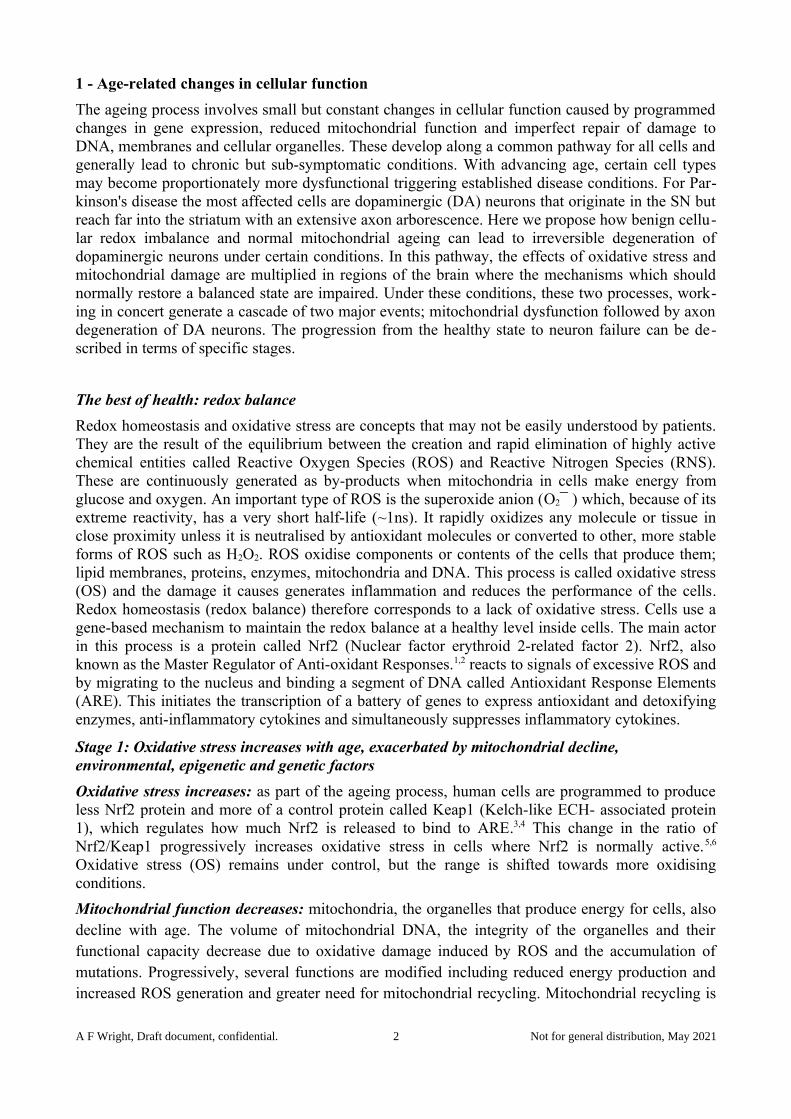
The ageing process involves small but constant changes in cellular function caused by programmedchanges in gene expression, reduced mitochondrial function and imperfect repair of damage toDNA, membranes and cellular organelles. These develop along a common pathway for all cells andgenerally lead to chronic but sub-symptomatic conditions. With advancing age, certain cell typesmay become proportionately more dysfunctional triggering established disease conditions. For Par-kinson's disease the most affected cells are dopaminergic (DA) neurons that originate in the SN butreach far into the striatum with an extensive axon arborescence. Here we propose how benign cellu-lar redox imbalance and normal mitochondrial ageing can lead to irreversible degeneration ofdopaminergic neurons under certain conditions. In this pathway, the effects of oxidative stress andmitochondrial damage are multiplied in regions of the brain where the mechanisms which shouldnormally restore a balanced state are impaired. Under these conditions, these two processes, work-ing in concert generate a cascade of two major events; mitochondrial dysfunction followed by axondegeneration of DA neurons. The progression from the healthy state to neuron failure can be de-scribed in terms of specific stages.
The best of health: redox balance
Redox homeostasis and oxidative stress are concepts that may not be easily understood by patients.They are the result of the equilibrium between the creation and rapid elimination of highly activechemical entities called Reactive Oxygen Species (ROS) and Reactive Nitrogen Species (RNS).These are continuously generated as by-products when mitochondria in cells make energy fromglucose and oxygen. An important type of ROS is the superoxide anion (O2¯ ) which, because of itsextreme reactivity, has a very short half-life (~1ns). It rapidly oxidizes any molecule or tissue inclose proximity unless it is neutralised by antioxidant molecules or converted to other, more stableforms of ROS such as H2O2. ROS oxidise components or contents of the cells that produce them;lipid membranes, proteins, enzymes, mitochondria and DNA. This process is called oxidative stress(OS) and the damage it causes generates inflammation and reduces the performance of the cells.Redox homeostasis (redox balance) therefore corresponds to a lack of oxidative stress. Cells use agene-based mechanism to maintain the redox balance at a healthy level inside cells. The main actorin this process is a protein called Nrf2 (Nuclear factor erythroid 2-related factor 2). Nrf2, alsoknown as the Master Regulator of Anti-oxidant Responses.1,2 reacts to signals of excessive ROS andby migrating to the nucleus and binding a segment of DNA called Antioxidant Response Elements(ARE). This initiates the transcription of a battery of genes to express antioxidant and detoxifyingenzymes, anti-inflammatory cytokines and simultaneously suppresses inflammatory cytokines.
Stage 1: Oxidative stress increases with age, exacerbated by mitochondrial decline, environmental, epigenetic and genetic factors
Oxidative stress increases: as part of the ageing process, human cells are programmed to produceless Nrf2 protein and more of a control protein called Keap1 (Kelch-like ECH- associated protein1), which regulates how much Nrf2 is released to bind to ARE.3,4 This change in the ratio ofNrf2/Keap1 progressively increases oxidative stress in cells where Nrf2 is normally active.5,6
Oxidative stress (OS) remains under control, but the range is shifted towards more oxidisingconditions.
Mitochondrial function decreases: mitochondria, the organelles that produce energy for cells, alsodecline with age. The volume of mitochondrial DNA, the integrity of the organelles and theirfunctional capacity decrease due to oxidative damage induced by ROS and the accumulation ofmutations. Progressively, several functions are modified including reduced energy production andincreased ROS generation and greater need for mitochondrial recycling. Mitochondrial recycling is
A F Wright, Draft document, confidential. 2 Not for general distribution, May 2021
the quality control process that extracts damaged components of mitochondria, creates neworganelles and removes debris.7–11
These two changes are both moving in the same direction; they are synergistic and self sustaining.
Other factors contributing to oxidative stress are toxins derived from food, poor diet or exposure topesticides or toxic chemicals as well as lack of exercise and obesity.12,13 Genetic mutations areknown to contribute to the development of familial and early onset Parkinson’s disease bycontributing to oxidative stress and damaging mitochondria.14–17 Finally epigenetic labelling mayalso be factor in driving age-related diseases by restricting the cell’s access to gene expression.18–20
Stage 2: Oxidative stress becomes a severe or chronic condition
All the factors contributing to oxidative stress and mitochondrial decline progress together to followa common pathway where oxidative stress begins to affect many different cell types. Chronicoxidative stress occurs when the levels of ROS regularly exceed safe values for long periods. Thisis the starting point for the pathogenesis of age-related diseases such as cardiovascular, urinary tractand neurodegenerative diseases.6,21,22 At this stage, although symptoms may be present, they not besufficiently well defined to lead to a specific diagnosis.
Stage 3: Zones with reduced mitochondrial QC capacity - “Cascades”
Chronic oxidative stress and mitochondrial decline can continue along this path producing reducedcellular function and increased inflammation in cells and tissues without triggering a specific dis-ease, because in the vast majority of of cases, the control and repair systems are able to restoreredox balance and mitochondrial quality control. The cases where these systems cannot restore suchbalance are rare and are specifically related the topology of the sites which limit the capacity oftransport of proteins and essential elements required for good control and maintenance between thenucleus and the site in question. Specifically, mitochondria are simply more vulnerable at siteswhere the control and repair systems can no longer keep pace with the demand.
One region where these systems are severely impaired is in the distal axons (axons far from the nuc-leus) and synapses of dopaminergic (DA) neurons. Each DA neuron has a very extended and com-plex axonal arborescence reaching from the SN far into the striatum, supplying dopamine to morethan a million synapses. This generates a considerable local energy demand which can only be satis-fied by mitochondria in the axons and synapses concerned. All the supplies required for the control,maintenance and repair must be transported along this route and the most distant axons present thegreatest difficulties in terms of just-in-time delivery. A sudden surge in ROS in mitochondria in adistal axon, requires a nano-second response by antioxidants if oxidative-stress damage is to beavoided. If local reserves of antioxidant molecules are insufficient, reinforcements must be gener-ated by Nrf2 action in the nucleus and transported to the point of use.
In this model, vulnerable zones where these conditions prevail are the physical departure points forneurodegenerative diseases from chronic oxidative stress and mitochondrial decline. In DAneurons, evolution can follow one of two trajectories; (i) if the maintenance systems can restoreredox balance and mitochondrial quality control, the axons will continue to function satisfactorily,maybe with reduced performance; (ii) if the maintenance systems can no longer restore redoxbalance and mitochondrial recycling, this will initiate the first of two destructive events occurringsuccessively. Uncontrolled ROS will induce lipid peroxidation23 and impairment of an enzyme(Complex I) essential for energy production by mitochondria and failure of the quality controlcycle.24–27 This in turn generates more ROS in a vicious circle that causes more mitochondrialdysfunction (cascade 3.1). When mitochondria can no longer meet the energy demand of the distalaxons in the striatum as a consequence of mitochondrial dysfunction, the most vulnerable axons andsynapses cease to function normally due to energy shortages (cascade 3.2).
A F Wright, Draft document, confidential. 3 Not for general distribution, May 2021
Fig 1: Schematic representation of the stepping stones and cascades in the basic model for the pathogenesis of Parkinson’s disease. OS: oxidative stress, mtD: mitochondrial dysfunction.
Cascade 3.1: Mitochondrial dysfunction and repair
Mitochondria are the main source of energy in cells, providing ATP through oxidativephosphorylation. Oxidative phosphorylation also generates reactive oxygen species (ROS) whichimpair the Complex I enzyme, the first of 4 enzymes enzymes in the electron transport chain. If thisROS is not rapidly quenched by the action of Nrf2, it will cause further damage to Complex I aspart of a vicious circle of oxidative stress and mitochondrial damage.9–11,24–29 The consequences of this condition, called secondary mitochondrial dysfunction (mtD), aremultiple: reduced ATP energy production, impaired calcium signalling30, increased oxidative stressand inflammation and a surge in demand to recycle damaged mitochondria and dispose of wastefragments (mitophagy). The onset of mtD is a critical physical transformation of the mitochondrialcondition which directly impacts the host cells. In this model we are focused on DA neurons in theSN which defines Parkinson’s disease, but mtD may also occur in other cell or neuron types andlead to additional complications or variants of PD. Other neurodegenerative or age-related diseasessuch as Alzheimer’s disease, cardiovascular diseases, urinary tract or digestive tract diseases mayalso be initiated by a similar failure of mtD.31–35
The process of recovery from dysfunctional to normal mitochondria is dependent on restoring redoxhomeostasis by the Nrf2/ARE pathway. In neurons where Nrf2 still has this capacity, mitochondrialpopulations can then recover normal function by activating the mitochondrial quality-control cycle.Mitochondrial dysfunction, which starves neurons of energy and is accompanied by increased OSand inflammation, very likely corresponds to the prodromal stage of Parkinson’s disease where non-specific and non-motor symptoms may be observed.
A F Wright, Draft document, confidential. 4 Not for general distribution, May 2021

Cascade 3.2: Degeneration of DA axons in the striatum
Neurons suffering chronic energy shortages through mitochondrial dysfunction will be unable tosustain full dopamine release in the most distant axonal regions. Mitochondria in these distal axonsare particularly vulnerable because of their very extended supply lines and by OS-induced impairedcalcium signalling which controls the transport of supplies along axons. There is growing evidencefrom post-mortem and imaging studies that axon degeneration in the striatum precedes the loss ofwhole SN neurons and is a predominant feature of early PD.36–38 If chronic oxidative stress persistsunabated, both mitochondrial dysfunction and axon degeneration will progress simultaneouslyleading to further loss of the axonal arborescence of DA neurons and the onset of both motor andnon-motor symptoms of PD.
Stage 4: Axon degeneration exceeds the 60% threshold for normal movement control
Stage 4 differs from stage 3 specifically in terms of the quantitative progression of mitochondrialdysfunction and DA axon degeneration. In particular, when DA axon degeneration or complete lossof DA neurons exceeds the critical threshold (>60% loss) in the striatum, normal motor functionwill be impaired, leading to the diagnosis of PD. This ultimately characterises fully-developed Par-kinson’s disease. This stage may not be detected until many years after the initial damage to axonsand DA neurons began.
2 - Evidence of oxidative stress and mitochondrial vulnerability in DA neurons
Oxidative stress, mitochondrial dysfunction, and energy deficiency are implicated in manyneurological and neurodegenerative diseases, despite these illnesses not being classified asmitochondrial diseases.28,39–42 Both of these conditions have been demonstrated by post-mortemexamination of brains and peripheral tissues of patients with PD.43 Furthermore, the role ofoxidative stress as a major contributing factor for mitochondrial dysfunction is now wellestablished.44,45 Oxidative stress is a common underlying condition of cellular dysfunction in geneticand idiopathic Parkinson’s disease.46 Increased levels of oxidised lipids, proteins and DNA, anddecreased levels of reduced glutathione have been systematically found in the SN of PD patients.47
Fig 2: Schematic representation of an SN dopaminergic neurons showing axonal projections and factors contributing to its high bioenergetic demands. Image gratefully reproduced from E Zampese and D J Surmeier, ref. 49.
A F Wright, Draft document, confidential. 5 Not for general distribution, May 2021

Impairment of Complex I of the electron transport chain is a common feature of mitochondriaisolated from PD patients.27 Magnetic Resonance Spectrometry in vivo has revealed thatmitochondrial dysfunction is not restricted to the substantia nigra, but can occur in almost allregions of the brains of Parkinson’s disease patients48.
The link between parkinsonism and mitochondria was identified in the early 1980s. Theneurotoxin, MPTP (1 methyl 4 phenyl 1,2,3,6 tetrahydropyridine) which causes a form of‐ ‐ ‐ ‐ ‐Parkinson’s disease inhibits complex I of the electron transport chain. Complex I is reported tobe reduced by about 30% in the SN and frontal cortex of Parkinson’s disease patients onautopsy.24,25
Dopaminergic neurons of the SN carry a very high bioenergetic burden because of the hugeaxonal branching that supplies dopamine to more than a million synapses.49,50 The extremelylong supply lines to these regions from the nucleus make redox control and mitochondrialquality control much more difficult to achieve in real time,51–54 making mitochondria in theseneurons particularly vulnerable to oxidative stress and mitochondrial dysfunction. This createsthe conditions for a closed loop vicious circle involving OS damage to Complex I, axonaltransport failure, mitochondrial dysfunction and finally energy shortages.
3 - The role of the Nrf2/ARE pathway in mitochondrial dysfunction
The role of Nrf2 in activating the antioxidant response (ARE) system to combat oxidative stress andinflammation in neurons is well established.1–4,55–58 Nrf2 also plays a role in sustainingmitochondrial function by facilitating quality control, increasing the availability of substrates forrespiration and ATP production and increasing the mitochondrial membrane potential.59–64 One ofthe most potent activators of Nrf2 is the natural isothiocyanate sulforaphane which is readilyavailable in broccoli seeds and sprouts as its precursor molecule glucoraphanin.22,55,65–67
In a landmark study of an animal model of Parkinson’s disease, A Jazwa, et al57 demonstrated thatupregulation of Nrf2 by sulforaphane provided protection from MPTP-induced parkinsonism inmice. The neurotoxin, MPTP inhibits Complex I of the electron transport chain and directly triggersmitochondrial dysfunction and parkinsonism24,25. Without sulforaphane treatment, analysis of brainsections showed a 60% loss of SN neurons and an 80% loss of dopamine in the striatum six daysafter injection of MPTP. Prior treatment with sulforaphane protected against 50% of the loss of SNneurons but only 20% of the loss of dopamine release in the striatum. The ratio of losses due toMPTP and the protection provided by sulforaphane indicates important role played by NRF2 incounteracting OS, but also the greater vulnerability of mitochondrial dysfunction in DA axons inthe striatum compared to the SN.
Upregulating Nrf2 attenuates non-motor PD symptoms
In a recent experiment designed and executed by the author, previously diagnosed with Parkinson’sdisease in 2018, a daily dose of a special broccoli seed tea containing sulforaphane considerably re-duced fatigue, apathy, sleep problems and urinary incontinence over a period of a few weeks, butmoderately increased tremor over the same period. Intrigued by this result, the author shared this in-formation with other PD patients who collectively established a protocol to standardize the experi-mental method and data recording. Eight Parkinson’s patients then carried out individual (n-of-1)studies to test their own symptom responses and subsequently shared their results.68
A F Wright, Draft document, confidential. 6 Not for general distribution, May 2021
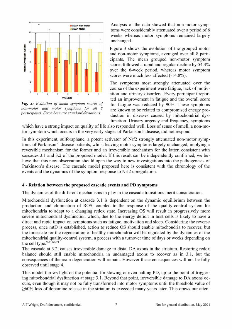
Analysis of the data showed that non-motor symp-toms were considerably attenuated over a period of 6weeks whereas motor symptoms remained largelyunchanged.
Figure 3 shows the evolution of the grouped motorand non-motor symptoms, averaged over all 8 parti-cipants. The mean grouped non-motor symptomscores followed a rapid and regular decline by 54.3%over the 6-week period, whereas motor symptomscores were much less affected (-14.8%).
The symptoms most strongly attenuated over thecourse of the experiment were fatigue, lack of motiv-ation and urinary disorders. Every participant repor-ted an improvement in fatigue and the overall scorefor fatigue was reduced by 90%. These symptomsare known to be related to compromised energy pro-duction in diseases caused by mitochondrial dys-function. Urinary urgency and frequency, symptoms
which have a strong impact on quality of life also responded well. Loss of sense of smell, a non-mo-tor symptom which occurs in the very early stages of Parkinson’s disease, did not respond.
In this experiment, sulforaphane, a potent activator of Nrf2 strongly attenuated non-motor symp-toms of Parkinson’s disease patients, whilst leaving motor symptoms largely unchanged, implying areversible mechanism for the former and an irreversible mechanism for the latter, consistent withcascades 3.1 and 3.2 of the proposed model. If this result can be independently confirmed, we be-lieve that this new observation should open the way to new investigations into the pathogenesis ofParkinson’s disease. The cascade model proposed here is consistent with the chronology of theevents and the dynamics of the symptom response to Nrf2 upregulation.
4 - Relation between the proposed cascade events and PD symptoms
The dynamics of the different mechanisms in play in the cascade transitions merit consideration.
Mitochondrial dysfunction at cascade 3.1 is dependent on the dynamic equilibrium between theproduction and elimination of ROS, coupled to the response of the quality-control system formitochondria to adapt to a changing redox state. Increasing OS will result in progressively moresevere mitochondrial dysfunction which, due to the energy deficit in host cells is likely to have adirect and rapid impact on symptoms such as fatigue, motivation and sleep. Considering the reverseprocess, once mtD is established, action to reduce OS should enable mitochondria to recover, butthe timescale for the regeneration of healthy mitochondria will be regulated by the dynamics of themitochondrial quality-control system, a process with a turnover time of days or weeks depending onthe cell type.9–11,69–71
The cascade at 3.2, causes irreversible damage to distal DA axons in the striatum. Restoring redoxbalance should still enable mitochondria in undamaged axons to recover as in 3.1, but theconsequences of the axon degeneration will remain. However these consequences will not be fullyobserved until stage 4.
This model throws light on the potential for slowing or even halting PD, up to the point of trigger-ing mitochondrial dysfunction at stage 3.1. Beyond that point, irreversible damage to DA axons oc-curs, even though it may not be fully transformed into motor symptoms until the threshold value of≥60% loss of dopamine release in the striatum is exceeded many years later. This draws our atten-
A F Wright, Draft document, confidential. 7 Not for general distribution, May 2021
Fig. 3: Evolution of mean symptom scores ofnon-motor and motor symptoms for all 8participants. Error bars are standard deviations.

tion to the importance of being able to recognise the signs and symptoms of stage 3.1 as early aspossible by developing reliable biological markers for this stage.
In the broccoli seed tea experiment, all 8 patients had reached the equivalent of Stage 4 of thismodel prior to the experiment. The lack of impact on motor symptoms suggests that upregulatingNrf2 had little or no additional effect on dopamine availability in striatal synapses. The strong im-pact on non-motor symptoms is however consistent with improved energy supply enabling betteroverall function of DA or other neuron types. The dynamics of the changes observed over 6 weeksin non-motor symptoms are consistent with the timescale of mitochondrial renewal and the lifecycle of mitochondria, equivalent to the slowing or halt of the mtD cascade at stage 3.1, and with nochange in axon degeneration at stage 3.2. It is important not to interpret the reduction of non-motorsymptoms in terms of disease reversal. Once stage 4 has been reached, non-motor symptoms mayhowever be a guide to the rate of disease progression. The aim should be to stabilise non-motorsymptoms at the lowest possible level.
Fatigue
Persistent fatigue, a sensation of global exhaustion unrelated to physical effort, is a commonsymptom reported by Parkinson’s disease patients, often occurring well before diagnosis andremaining throughout its progression. Fatigue has a major impact on quality of life of PD patients,but remains one of the least documented and least researched symptoms of PD 72–74. Fatigue is also ahallmark symptom of mitochondrial disease.75 Markers of elevated oxidative stress andmitochondrial dysfunction correlate with disease severity of patients diagnosed with ChronicFatigue Syndrome.
A chronological model for Parkinson’s disease
Figure 4 shows the timeline for the progression of PD according to this model and the points atwhich different factors may contribute or attenuate disease progression. The major contributions tothe pathogenesis of PD occur in the very early stages when there are no clearly defined symptoms.Fully preventing PD can only be achieved at stages 1 and 2 through exercise, good diet, low stress,calorie restriction and preventative measures to avoid triggering mitochondrial dysfunction at stage3.1. Even so, success is not guaranteed and will depend on the factors driving mitochondrial ageingand OS forward. The importance of identifying reliable markers for stage 2 of this model cannot beoverestimated. Similar arguments are likely to apply for other neurodegenerative diseases such asAlzheimer’s disease.
Parkinson’s disease is already engaged at stage 3.1 when the early signs appear, but by vigorousaction to combat OS and mitochondrial dysfunction simultaneously, it may be possible to slow thedisease to the point of avoiding the critical threshold of >60% loss of dopamine release in thestriatum. If this can be achieved, then quality of life for patients should be very good.
5 - Implications for patients
How can patients know where they are on this journey?
The delay between the reaching the various stages in this model of PD and the occurrence ofsymptoms is a major handicap to identifying at which stage patients may find themselves. The mostwell-defined stage is at the onset of motor symptoms which is usually when patients are clinicallydiagnosed. At this point, both cascade events are already well advanced and loss of striatal axons issevere. We should therefore look at what occurs both before and at this point and investigatepossible measures to modify the progress of events.
A F Wright, Draft document, confidential. 8 Not for general distribution, May 2021
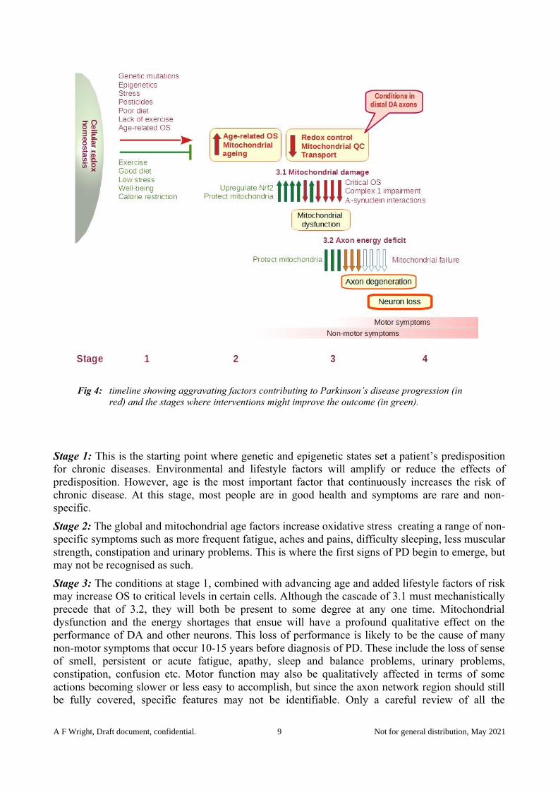
Fig 4: timeline showing aggravating factors contributing to Parkinson’s disease progression (in red) and the stages where interventions might improve the outcome (in green).
Stage 1: This is the starting point where genetic and epigenetic states set a patient’s predispositionfor chronic diseases. Environmental and lifestyle factors will amplify or reduce the effects ofpredisposition. However, age is the most important factor that continuously increases the risk ofchronic disease. At this stage, most people are in good health and symptoms are rare and non-specific.
Stage 2: The global and mitochondrial age factors increase oxidative stress creating a range of non-specific symptoms such as more frequent fatigue, aches and pains, difficulty sleeping, less muscularstrength, constipation and urinary problems. This is where the first signs of PD begin to emerge, butmay not be recognised as such.
Stage 3: The conditions at stage 1, combined with advancing age and added lifestyle factors of riskmay increase OS to critical levels in certain cells. Although the cascade of 3.1 must mechanisticallyprecede that of 3.2, they will both be present to some degree at any one time. Mitochondrialdysfunction and the energy shortages that ensue will have a profound qualitative effect on theperformance of DA and other neurons. This loss of performance is likely to be the cause of manynon-motor symptoms that occur 10-15 years before diagnosis of PD. These include the loss of senseof smell, persistent or acute fatigue, apathy, sleep and balance problems, urinary problems,constipation, confusion etc. Motor function may also be qualitatively affected in terms of someactions becoming slower or less easy to accomplish, but since the axon network region should stillbe fully covered, specific features may not be identifiable. Only a careful review of all the
A F Wright, Draft document, confidential. 9 Not for general distribution, May 2021

symptoms and their progression can help to define this phase. This is where markers of PD andmitochondrial dysfunction are sorely needed.
As stage 3.2 advances towards the point of clinical diagnosis, all PD symptoms and will becomemore clearly identifiable. The increased severity of non-motor symptoms plus the first clear motorsymptoms should be the signal. At this point, the cascade of 3.1 is severely affecting theperformance of DA neurons and causing non-motor symptoms, but despite this, the network ofaxons and synapses in the striatum may still be sufficient to control motor function.
At the point of clinical diagnosis, the cascades of 3.1 and 3.2 will have been fully operational formany years and the threshold of lost DA axons and neurons required to maintain full motor networkcoverage has already been exceeded. Motor and non-motor symptoms are both fully present.
This is usually the point at which patients are offered Levodopa or dopamine agonist therapy. Thesetherapies considerably help re-establish motor function by making sufficient dopamine available todistal axon terminals. They do not however solve the problem of possible gaps in the some regionsof the neural network where many axons may have been lost, nor that of the poor performance ofDA or other types of neurons due to mtD and energy shortages. Dopamine therapy does not addressthese aspects of Parkinson’s disease. As a consequence, many patients on levodopa still sufferserious non-motor symptoms.
Stage 4 occurs when the cascades of stage 3 continue unabated thus reducing neuron performanceand further increasing axon loss beyond the threshold needed to maintain full motor control.
Conclusions
This simplified model for the pathogenesis of Parkinson’s disease is supported by a very substantialbase of experimental evidence covering every step in the direction of increasing disease severity.Nevertheless, the cascade stage 3.1 leading to mitochondrial dysfunction and the ensuing energydeficit needs to be tested in clinical trials in the reverse direction; that of restoring redox balanceand mitochondrial quality control to normal levels. If this model can be confirmed, stage 3.1represents an important opportunity for attenuating disease progression. It opens the way to disease-modifying therapy by simultaneously addressing both oxidative stress and mitochondrialdysfunction. These two processes call for different therapeutic approaches which if applied togethercould transform the synergistic vicious circle driving Parkinson’s disease progression into avirtuous circle to slow or even halt it.
Further work
In a future article, the question of which therapeutic methods might be considered to address bothoxidative stress and mitochondrial dysfunction will be discussed.
References
(1) Vomund, S.; Schäfer, A.; Parnham, M. J.; Brüne, B.; Von Knethen, A. Nrf2, the Master Regulator of Anti-Oxidative Responses. International Journal of Molecular Sciences 2017, 18(12), 2772. https://doi.org/10.3390/ijms18122772.
(2) Tonelli, C.; Chio, I. I. C.; Tuveson, D. A. Transcriptional Regulation by Nrf2. Antioxidants & Redox Signaling 2017, 29 (17), 1727–1745. https://doi.org/10.1089/ars.2017.7342.
A F Wright, Draft document, confidential. 10 Not for general distribution, May 2021
(3) Zhang, H.; Davies, K. J. A.; Forman, H. J. Oxidative Stress Response and Nrf2 Signaling in Aging. Free Radical Biology and Medicine 2015, 88, 314–336. https://doi.org/10.1016/j.freeradbiomed.2015.05.036.
(4) Zhou, L.; Zhang, H.; Davies, K. J. A.; Forman, H. J. Aging-Related Decline in the Induction of Nrf2-Regulated Antioxidant Genes in Human Bronchial Epithelial Cells. Redox Biology 2018, 14, 35–40. https://doi.org/10.1016/j.redox.2017.08.014.
(5) Schmidlin, C. J.; Dodson, M. B.; Madhavan, L.; Zhang, D. D. Redox Regulation by NRF2 in Aging and Disease. Free Radical Biology and Medicine 2019, 134, 702–707. https://doi.org/10.1016/j.freeradbiomed.2019.01.016.
(6) Tan, B. L.; Norhaizan, M. E.; Liew, W.-P.-P.; Sulaiman Rahman, H. Antioxidant and Oxidative Stress: A Mutual Interplay in Age-Related Diseases. Front. Pharmacol. 2018, 9. https://doi.org/10.3389/fphar.2018.01162.
(7) Sun, N.; Youle, R. J.; Finkel, T. The Mitochondrial Basis of Aging. Mol Cell 2016, 61 (5), 654–666. https://doi.org/10.1016/j.molcel.2016.01.028.
(8) Chistiakov, D. A.; Sobenin, I. A.; Revin, V. V.; Orekhov, A. N.; Bobryshev, Y. V. Mitochondrial Aging and Age-Related Dysfunction of Mitochondria. Biomed Res Int 2014, 2014. https://doi.org/10.1155/2014/238463.
(9) Suliman, H. B.; Piantadosi, C. A. Mitochondrial Quality Control as a Therapeutic Target. Pharmacol Rev 2016, 68 (1), 20–48. https://doi.org/10.1124/pr.115.011502.
(10) Cho, B.; Kim, T.; Huh, Y.-J.; Lee, J.; Lee, Y.-I. Amelioration of Mitochondrial Quality Control and Proteostasis by Natural Compounds in Parkinson’s Disease Models. Int J Mol Sci 2019, 20 (20). https://doi.org/10.3390/ijms20205208.
(11) Picca, A.; Calvani, R.; Coelho-Junior, H. J.; Landi, F.; Bernabei, R.; Marzetti, E. Mitochondrial Dysfunction, Oxidative Stress, and Neuroinflammation: Intertwined Roads to Neurodegeneration. Antioxidants (Basel) 2020, 9 (8). https://doi.org/10.3390/antiox9080647.
(12) Dick, F. D.; Palma, G. D.; Ahmadi, A.; Scott, N. W.; Prescott, G. J.; Bennett, J.; Semple, S.; Dick, S.; Counsell, C.; Mozzoni, P.; Haites, N.; Wettinger, S. B.; Mutti, A.; Otelea, M.; Seaton, A.; Söderkvist, P.; Felice, A. Environmental Risk Factors for Parkinson’s Disease and Parkinsonism: The Geoparkinson Study. Occupational and Environmental Medicine 2007, 64 (10), 666–672. https://doi.org/10.1136/oem.2006.027003.
(13) Zhang, P.; Tian, B. Metabolic Syndrome: An Important Risk Factor for Parkinson’s Disease. Oxidative Medicine and Cellular Longevity 2014, 2014, e729194. https://doi.org/10.1155/2014/729194.
(14) Dias, V.; Junn, E.; Mouradian, M. M. The Role of Oxidative Stress in Parkinson’s Disease. Journal of Parkinson’s Disease 2013, 3 (4), 461–491. https://doi.org/10.3233/JPD-130230.
(15) Angeles, D. C.; Gan, B.-H.; Onstead, L.; Zhao, Y.; Lim, K.-L.; Dachsel, J.; Melrose, H.; Farrer, M.; Wszolek, Z. K.; Dickson, D. W.; Tan, E.-K. Mutations in LRRK2 Increase Phosphorylation of Peroxiredoxin 3 Exacerbating Oxidative Stress-Induced Neuronal Death. Human Mutation 2011, 32 (12), 1390–1397. https://doi.org/10.1002/humu.21582.
(16) Li, H.; Ham, A.; Ma, T. C.; Kuo, S.-H.; Kanter, E.; Kim, D.; Ko, H. S.; Quan, Y.; Sardi, S. P.; Li, A.; Arancio, O.; Kang, U. J.; Sulzer, D.; Tang, G. Mitochondrial Dysfunction and Mitophagy Defect Triggered by Heterozygous GBA Mutations. Autophagy 2019, 15 (1), 113–130. https://doi.org/10.1080/15548627.2018.1509818.
(17) Park, J.-S.; Davis, R. L.; Sue, C. M. Mitochondrial Dysfunction in Parkinson’s Disease: New Mechanistic Insights and Therapeutic Perspectives. Curr Neurol Neurosci Rep 2018, 18 (5), 21. https://doi.org/10.1007/s11910-018-0829-3.
(18) McIntyre, R. L.; Daniels, E. G.; Molenaars, M.; Houtkooper, R. H.; Janssens, G. E. From Molecular Promise to Preclinical Results: HDAC Inhibitors in the Race for Healthy Aging
A F Wright, Draft document, confidential. 11 Not for general distribution, May 2021

Drugs. EMBO Molecular Medicine 2019, 11 (9), e9854. https://doi.org/10.15252/emmm.201809854.
(19) Sharma, S.; Taliyan, R. Targeting Histone Deacetylases: A Novel Approach in Parkinson’s Disease. Parkinson’s Disease 2015, 2015, e303294. https://doi.org/10.1155/2015/303294.
(20) Lacal, I.; Ventura, R. Epigenetic Inheritance: Concepts, Mechanisms and Perspectives. Front. Mol. Neurosci. 2018, 11. https://doi.org/10.3389/fnmol.2018.00292.
(21) Liguori, I.; Russo, G.; Curcio, F.; Bulli, G.; Aran, L.; Della-Morte, D.; Gargiulo, G.; Testa, G.;Cacciatore, F.; Bonaduce, D.; Abete, P. Oxidative Stress, Aging, and Diseases. CIA 2018, 13, 757–772. https://doi.org/10.2147/CIA.S158513.
(22) Cuadrado, A.; Manda, G.; Hassan, A.; Alcaraz, M. J.; Barbas, C.; Daiber, A.; Ghezzi, P.; León, R.; López, M. G.; Oliva, B.; Pajares, M.; Rojo, A. I.; Robledinos-Antón, N.; Valverde, A. M.; Guney, E.; Schmidt, H. H. H. W. Transcription Factor NRF2 as a Therapeutic Target for Chronic Diseases: A Systems Medicine Approach. Pharmacol Rev 2018, 70 (2), 348–383. https://doi.org/10.1124/pr.117.014753.
(23) Angelova, P. R.; Esteras, N.; Abramov, A. Y. Mitochondria and Lipid Peroxidation in the Mechanism of Neurodegeneration: Finding Ways for Prevention. Med Res Rev 2021, 41 (2), 770–784. https://doi.org/10.1002/med.21712.
(24) Parker, W. D.; Parks, J. K.; Swerdlow, R. H. Complex I Deficiency in Parkinson’s Disease Frontal Cortex. Brain Research 2008, 1189, 215–218. https://doi.org/10.1016/j.brainres.2007.10.061.
(25) Exner, N.; Lutz, A. K.; Haass, C.; Winklhofer, K. F. Mitochondrial Dysfunction in Parkinson’s Disease: Molecular Mechanisms and Pathophysiological Consequences: Mitochondrial Dysfunction in Parkinson’s Disease. The EMBO Journal 2012, 31 (14), 3038–3062. https://doi.org/10.1038/emboj.2012.170.
(26) Guo, C.; Sun, L.; Chen, X.; Zhang, D. Oxidative Stress, Mitochondrial Damage and Neurodegenerative Diseases. Neural Regen Res 2013, 8 (21), 2003–2014. https://doi.org/10.3969/j.issn.1673-5374.2013.21.009.
(27) Keeney, P. M.; Xie, J.; Capaldi, R. A.; Bennett, J. P. Parkinson’s Disease Brain Mitochondrial Complex I Has Oxidatively Damaged Subunits and Is Functionally Impaired and Misassembled. J. Neurosci. 2006, 26 (19), 5256–5264. https://doi.org/10.1523/JNEUROSCI.0984-06.2006.
(28) Bhatti, J. S.; Bhatti, G. K.; Reddy, P. H. Mitochondrial Dysfunction and Oxidative Stress in Metabolic Disorders — A Step towards Mitochondria Based Therapeutic Strategies. Biochimica et Biophysica Acta (BBA) - Molecular Basis of Disease 2017, 1863 (5), 1066–1077. https://doi.org/10.1016/j.bbadis.2016.11.010.
(29) Di Giacomo, M.; Zara, V.; Bergamo, P.; Ferramosca, A. Crosstalk between Mitochondrial Metabolism and Oxidoreductive Homeostasis: A New Perspective for Understanding the Effects of Bioactive Dietary Compounds. Nutr. Res. Rev. 2020, 33 (1), 90–101. https://doi.org/10.1017/S0954422419000210.
(30) Zaichick, S. V.; McGrath, K. M.; Caraveo, G. The Role of Ca2+ Signaling in Parkinson’s Disease. Dis Model Mech 2017, 10 (5), 519–535. https://doi.org/10.1242/dmm.028738.
(31) Cui, H.; Kong, Y.; Zhang, H. Oxidative Stress, Mitochondrial Dysfunction, and Aging. Journal of Signal Transduction 2011, 2012, e646354. https://doi.org/10.1155/2012/646354.
(32) Misrani, A.; Tabassum, S.; Yang, L. Mitochondrial Dysfunction and Oxidative Stress in Alzheimer’s Disease. Front. Aging Neurosci. 2021, 13. https://doi.org/10.3389/fnagi.2021.617588.
(33) Peoples, J. N.; Saraf, A.; Ghazal, N.; Pham, T. T.; Kwong, J. Q. Mitochondrial Dysfunction and Oxidative Stress in Heart Disease. Experimental & Molecular Medicine 2019, 51 (12), 1–13. https://doi.org/10.1038/s12276-019-0355-7.
A F Wright, Draft document, confidential. 12 Not for general distribution, May 2021

(34) Monzio Compagnoni, G.; Di Fonzo, A.; Corti, S.; Comi, G. P.; Bresolin, N.; Masliah, E. The Role of Mitochondria in Neurodegenerative Diseases: The Lesson from Alzheimer’s Disease and Parkinson’s Disease. Mol Neurobiol 2020, 57 (7), 2959–2980. https://doi.org/10.1007/s12035-020-01926-1.
(35) Nicoletti, V.; Palermo, G.; Del Prete, E.; Mancuso, M.; Ceravolo, R. Understanding the Multiple Role of Mitochondria in Parkinson’s Disease and Related Disorders: Lesson From Genetics and Protein–Interaction Network. Front Cell Dev Biol 2021, 9. https://doi.org/10.3389/fcell.2021.636506.
(36) Tagliaferro, P.; Burke, R. E. Retrograde Axonal Degeneration in Parkinson Disease. Journal of Parkinson’s Disease 2016, 6 (1), 1–15. https://doi.org/10.3233/JPD-150769.
(37) Berthet, A.; Margolis, E. B.; Zhang, J.; Hsieh, I.; Zhang, J.; Hnasko, T. S.; Ahmad, J.; Edwards, R. H.; Sesaki, H.; Huang, E. J.; Nakamura, K. Loss of Mitochondrial Fission Depletes Axonal Mitochondria in Midbrain Dopamine Neurons. J. Neurosci. 2014, 34 (43), 14304–14317. https://doi.org/10.1523/JNEUROSCI.0930-14.2014.
(38) Salvadores, N.; Sanhueza, M.; Manque, P.; Court, F. A. Axonal Degeneration during Aging and Its Functional Role in Neurodegenerative Disorders. Front. Neurosci. 2017, 11. https://doi.org/10.3389/fnins.2017.00451.
(39) Morris, G.; Berk, M. The Many Roads to Mitochondrial Dysfunction in Neuroimmune and Neuropsychiatric Disorders. BMC Medicine 2015, 13 (1), 68. https://doi.org/10.1186/s12916-015-0310-y.
(40) Morris, G.; Walder, K. R.; Berk, M.; Marx, W.; Walker, A. J.; Maes, M.; Puri, B. K. The Interplay between Oxidative Stress and Bioenergetic Failure in Neuropsychiatric Illnesses: Can We Explain It and Can We Treat It? Mol Biol Rep 2020, 47 (7), 5587–5620. https://doi.org/10.1007/s11033-020-05590-5.
(41) Stanga, S.; Caretto, A.; Boido, M.; Vercelli, A. Mitochondrial Dysfunctions: A Red Thread across Neurodegenerative Diseases. International Journal of Molecular Sciences 2020, 21 (10), 3719. https://doi.org/10.3390/ijms21103719.
(42) Perier, C.; Vila, M. Mitochondrial Biology and Parkinson’s Disease. Cold Spring Harb Perspect Med 2012, 2 (2), a009332. https://doi.org/10.1101/cshperspect.a009332.
(43) Ciccone, S.; Maiani, E.; Bellusci, G.; Diederich, M.; Gonfloni, S. Parkinson’s Disease: A Complex Interplay of Mitochondrial DNA Alterations and Oxidative Stress. International Journal of Molecular Sciences 2013, 14 (2), 2388–2409. https://doi.org/10.3390/ijms14022388
(44) Zhou, C.; Huang, Y.; Przedborski, S. Oxidative Stress in Parkinson’s Disease. Annals of the New York Academy of Sciences 2008, 1147 (1), 93–104. https://doi.org/10.1196/annals.1427.023.
(45) Rose, S.; Frye, R. E.; Slattery, J.; Wynne, R.; Tippett, M.; Pavliv, O.; Melnyk, S.; James, S. J. Oxidative Stress Induces Mitochondrial Dysfunction in a Subset of Autism Lymphoblastoid Cell Lines in a Well-Matched Case Control Cohort. PLOS ONE 2014, 9 (1), e85436. https://doi.org/10.1371/journal.pone.0085436.
(46) Nakabeppu, Y.; Tsuchimoto, D.; Yamaguchi, H.; Sakumi, K. Oxidative Damage in Nucleic Acids and Parkinson’s Disease. Journal of Neuroscience Research 2007, 85 (5), 919–934. https://doi.org/10.1002/jnr.21191.
(47) Zeevalk, G. D.; Razmpour, R.; Bernard, L. P. Glutathione and Parkinson’s Disease: Is This theElephant in the Room? Biomedicine & Pharmacotherapy 2008, 62 (4), 236–249. https://doi.org/10.1016/j.biopha.2008.01.017.
(48) Rango, M.; Bonifati, C.; Bresolin, N. Parkinson’s Disease and Brain Mitochondrial Dysfunction: A Functional Phosphorus Magnetic Resonance Spectroscopy Study. J Cereb Blood Flow Metab 2006, 26 (2), 283–290. https://doi.org/10.1038/sj.jcbfm.9600192.
A F Wright, Draft document, confidential. 13 Not for general distribution, May 2021
(49) Zampese, E.; Surmeier, D. J. Calcium, Bioenergetics, and Parkinson’s Disease. Cells 2020, 9 (9), 2045. https://doi.org/10.3390/cells9092045.
(50) Giguère, N.; Burke Nanni, S.; Trudeau, L.-E. On Cell Loss and Selective Vulnerability of Neuronal Populations in Parkinson’s Disease. Front. Neurol. 2018, 9. https://doi.org/10.3389/fneur.2018.00455.
(51) Reeve, A. K.; Grady, J. P.; Cosgrave, E. M.; Bennison, E.; Chen, C.; Hepplewhite, P. D.; Morris, C. M. Mitochondrial Dysfunction within the Synapses of Substantia Nigra Neurons in Parkinson’s Disease. npj Parkinson’s Disease 2018, 4 (1), 1–10. https://doi.org/10.1038/s41531-018-0044-6.
(52) Grünewald, A.; Kumar, K. R.; Sue, C. M. New Insights into the Complex Role of Mitochondria in Parkinson’s Disease. Progress in Neurobiology 2019, 177, 73–93. https://doi.org/10.1016/j.pneurobio.2018.09.003.
(53) Pacelli, C.; Giguère, N.; Bourque, M.-J.; Lévesque, M.; Slack, R. S.; Trudeau, L.-É. Elevated Mitochondrial Bioenergetics and Axonal Arborization Size Are Key Contributors to the Vulnerability of Dopamine Neurons. Current Biology 2015, 25 (18), 2349–2360. https://doi.org/10.1016/j.cub.2015.07.050.
(54) Johnson, D. A.; Johnson, J. A. Nrf2—a Therapeutic Target for the Treatment of Neurodegenerative Diseases. Free Radical Biology and Medicine 2015, 88, 253–267. https://doi.org/10.1016/j.freeradbiomed.2015.07.147.
(55) Dinkova‐Kostova, A. T.; Kostov, R. V.; Kazantsev, A. G. The Role of Nrf2 Signaling in Counteracting Neurodegenerative Diseases. The FEBS Journal 2018, 285 (19), 3576–3590. https://doi.org/10.1111/febs.14379.
(56) Zhang, M.; An, C.; Gao, Y.; Leak, R. K.; Chen, J.; Zhang, F. Emerging Roles of Nrf2 and Phase II Antioxidant Enzymes in Neuroprotection. Progress in Neurobiology 2013, 100, 30–47. https://doi.org/10.1016/j.pneurobio.2012.09.003.
(57) Jazwa, A.; Rojo, A. I.; Innamorato, N. G.; Hesse, M.; Fernández-Ruiz, J.; Cuadrado, A. Pharmacological Targeting of the Transcription Factor Nrf2 at the Basal Ganglia Provides Disease Modifying Therapy for Experimental Parkinsonism. Antioxidants & Redox Signaling 2011, 14 (12), 2347–2360. https://doi.org/10.1089/ars.2010.3731.
(58) Dinkova-Kostova, A. T.; Abramov, A. Y. The Emerging Role of Nrf2 in Mitochondrial Function. Free Radical Biology and Medicine 2015, 88, 179–188. https://doi.org/10.1016/j.freeradbiomed.2015.04.036.
(59) Holmström, K. M.; Kostov, R. V.; Dinkova-Kostova, A. T. The Multifaceted Role of Nrf2 in Mitochondrial Function. Current Opinion in Toxicology 2016, 1, 80–91. https://doi.org/10.1016/j.cotox.2016.10.002.
(60) Holmström, K. M.; Baird, L.; Zhang, Y.; Hargreaves, I.; Chalasani, A.; Land, J. M.; Stanyer, L.; Yamamoto, M.; Dinkova-Kostova, A. T.; Abramov, A. Y. Nrf2 Impacts Cellular Bioenergetics by Controlling Substrate Availability for Mitochondrial Respiration. Biology Open 2013, 2 (8), 761–770. https://doi.org/10.1242/bio.20134853.
(61) Esteras, N.; Dinkova-Kostova, A. T.; Abramov, A. Y. Nrf2 Activation in the Treatment of Neurodegenerative Diseases: A Focus on Its Role in Mitochondrial Bioenergetics and Function. Biological Chemistry 2016, 397 (5), 383–400. https://doi.org/10.1515/hsz-2015-0295.
(62) Bento‐Pereira, C.; Dinkova‐Kostova, A. T. Activation of Transcription Factor Nrf2 to Counteract Mitochondrial Dysfunction in Parkinson’s Disease. Medicinal Research Reviews 2021, 41 (2), 785–802. https://doi.org/10.1002/med.21714.
(63) Diaz, F.; Moraes, C. T. Mitochondrial Biogenesis and Turnover. Cell Calcium 2008, 44 (1), 24–35. https://doi.org/10.1016/j.ceca.2007.12.004.
(64) Ploumi, C.; Daskalaki, I.; Tavernarakis, N. Mitochondrial Biogenesis and Clearance: A Balancing Act. The FEBS Journal 2017, 284 (2), 183–195. https://doi.org/10.1111/febs.13820.
A F Wright, Draft document, confidential. 14 Not for general distribution, May 2021
(65) Schepici, G.; Bramanti, P.; Mazzon, E. Efficacy of Sulforaphane in Neurodegenerative Diseases. International Journal of Molecular Sciences 2020, 21, 8637. https://doi.org/10.3390/ijms21228637.
(66) Yagishita, Y.; Fahey, J. W.; Dinkova-Kostova, A. T.; Kensler, T. W. Broccoli or Sulforaphane: Is It the Source or Dose That Matters? Molecules 2019, 24 (19), 3593. https://doi.org/10.3390/molecules24193593.
(67) Fahey, J. W.; Kensler, T. W. The Challenges of Designing and Implementing Clinical Trials With Broccoli Sprouts… and Turning Evidence Into Public Health Action. Front. Nutr. 2021, 8. https://doi.org/10.3389/fnut.2021.648788.
(68) Wright, A. F. A Pilot Study of a Broccoli-Seed Tea by Eight Parkinson’s Disease Patients. 2021. https://doi.org/10.13140/RG.2.2.12812.03207/2.
(69) Menzies, R. A.; Gold, P. H. The Turnover of Mitochondria in a Variety of Tissues of Young Adult and Aged Rats. J Biol Chem 1971, 246 (8), 2425–2429.
(70) Rugarli, E. I.; Langer, T. Mitochondrial Quality Control: A Matter of Life and Death for Neurons. The EMBO Journal 2012, 31 (6), 1336–1349. https://doi.org/10.1038/emboj.2012.38.
(71) Friedman, J. H.; Beck, J. C.; Chou, K. L.; Clark, G.; Fagundes, C. P.; Goetz, C. G.; Herlofson, K.; Kluger, B.; Krupp, L. B.; Lang, A. E.; Lou, J.-S.; Marsh, L.; Newbould, A.; Weintraub, D. Fatigue in Parkinson’s Disease: Report from a Multidisciplinary Symposium. npj Parkinson’s Disease 2016, 2 (1), 1–6. https://doi.org/10.1038/npjparkd.2015.25.
(72) Filler, K.; Lyon, D.; Bennett, J.; McCain, N.; Elswick, R.; Lukkahatai, N.; Saligan, L. N. Association of Mitochondrial Dysfunction and Fatigue: A Review of the Literature. BBA Clinical 2014, 1, 12–23. https://doi.org/10.1016/j.bbacli.2014.04.001.
(73) Smits, B.; van den Heuvel, L.; Knoop, H.; Küsters, B.; Janssen, A.; Borm, G.; Bleijenberg, G.;Rodenburg, R.; van Engelen, B. Mitochondrial Enzymes Discriminate between Mitochondrial Disorders and Chronic Fatigue Syndrome. Mitochondrion 2011, 11 (5), 735–738. https://doi.org/10.1016/j.mito.2011.05.005.
(74) Siciliano, M.; Trojano, L.; Santangelo, G.; Micco, R. D.; Tedeschi, G.; Tessitore, A. Fatigue inParkinson’s Disease: A Systematic Review and Meta-Analysis. Movement Disorders 2018, 33 (11), 1712–1723. https://doi.org/10.1002/mds.27461.
(75) Ferrer, I. Neuropathology and Neurochemistry of Nonmotor Symptoms in Parkinson’s Disease. Parkinson’s Disease 2011, 2011, e708404. https://doi.org/10.4061/2011/708404.
A F Wright, Draft document, confidential. 15 Not for general distribution, May 2021