alpha-synuclein-induced mitochondrial dysfunction is
TRANSCRIPT

RESEARCH ARTICLE Open Access
Alpha-synuclein-induced mitochondrialdysfunction is mediated via a sirtuin 3-dependent pathwayJae-Hyeon Park1, Jeremy D. Burgess1,2, Ayman H. Faroqi1, Natasha N. DeMeo1, Fabienne C. Fiesel1,Wolfdieter Springer1,2, Marion Delenclos1* and Pamela J. McLean1,2*
Abstract
Background: Misfolding and aggregation of the presynaptic protein alpha-synuclein (αsyn) is a hallmark of Parkinson’sdisease (PD) and related synucleinopathies. Although predominantly localized in the cytosol, a body of evidence hasshown that αsyn localizes to mitochondria and contributes to the disruption of key mitochondrial processes.Mitochondrial dysfunction is central to the progression of PD and mutations in mitochondrial-associated proteins arefound in familial cases of PD. The sirtuins are highly conserved nicotinamide adenine dinucleotide (NAD+)-dependentenzymes that play a broad role in cellular metabolism and aging. Interestingly, mitochondrial sirtuin 3 (SIRT3) plays amajor role in maintaining mitochondrial function and preventing oxidative stress, and is downregulated in aging andage-associated diseases such as neurodegenerative disorders. Herein, we hypothesize that αsyn is associated withdecreased SIRT3 levels contributing to impaired mitochondrial dynamics and biogenesis in PD.
Methods: The level of mitochondrial SIRT3 was assessed in cells expressing oligomeric αsyn within the cytosolic andmitochondrial-enriched fractions. Mitochondrial integrity, respiration, and health were examined using several markersof mitochondrial dynamics and stress response and by measuring the rate of oxygen consumption (OCR). Our findingswere validated in a rodent model of PD as well as in human post-mortem Lewy body disease (LBD) brain tissue.
Results: Here, we demonstrate that αsyn associates with mitochondria and induces a decrease in mitochondrial SIRT3levels and mitochondrial biogenesis. We show that SIRT3 downregulation is accompanied by decreasedphosphorylation of AMPK and cAMP-response element binding protein (CREB), as well as increased phosphorylation ofdynamin-related protein 1 (DRP1), indicative of impaired mitochondrial dynamics. OCR was significantly decreasedsuggesting a mitochondria respiratory deficit. Interestingly treatment with AMPK agonist 5-aminoimidazole-4-carboxamide-1-β-d-ribofuranoside (AICAR) restores SIRT3 expression, improves mitochondrial function, and decreasesαsyn oligomer formation in a SIRT3-dependent manner.
Conclusions: Together, our findings suggest that pharmacologically increasing SIRT3 levels can counteract αsyn-induced mitochondrial dysfunction by reducing αsyn oligomers and normalizing mitochondrial bioenergetics. Thesedata support a protective role for SIRT3 in PD-associated pathways and contribute significant mechanistic insight intothe interplay of SIRT3 and αsyn.
Keywords: α-Synuclein, Sirtuin 3, Mitochondria dysfunction, Parkinson’s disease
© The Author(s). 2020 Open Access This article is distributed under the terms of the Creative Commons Attribution 4.0International License (http://creativecommons.org/licenses/by/4.0/), which permits unrestricted use, distribution, andreproduction in any medium, provided you give appropriate credit to the original author(s) and the source, provide a link tothe Creative Commons license, and indicate if changes were made. The Creative Commons Public Domain Dedication waiver(http://creativecommons.org/publicdomain/zero/1.0/) applies to the data made available in this article, unless otherwise stated.
* Correspondence: [email protected]; [email protected] of Neuroscience, Mayo Clinic, 4500 San Pablo Road,Jacksonville, FL 32224, USAFull list of author information is available at the end of the article
Park et al. Molecular Neurodegeneration (2020) 15:5 https://doi.org/10.1186/s13024-019-0349-x

BackgroundAlpha-synuclein (αsyn) accumulation is believed to be akey step in the pathogenesis of Parkinson’s disease (PD)and related alpha-synucleinopathies. Despite predominantlocalization in the cytosol, αsyn is found localized to mito-chondria in post-mortem PD brain [1]. Mitochondrial ac-cumulation of αsyn has been associated with impairedcomplex-I dependent respiration, decreased mitochondrialmembrane potential, and increased levels of mitochondrialreactive oxygen species (mtROS) in multiple cellularmodels [1–4]. The evidence supporting the contributionof abnormal accumulation of αsyn to disruption of mito-chondrial processes is compelling and indicates a crucialrole for αsyn-induced mitochondrial dysfunction in PDpathogenesis and alpha-synucleopathies.The sirtuins (SIRTs) are a family of nicotinamide aden-
ine dinucleotide (NAD+)-dependent deacetylases and/oradenosine diphosphate (ADP)-ribosyltransferases thathave long been recognized as essential for cell survival,metabolism, and longevity [5]. In mammals there areseven human SIRT homologs (SIRT1–7) with varied en-zymatic activities. SIRT1, SIRT6, and SIRT7 predomin-antly reside in the nucleus whereas SIRT2 is located in thecytoplasm, and SIRT3, 4, and 5 reside in the mitochon-dria. SIRTs have been previously implicated in mecha-nisms of PD in a number of in vitro and in vivo studies[6–8]. The use of pharmacological activators and inhibi-tors of SIRTs in PD models have revealed neuroprotectiveand beneficial effects. For example, resveratrol, an activa-tor of SIRT1, protects against cell death in neurotoxin-induced PD animal models [9, 10] and we have shownthat SIRT2 inhibitors can rescue αsyn-mediated toxicity incellular PD models [8]. SIRT3 is the predominant mito-chondrial sirtuin and the major regulator of mitochondrialprotein acetylation [11–13]. SIRT3 is expressed at highlevels in the brain [14, 15] and plays an important role inmaintaining mitochondrial integrity, energy metabolism,and regulating mitochondrial oxidative pathways [16, 17].SIRT3-mediated deacetylation activates enzymes respon-sible for the reduction of ROS production, such as super-oxide dismutase 2 (SOD2) [18]. Interestingly, SIRT3 actsas a pro-survival factor in neurons exposed to excitotoxicinjury [19] and recent studies demonstrate a neuroprotec-tive effect of SIRT3 in cell culture models of stroke, Hun-tington’s disease (HD), and Alzheimer’s disease (AD) [20–22]. Importantly, and relevant to the present study, over-expression of SIRT3 was recently demonstrated to preventdopaminergic cell loss in a rodent model of PD [11].Experimental evidence supports SIRT3-induced pro-
tection against oxidative stress by enhancement of mito-chondrial biogenesis and integrity [23]. The multifacetedmitochondrial health-enhancing capabilities of SIRT3thus make it an attractive therapeutic target for neuro-degenerative diseases where mitochondrial dysfunction
contributes to disease pathogenesis. Herein, we investigatea role for SIRT3 in PD pathogenesis and identify a poten-tial mechanistic interaction between SIRT3 and αsyn. Wehypothesize that the association of αsyn with mitochon-dria reduces SIRT3 deacetylase activity and contributes tomitochondrial dysfunction and pathogenesis in PD and re-lated alpha-synucleinopathies. The data presented hereinsignificantly advances our mechanistic understanding ofSIRT3 in mitochondrial dysfunction and validates a pro-tective role for SIRT3 in PD. Overall we confirm the po-tential application of SIRT3 activators as future targets forpharmacological strategies against neurodegeneration inPD and related alpha-synucleinopathies.
MethodsCell cultureA stable cell line co-expressing human αsyn fused to ei-ther the amino-terminal (SL1) or carboxy-terminal frag-ment (SL2) of humanized Gaussia princeps luciferase wasgenerated and described previously [24]. H4 SL1&SL2 andwt-αsyn cells were maintained at 37 °C in a 95% air/5%CO2 humidified incubator in Opti-MEM supplementedwith 10% FBS. Stock cultures were kept in the presence of1 μg/ml tetracycline (Invitrogen) to block the expressionof the transgenes (SL1&SL2, wt--αsyn). αSyn expression isturned on or off by the absence (Tet- cells) or presence(Tet + cells) of tetracycline respectively.Embryonic primary cortical neurons were prepared
from E15 CD1 wildtype mice (Charles River, Wilming-ton, MA). Briefly, brains of E15 embryos were dissectedin calcium and magnesium free HBSS, dissociated with0.25% trypsin-EDTA (Life Technologies, Grand Island,NY), and seeded on poly-D-lysine coated 6 cm dishes at0.95 × 105 cells per cm2 (2 × 10⁶ cells per dish) in Neuro-basal media containing 10% FBS, 1% pen/strep and 1%glutamax. After 1 h, media was exchanged for Neuroba-sal containing B-27 supplement, 1% pen/strep and 1%glutamax. Neurons were maintained at 37 °C in a hu-midified incubator with 5% CO2/95% air. At day 7in vitro (DIV) neurons were transduced with adeno-associated-virus (AAV) serotype2/8 expressing wt-αsynor venusYFP under the chicken beta actin promoter.
Rodent stereotaxic surgeryAdult female Sprague Dawley rats (225-250 g, Envigo,USA) were housed and treated in accordance with theNIH Guide for Care and Use of Laboratory animals. Allanimal procedures were approved by the Mayo Institu-tional Animal Care and Use Committee and are in accord-ance with the NIH Guide for Care and Use of Laboratoryanimals. All viral vector delivery surgical procedures andtissue processing was performed as previously describedby our group [25]. Briefly, AAVs serotype 2/8 expressinghuman αsyn fused with either the C-terminus (AAV-SL1)
Park et al. Molecular Neurodegeneration (2020) 15:5 Page 2 of 19

or N-terminus (AAV-SL2) of Gaussia princeps luciferasewere produced by plasmid triple transfection with helperplasmids in HEK293T cells. 48 h later, cells were harvestedand lysed in the presence of 0.5% sodium deoxycholate and50U/ml Benzonase (Sigma-Aldrich, St. Louis, MO) byfreeze-thawing, and the virus was isolated using a discon-tinuous iodixanol gradient. The genomic titer of each viruswas determined by quantitative PCR. A combination ofAAV-SL1 (8.10e12gc/ml) + AAV-SL2 (8.10e12 gc/ml) wasdelivered directly to the right substantia nigra/midbrain(SN) using stereotaxic surgery (coordinates: AP − 5.2mm,ML + 2.0mm, DV + 7.2mm from dura) [26]. A mix ofAAVs were infused at a rate of 0.4 μL/min (final volume2 μL) using a microinjector (Stoelting). A group of controlanimals were injected with 2 μL of AAV8 expressingfull length of humanized Gaussia princeps luciferase(AAV8-Hgluc).
Human brain tissueFrozen human post-mortem brain was provided by theMayo Clinic brain bank at the Mayo Clinic in Jacksonville.For this study, striatum (STR) samples from 10 control pa-tients (6 females, 4 males) and 10 patients diagnosed withLewy body disease (LBD) (4 females and 6 males) wereincluded. Detailed information of brain tissue is provided inTable 1. Each frozen brain sample was weighed and ho-mogenized in 10X volume of radio-immunoprecipitationassay (RIPA) lysis buffer (0.5M Tris-HCl, pH 7.4, 1.5MNaCl, 2.5% deoxycholic acid, 10% NP-40, 10mM EDTA,20–188) containing 1mM phenylmethylsulfonyl fluoride(PMSF), protease inhibitor cocktail, and halt phosphataseinhibitor cocktail, followed by sonication and centrifugationfor 15min at 16,000×g at 4 °C to remove cellular debris. Su-pernatants were collected, protein concentration was deter-mined by Bradford assay, and samples were processed forimmunoblotting.
ImmunofluorescenceCells were cultured on 12-mm glass coverslips with orwithout 1 μg/ml tetracycline for 72 h. Cells were washedwith phosphate-buffered saline (PBS) and incubated with300 nM with MitoTracker-Green (Molecular Probes,Inc., Eugene, OR, USA) according to the manufacturer’sprotocol to visualize mitochondria. Cells were fixed with4% paraformaldehyde for 10 min at room temperature(RT) and washed three times in 1X Tris-buffered saline(TBS) (500 mM NaCl, 20 mM Tris, pH 7.4), blocked for1 h in 1.5% goat serum, 0.5% Triton X-100 in 1X TBSand incubated overnight at 4 °C with primary antibodies(SIRT3 and human αsyn). The following day cells werewashed and treated with Alexa Fluor® 488 and 568 sec-ondary antibodies for 1 h at RT (see Table 2, for detailsof the antibodies used in the study). Coverslips weremounted on Super Frost Plus slides with Vectashield
Hardset (Vector Labs, Burlingame, CA) and cells werevisualized using an Axio observer inverted microscope(Carl Zeiss, Germany).
Gaussia luciferase protein-fragment complementationassaysLuciferase activity was measured in 15μg cell lysate or infreshly homogenized STR and SN rat tissue in multilabelplate reader at 480 nm (EnVision, PerkinElmer; Wal-tham, MA, USA) following the injection of the substrate,coelenterazine (40 μM, NanoLight tech, AZ, USA) with asignal integration of 2 s.
Western blotting analysisTo prepare whole cell lysates, cells were washed twicewith ice-cold PBS and total proteins were isolated by in-cubating H4 cells in RIPA lysis buffer (50 mM Tris–HCl,pH 7.4, 150 mM NaCl, 1 mM EDTA, 1 mM EGTA, 1.2%Triton X-100, 0.5% sodium deoxycholate, and 0.1% SDS,ADI-80-1496,1 mM PMSF) or primary neurons in atriton-X based lysis buffer (150mM NaCl, 1 mM EDTA,20mM Tris-HCL,1% triton-X pH 7.4). Both buffers were
Table 1 Human brain samples
Case Pathology Dx Thal Braak Clinical Dx Age at Death Sex
1 Normal 2 aMCI 70 Male
2 Normal 0 Normal 56 Female
3 Normal 0 Normal 57 Female
4 Normal 0 2 AD v DLB 69 Male
5 Normal 1 1 DA 64 Female
6 Normal 0 3 DLB v FTD 63 Male
7 Normal 0 Normal 61 Female
8 Normal 1 1 NAIM 60 Female
9 Normal 0 1 PSP/PLS 56 Female
10 Normal 0 1 TD 61 Male
1 DLBD 0 0 DLB 60 Male
2 DLBD 0 2 DLB 61 Male
3 DLBD 0 2 PDD 66 Male
4 DLBD 0 0 PDD 68 Female
5 DLBD 1 2 PSP 72 Female
6 DLBD 1 2 DLB (RBD) 70 Male
7 DLBD 1 2 PDD 56 Male
8 DLBD 1 2.5 PDD 62 Female
9 DLBD 0 2 PD-MCI 66 Male
10 DLBD 2 1 PDD v CBD 69 Female
Dx Diagnosis, AD Alzheimer’s diseases, aMCI Amnestic mild cognitiveimpairment, CBD Corticobasal degeneration, DA Dysautonomia, DLBD Diffuselewy body disease, DLB Dementia with lewy bodies, FTD Frontotemporaldementia, NAIM Nonvasculitic autoimmune inflammatory meningoencephalitis,PD Parkinson’s disease, PDD Parkinson’s disease with dementia, PLS Primarylateral sclerosis, PSP Progressive supranuclear palsy, RBD REM sleep behaviordisorder, TD Torsion dystonia
Park et al. Molecular Neurodegeneration (2020) 15:5 Page 3 of 19

supplemented with protease inhibitor cocktail, and haltphosphatase inhibitor cocktail. Collected cells were cen-trifuged at 10,000×g for 10 min at 4 °C. The protein con-centration was determined with Bradford reagent. 15 μgproteins were separated on Bis-Tris polyacrylamide gra-dient gels (NuPAGE Novex 4–12% Bis-Tris Gel, Lifetech) and transferred to nitrocellulose membranes.Membranes were then blocked for 1 h at RT in TBS-T(500 mM NaCl, 20 mM Tris, 0.1% Tween 20, pH 7.4)supplemented with 10% non-fat dried milk. Subse-quently membranes were incubated overnight at 4 °Cwith primary antibodies followed by 1 h at RT withHRP-conjugated secondary antibodies or IRDye® conju-gated secondaries (LI-COR®) (Table 2). Proteins were de-tected using an enhanced chemiluminescent detection
system (ECL, EMD Millipore) and a CCD imaging sys-tem (LAS-4000, Fujifilm, Japan) or Odyssey® CLx Im-aging System (LI-COR®, USA).
Mitochondria/cytosol fractionationCells were homogenized in buffer A (0.25M sucrose, 10mM Tris–HCl [pH 7.5], 10 mM KCl, 1.5 mM MgCl2, 1mM EDTA, 1 mM dithiothreitol, and 0.1 mM PMSF).Homogenates were centrifuged at 700×g for 5 min at4 °C, and supernatants were collected and centrifuged at10,000×g for 30 min at 4 °C. The supernatant was desig-nated the cytosolic fraction, and the pellet was used asthe mitochondrial enriched fraction. The pellets were re-suspended in buffer B (0.25M sucrose, 10 mM Tris–HCl[pH 7.5], 10 mM KCl, 1.5 mM MgCl2, 1 mM EDTA, 1
Table 2 Antibodies used for western blot and immunocyhistochemistry
Antibody Source Dilution
α-Synuclein (mouse) BD Transduction Laboratories (61078) 1:2000 (WB)
α-Synuclein (mouse) Biolegend (SIG39730) 1:2000 (WB, ICC)
α-Synuclein, clone 5G4 (mouse) Millipore (MABN389) 1:1000 (WB)
Oligomer A11 (rabbit) Thermo Fisher Scientific (AHB0052) 1:1000 (WB)
SIRT3 (mouse) Santa Cruz (sc-135,796) 1:1000 (WB)
SIRT3 (rabbit) Cell Signaling (2627 s) 1:2000 (WB)
SIRT3 (rabbit) Novus Biologicals (NBP1–31029) 1:1000 (WB)
1:500 (ICC)
SIRT3 (rabbit) Cell Signaling (5490) 1:1000 (WB)
Heme oxygenase-1 (rabbit) Cell Signaling (5853 s) 1:1000 (WB)
AMPK (rabbit) Cell Signaling (5831 T) 1:1000 (WB)
Phospho-AMPK (rabbit) Cell Signaling (2535 s) 1:1000 (WB)
CREB (rabbit) Cell Signaling (4820 s) 1:1000 (WB)
Phospho-CREB (rabbit) EMD Millipore (06–519) 1:2000 (WB)
DRP1 (rabbit) Bethyl Laboratories (A303-410A-M) 1:2000 (WB)
Phospho-DRP1 (rabbit) Cell Signaling (3455 s) 1:1000 (WB)
SOD2 (rabbit) abcam (ab13533) 1:5000 (WB)
SOD2 (acetyl K68) (rabbit) abcam (ab137037) 1:2000 (WB)
COX IV (rabbit) Cell Signaling (4850 s) 1:1000 (WB)
GAPDH (rabbit) Santa Cruz (sc-25,778) 1:4000 (WB)
Abgent (AP7873a) 1:4000 (WB)
Actin (mouse) Sigma (A5316) 1:7500 (WB)
GM130 (rabbit) abcam (ab52649) 1:2000 (WB)
Alexa Fluor 488 (goat anti-mouse) Thermo Fisher Scientific (A11001) 1:500 (ICC)
Alexa Fluor 568 (goat anti-rabbit) Thermo Fisher Scientific (A11011) 1:500 (ICC)
Goat anti-mouse HRP Southern biotech (1010–05) 1:5000 (WB)
Goat anti-rabbit HRP Southern biotech (4010–05) 1:5000 (WB)
IRDye® 680RD Goat anti-Mouse IgG (H + L) LI-COR® (926–68,070) 1:10000 (WB)
IRDye® 800CW Goat anti-Mouse IgG (H + L), 0.5 mg LI-COR® (926–32,210) 1:10000 (WB)
IRDye® 800CW Goat anti-Rabbit IgG (H + L), 0.5 mg LI-COR® (926–32,211) 1:10000 (WB)
WB Western blot, ICC Immunocytochemistry
Park et al. Molecular Neurodegeneration (2020) 15:5 Page 4 of 19

Fig. 1 (See legend on next page.)
Park et al. Molecular Neurodegeneration (2020) 15:5 Page 5 of 19
mM dithiothreitol, 0.1 mM PMSF, and 1% NP 40). Toconfirm the purity of the mitochondrial fraction, the ly-sates were probed for the specific mitochondria markercytochrome c oxidase IV (COXIV).
Dot blot assayTris-buffered saline (TBS)-wetted nitrocellulose mem-brane (NC membrane, 0.45 μm pore) was mounted onthe Bio-Dot microfiltration apparatus (cat. no. 1706545,Bio-Rad). 15 μg proteins were loaded into the wells of amicrofiltration apparatus under mild vacuum. Afterwashing with TBS, the NC membranes were blockedwith TBS-T (500 mM NaCl, 20 mM Tris, 0.1% Tween20, pH 7.4) supplemented with 10% non-fat dried milkand incubated overnight at 4 °C with primary antibodies(anti-oligomer A11, cat. no. AHB0052; anti-αsyn 5G4,cat. no. MABN389 followed by 1 h at RT with HRP-conjugated secondary antibodies. Proteins were detectedusing an enhanced chemiluminescent detection system(ECL, EMD Millipore) and ChemiDoc MP Imaging Sys-tem (Bio-Rad, 170–01402, USA).
Isolation of rat brain mitochondriaSN were dissected and homogenized in 0.5mL of ice-coldMIBA (10mM Tris–HCl [pH 7.4], 1 mM EDTA, 0.2M D-mannitol, 0.05M sucrose, 0.5 mM sodium orthovanadate,1 mM sodium fluoride and dissolved in water) containing1X protease inhibitors with a hand-held homogenizer for40 strokes on ice. The homogenate was transferred into1.5 mL tubes and then centrifuged at 500×g for 5 min.The pellet was discarded, and remaining supernatant wascentrifuged at 11,000×g for 20min at 4 °C, yielding theheavy mitochondrial (HM, pellet) and the light mitochon-drial (LM, supernatant) fraction. The HM pellet waswashed twice with 1mL ice-cold MIBA buffer it wasresuspended in 0.1–0.3mL of MIBA to yield the finalsolution enriched in mitochondria.
Mitochondrial respiration analysisThe oxygen consumption rate (OCR) was assessed usinga Seahorse Bioscience XF96 analyzer (Seahorse Bio-science, Billerica, MA, USA) in combination with theSeahorse Bioscience XF Cell Mito Stress Test assay kitaccording to the manufacturer’s recommendations. H4
SL1&SL2 cells were seeded in 12-wells of a XF 96-wellcell culture microplate (Seahorse Bioscience, 102,601–100) and grown to 70% confluency in 200 μL of growthmedium prior to analysis. On the day of assay, culturemedia were changed to assay medium with 175 μL (Dul-becco’s Modified Eagle’s Medium, D5030), supplementedwith 25mM glucose, 2 mM glutamine, and 2mM pyru-vate. Prior to assay, plates were incubated at 37 °C for 1 hwithout CO2. Thereafter successive OCR measurementswere performed consisting of basal OCR, followed byOCR level after the automated injection of 25 μl oligomy-cin (20 μM), 25 μl carbonyl cyanide 4-(trifluoromethoxy)phenylhydrazone (FCCP) (20 μM), and a combination of25 μl rotenone + antimycin A (12 μM), respectively. Afterthe assays, plates were saved and OCR was normalized tothe total protein amount per well.
SIRT3 siRNA transfectionSmall interfering RNAs (siRNAs) for human SIRT3 (sc-61,555, Santa Cruz Biotechnology, CA, USA) and controlnon-target siRNA (SN-1003, Negative Control, Bioneer,Daejeon, Korea) were reconstituted in siRNA buffer (Qia-gen, CA) following the manufacturer’s instructions andtransfections of conducted using Lipofectamine 2000 re-agent (Invitrogen, CA, USA). Briefly H4 SL1&SL2 cellswere seeded in 60-mm culture dishes for 24 h beforetransfection. Subconfluent cells were treated either withSIRT3 siRNA (100 nM) or non-targeting siRNA (20 nM)complexed with lipofectamine for 2 h. The extent ofknockdown was evaluated by western blot analysis.
Determination of mitochondrial ROSMitoSOX™ Red fluorescent probe (Molecular Probes, Inc.,Eugene, OR, USA) was used to visualize mitochondrialsuperoxide production according to the manufacturer’sprotocol. Briefly, H4 SL1&SL2 grown on 12-mm glass werewashed twice with PBS to remove the medium and incu-bated with 2.5 μM MitoSOX-Red reagent in the dark at37 °C. Cells were washed gently three times with warm PBSbuffer and imaged immediately after, under fluorescencemicroscopy. To confirm mitochondrial localization ofMitoSOX-Red, cells were loaded with 300 nMMitoTracker-Green (Molecular Probes, Inc., Eugene, OR,USA) for 30min. The mean fluorescence intensities of
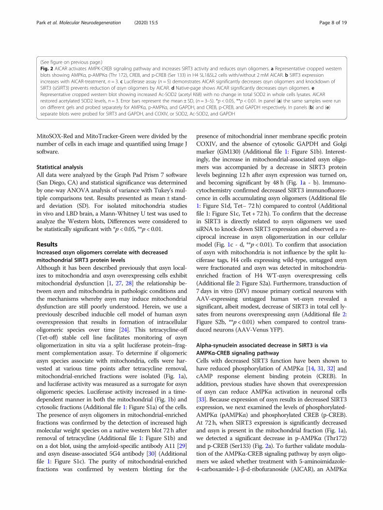
(See figure on previous page.)Fig. 1 αSyn is found in mitochondria-enriched fraction of H4 SL1&SL2 cells and induces a decrease in SIRT3 expression. a Representative croppedwestern blots showing αsyn and SIRT3 in cytosolic and mitochondrial fractions at different time points (0 – 72 h) (b) αsyn oligomers are apparentafter 24 h by luciferase assay (RLU: relative luminescence units), n = 5. Quantification of SIRT3 protein level in mitochondria demonstratessignificant decrease in SIRT3 at 48 h and 72 h. c Whole cells lysates from H4 SL1&SL2 cells demonstrate decreased SIRT3 expression aftertransfection with SIRT3 siRNA n = 6 (d) Luciferase activity from αsyn oligonmerization is significantly increased in cells transfected with SIRT3siRNA (siSIRT3) compared to control siRNA (siCtrl). Error bars represent the mean ± SD. *p < 0.05, **p < 0.01. Note: In (a) αsyn and SIRT3 bands arefrom different experiments run on different gels. COXIV, GAPDH, and SIRT3 are all from same samples and immunoblot. Loading controls for αsynblot are not shown. In panel (c) αsyn, SIRT3, and GAPDH are detected on same immunoblot
Park et al. Molecular Neurodegeneration (2020) 15:5 Page 6 of 19

Fig. 2 (See legend on next page.)
Park et al. Molecular Neurodegeneration (2020) 15:5 Page 7 of 19
MitoSOX-Red and MitoTracker-Green were divided by thenumber of cells in each image and quantified using Image Jsoftware.
Statistical analysisAll data were analyzed by the Graph Pad Prism 7 software(San Diego, CA) and statistical significance was determinedby one-way ANOVA analysis of variance with Tukey’s mul-tiple comparisons test. Results presented as mean ± stand-ard deviation (SD). For isolated mitochondria studiesin vivo and LBD brain, a Mann-Whitney U test was used toanalyze the Western blots, Differences were considered tobe statistically significant with *p < 0.05, **p < 0.01.
ResultsIncreased αsyn oligomers correlate with decreasedmitochondrial SIRT3 protein levelsAlthough it has been described previously that αsyn local-izes to mitochondria and αsyn overexpressing cells exhibitmitochondrial dysfunction [1, 27, 28] the relationship be-tween αsyn and mitochondria in pathologic conditions andthe mechanisms whereby αsyn may induce mitochondrialdysfunction are still poorly understood. Herein, we use apreviously described inducible cell model of human αsynoverexpression that results in formation of intracellularoligomeric species over time [24]. This tetracycline-off(Tet-off) stable cell line facilitates monitoring of αsynoligomerization in situ via a split luciferase protein–frag-ment complementation assay. To determine if oligomericαsyn species associate with mitochondria, cells were har-vested at various time points after tetracycline removal,mitochondrial-enriched fractions were isolated (Fig. 1a),and luciferase activity was measured as a surrogate for αsynoligomeric species. Luciferase activity increased in a time-dependent manner in both the mitochondrial (Fig. 1b) andcytosolic fractions (Additional file 1: Figure S1a) of the cells.The presence of αsyn oligomers in mitochondrial-enrichedfractions was confirmed by the detection of increased highmolecular weight species on a native western blot 72 h afterremoval of tetracycline (Additional file 1: Figure S1b) andon a dot blot, using the amyloid-specific antibody A11 [29]and αsyn disease-associated 5G4 antibody [30] (Additionalfile 1: Figure S1c). The purity of mitochondrial-enrichedfractions was confirmed by western blotting for the
presence of mitochondrial inner membrane specific proteinCOXIV, and the absence of cytosolic GAPDH and Golgimarker (GM130) (Additional file 1: Figure S1b). Interest-ingly, the increase in mitochondrial-associated αsyn oligo-mers was accompanied by a decrease in SIRT3 proteinlevels beginning 12 h after αsyn expression was turned on,and becoming significant by 48 h (Fig. 1a - b). Immuno-cytochemistry confirmed decreased SIRT3 immunofluores-cence in cells accumulating αsyn oligomers (Additional file1: Figure S1d, Tet– 72 h) compared to control (Additionalfile 1: Figure S1c, Tet + 72 h). To confirm that the decreasein SIRT3 is directly related to αsyn oligomers we usedsiRNA to knock-down SIRT3 expression and observed a re-ciprocal increase in αsyn oligomerization in our cellularmodel (Fig. 1c - d, **p < 0.01). To confirm that associationof αsyn with mitochondria is not influence by the split lu-ciferase tags, H4 cells expressing wild-type, untagged αsynwere fractionated and αsyn was detected in mitochondria-enriched fraction of H4 WT-αsyn overexpressing cells(Additional file 2: Figure S2a). Furthermore, transduction of7 days in vitro (DIV) mouse primary cortical neurons withAAV-expressing untagged human wt-αsyn revealed asignificant, albeit modest, decrease of SIRT3 in total cell ly-sates from neurons overexpressing αsyn (Additional file 2:Figure S2b, **p < 0.01) when compared to control trans-duced neurons (AAV-Venus YFP).
Alpha-synuclein associated decrease in SIRT3 is viaAMPKα-CREB signaling pathwayCells with decreased SIRT3 function have been shown tohave reduced phosphorylation of AMPKα [14, 31, 32] andcAMP response element binding protein (CREB). Inaddition, previous studies have shown that overexpressionof αsyn can reduce AMPKα activation in neuronal cells[33]. Because expression of αsyn results in decreased SIRT3expression, we next examined the levels of phosphorylated-AMPKα (pAMPKα) and phosphorylated CREB (p-CREB).At 72 h, when SIRT3 expression is significantly decreasedand αsyn is present in the mitochondrial fraction (Fig. 1a),we detected a significant decrease in p-AMPKα (Thr172)and p-CREB (Ser133) (Fig. 2a). To further validate modula-tion of the AMPKα-CREB signaling pathway by αsyn oligo-mers we asked whether treatment with 5-aminoimidazole-4-carboxamide-1-β-d-ribofuranoside (AICAR), an AMPKα
(See figure on previous page.)Fig. 2 AICAR activates AMPK-CREB signaling pathway and increases SIRT3 activity and reduces αsyn oligomers. a Representative cropped westernblots showing AMPKα, p-AMPKα (Thr 172), CREB, and p-CREB (Ser 133) in H4 SL1&SL2 cells with/without 2 mM AICAR. b SIRT3 expressionincreases with AICAR-treatment, n = 3. c Luciferase assay (n = 5) demonstrates AICAR significantly decreases αsyn oligomers and knockdown ofSIRT3 (siSIRT3) prevents reduction of αsyn oligomers by AICAR. d Native-page shows AICAR significantly decreases αsyn oligomers. eRepresentative cropped western blot showing increased Ac-SOD2 (acetyl K68) with no change in total SOD2 in whole cells lysates. AICARrestored acetylated SOD2 levels, n = 3. Error bars represent the mean ± SD, (n = 3–5). *p < 0.05, **p < 0.01. In panel (a) the same samples were runon different gels and probed separately for AMPKα, p-AMPKα, and GAPDH, and CREB, p-CREB, and GAPDH respectively. In panels (b) and (e)separate blots were probed for SIRT3 and GAPDH, and COXIV, or SOD2, Ac-SOD2, and GAPDH
Park et al. Molecular Neurodegeneration (2020) 15:5 Page 8 of 19
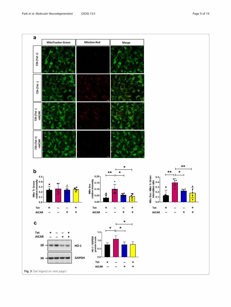
Fig. 3 (See legend on next page.)
Park et al. Molecular Neurodegeneration (2020) 15:5 Page 9 of 19

agonist, can rescue αsyn-induced changes in mitochondrialSIRT3 and associated signaling proteins. Following treat-ment with 2mM AICAR [34], a significant increase in p-AMPKα and p-CREB levels was observed after 72 h (Fig. 2a,*p < 0.05) and importantly, a significant restoration ofSIRT3 levels (Fig. 2b, *p < 0.05) accompanied by a signifi-cant decrease in αsyn oligomers observed by luciferaseassay (Fig. 2c) and detected by native PAGE as well (Fig. 2d).AICAR failed to reduce αsyn oligomers when SIRT3 ex-pression is knocked down using siRNA, indicating that the
effect of AICAR treatment is dependent on the presence ofSIRT3 (Fig. 2c). Because SIRT3 is a major mitochondrialdeacetylase, we examined the acetylation state of a knownsubstrate, superoxide dismutase 2 (SOD2) [35], in cellsoverexpressing αsyn oligomers. Acetylated SOD2 (K68) issignificantly increased in cells overexpressing αsyn com-pared to control (Fig. 2e, **p < 0.01), consistent with re-duced SIRT3 activity, and AICAR treatment decreases theacetylation status, consistent with a restoration of SIRT3levels. Of note, αsyn overexpression had no effect on total
(See figure on previous page.)Fig. 3 Activation of SIRT3 by AICAR attenuates ROS production. a Fluorescence microscopy images of MitoSox-Red and Mitotracker-Greenstaining in fixed H4 SL1&SL2 cells. MitoSox-Red fluorescence increases when αsyn is overexpressed and AICAR treatment attenuates mtROS.Representative images from 3 experiments. MitoTracker-Green (mitochondria; green); MitoSox-Red (mitochondria; red); merged images (yellow).Scale bar = 10 μm. b Relative intensity of MitoTracker-Green signal between conditions. Quantification of MitoSOX-Red signal intensities (n = 5),and mean intensity of MitoSOX-Red normalized to MitoTracker-Green intensity (c) Representative cropped western blot of HO-1 and GAPDH inwhole cells lysates from H4 SL1&SL2 cells. HO-1 level increases at 72 h and is reduced after AICAR-treatment, n = 5. Error bars represent themean ± SD. *p < 0.05, **p < 0.01
Fig. 4 αSyn expression affects mitochondrial dynamics and can be rescued with AICAR. a Representative cropped western blot showing DRP1 incytosol and mitochondria from H4 SL1&SL2 cells over time (0 – 72 h). Quantitation of protein levels for DRP1 in cytosol and mitochondria (n = 3).DRP1 band was normalized to respective loading controls GAPDH and COXIV. b Representative cropped western blot from showing DRP1and p-DRP1 levels in cells treated with or without AICAR (n = 4). Error bars represent the mean ± SD. *p < 0.05, **p < 0.01
Park et al. Molecular Neurodegeneration (2020) 15:5 Page 10 of 19

Fig. 5 (See legend on next page.)
Park et al. Molecular Neurodegeneration (2020) 15:5 Page 11 of 19
SOD2 levels which remained consistent in all conditions(Fig. 2e).
SIRT 3 activation attenuates αsyn-induced mitochondrialROSBecause SIRT3 plays a crucial role in modulating reactiveoxygen species (ROS) and limiting oxidative damage ofcellular components [36], we asked whether the presenceof αsyn in mitochondrial fraction is associated with in-creased oxidative stress that can be rescued with SIRT3activation. Cells overexpressing αsyn were stained withMitotracker-Green, to visualize mitochondria and controlfor total levels of mitochondria, and MitoSOX-Red tomonitor mitochondrial ROS production. Fluorescence mi-croscopy revealed increased ROS at 72 h compared tocontrol conditions (Tet + 72 h) (Fig. 3a - b), and as pre-dicted, AICAR-treatment reduced ROS production (Fig. 3aand b, bottom row). Of note, neither expression of αsynnor AICAR treatment altered the total number of mito-chondria (Fig. 3b). Increased oxidative stress and ROS caninduce the expression of heme oxygenase-1 (HO-1) [37]and increased HO-1 mRNA and protein expression havebeen reported in a wide spectrum of diseases includingneurodegenerative diseases such as Parkinson disease [38,39]. In line with these data, we found a significant increaseof HO-1 in cells expressing αsyn for 72 h (Fig. 3c), and aconcomitant decrease of HO-1 in cells treated withAICAR compared to control (Tet + 72 h) (Fig. 3c).
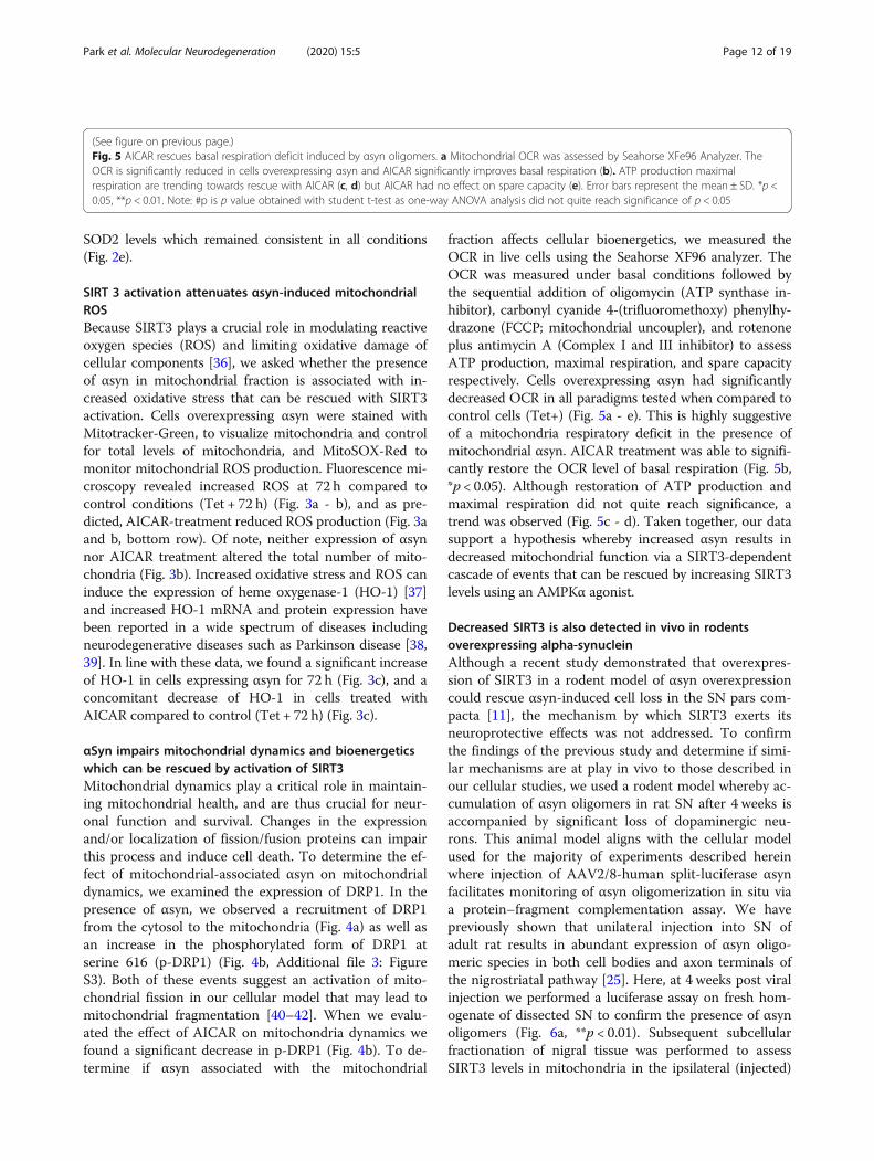
αSyn impairs mitochondrial dynamics and bioenergeticswhich can be rescued by activation of SIRT3Mitochondrial dynamics play a critical role in maintain-ing mitochondrial health, and are thus crucial for neur-onal function and survival. Changes in the expressionand/or localization of fission/fusion proteins can impairthis process and induce cell death. To determine the ef-fect of mitochondrial-associated αsyn on mitochondrialdynamics, we examined the expression of DRP1. In thepresence of αsyn, we observed a recruitment of DRP1from the cytosol to the mitochondria (Fig. 4a) as well asan increase in the phosphorylated form of DRP1 atserine 616 (p-DRP1) (Fig. 4b, Additional file 3: FigureS3). Both of these events suggest an activation of mito-chondrial fission in our cellular model that may lead tomitochondrial fragmentation [40–42]. When we evalu-ated the effect of AICAR on mitochondria dynamics wefound a significant decrease in p-DRP1 (Fig. 4b). To de-termine if αsyn associated with the mitochondrial
fraction affects cellular bioenergetics, we measured theOCR in live cells using the Seahorse XF96 analyzer. TheOCR was measured under basal conditions followed bythe sequential addition of oligomycin (ATP synthase in-hibitor), carbonyl cyanide 4-(trifluoromethoxy) phenylhy-drazone (FCCP; mitochondrial uncoupler), and rotenoneplus antimycin A (Complex I and III inhibitor) to assessATP production, maximal respiration, and spare capacityrespectively. Cells overexpressing αsyn had significantlydecreased OCR in all paradigms tested when compared tocontrol cells (Tet+) (Fig. 5a - e). This is highly suggestiveof a mitochondria respiratory deficit in the presence ofmitochondrial αsyn. AICAR treatment was able to signifi-cantly restore the OCR level of basal respiration (Fig. 5b,*p < 0.05). Although restoration of ATP production andmaximal respiration did not quite reach significance, atrend was observed (Fig. 5c - d). Taken together, our datasupport a hypothesis whereby increased αsyn results indecreased mitochondrial function via a SIRT3-dependentcascade of events that can be rescued by increasing SIRT3levels using an AMPKα agonist.
Decreased SIRT3 is also detected in vivo in rodentsoverexpressing alpha-synucleinAlthough a recent study demonstrated that overexpres-sion of SIRT3 in a rodent model of αsyn overexpressioncould rescue αsyn-induced cell loss in the SN pars com-pacta [11], the mechanism by which SIRT3 exerts itsneuroprotective effects was not addressed. To confirmthe findings of the previous study and determine if simi-lar mechanisms are at play in vivo to those described inour cellular studies, we used a rodent model whereby ac-cumulation of αsyn oligomers in rat SN after 4 weeks isaccompanied by significant loss of dopaminergic neu-rons. This animal model aligns with the cellular modelused for the majority of experiments described hereinwhere injection of AAV2/8-human split-luciferase αsynfacilitates monitoring of αsyn oligomerization in situ viaa protein–fragment complementation assay. We havepreviously shown that unilateral injection into SN ofadult rat results in abundant expression of αsyn oligo-meric species in both cell bodies and axon terminals ofthe nigrostriatal pathway [25]. Here, at 4 weeks post viralinjection we performed a luciferase assay on fresh hom-ogenate of dissected SN to confirm the presence of αsynoligomers (Fig. 6a, **p < 0.01). Subsequent subcellularfractionation of nigral tissue was performed to assessSIRT3 levels in mitochondria in the ipsilateral (injected)
(See figure on previous page.)Fig. 5 AICAR rescues basal respiration deficit induced by αsyn oligomers. a Mitochondrial OCR was assessed by Seahorse XFe96 Analyzer. TheOCR is significantly reduced in cells overexpressing αsyn and AICAR significantly improves basal respiration (b). ATP production maximalrespiration are trending towards rescue with AICAR (c, d) but AICAR had no effect on spare capacity (e). Error bars represent the mean ± SD. *p <0.05, **p < 0.01. Note: #p is p value obtained with student t-test as one-way ANOVA analysis did not quite reach significance of p < 0.05
Park et al. Molecular Neurodegeneration (2020) 15:5 Page 12 of 19
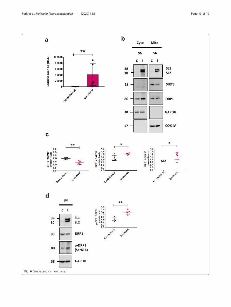
Fig. 6 (See legend on next page.)
Park et al. Molecular Neurodegeneration (2020) 15:5 Page 13 of 19
side of the rat brain. Consistent with our in vitro data,accumulation of αsyn in SN was accompanied by a sig-nificant decrease in SIRT3 (Fig. 6b - c, **p < 0.01, *p <0.05). Of note there was no difference in SIRT3 levels inSN in control animals that received an injection ofAAV8 expressing gaussia luciferase only (Additional file 4:Figure S4a). Surprisingly, despite a decrease of SIRT3level, we did not observed a decrease of cytosolic DRP1as previously described but rather a significant increase(Fig. 6c). However, examination of DRP1 levels in ourrodent model revealed a significant increase of p-DRP1in the injected SN (Fig. 6d, **p < 0.01), mimicking onceagain our in vitro observation. Lastly, AMPKα-CREB sig-naling was downregulated in the SN of these animals(Additional file 4: Figure S4b).
SIRT3 levels are decreased in human Lewy body diseasebrainsLastly, we assessed the level of SIRT3 in human post mor-tem brain with a confirmed neuropathological diagnosisof Lewy body disease (LBD) (Table 1). Frozen striatal tis-sue from 10 LBD and 10 age-matched healthy controlswere homogenized, run on SDS-PAGE, and probed withantibodies to detect αsyn, SIRT3, and DRP1. Western blotanalyses showed significantly reduced expression of SIRT3in LBD brains compared to controls but no significant dif-ference in the level of αsyn and DRP1 compared to con-trols (Fig. 7a - b). Brains from both sexes were utilized butthere was no difference in the interpretation of the datawhen stratified by sex (data not shown). Interestingly, sub-cellular fractionation of LBD brain homogenates also re-vealed a decrease in cytosolic levels of DRP1 and αsyn inLBD samples (Fig. 7c - d), with a corresponding increasein levels of mitochondrial localized DRP1 and αsyn (Fig. 7e- f) when compared to controls. Together, these resultsare consistent with our findings from cell and rodentmodels where decreased SIRT3 protein levels were ob-served when αsyn localizes to the mitochondria.
DiscussionHerein, we identify a cellular mechanism that illuminateshow αsyn-associated mitochondria may lead to mitochon-drial dysfunction and the initiation of a self-perpetuatingcycle of aggregation, deficient cellular metabolism, and
eventually cell death (Fig. 8). For the first time we identifyαsyn oligomers in the mitochondrial enriched fraction ofcells and a consequent decrease in SIRT3 activity in mul-tiple model systems including cell culture, rodent models,and human post-mortem brains with a neuropathologicaldiagnosis of LBD. We demonstrate that the presence ofαsyn oligomers in mitochondrial-enriched fractions corre-lates with decreased mitochondrial function and decreasedSIRT3 expression and function. Interestingly, we show thatSIRT3 downregulation is accompanied by dysregulation ofthe AMPK signaling pathway, perturbation of fission mech-anisms, and impairment of basal respiration, all of whichcontribute to increased ROS and mitochondrial dysfunc-tion. These findings are observed not only in an experimen-tal cellular model, but also a rodent model of αsynaccumulation, and importantly, in human post mortemLBD brain. Lastly, treatment with an AMPK agonist,AICAR, appears to improve αsyn-associated mitochondrialdysfunction by decreasing αsyn oligomer formation and in-creasing SIRT3 expression. Overall, these results confirmthe health enhancing capabilities of SIRT3 and validate itspotential as a new therapeutic target for PD and relateddisorders.Mitochondrial dysfunction has been linked to the
pathogenesis of neurodegenerative diseases includingPD, with mutations identified in mitochondrial-associated proteins such as PINK1 and parkin causingfamilial PD [43, 44]. αSyn, a major neuropathologicalhallmark of PD and alpha-synucleinopathies can perturbmitochondria and previous studies have shown thatoverexpression of αsyn has dramatic effects on mito-chondrial morphology, reduces respiratory chain com-plex activity, and impairs mitochondrial functionsin vitro and in vivo [45, 46]. Accumulation of wild-typeαsyn and truncated species within the mitochondria hasbeen described [47, 48] however, no study has directlydemonstrated that oligomeric αsyn species can associatemitochondria. Here, we used a split luciferase proteincomplementation assay to demonstrate accumulation ofαsyn oligomers in the mitochondrial-enriched fraction ofcells in culture and rat brain homogenates. We speculatethat the presence of oligomeric αsyn species triggers acascade of events leading to mitochondria malfunctionassociated with PD pathogenesis. However, our data do
(See figure on previous page.)Fig. 6 αSyn expression decreases SIRT3 and alters mitochondrial dynamics in vivo. a αSyn oligomers were quantified via luciferase assay in ratbrain homogenates at 4 weeks after stereotaxic injection of AAV8-SL1&SL2 in SN, n = 5. b Representative cropped western blots showing αsyn,SIRT3, DRP1 in cytosol and mitochondria from SN of rats 4 weeks after stereotaxic injection of AAV8-SL1 and AAV8-SL2. (C). c Quantification ofαsyn, SIRT3, DRP1, protein levels in cytosol and mitochondria from two separate blots for each of 4–5 rats. All bands were normalized torespective loading controls GAPDH and COXIV. d Representative cropped western blot of αsyn, DRP1, and p-DRP1 (Ser 616) in SN lysate of AAV8-SL1&SL2 injected rat. αSyn expression leads to increased p-DRP1 protein levels in ipsilateral (I) injected SN compared to contralateral (C)uninjected SN, n = 5 rats total. In panel (a) the same samples were run on one blot that was cropped prior to immunoblotting for αsyn, SIRT3,COXIV, DRP1, and GAPDH. Error bars represent the mean ± SD, (n = 4–5 rats). *p < 0.05, **p < 0.01
Park et al. Molecular Neurodegeneration (2020) 15:5 Page 14 of 19

Fig. 7 (See legend on next page.)
Park et al. Molecular Neurodegeneration (2020) 15:5 Page 15 of 19

not preclude the possibility that cytosolic αsyn may con-tribute to the observed mitochondrial dysfunction orthat the observed deficits are induced by overexpressedmonomeric αsyn. While we know that αsyn oligomers as-sociate with mitochondria because luciferase activity isonly be detected when oligomers are present, we cannotrule out the possibility that undetected monomers of αsynare also present and responsible for some of the observedeffects. We also cannot rule out the possibility that higherorder αsyn species detected via native PAGE are actuallyrepresentative of a protein complex containing αsyn. To
address these concerns will require methods to selectivelydeplete cytosolic αsyn in cultured cells as well as methodsto distinguish between monomers and oligomers whenαsyn is expressed in cells. To our knowledge such toolshave yet to be developed.In line with our results, deficiency of SIRT3 is observed
in cellular models of HD [20] and down regulation ofSIRT3 increases dopaminergic cell death in an 1-methyl-4-phenyl-1,2,3,6-tetrahydropyridine (MPTP) mouse modelof PD [6]. Most recently, overexpression of SIRT3 wasdemonstrated to prevent αsyn-induced neurodegeneration
(See figure on previous page.)Fig. 7 SIRT3 is decreased in human post-mortem brain of neuropathologically confirmed Lewy body disease individuals. a Representativecropped western blot from n = 3 showing decreased SIRT3 in total brain lysates from ten LBD brains compared to ten age-matched healthycontrols. b Quantification of αsyn, SIRT3 protein levels from n = 3 western blots. c, d Representative western blot of cytosolic fraction. DRP1 andαsyn were quantified using GAPDH as a loading control. e, f Representative western blot of mitochondrial fraction. DRP1 and αsyn werequantified using COXIV as a loading control. Error bars represent the mean ± SD. **p < 0.01
Fig. 8 Cartoon representing of αsyn-induced effect on SIRT3 and mitochondrial dysfunction. Consequences of decreased SIRT3 include decreasedAMPK-CREB signaling, impairment in mitochondrial bioenergetics and dynamics, and increased acetylation of SIRT3 substrates such as SOD2 all ofwhich contribute to increased ROS production and neurodegeneration. Question mark indicates pathway not supported by data inthis manuscript
Park et al. Molecular Neurodegeneration (2020) 15:5 Page 16 of 19
in a rodent AAV model [11]. In humans, downregulationof SIRT3 has been previously reported in post-mortemhuman AD brain [49, 50].SIRT3 is emerging as an important regulator of cellu-
lar biogenesis and oxidative stress. Recent evidence sup-ports attenuation of ROS and improved mitochondrialbioenergetics upon activation of SIRT3 [51], while SIRT3knockdown exacerbates ROS production [52]. Thecurrent school of thought is that SIRT3 induces neuro-protection by enhancing mitochondrial biogenesis andintegrity, perhaps by increasing mitochondrial DNA con-tent and suppressing SOD activity [23, 52–54]. SIRT3also seems to be under the control of AMPK/CREB-PGC-1α signaling pathway which is known to play a rolein regulation of mitochondrial biogenesis and function,activating mitochondrial enzymes involved in antioxi-dant defenses and metabolism [17, 32, 55]. Here, wetested the hypothesis that the AMPK/CREB signalingpathway plays a role in αsyn-associated SIRT3 down-regulation. Decreased levels of p-AMPKα and p-CREBin the presence of αsyn were restored with AICAR,which also increased mitochondrial SIRT3 protein ex-pression. Evidence in the literature supports an increaseof SIRT3 activity directly by AICAR [56]. By contrast,previous studies confirm a role for AMPKα in the regu-lation of SIRT3 protein [57]. Herein, our data supportthe effect of AICAR being dependent on SIRT3 sinceknockdown of SIRT3 with siRNA precludes rescue byAICAR. Importantly, we found that activating AMPKαsignificantly reduced αsyn oligomers in our cellularmodel system. These data indicate that modulatingSIRT3 levels may represent a target to alleviate αsyn-induced pathology and slow or halt αsyn-induced cellu-lar dysfunction in Parkinson’s disease and related alpha-synucleinopathies.Mitochondria are dynamic organelles that continu-
ously undergo fission and fusion, processes necessary forcell survival and adaptation to changing energy require-ments for cell growth, division, and distribution of mito-chondria during differentiation [58]. Consistent with aprevious study [59], our results demonstrate that mito-chondrial dynamics are modified by the association ofαsyn oligomers with mitochondria. Our data demon-strating increased recruitment of DRP1 to mitochondriaand increased pDRP1 support increased mitochondriafission. Under stress conditions, DRP1 is recruited tomitochondria where it initiates mitochondrial fissionand induces mitochondrial dysfunction. DRP1 activity isregulated by several post-translational modifications in-cluding phosphorylation at Serine 616 [41], which rap-idly activates DRP1 and stimulates mitochondrial fissionduring mitosis [42, 60]. AICAR-treatment was able torestore DRP1 protein expression to control levels, im-proving mitochondrial function and indicating that
SIRT3 plays an important role in regulating of mainten-ance of mitochondrial function during stress. Our resultsare consistent with a recent study demonstrating inter-action of αsyn with ATP synthase in the mitochondriaand impairment of complex I-dependent respiration [3].Additionally, previous studies have shown that αsyn in-teracts with TOM20 [61], a protein required for mito-chondrial protein import, and decreases its function.SIRT3 is reported to exist in the cytoplasm in an inactiveform and is recruited to the mitochondria upon stress[62], although the data presented here do not supportcytosolic SIRT3 expression. It is tempting to speculatethat TOM20 plays a role in the translocation of SIRT3to mitochondria and that αsyn-induced deficit in proteinimport results in reduced mitochondrial SIRT3 levelsthereby initiating the cascade of mitochondrial dysfunc-tion that results in decreased mitochondrialbioenergetics.
ConclusionsOur results identify a mechanism whereby the associ-ation of αsyn oligomers with mitochondria contribute toimpaired mitochondrial respiration and impaired mito-chondrial dynamics by disrupting AMPK/CREB/SIRT3signaling (Fig. 8). Taken together, our study opens thedoor to the use of SIRT3 activators as potential thera-peutics for restoration of mitochondrial deficits and de-crease in αsyn-induced pathophysiology. Further studieswill be necessary to address the role of SIRT3 deacetyla-tion substrates as possible players in PD pathogenesis.
Supplementary informationSupplementary information accompanies this paper at https://doi.org/10.1186/s13024-019-0349-x.
Additional file 1: Figure S1. (a) Luciferase activity in cytosolic fractionsof H4 SL1&SL2 cells over time, n = 5 (b) Representative cropped westernblots of denaturing SDS-page (left) and native-page (right) performed at72 h to confirm the purity of the mitochondrial-enriched fractions, the ly-sates were probed for the golgi marker (GM130), inner mitochondrialmembrane marker (COXIV), GAPDH and αsyn in cytosol and mitochondriafrom H4 SL1&SL2 cells. (c) Representative image of dot-blot assay of themitochondrial fractions at 72 h probed for amyloid-specific A11 and αsyndisease-associated (5G4) antibodies. (d) SIRT3 expression was detected byimmune-fluorescence in mitochondria. SIRT3 expression is decreased at72 h in cells overexpressing αsyn. Representative image from 3 experi-ments. DAPI (nucleus; blue); αsyn (green); SIRT3 (mitochondria; orange);merged images (yellow). Scale bar = 10 μm. Error bars represent themean ± SD (n = 3–5). **p < 0.01.
Additional file 2: Figure S2. (a) Representative cropped western blotsshowing αsyn in cytosolic and mitochondrial fractions from H4 WT-αsynoverexpressing cells. αSyn, GAPDH, and COXIV are all from same samplesand immunoblot. (b) Western blot and quantification for SIRT3 in primaryembryonic mouse neurons treated at DIV7 for 5 days with either AAV2/8WT-αsyn or AAV2/8 venus control. Untagged αsyn overexpression showsa significant decrease in SIRT3 level compared to control. Error bars repre-sent the mean ± SD. **p < 0.01 (n = 4).
Additional file 3: Figure S3. (a) Representative cropped western blotshowing DRP1 and p-DRP1 (Ser 616) in whole cells lysates from H4
Park et al. Molecular Neurodegeneration (2020) 15:5 Page 17 of 19

SL1&SL2 cells over time (n = 4). Removal of tet leads to significantly in-creased p-DRP1 by 72 h. (b) Quantification of p-DRP1/DRP1 protein ratioin whole cell lysates. Error bars represent the mean ± SD. **p < 0.01.
Additional file 4: Figure S4. (a) Representative cropped western blotof lysates from dissected substantia nigra/midbrain (SN) of three ratsinjected unilaterally with control virus, AAV8-Hgluc (gaussia luciferase)demonstrates no change in SIRT3 protein levels when contralateral (C)uninjected SN is compared to ipsilateral (I) injected SN (b) Representativecropped western blot of AMPKα, p-AMPKα (Thr 172), CREB, and p-CREB(Ser 133) in SN lysate of AAV8-SL1&SL2 injected rat. αSyn expression leadsto decreased p-AMPK and p-CREB protein levels in ipsilateral (I) injectedSN compared to contralateral (C) uninjected SN, n = 5 rats total. Error barsrepresent the mean ± SD. *p < 0.05, **p < 0.01.
AbbreviationsAD: Alzheimer’s disease; AICAR: 5-aminoimidazole-4-carboxamide-1-β-d-ribofuranoside; AMPK: Adenosine monophosphate activated protein kinase;CREB: CAMP-response element binding protein; DRP1: Dynamin-relatedprotein 1; HD: Huntington’s disease; HO-1: Heme oxygenase-1; LBD: Lewybody disease; mtROS: Mitochondrial reactive oxygen species; OCR: Oxygenconsumption rate; PD: Parkinson disease; SIRT3: Sirtuin 3; SOD2: Superoxidedismutase 2; α-synuclein: αsyn
AcknowledgementsWe thank Dr. Dennis Dickson, Dr. Michael DeTure, and the Mayo Clinic Brainbank for human post-mortem brain samples used in this study.
Authors’ contributionsJHP conceived and designed the study, performed the measurements anddata analysis and wrote the manuscript. JDB performed experiments andcontributed to interpretation of data. AHF, NND, FF, and WS madesubstantial contributions to acquisition and/or interpretation of data. MDperformed the animal studies and contributed to manuscript writing. PJMconceived of the study, obtained study funding, and assisted withmanuscript preparation. All authors read and approved the final manuscript.
FundingFunded in part by NIH/NINDS NS110085 to PJM and WS, and the MayoFoundation for Medical Research. MD was supported by the MangurianFoundation for LBD research.
Availability of data and materialsAll data generated or analyzed during this study are included in thispublished article and the supplementary information files.
Ethics approval and consent to participateAll applicable international, national, and/or institutional guidelines for thecare and use of animals were followed.
Consent for publicationNot applicable.
Competing interestsThe authors declare that they have no competing interests.
Author details1Department of Neuroscience, Mayo Clinic, 4500 San Pablo Road,Jacksonville, FL 32224, USA. 2Neuroscience PhD Program, Mayo ClinicGraduate School of Biomedical Sciences, Mayo Clinic College of Medicine,4500 San Pablo Road, Jacksonville, FL 32224, USA.
Received: 14 October 2019 Accepted: 29 November 2019
References1. Devi L, Raghavendran V, Prabhu BM, Avadhani NG, Anandatheerthavarada
HK. Mitochondrial import and accumulation of alpha-synuclein impaircomplex I in human dopaminergic neuronal cultures and Parkinson diseasebrain. J Biol Chem. 2008;283:9089–100.
2. Hsu LJ, Sagara Y, Arroyo A, Rockenstein E, Sisk A, Mallory M, Wong J,Takenouchi T, Hashimoto M, Masliah E. Alpha-synuclein promotesmitochondrial deficit and oxidative stress. Am J Pathol. 2000;157:401–10.
3. Ludtmann MHR, Angelova PR, Horrocks MH, Choi ML, Rodrigues M, BaevAY, Berezhnov AV, Yao Z, Little D, Banushi B, et al. α-Synuclein oligomersinteract with ATP synthase and open the permeability transition pore inParkinson's disease. Nat Commun. 2018;9:2293.
4. Reeve AK, Ludtmann MH, Angelova PR, Simcox EM, Horrocks MH,Klenerman D, Gandhi S, Turnbull DM, Abramov AY. Aggregated α-synucleinand complex I deficiency: exploration of their relationship in differentiatedneurons. Cell Death Dis. 2015;6:e1820.
5. Kyrylenko S, Baniahmad A. Sirtuin family: a link to metabolic signaling andsenescence. Curr Med Chem. 2010;17:2921–32.
6. Liu L, Peritore C, Ginsberg J, Kayhan M, Donmez G. SIRT3 attenuates MPTP-induced nigrostriatal degeneration via enhancing mitochondrial antioxidantcapacity. Neurochem Res. 2015a;40:600–8.
7. Liu L, Peritore C, Ginsberg J, Shih J, Arun S, Donmez G. Protective role of SIRT5against motor deficit and dopaminergic degeneration in MPTP-induced micemodel of Parkinson’s disease. Behav Brain Res. 2015b;281:215–21.
8. Outeiro TF, Kontopoulos E, Altmann SM, Kufareva I, Strathearn KE, AmoreAM, Volk CB, Maxwell MM, Rochet JC, McLean PJ, et al. Sirtuin 2 inhibitorsrescue alpha-synuclein-mediated toxicity in models of Parkinson's disease.Science. 2007;317:516–9.
9. Jin F, Wu Q, Lu YF, Gong QH, Shi JS. Neuroprotective effect of resveratrol on 6-OHDA-induced Parkinson’s disease in rats. Eur J Pharmacol. 2008;600:78–82.
10. Lofrumento DD, Nicolardi G, Cianciulli A, De Nuccio F, La Pesa V, CarofiglioV, Dragone T, Calvello R, Panaro MA. Neuroprotective effects of resveratrolin an MPTP mouse model of Parkinson’s-like disease: possible role of SOCS-1 in reducing pro-inflammatory responses. Innate Immun. 2014;20:249–60.
11. Gleave JA, Arathoon LR, Trinh D, Lizal KE, Giguère N, Barber JHM, Najarali Z,Khan MH, Thiele SL, Semmen MS, Koprich JB, Brotchie JM, Eubanks JH,Trudeau LE, Nash JE. Sirtuin 3 rescues neurons through the stabilisation ofmitochondrial biogenetics in the virally-expressing mutant α-synuclein ratmodel of parkinsonism. Neurobiol Dis. 2017;106:133–46.
12. Hebert AS, Dittenhafer-Reed KE, Yu W, Bailey DJ, Selen ES, Boersma MD,Carson JJ, Tonelli M, Balloon AJ, Higbee AJ, et al. Calorie restriction andSIRT3 trigger global reprogramming of the mitochondrial proteinacetylome. Mol Cell. 2013;49:186–99.
13. Herskovits AZ, Guarente L. Sirtuin deacetylases in neurodegenerativediseases of aging. Cell Res. 2013;23:746–58.
14. Lombard DB, Alt FW, Cheng HL, Bunkenborg J, Streeper RS, Mostoslavsky R,Kim J, Yancopoulos G, Valenzuela D, Murphy A, et al. Mammalian Sir2homolog SIRT3 regulates global mitochondrial lysine acetylation. Mol CellBiol. 2007;27:8807–14.
15. López-Otín C, Blasco MA, Partridge L, Serrano M, Kroemer G. The hallmarksof aging. Cell. 2013;153:1194–217.
16. Bause AS, Haigis MC. SIRT3 regulation of mitochondrial oxidative stress. ExpGerontol. 2013;48:634–9.
17. Kong X, Wang R, Xue Y, Liu X, Zhang H, Chen Y, Fang F, Chang Y. Sirtuin 3a new target of PGC-1α, plays an important role in the suppression of ROSand mitochondrial biogenesis. PLoS One. 2010;5:e11707.
18. Ansari A, Rahman MS, Saha SK, Saikot FK, Deep A, Kim KH. Function of theSIRT3 mitochondrial deacetylase in cellular physiology, cancer, andneurodegenerative disease. Aging Cell. 2017;16:4–16.
19. Kim SH, Lu HF, Alano CC. Neuronal Sirt3 protects against excitotoxic injuryin mouse cortical neuron culture. PLoS One. 2011;6:e14731.
20. Fu J, Jin J, Cichewicz RH, Hageman SA, Ellis TK, Xiang L, et al. Trans-(−)-ε-Viniferin increases mitochondrial sirtuin 3 (SIRT3), activates AMP-activatedprotein kinase (AMPK), and protects cells in models of Huntington disease. JBiol Chem. 2012;287:24460–72.
21. Weir HJ, Murray TK, Kehoe PG, Love S, Verdin EM, O'Neill MJ, et al. CNSSIRT3 expression is altered by reactive oxygen species and in Alzheimer'sdisease. PLoS One. 2012;7:e48225.
22. Yin J, Han P, Tang Z, Liu Q, Shi J. Sirtuin 3 mediates neuroprotection ofketones against ischemic stroke. J Cereb Blood Flow Metab. 2015;35:1783–9.
23. Liu J, Li D, Zhang T, Tong Q, Ye RD, Lin L. SIRT3 protects hepatocytes fromoxidative injury by enhancing ROS scavenging and mitochondrial integrity.Cell Death Dis. 2017;8:e3158.
24. Moussaud S, Malany S, Mehta A, Vasile S, Smith LH, McLean PJ. Targeting α-synuclein oligomers by protein-fragment complementation for drugdiscovery in synucleinopathies. Expert Opin Ther Targets. 2015;19:589–603.
Park et al. Molecular Neurodegeneration (2020) 15:5 Page 18 of 19

25. Delenclos M, Trendafilova T, Jones DR, Moussaud S, Baine AM, Yue M, HirstWD, McLean PJ. A Rapid, Semi-Quantitative Assay to Screen for Modulatorsof Alpha-Synuclein Oligomerization Ex vivo. Front Neurosci. 2016;9:511.
26. Paxinos G, Watson C. The rat brain in stereotaxic coordinates. 4th ed. SanDiego: Academic Press; 1998.
27. Marongiu R, Spencer B, Crews L, Adame A, Patrick C, Trejo M, et al. MutantPink1 induces mitochondrial dysfunction in a neuronal cell model ofParkinson’s disease by disturbing calcium flux. J Neurochem. 2009;108:1561–74.
28. Nakamura K, Nemani VM, Azarbal F, Skibinski G, Levy JM, Egami K,Munishkina L, Zhang J, Gardner B, Wakabayashi J, Sesaki H, Cheng Y,Finkbeiner S, Nussbaum RL, Masliah E, Edwards RH. Direct membraneassociation drives mitochondrial fission by the Parkinson disease-associatedprotein alpha-synuclein. J Biol Chem. 2011;286:20710–26.
29. Kayed R, Head E, Thompson JL, McIntire TM, Milton SC, Cotman CW, GlabeCG. Common structure of soluble amyloid oligomers implies commonmechanism of pathogenesis. Science. 2003;300:486–9.
30. Kovacs GG, Wagner U, Dumont B, Pikkarainen M, Osman AA, StreichenbergerN, Leisser I, Verchère J, Baron T, Alafuzoff I, Budka H, Perret-Liaudet A,Lachmann I. An antibody with high reactivity for disease-associated α-synuclein reveals extensive brain pathology. Acta Neuropathol. 2012;124:37–50.
31. Pillai VB, Sundaresan NR, Kim G, Gupta M, Rajamohan SB, Pillai JB, et al.Exogenous NAD blocks cardiac hypertrophic response via activation of theSIRT3-LKB1-AMP-activated kinase pathway. J Biol Chem. 2010;285:3133–44.
32. Shi T, Wang F, Stieren E, Tong Q. SIRT3, a mitochondrial sirtuin deacetylase,regulates mitochondrial function and thermogenesis in brown adipocytes. JBiol Chem. 2005;280:13560–7.
33. Dulovic M, Jovanovic M, Xilouri M, Stefanis L, Harhaji-Trajkovic L, Kravic-Stevovic T, Paunovic V, Ardah MT, El-Agnaf OM, Kostic V, Markovic I,Trajkovic V. The protective role of AMP-activated protein kinase in alpha-synuclein neurotoxicity in vitro. Neurobiol Dis. 2014;63:1–11.
34. Takeuchi K, Morizane Y, Kamami-Levy C, Suzuki J, Kayama M, Cai W, et al. AMP-dependent kinase inhibits oxidative stress-induced caveolin-1 phosphorylationand endocytosis by suppressing the dissociation between c-Abl and Prdx1proteins in endothelial cells. J Biol Chem. 2013;288:20581–91.
35. Qiu X, Brown K, Hirschey MD, Verdin E, Chen D. Calorie restriction reducesoxidative stress by SIRT3-mediated SOD2 activation. Cell Metab. 2010;12:662–7.
36. Torrens-Mas M, Oliver J, Roca P, Sastre-Serra J. SIRT3: Oncogene and TumorSuppressor in Cancer. Cancers (Basel). 2017;9:90.
37. Bansal S, Biswas G, Avadhani NG. Mitochondria-targeted heme oxygenase-1induces oxidative stress and mitochondrial dysfunction in macrophages,kidney fibroblasts and in chronic alcohol heaptotoxicity. Redox Biol. 2013;2:273–83.
38. Shipper HM, Liberman A, Stopa EG. Neural heme oxygenase-1 expression inidiopathic Parkinson's disease. Exp Neurol. 1998;150:60–8.
39. Song W, Patel A, Qureshi HY, Han D, Schipper HM, Paudel HK. The Parkinsondisease-associated A30P mutation stabilizes alpha-synuclein againstproteasomal degradation triggered by heme oxygenase-1 over-expressionin human neuroblastoma cells. J Neurochem. 2009;110:719–33.
40. Alaimo A, Gorojod RM, Beauquis J, Muñoz MJ, Saravia F, Kotler ML.Deregulation of mitochondria-shaping proteins Opa-1 and Drp-1 inmanganese-induced apoptosis. PLoS One. 2014;9:e91848.
41. Elgass K, Pakay J, Ryan MT, Palmer CS. Recent advances into the understandingof mitochondrial fission. Biochim Biophys Acta. 1833;2013:150–61.
42. Taguchi N, Ishihara N, Jofuku A, Oka T, Mihara K. Mitotic phosphorylation ofdynamin-related GTPase Drp1 participates in mitochondrial fission. J BiolChem. 2007;282:11521–9.
43. Dawson TM, Dawson VL. Molecular pathways of neurodegeneration inParkinson’s disease. Science. 2003;302:819–22.
44. Schapira AH, Hartley A, Cleeter MW, Cooper JM. Free radicals and mitochondrialdysfunction in Parkinson's disease. Biochem Soc Trans. 1993;21:367–70.
45. Bobela W, Nazeeruddin S, Knott G, Aebischer P, Schneider BL. Modulatingthe catalytic activity of AMPK has neuroprotective effects against α-synuclein toxicity. Mol Neurodegener. 2017;12:80.
46. Siddiqui A, Chinta SJ, Mallajosyula JK, Rajagopolan S, Hanson I, Rane A,Melov S, Andersen JK. Selective binding of nuclear alpha-synuclein to thePGC1alpha promoter under conditions of oxidative stress may contribute tolosses in mitochondrial function: implications for Parkinson's disease. FreeRadic Biol Med. 2012;53:993–1003.
47. Sarafian TA, Ryan CM, Souda P, Masliah E, Kar UK, Vinters HV, Mathern GW,Faull KF, Whitelegge JP, Watson JB. Impairment of mitochondria in adult
mouse brain overexpressing predominantly full-length, N-terminallyacetylated human α-synuclein. PLoS One. 2013;8:e63557.
48. Subramaniam SR, Vergnes L, Franich NR, Reue K, Chesselet MF. Regionspecific mitochondrial impairment in mice with widespread overexpressionof alpha-synuclein. Neurobiol Dis. 2014;70:204–13.
49. Han P, Tang Z, Yin J, Maalouf M, Beach TG, Reiman EM, Shi J. Pituitaryadenylate cyclase-activating polypeptide protects against β-amyloid toxicity.Neurobiol Aging. 2014;35:2064–71.
50. Lee J, Kim Y, Liu T, Hwang YJ, Hyeon SJ, Im H, Lee K, Alvarez VE, McKee AC,Um SJ, et al. SIRT3 deregulation is linked to mitochondrial dysfunction inAlzheimer's disease. Aging Cell. 2018;17:e12679.
51. Ramesh S, Govindarajulu M, Lynd T, Briggs G, Adamek D, Jones E, Heiner J,Majrashi M, Moore T, Amin R, Suppiramaniam V, Dhanasekaran M. SIRT3activator Honokiol attenuates β-amyloid by modulating amyloidogenicpathway. PLoS One. 2018;13:e0190350.
52. Zhang JY, Deng YN, Zhang M, Su H, Qu QM. SIRT3 acts as aNeuroprotective agent in rotenone-induced Parkinson cell model.Neurochem Res. 2016;41:1761–73.
53. Dai SH, Chen T, Wang YH, Zhu J, Luo P, Rao W, Yang YF, Fei Z, Jiang XF.Sirt3 protects cortical neurons against oxidative stress via regulatingmitochondrial Ca2+ and mitochondrial biogenesis. Int J Mol Sci. 2014a;15:14591–609.
54. Dai SH, Chen T, Wang YH, Zhu J, Luo P, Rao W, Yang YF, Fei Z, Jiang XF.Sirt3 attenuates hydrogen peroxide induced oxidative stress through thepreservation of mitochondrial function in HT22 cells. Int J Mol Med. 2014b;34:1159–68.
55. Abdel Khalek W, Cortade F, Ollendorff V, Lapasset L, Tintignac L, Chabi BWrutniak-Cabello C. SIRT3, a mitochondrial NAD+-dependent deacetylase, isinvolved in the regulation of myoblast differentiation. PLoS One. 2014;9:e114388.
56. Morigi M, Perico L, Rota C, Longaretti L, Conti S, Rottoli D, Novelli R, Remuzzi G,Benigni A. Sirtuin 3-dependent mitochondrial dynamic improvements protectagainst acute kidney injury. J Clin Invest. 2015;2:715-26.
57. Brandauer J, Andersen MA, Kellezi H, Risis S, Frøsig C, Vienberg SG, TreebakJT. AMP-activated protein kinase controls exercise training- and AICAR-induced increases in SIRT3 and MnSOD. Front Physiol. 2015;6:85.
58. van der Bliek AM, Shen Q, Kawajiri S. Mechanisms of mitochondrial fissionand fusion. Cold Spring Harb Perspect Biol. 2013;5:a011072.
59. Martinez JH, Alaimo A, Gorojod RM, Porte Alcon S, Fuentes F, ColuccioLeskow F, Kotler ML. Drp-1 dependent mitochondrial fragmentation andprotective autophagy in dopaminergic SH-SY5Y cells overexpressing alpha-synuclein. Mol Cell Neurosci; 2018;88:107-17.
60. Sanchis-Gomar F, Derbré F. Mitochondrial fission and fusion in humandiseases. N Engl J Med. 2014;370:1073-4.
61. Di Maio R, Barrett PJ, Hoffman EK, Barrett CW, Zharikov A, Borah A, Hu X,McCoy J, Chu CT, Burton EA, Hastings TG, Greenamyre JT. α- Synucleinbinds to TOM20 and inhibits mitochondrial protein import in Parkinson'sdisease. Sci Transl. 2016;8:342ra78.
62. Anamika, Khanna A, Acharjee P, Acharjee A, Trigun SK. Mitochondrial SIRT3and neurodegenerative brain disorders. J Chem Neuroanat. 2019;95:43-53.
Publisher’s NoteSpringer Nature remains neutral with regard to jurisdictional claims inpublished maps and institutional affiliations.
Park et al. Molecular Neurodegeneration (2020) 15:5 Page 19 of 19