peripheral giant cell granuloma: immunohistochemical...
TRANSCRIPT
AVANCES EN ODONTOESTOMATOLOGÍA/189
Falaschini S, Ciavarella D, Mazzanti R, Di Cosola M, Turco M, Escudero N, Bascones A, Lo Muzio L.Peripheral giant cell granuloma: immunohistochemical analysis of different markers. Study of three cases
Peripheral giant cell granuloma:immunohistochemical analysis of differentmarkers. Study of three cases
Falaschini S*, Ciavarella D*, Mazzanti R**, Di Cosola M***, Turco M***, Escudero N,Bascones A****, Lo Muzio L*
SUMMARY
Peripheral giant cell granuloma (PGCG) is a non-neoplastic lesion representing a local hyperplastic reaction toinjury or inflammation. It is known to be a reactive soft tissue lesion that develops only within the oral cavity,with a slightly predilection for female sex. The usual localization for PGCG is the premolar region and the crestof the edentulous ridge.This study presents three cases of PGCG, including 2 male and 1 female, with an age comprised between 25and 35 years. All patients were treated with resection biopsy and no one relapsed.With the aim of determine the probable origin of stromal mononuclear cells and multinuclear giant cells, eachcase was then studied by immunohistochemistry to evaluate the expression of endothelial and monocyte/macrophage lineage.Immunohistochemical results showed a strong diffuse positivity for CD-68 in round mononuclear stromal cellsand in multinucleate giant cells. These latter were immunonegative for CD-34 and only focally positive for á-1antitrypsin.These results suggest that multinucleated giant cell shows an osteoclast phenotype and that probably derivefrom monocyte/macrophage lineage and that do not derive from the endothelial cells of the capillary.In second instance, we underlined the importance of an exhaustive dia.
Key words: Peripheral Giant Cell Granuloma (PGCG), immunohistochemistry.
Received: April 2007.Acepted for publication: May 2007.
* Department of Surgical Sciences, “Universitá di Foggia”, Foggia, Italia.** Department of Dental Sciences, University of Palermo, Palermo, Italy.*** Institute of Pathology, University of Ancona, Ancona, Italy.**** U.O. Odontostomatologia e Chirurgia Orale Z.T 6 e 7 ASUR MARCHE, Italy.
Falaschini S, Ciavarella D, Mazzanti R, Di Cosola M, Turco M, Escudero N, Bascones A, Lo Muzio L. Peripheralgiant cell granuloma: immunohistochemical analysis of different markers. Study of three cases. Av.Odontoestomatol 2007; 23 (4): 189-196.
INTRODUCTION
Peripheral giant cell granuloma is an infrequentreactive, exophytic lesion of the oral cavity, also knownas giant-cell epulis, osteoclastoma, giant cell
reparative granuloma, or giant cell hyperplasia. It isthe most frequent giant cell lesion of the jaws, andoriginates from the connective tissue of theperiosteum or from the periodontal membrane, inresponse to local irritation or chronic trauma (1). It is

AVANCES EN ODONTOESTOMATOLOGÍAVol. 23 - Núm. 4 - 2007
/AVANCES EN ODONTOESTOMATOLOGÍA190
more frequent in women than in men, with a slightlyhigher prevalence in the 30- to 70-year-old-age group,and affects largely the lower jaw (55%) than in theupper jaw (the reported proportion being 2,4:1) (2).
Cases of PGCG have been documented in children,where the lesion appears to be more aggressive, withabsorption of the interproximal crest area,displacement of the adjacent teeth and multiplerecurrences (3).
Clinically, it manifests as a soft to firm, bright noduleor as a sessile or pediculate mass, which ispredominantly bluish red with a smooth shiny ormamillated surface, localized in the gingival tissue oralveolar processes of the incisor and canine region(1), through according to Pindborg the preferentiallocation is the premolar and molar zone (4). Thelesion ranges in size from small papules to enlargedmasses, though reportedly rarely exceeding 2 cm indiameter, and are generally located in the interdentalpapilla, edentulous alveolar margin, or at marginalgum level (5). It is basically asymptomatic, in factpain is not a common characteristic, and lesiongrowth in most cases is induced by repeated traumasuch as with occlusion, in which case it may ulcerateand becomes infected (6, 7).
Although the pathogenesis of oral cavity PGCGs isstill uncertain, local irritants such as calculus, bacterialplaque, periodontitis, periodontal surgery, ill-fittingdentures, overhanging restorations and toothextractions are suggested as the etiological causes(8-10). These are soft tissue lesions that rarely affectthe underlying bone, though the latter may suffererosion (5, 11). Treatment comprises surgicalresection, with extensive clearing of the base of thelesion to avoid relapses (12).
The present study describes three clinical cases ofperipheral giant cell granuloma located in differentareas. We analyzed the presence and tissuelocalization of several markers such as CD68, CD34and α1- antitrypsin by immunohistochemistry, for thepurpose of evaluating the origin of stromal mononu-clear cells and multinucleate giant cells. Finally weunderlined the importance of a early clinic,radiographic and histologic diagnosis to preventpossible damages to the teeth and adjacent bone
CLINICAL CASES
Case 1
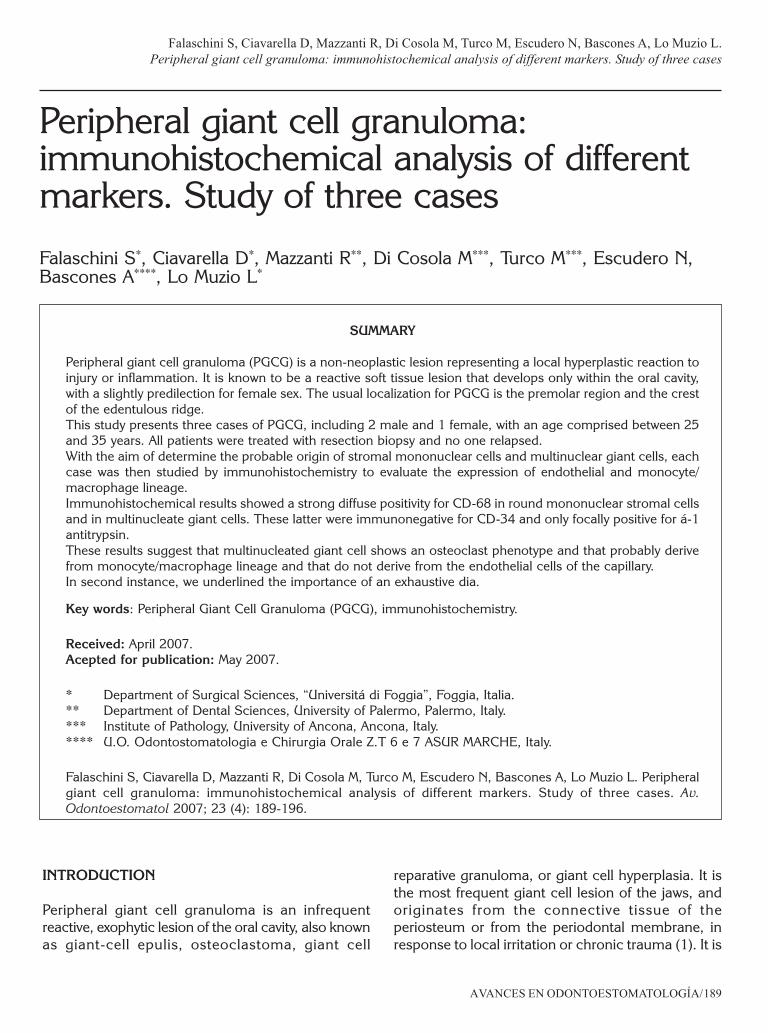
A 27-years-old Bolivian woman without diseaseantecedents of interest or known drug allergiesreferred for resection-biopsy of a gingival epulis. Thelesion was located between the second and thirdmolar, measured 1.8×1.5 cm in size, was purple incolour and bleeding if touched (fig. 1a). The periapi-cal X-rays showed a bone loss between 1.7 and 1.8,demonstrating the possible involvement of the pe-riodontal ligament (fig. 1b).
Treatment consists of lesion resection underinfiltrating anaesthesia, followed by the total avulsionof 17 and 18. The histological study confirmed thediagnosis of peripheral giant cell granuloma,described as ulcerated fibrous epulis with moderatechronic lymphoplasmacellular inflammatory infiltrate.Control examination after six days showed the com-plete restitution ad integrum of the tissue; six monthsafter resection there were not signs of relapse.
Case 2
A 25-years-old Italian male referred for removal of atooth, 15, because it is very damaged and difficult to
Fig. 1a. Case 1: The peripheral giant cell granuloma localizedbetween second premolar and third top molar.
AVANCES EN ODONTOESTOMATOLOGÍA/191
Falaschini S, Ciavarella D, Mazzanti R, Di Cosola M, Turco M, Escudero N, Bascones A, Lo Muzio L.Peripheral giant cell granuloma: immunohistochemical analysis of different markers. Study of three cases
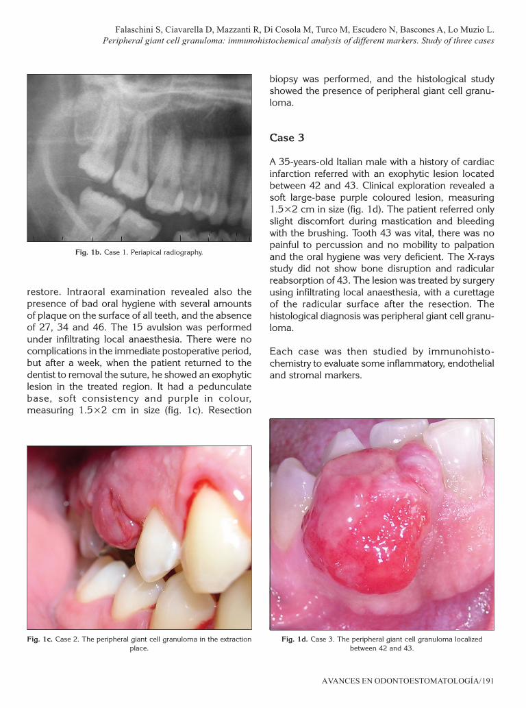
restore. Intraoral examination revealed also thepresence of bad oral hygiene with several amountsof plaque on the surface of all teeth, and the absenceof 27, 34 and 46. The 15 avulsion was performedunder infiltrating local anaesthesia. There were nocomplications in the immediate postoperative period,but after a week, when the patient returned to thedentist to removal the suture, he showed an exophyticlesion in the treated region. It had a pedunculatebase, soft consistency and purple in colour,measuring 1.5×2 cm in size (fig. 1c). Resection
biopsy was performed, and the histological studyshowed the presence of peripheral giant cell granu-loma.
Case 3
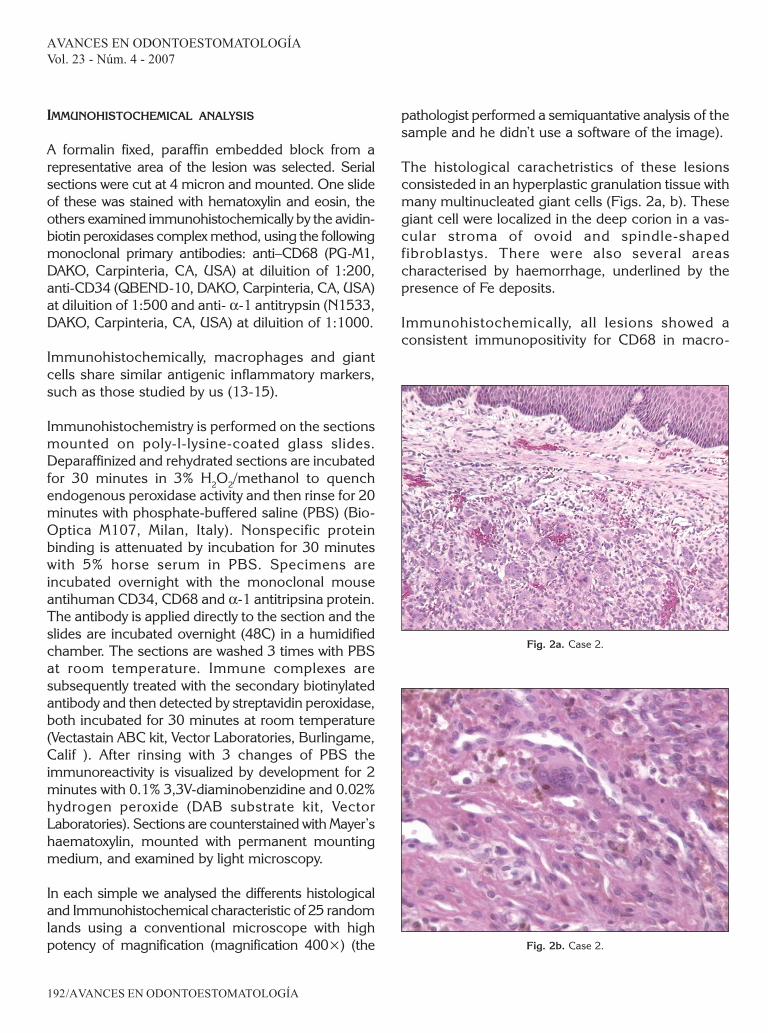
A 35-years-old Italian male with a history of cardiacinfarction referred with an exophytic lesion locatedbetween 42 and 43. Clinical exploration revealed asoft large-base purple coloured lesion, measuring1.5×2 cm in size (fig. 1d). The patient referred onlyslight discomfort during mastication and bleedingwith the brushing. Tooth 43 was vital, there was nopainful to percussion and no mobility to palpationand the oral hygiene was very deficient. The X-raysstudy did not show bone disruption and radicularreabsorption of 43. The lesion was treated by surgeryusing infiltrating local anaesthesia, with a curettageof the radicular surface after the resection. Thehistological diagnosis was peripheral giant cell granu-loma.
Each case was then studied by immunohisto-chemistry to evaluate some inflammatory, endothelialand stromal markers.
Fig. 1b. Case 1. Periapical radiography.
Fig. 1c. Case 2. The peripheral giant cell granuloma in the extractionplace.
Fig. 1d. Case 3. The peripheral giant cell granuloma localizedbetween 42 and 43.
AVANCES EN ODONTOESTOMATOLOGÍAVol. 23 - Núm. 4 - 2007
/AVANCES EN ODONTOESTOMATOLOGÍA192
Fig. 2b. Case 2.
Fig. 2a. Case 2.
IMMUNOHISTOCHEMICAL ANALYSIS
A formalin fixed, paraffin embedded block from arepresentative area of the lesion was selected. Serialsections were cut at 4 micron and mounted. One slideof these was stained with hematoxylin and eosin, theothers examined immunohistochemically by the avidin-biotin peroxidases complex method, using the followingmonoclonal primary antibodies: anti–CD68 (PG-M1,DAKO, Carpinteria, CA, USA) at diluition of 1:200,anti-CD34 (QBEND-10, DAKO, Carpinteria, CA, USA)at diluition of 1:500 and anti- α-1 antitrypsin (N1533,DAKO, Carpinteria, CA, USA) at diluition of 1:1000.
Immunohistochemically, macrophages and giantcells share similar antigenic inflammatory markers,such as those studied by us (13-15).
Immunohistochemistry is performed on the sectionsmounted on poly-l-lysine-coated glass slides.Deparaffinized and rehydrated sections are incubatedfor 30 minutes in 3% H2O2/methanol to quenchendogenous peroxidase activity and then rinse for 20minutes with phosphate-buffered saline (PBS) (Bio-Optica M107, Milan, Italy). Nonspecific proteinbinding is attenuated by incubation for 30 minuteswith 5% horse serum in PBS. Specimens areincubated overnight with the monoclonal mouseantihuman CD34, CD68 and α-1 antitripsina protein.The antibody is applied directly to the section and theslides are incubated overnight (48C) in a humidifiedchamber. The sections are washed 3 times with PBSat room temperature. Immune complexes aresubsequently treated with the secondary biotinylatedantibody and then detected by streptavidin peroxidase,both incubated for 30 minutes at room temperature(Vectastain ABC kit, Vector Laboratories, Burlingame,Calif ). After rinsing with 3 changes of PBS theimmunoreactivity is visualized by development for 2minutes with 0.1% 3,3V-diaminobenzidine and 0.02%hydrogen peroxide (DAB substrate kit, VectorLaboratories). Sections are counterstained with Mayer’shaematoxylin, mounted with permanent mountingmedium, and examined by light microscopy.
In each simple we analysed the differents histologicaland Immunohistochemical characteristic of 25 randomlands using a conventional microscope with highpotency of magnification (magnification 400×) (the
pathologist performed a semiquantative analysis of thesample and he didn’t use a software of the image).
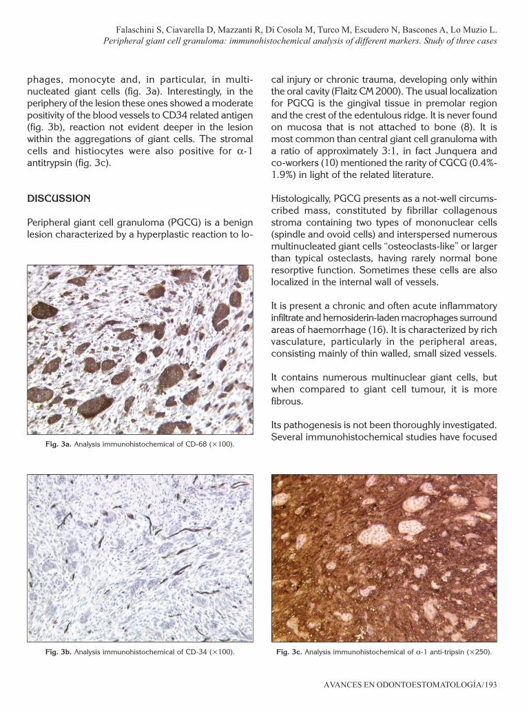
The histological carachetristics of these lesionsconsisteded in an hyperplastic granulation tissue withmany multinucleated giant cells (Figs. 2a, b). Thesegiant cell were localized in the deep corion in a vas-cular stroma of ovoid and spindle-shapedfibroblastys. There were also several areascharacterised by haemorrhage, underlined by thepresence of Fe deposits.
Immunohistochemically, all lesions showed aconsistent immunopositivity for CD68 in macro-
AVANCES EN ODONTOESTOMATOLOGÍA/193
Falaschini S, Ciavarella D, Mazzanti R, Di Cosola M, Turco M, Escudero N, Bascones A, Lo Muzio L.Peripheral giant cell granuloma: immunohistochemical analysis of different markers. Study of three cases
Fig. 3a. Analysis immunohistochemical of CD-68 (×100).
Fig. 3b. Analysis immunohistochemical of CD-34 (×100). Fig. 3c. Analysis immunohistochemical of α-1 anti-tripsin (×250).
phages, monocyte and, in particular, in multi-nucleated giant cells (fig. 3a). Interestingly, in theperiphery of the lesion these ones showed a moderatepositivity of the blood vessels to CD34 related antigen(fig. 3b), reaction not evident deeper in the lesionwithin the aggregations of giant cells. The stromalcells and histiocytes were also positive for α-1antitrypsin (fig. 3c).
DISCUSSION
Peripheral giant cell granuloma (PGCG) is a benignlesion characterized by a hyperplastic reaction to lo-
cal injury or chronic trauma, developing only withinthe oral cavity (Flaitz CM 2000). The usual localizationfor PGCG is the gingival tissue in premolar regionand the crest of the edentulous ridge. It is never foundon mucosa that is not attached to bone (8). It ismost common than central giant cell granuloma witha ratio of approximately 3:1, in fact Junquera andco-workers (10) mentioned the rarity of CGCG (0.4%-1.9%) in light of the related literature.
Histologically, PGCG presents as a not-well circums-cribed mass, constituted by fibrillar collagenousstroma containing two types of mononuclear cells(spindle and ovoid cells) and interspersed numerousmultinucleated giant cells “osteoclasts-like” or largerthan typical osteclasts, having rarely normal boneresorptive function. Sometimes these cells are alsolocalized in the internal wall of vessels.
It is present a chronic and often acute inflammatoryinfiltrate and hemosiderin-laden macrophages surroundareas of haemorrhage (16). It is characterized by richvasculature, particularly in the peripheral areas,consisting mainly of thin walled, small sized vessels.
It contains numerous multinuclear giant cells, butwhen compared to giant cell tumour, it is morefibrous.
Its pathogenesis is not been thoroughly investigated.Several immunohistochemical studies have focused
AVANCES EN ODONTOESTOMATOLOGÍAVol. 23 - Núm. 4 - 2007
/AVANCES EN ODONTOESTOMATOLOGÍA194
on identifying the nature and the interrelationsbetween cellular components in the formation ofGCG: the results have suggested that the mononu-clear stromal cells may originate from fibroblasts andcells of histiocytic origin whereas the origin of giantcells has still been a source of controversy: in factsome authors suggest that they arise secondary toan alteration of the endothelial cells of the capillaries(Flaitz CM 2000), others as a consequence of atraumatic mechanism (some similarities to theosteoclasts) (17, 18).
Palacios et co-workers suggested that giant cellformation to be a fusion of hystiocytes, endothelialcells and fibroblasts (18).
Our immunohistochemical evaluation revealed adiffuse presence of CD68 (antigen most widelydistributed in monocyte/macrophages lineage atvarious differentiation stages as well dendritic cellsand osteoclasts) in a fraction of round mononuclearstromal cells and in mononuclear giant cells. Thisresult confirms that these latter may derive fromosteoclasts, according to previous study (19).
In addition, it is interesting to show the stainingpattern of the blood vessels to CD34 related antigenin the peripheral giant cell granuloma: the capillarieson the periphery of the lesions were strongly positivefor this antibody (fig. 3b), reaction product not evidentin the lesion within the aggregations of multinucleategiant cells. This data may suggest that multinucleategiant cell not arise from endothelial cells of thecapillaries.
The present case study was performed also toevaluate the role of the alpha1-antitrypsin (α1-AT) inpatients with PGCG. α1-AT is a physiologicalinhibitor of activated protein C and thereforedecreases activated protein C activity. α1-AT, a52,000 D glycoprotein, is secreted mostly byhepatocytes, lung epithelial cells and phagocytes.α1-AT inhibits a variety of serine proteinases by itsactive site (Met358-Ser359), but its preferentialtarget is human neutrophil elastase (HNE) asdemonstrated by the high association rate constant(Kass) for this proteinase. The immunohistochemicalanalysis showed a diffuse, strong immunopositiveof mononuclear stromal cells ad only focally for
multinuclear giant cells for α-1 antitrypsin (fig. 3c).These data pointed out that this antigen is able toinhibit the activity of human neutrophil elastase(fig. 3c).
The differential diagnosis of PGCG particularlyinvolves giant cell tumour (Chaparro-Avendano AV2005): nonossifying fibroma which differs from PGCGlesions in consistency and colour; pyogenic granulo-ma which is difficult to distinguish from PCGC lesions;CGCG which is an expansive and destructiveintraosseous lesion that can perforate the cortex,mimicking PGCG; chondroblastoma which,localized in the gum, may provoke irregular bonedestruction below the exophytic lesion; odontogeniccyst; parulis, which is frequently associated with anecrotic tooth or with periodontal disorder;haemangioma cavernosum, which is distinguishedfrom PGCG lesions by their pulsatile nature; fissuredepulis (9).
The treatment of choice is surgical excision withthe suppression of the underlying etiologic factors(5). The periosteum must be included in theexcision to prevent recurrences; in fact recurrenceis frequent and is observed in 5% and 11% of ca-ses according to Eversole (22) and Mighell (23)respectively. Curettage in addition to the excisionto remove the base of the lesion also has beensuggested. The recurrence rate of PCGC has beenreported to range from 5-70,6%. This wide variationmay be attributed to the surgical technique usedin excision (8-10).
In conclusion our immunohistochemical studysuggests, according with previous immunohisto-chemical study, that multinucleated giant cell showsan osteoclast phenotype and that probably derivefrom monocyte/macrophage lineage and that giantcells do not derive from the endothelial cells of thecapillary.
In second instance an early and precise diagnosis ofPGCG, based on the clinical, radiological findingsand histological study, allows conservativemanagement with a lower risk for the teeth andadjacent bone. In all cases described here, thetreatment applied is that reported in the literature (5,7), consisting of surgical excision and subsequent
AVANCES EN ODONTOESTOMATOLOGÍA/195
Falaschini S, Ciavarella D, Mazzanti R, Di Cosola M, Turco M, Escudero N, Bascones A, Lo Muzio L.Peripheral giant cell granuloma: immunohistochemical analysis of different markers. Study of three cases
curettage to remove the base of the lesion and allassociated irritant factors (9).
REFERENCES
1. Shafer WG, Hine MK, Levi BM. Tratado depatologìa Bucal. eds. 4th. Rio de Janeiro:Guanabara-Koogan; 1987. p.143-5.
2. Reichart PA, Philipsen H. Atlas de Patologìa Oral.eds.1 Masson; 2000. p. 164.
3. Wolfson L, Tal H, Covo S. Peripheral giant cellgranuloma during orthodontic treatment. Am JOrthod Dentofac Orthop 96: 519-23.
4. Bagàn Sebastiàn JV. Atlas de enfermedales de lamucosa oral. eds. 5th Barcelona; 1995. p. 186.
5. Flaitz CM. Peripheral giant cell granuloma: apotentially aggressive lesion in children. PediatrDent 2000; 22: 232-3.
6. Nedir R, Lombardi T, Samson J. Recurrentperipheral giant cell granuloma associated withcervical resorption. J Periodontol 1997; 67:381-4.
7. Pandolfi PJ, Felefli S, Flaitz CM, Johnson JV.Anaggressive peripheral giant cell granuloma in achild. J Clin Pediatr Dent 1999; 23: 353-5.
8. Breault LG, Fowler EB, Wolfgang MJ, Lewis DM.Peripheral giant cell granuloma: a case report.Gen Dent 2000; 48:716-9.
9. Gandara-Rey JM, Pacheco Martins Carneiro JL,Gandara-Vila P, Blanco-Carrion A, Garcia-GarciaA, Madrinan-Grana P, Martin MS. Peripheral giantcell granuloma. Review of 13 cases. Med Oral2002;7:254-9.
10. Junquera LM, Lupi E, Lombardia E, Fresno MF.Multiple and synchronous peripheral giant cellgranulomas of the gums. Ann Otol RhinolLaryngol 2002; 111: 751-3.
11. Ceballos-Salobrena A. Tumores benignos de la mu-cosa oral. En: Bagàn-Sebastian JV, Ceballos-
Salobrena A, Bermejo-Fenoll A, Aguirre-Urizar JV,Penarrocha-Diago M. Medicina Oral; 1995. p.182-3.
12. Kfir Y, Buchner A, Hansen LS. Reactive lesions ofthe gingiva. A clinicopathological study of 741cases. J Periodontol 1980; 51: 655-61.
13. Regezi JA, Zarbo RJ, Lloyd RV. Muramidase,alpha-1 antitrypsin, alpha-1 antichymotrypsin,and S-100 protein immunoreactivity in giant celllesions. Cancer. 1987 Jan 1;59(1):64-8
14. Regezi JA, Nickoloff BJ, Headington JT Oralsubmucosal dendrocytes: factor XIIIa+ andCD34+ dendritic cell populations in normal tissueand fibrovascular lesions. J Cutan Pathol. 1992Oct;19(5):398-406.
15. Panico L, Passeretti U, De Rosa N, D’Antonio A,De Rosa G. Giant cell reparative granuloma ofthe distal skeletal bones. A report of five caseswith immunohistochemical findings. VirchowsArch. 1994;425(3):315-20.
16. Wood NK. Diagnòstico diferencial de las lesio-nes orales y maxilofaciales. Madrid: HarcourtBrace de Espana SA;1998. p. 141-2.
17. Matsumura T, Sugahara T, Wada T, Kawakatsu K.Recurrent giant-cell reparative granuloma: reportof case and histochemical patterns. J Oral Surg1971; 29: 212-6.
18. Sapp JP. Ultrastructure and histogenesis ofperipheral giant cell reparative granuloma of thejaws. Cancer 1972; 30: 1119-29.
19. Palacios E, Valvassori G. Giant cell reparativegranuloma. Ear Nose Throat J 2000; 79: 688.
20. Burghaus B, Langer C, Thedieck S, Nowak-GottlU. Elevated alpha1-antitrypsin is a risk factor forarterial ischemic stroke in childhood. ActaHaematol. 2006;115(3-4):186-91
21. Chaparro-Avendano AV, Berini-Aytes L, Gay-Es-coda C. Peripheral giant cell granuloma. A reportof five cases and review of the literature. MedOral Patol Oral Cir Bucal. 2005; 10(1): 53-7.
AVANCES EN ODONTOESTOMATOLOGÍAVol. 23 - Núm. 4 - 2007
/AVANCES EN ODONTOESTOMATOLOGÍA196
22. Eversole LF, Rovin S. Reactive lesions of the gin-giva. J Oral Pathol 1972; 1: 30-8.
23. Mighell AJ, Robinson PA, Hume WJ. Peripheralgiant cell granuloma: a clinical study of 77 casesfrom 62 patients, and literature review. OralDis1995;1: 12-9.
CORRESPONDENCE
Prof. Lorenzo Lo MuzioVia Carelli 2871100 Foggia – Italia.Phone nunber and Fax 0039 0881 685809Correo electrónico: [email protected] o [email protected]