percutaneous drainage of abscess and post operative collections
TRANSCRIPT

Percutaneous Drainage Of Collections and Abscess
Dr.Suhas Basavaiah
Resident (MD Radio-Diagnosis)

INTRODUCTIONPercutaneous abscess drainage (PAD) has evolved from revolutionary
to routine, replacing open surgical abscess drainage in all but the most difficult or inaccessible cases.
Originally only patients with simple fluid collections were candidates for PAD; however, researchers have convincingly demonstrated that both septated and viscous fluid collections may be successfully treated percutaneously, particularly with the adjunctive use of lytic agents.
An aggressive practical approach with relatively simple devices and techniques may yield a high success rate with few complications.
Marked growth in last 20 years
All types of simple and complex collections drained in the chest,abdomen and pelvis
Requires ability to assess CT and US images and familiarity with drainage equipment

Collection Assessment-Imaging
Aim - shortest, safest route to site drain in the most dependent position
Avoid major vessels
Avoid transgressing bowel
Assessment of nature of fluid-echogenicity ; septations

IMAGING – US or CT
CT
good visualisation
opacified bowel
not limited by ileus or depth
US
real time
portable
operator dependent
Size+site of collection ; operator preference

Ultrasound guided percutaneous drainage is one form of image guided procedure, allowing minimally invasive treatment of collections that are accessible by ultrasound study.
It has several advantages and disadvantages over CT, which include:
Advantages
is a dynamic study, allowing greater precision to control needle insertion
not exposes patient to ionising radiation
does not require as wide a range of staff, compared to CT-guided procedures
Disadvantages
deeper targets may not be as well visualised on ultrasound (e.g. retroperitoneal nodes)
bowel gas may obscure visualization
attenuation of the sound beam on larger patients

IndicationsIndications for percutaneous drainage are broad: essentially any abnormal fluid collection in the patient which can be accessible. Examples include:
complicated diverticular abscess
Crohn disease related abscess
complicated appendicitis with appendicular abscess
tuboovarian abscess
post-surgical fluid collections
hepatic abscess (e.g. amebic or post-operative)
renal abscess or retroperitoneal abscess.
splenic abscess
Contraindications
The only common contraindications are:
biopsy target is not accessible
patient has a bleeding diathesis

Laboratory parameters for a safe procedureInterventional procedures like percutaneous drainage require special attention to coagulation indices. There are widely divergent opinions about the safe values of these indices for percutaneous biopsies. The values suggested below were considered based on a literature review.
Complete blood count: Platelet > 50000/mm3 (Some institutions determine other values between 50000-100000/mm3)
Coagulation profile:
international normalized ratio (INR) ≤1.5 1
normal prothrombin time (PT), partial thromboplastin time (PTT)
Some studies show that having a normal INR or prothrombin time is no reassurance that the patient will not bleed after the procedure

Pre-procedure evaluation
Review other diagnostic studies first to clarify the collection that is requested to be drained.
An ultrasound study should be done prior to biopsy to decide the access angle and check the relationship of the collection to adjacent structures.
In general, the shortest possible route is preferred, as long as it does not traversing other structures.

EquipmentNeedle
Typical abscess fluid is readily aspirated through an 18-gauge needle
Viscous or debris-laden fluid is more likely to cause a false-negative aspirate with a 21-gauge needle as only clear supernatant may return through the needle.
An 18-gauge needle is easier to control and image and accepts a 0.038-inch guidewire.
There is no clinically significant difference in needle trauma between the 18 and 22-gauge needles when a catheter is placed through the same tract.
The 21-gauge needle may be used to minimize trauma for a challenging localization, low-probability fluid collection, or personal preference.
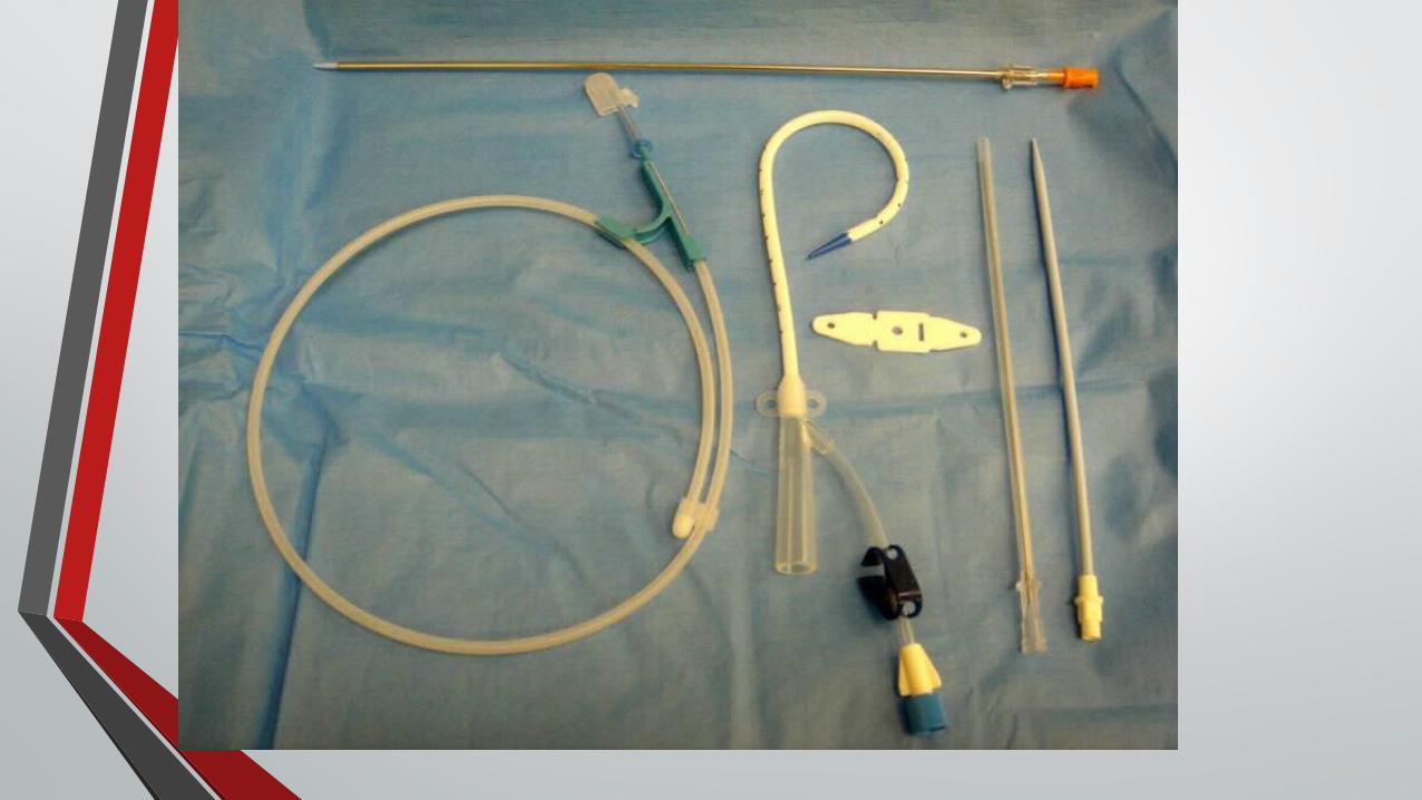

Equipment (contd.)
Catheters
6F-24F catheters
Locking or non-locking-VIP at removal
Sump or non-sump-2nd lumen containing air which prevents cavity collapsing around catheter tip


Guidewire
A variety of guidewires are available for PAD with different properties and prices. Guidewires should meet the following specifications:
Stiff enough to guide dilators and catheter into abscess
Not too stiff to prevent easy coiling of wire shaft within abscess
Floppy tipped enough to encourage wire to coil within the abscess and not perforate the abscess wall
Short enough to make use convenient


Localization Techniques Any modality may be used to assist needle placement.
Prior to ready availability of CT fluoroscopy, patient assessment may be performed with CT scanning, with the PAD procedure performed with US localization.
Conventional fluoroscopy can be used as an adjunct to US.
US guidance allows real-time imaging and does not involve radiation exposure.
CT fluoroscopy is increasingly available and facilitates "one-stop-shopping."
The diagnostic CT and PAD may now be performed readily in one setting.

PATIENT PREPARATION
IV access
Fasted for > 2 hours
Coagulopathy excluded
Informed consent

PROCEDURE
Ultrasound guided percutaenous drainage may be performed with a single or multiple stage technique.
Consider conscious sedation
Clean skin
Anaesthetise skin
Skin incision large enough for passage of catheter
Consider tract dissection

TROCAR TECHNIQUE
Reference needle in collection
Catheter assembly advanced to the same depth ,in the same plane
Remove stylet and aspirate
Advance catheter over stationary stiffener

SELDINGER TECHNIQUE
18g needle in collection
Pass 0.035 wire into collection
Dilate tract
Pass catheter and stiffener over wire
When inside collection pass catheter alone
POST-INSERTION OF DRAIN
Aspirate fluid
Re-image:?need for 2nd drain
Secure drain-it is always more difficult to re-puncture a partially drained collection

POST-PROCEDURE CAREPost-procedure care The patient's basic vital signs should be monitored for 4 hours post
procedure (pulse, blood pressure, SpO2), or as long as deemed necessary.
Aspirate 8hrly with a 50ml. Syringe
Irrigate with 10ml. of saline
Dependent position of bag
The patient should remain in bed for 2 hours. After this time period mobilization and oral intake is permitted.
Removal-clinical improvement and drainage of <10ml. per day or collection resolved on re-imaging
The entry site should be reviewed on a daily basis. If output from the collection ceases, it may mean that the collection is no longer present or that the drain is clogged.

TIPS - INSERTION
Ensure adequate skin incision
Avoid kinking wire(no fluoroscopy)
Ideal wire-stiff enough to allow passage of dilators and catheter but will coil within abscess and not perforate posterior wall
Cut thread flush with catheter hub
3-way tap

IF COLLECTION PERSISTS WITH LOW FLOWS
Catheter displacement
Catheter/tubing blocked or kinked
Upsizing catheter
Septation/loculation

If Collection Persists with high flows
Expect to find a fistula
Can occur from bowel, bile and pancreatic duct, renal tract
Exclude distal obstruction ; underlying bowel disease ; proximal diversion ; parenteral feeding
Bile leak postlap.chole.- drain plus cbd stent
IF THERE IS PRESENCE OF GROSS BLOOD
Place the catheter
Let the blood drain into the bag
Since blood is a potent irritant and toxic to omentum, it has to be drained regardless to avoid fatal complications like peritonitis and adhesions

COMPLICATIONS
Viscus perforation
Catheter dislodgement
Damage to vessels
Peritonitis
Diaphragm rupture

MINIMIZING COMPLICATIONS
Broad spectrum antibiotics
Correct coagulopathy
Adequate sedation + analgesia-beware the restless patient
Good bowel opacification at CT
Post procedure catheter management
Beware collections adjacent to implants-aspirate>drain
Discuss cases with clinical team

PITFALLS
The procedure was not indicated.
Failure to obtain informed consent
Failure to perform the procedure in a reasonable manner and deviation from the standard of care.
Failure to promptly recognize and react to a complication.
Failure to adequately treat the complication according to an adequate standard of care.

CONCLUSION
Assess pre-procedure imaging
Minimise complications related to PAD
Involvement in post procedure catheter management
Practical knowledge of needles, wires and catheters

References
Emedicine , percutaneous drainage of abscess and post operative collections
Radiopedia , USG guided percutaneous drainage
American College of Radiology. Percutaneous catheter drainage of infected intra-abdominal fluid collections
Haaga JR, Weinstein AJ. CT-guided percutaneous aspiration and drainage of abscesses.
Lang EK, Springer RM, Giorioso LW, Cammarata CA. Abdominal abscess drainage under radiologic guidance: causes of failure.
Thank You