pathology of parasitic infections - scacm
TRANSCRIPT
Pathology of
Parasitic Infections
Julie A Ribes, MD, PhD
Director of Clinical Microbiology
UK HealthCare
1
Objectives
• Identify structures in tissues/fluids that should make
you think of parasites
• Describe inflammatory changes that may result from
parasitic infections
• Delineate parasite-tissue tropisms
2

Measure about 110-150 uM in length
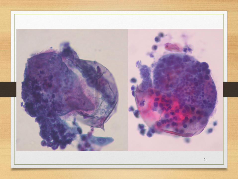
A Urine was submitted to cytology (PAP stain) from
a 36-year-old emigrant from the Republic of Congo
for evaluation of recent onset of dysuria, hematuria
and increased urinary frequency
130 uM
3

~140 uM
4
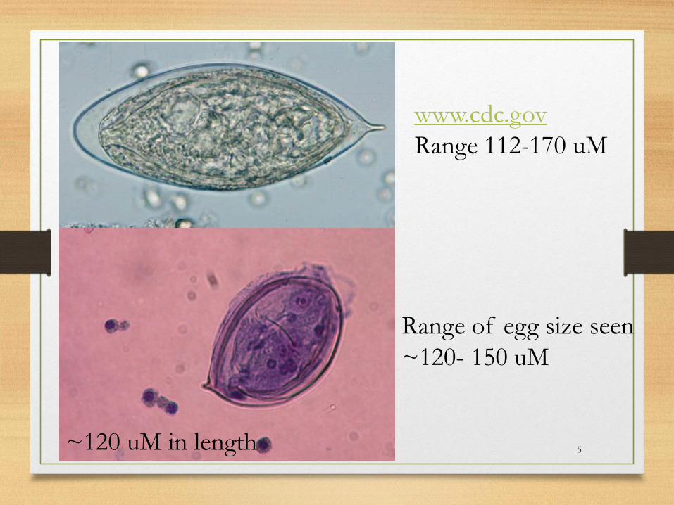
~120 uM in length
www.cdc.gov
Range 112-170 uM
Range of egg size seen
~120- 150 uM
5
6

Schistosomiasis:
Stages Found in
Human Tissues and
Fluids
1. Adult worms
2. Eggs
3. Egg contents
(miracidia)
7
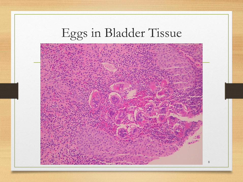
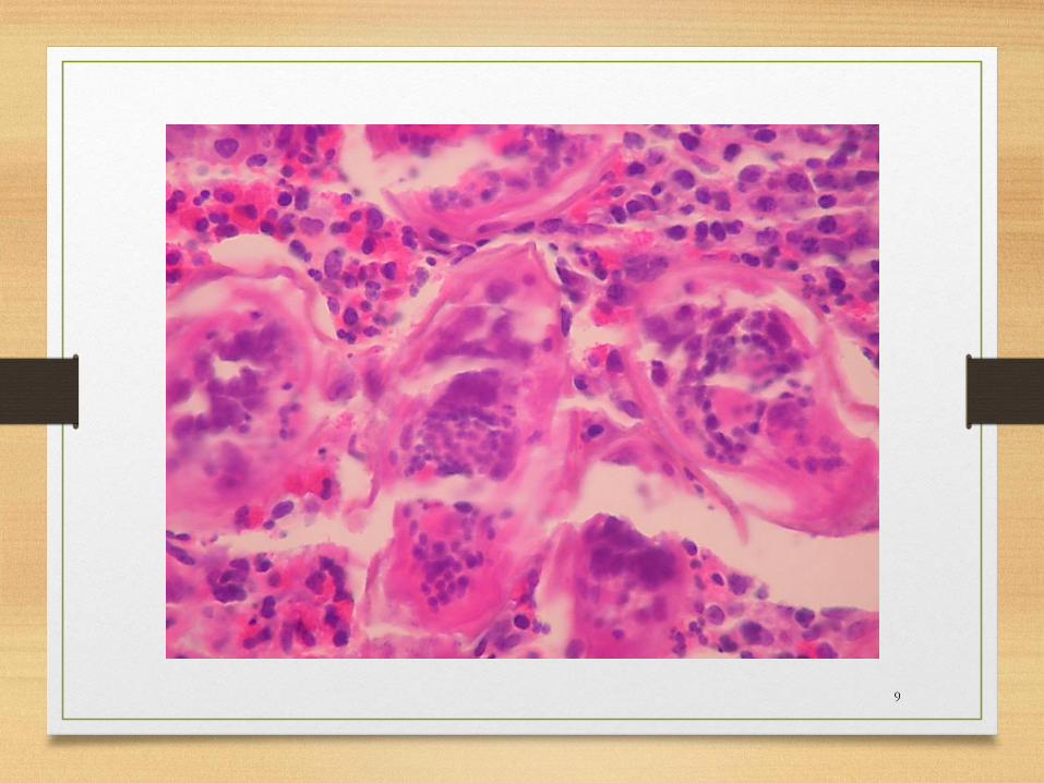
Eggs in Bladder Tissue
8
9
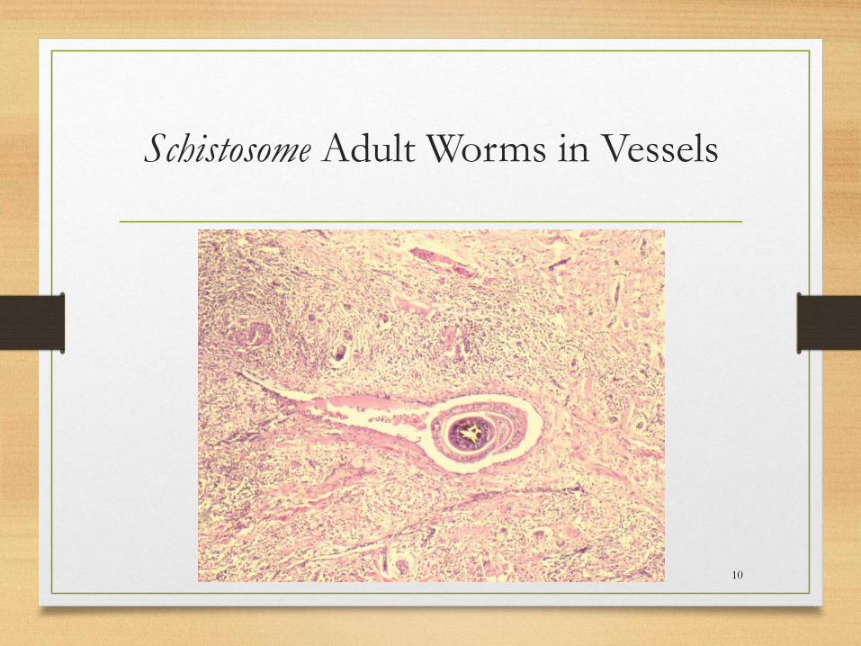
Schistosome Adult Worms in Vessels
10

Schistosome Tropism
Species Adult Location Egg Location
Schistosoma mansoni Veins of colon and lower
ileum
Portal veins of liver
Shed in stool
Seen in colonic mucosa and
liver
Schistosoma haemotobium Veins of the bladder and
lower rectum
Shed in urine and infrequently
seen in stool
Seen in bladder wall
Schistosoma intercalatum Veins of rectum Shed only in stool, not urine
Seen in rectal mucosa
Schistosoma japonicum
Schistosoma mekongi
Veins of the small
intestine
Shed in stool
Seen in intestinal mucosa
11
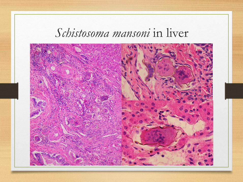
Schistosoma mansoni in liver
12
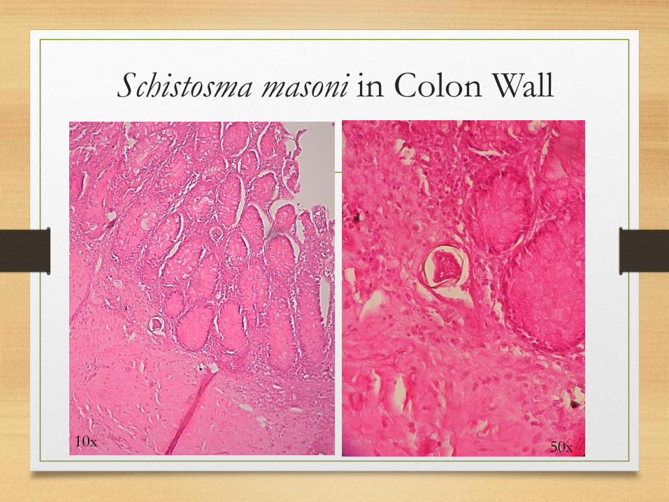
Schistosma masoni in Colon Wall
1310x 50x
Schistosomiasis Summary
• Eosinophils in blood, fluids, tissues should make you think of parasitic infection with helminths
• Eggs in urine/stool with knobs and spines should make you think of a schistosomal infection
• Male and female worms in vessels should make you think of schistosomes
• Tissue tropism will suggest the specific schistosome causing the infection
• Ciliates in urine should make you think of Schistosoma haematobium
• Eggs are often distorted and fractured so ID of diagnostic knobs and spines may be difficult – egg size and tropism may be helpful
14
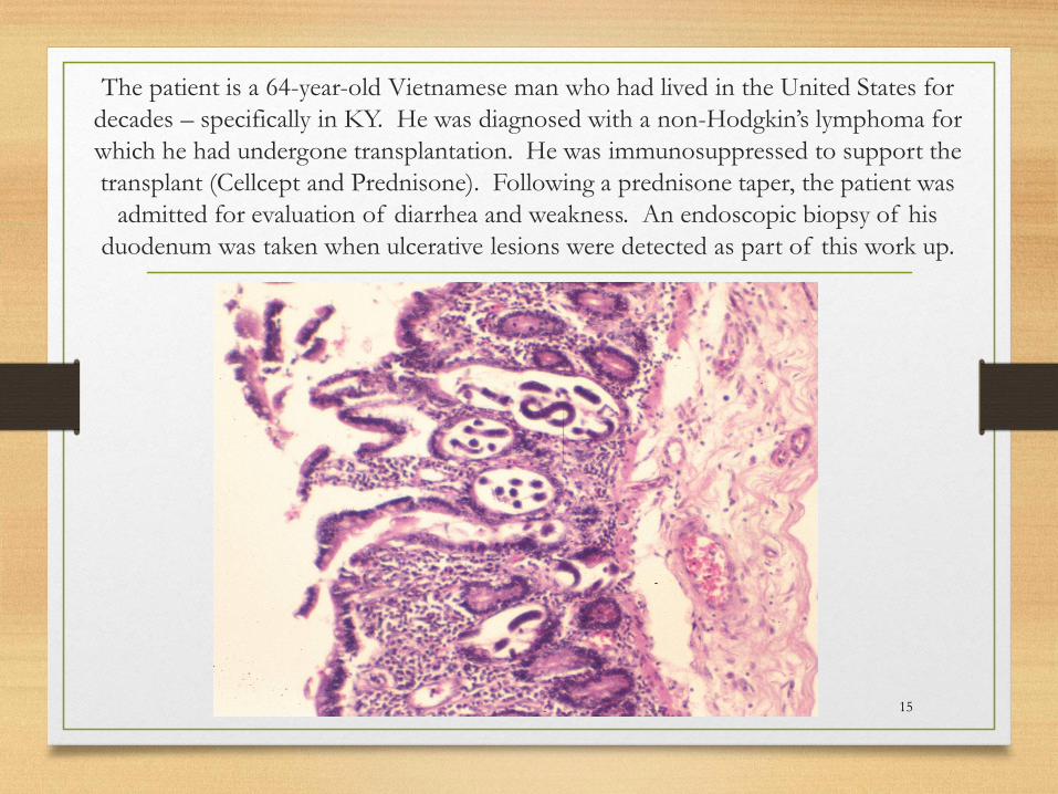
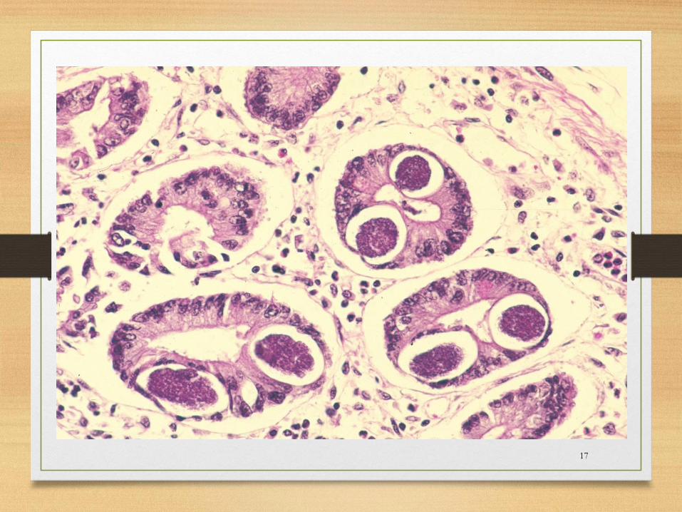
The patient is a 64-year-old Vietnamese man who had lived in the United States for
decades – specifically in KY. He was diagnosed with a non-Hodgkin’s lymphoma for
which he had undergone transplantation. He was immunosuppressed to support the
transplant (Cellcept and Prednisone). Following a prednisone taper, the patient was
admitted for evaluation of diarrhea and weakness. An endoscopic biopsy of his
duodenum was taken when ulcerative lesions were detected as part of this work up.
15
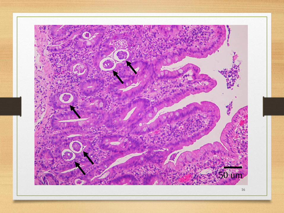
16
50 um
17
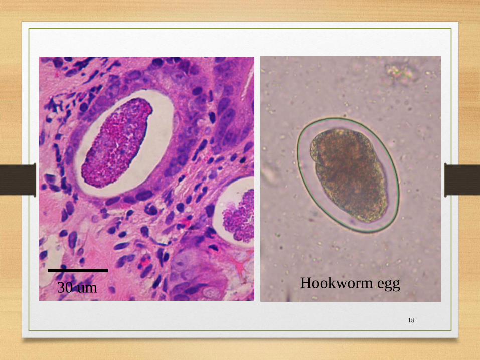
18
30 um Hookworm egg
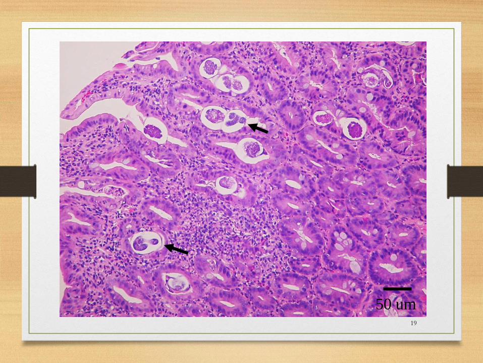
19
50 um
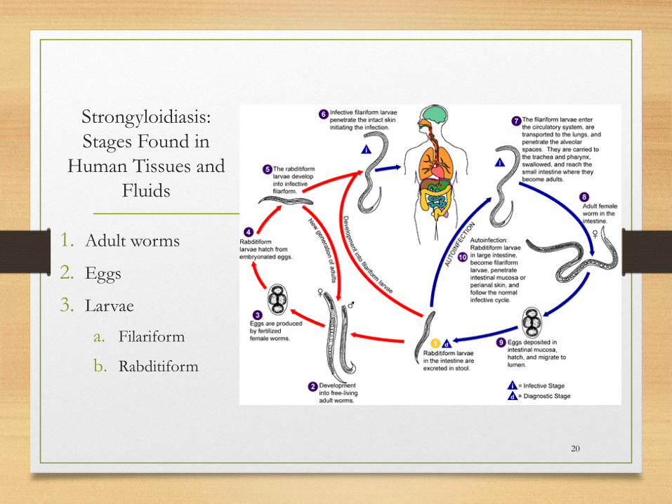
Strongyloidiasis:
Stages Found in
Human Tissues and
Fluids
1. Adult worms
2. Eggs
3. Larvae
a. Filariform
b. Rabditiform
20

www.cdc.gov
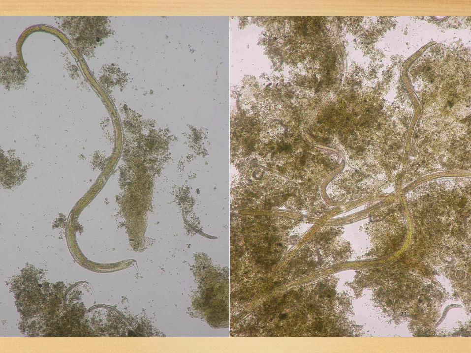
Larvae of Strongyloides and Hook worm
21

www.cdc.gov
Larvae of Strongyloides and Hook worm
22
23
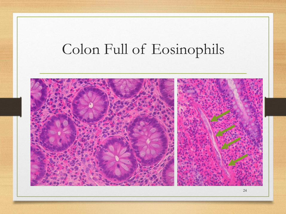
Colon Full of Eosinophils
24
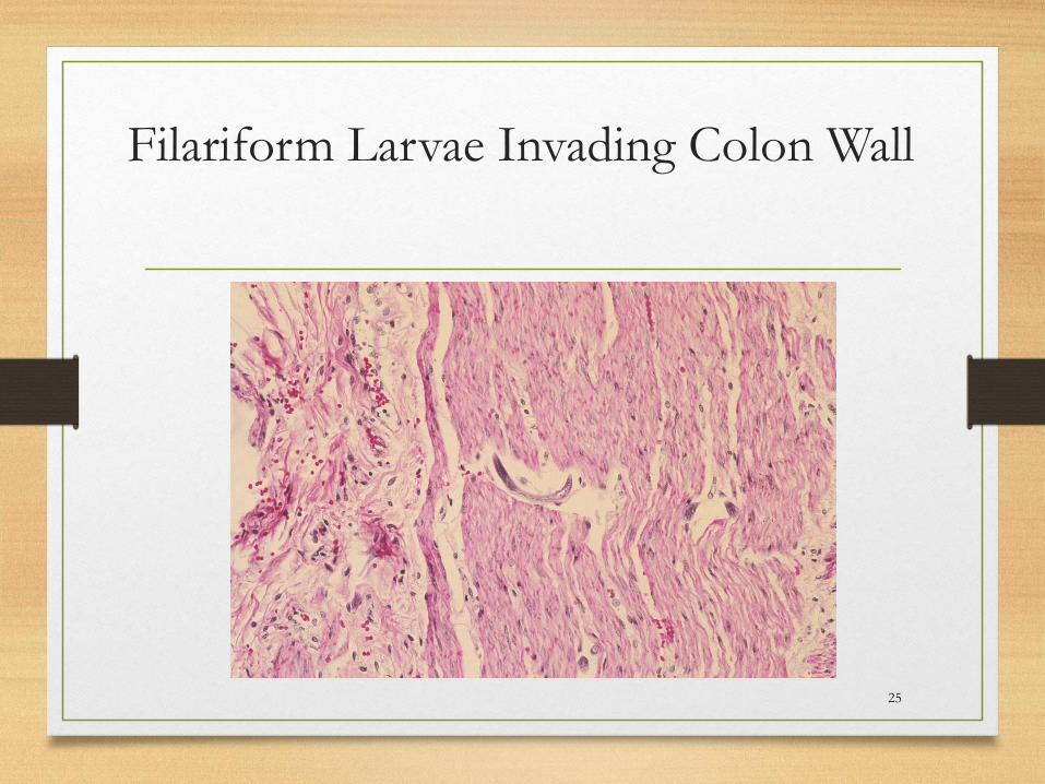
Filariform Larvae Invading Colon Wall
25

Strongyloides stercoralis Hyper infection
Syndrome
• Strong association with steroids
• Filariform larvae may be found in any tissue
• Sepsis or fungemia often noted
• Meningitis with GNR, Enterococci, or non-
cryptococcal yeasts should raise suspicion
• Often fatal
26

Filariform Larvae Transiting
Through Lung
Wright’s Giemsa GMS
27
Pap
Strongyloidiasis Summary
• Eosinophils in blood, fluids, tissues should make you think of parasitic infection with helminths – for Strongyloides, eosinophilia is variable
• Inflammation is variable – patients are often profoundly immunocompromised
• Female worms in small intestinal crypts should make you think of Strongyloides
• Eggs resembling those of Hook worm retained within the intestinal crypts should make you think of Strongyloides
• Embryonated eggs in the intestinal crypts should make you think Strongyloides
• History of recent steroid use should make you think of Strongyloides hyper infection symdrome
• Filariform larvae look like “speckled bands” on H&E may be seen in colon and other tissues in hyper infection syndrome
• Filariform larvae may be seen in BAL and sputum
28
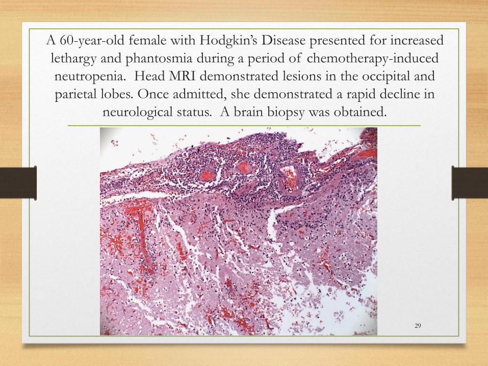
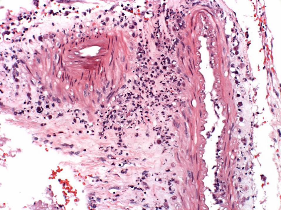
A 60-year-old female with Hodgkin’s Disease presented for increased
lethargy and phantosmia during a period of chemotherapy-induced
neutropenia. Head MRI demonstrated lesions in the occipital and
parietal lobes. Once admitted, she demonstrated a rapid decline in
neurological status. A brain biopsy was obtained.
29
30

31
32
33
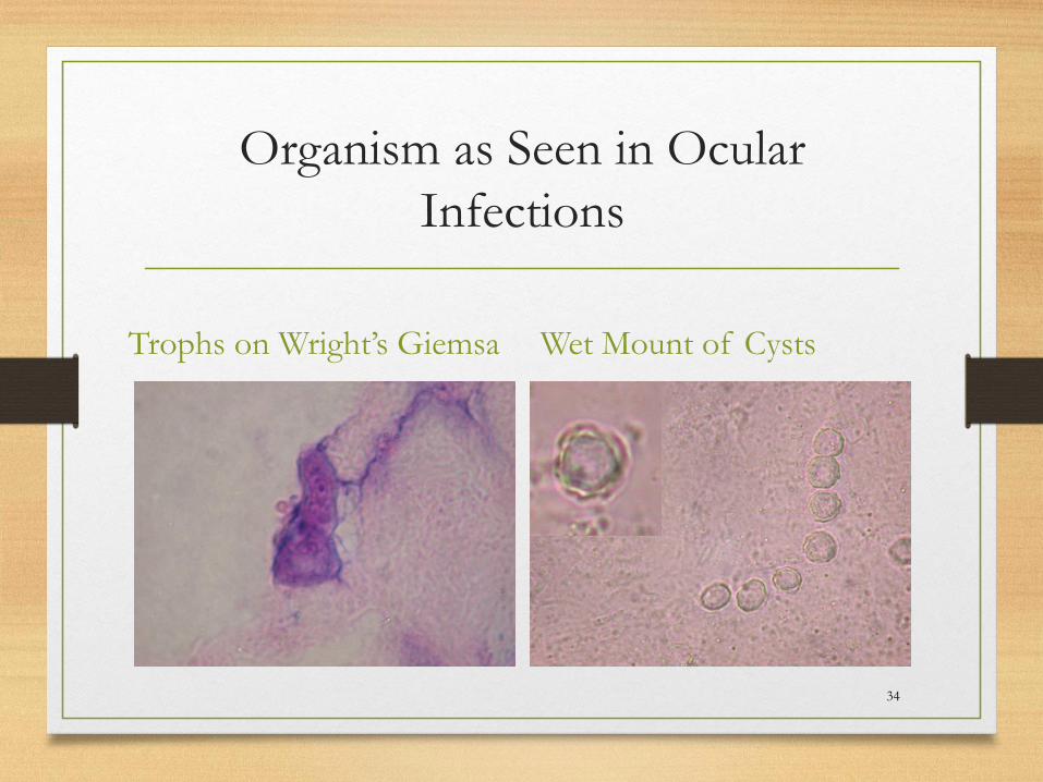
Organism as Seen in Ocular
Infections
Trophs on Wright’s Giemsa Wet Mount of Cysts
34
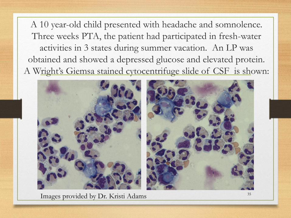
A 10 year-old child presented with headache and somnolence.
Three weeks PTA, the patient had participated in fresh-water
activities in 3 states during summer vacation. An LP was
obtained and showed a depressed glucose and elevated protein.
A Wright’s Giemsa stained cytocentrifuge slide of CSF is shown:
35Images provided by Dr. Kristi Adams

A 10 year-old child presented with headache and somnolence. Three weeks
PTA, the patient had participated in fresh-water activities in 3 states during
summer vacation. An LP was obtained and showed a depressed glucose and
elevated protein and many neutrophils. A second CSF was obtained 2 days
later and stained with Wright’s Giemsa. Specimen was sent to the CDC for
additional testing.
36
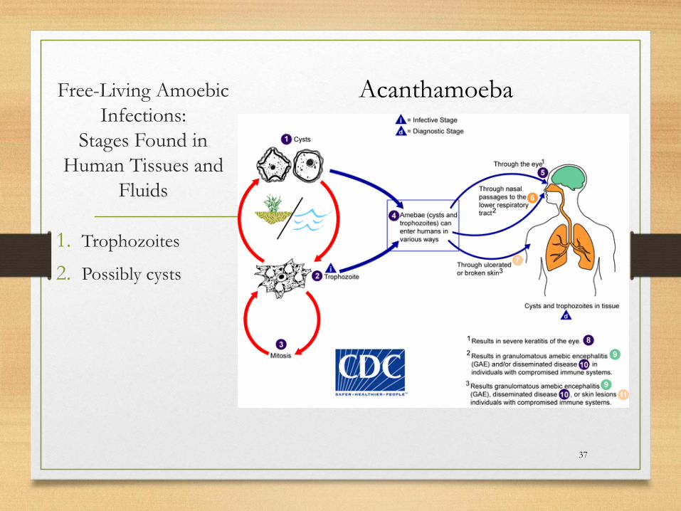
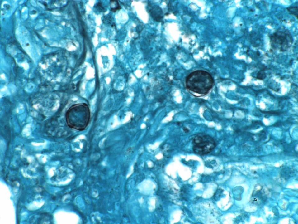
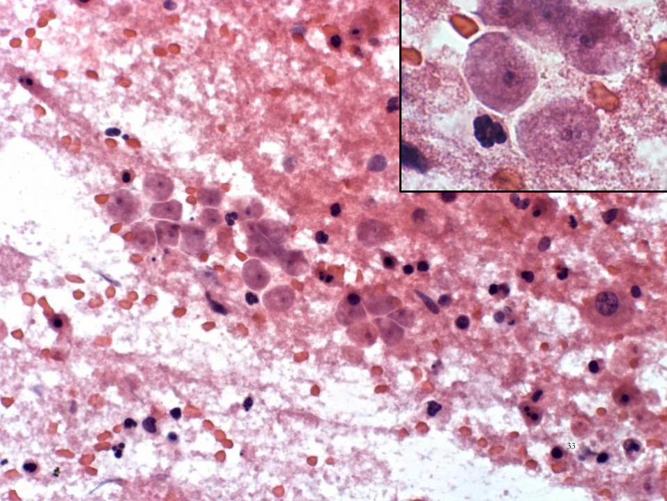
Free-Living Amoebic
Infections:
Stages Found in
Human Tissues and
Fluids
1. Trophozoites
2. Possibly cysts
37
Acanthamoeba
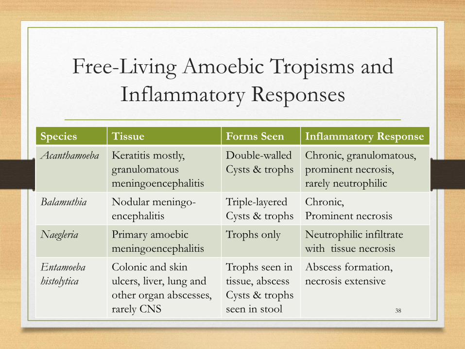
Free-Living Amoebic Tropisms and
Inflammatory Responses
Species Tissue Forms Seen Inflammatory Response
Acanthamoeba Keratitis mostly,
granulomatous
meningoencephalitis
Double-walled
Cysts & trophs
Chronic, granulomatous,
prominent necrosis,
rarely neutrophilic
Balamuthia Nodular meningo-
encephalitis
Triple-layered
Cysts & trophs
Chronic,
Prominent necrosis
Naegleria Primary amoebic
meningoencephalitis
Trophs only Neutrophilic infiltrate
with tissue necrosis
Entamoeba
histolytica
Colonic and skin
ulcers, liver, lung and
other organ abscesses,
rarely CNS
Trophs seen in
tissue, abscess
Cysts & trophs
seen in stool
Abscess formation,
necrosis extensive
38
Free-Living Amoeba Summary
• Trophozoites in tissue/fluids may be very subtle
• Trophozoites in tissue/fluids may be decomposed
• Cysts in tissues/fluids may not be present based on the organism
• The inflammatory response varies from acute neutrophilic, to chronic, granulomatous, or necrotic based on organism
• CDC has special stains and PCRs to assist with dx.
39
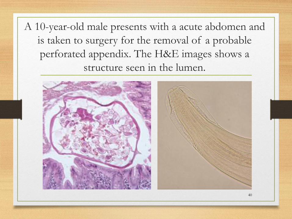
A 10-year-old male presents with a acute abdomen and
is taken to surgery for the removal of a probable
perforated appendix. The H&E images shows a
structure seen in the lumen.
40

Enterobiasis:
Stages Found in Human
Tissues and Fluids
1. Adult worms
2. Eggs
1. Non-embryonated
2. Embryonated
41
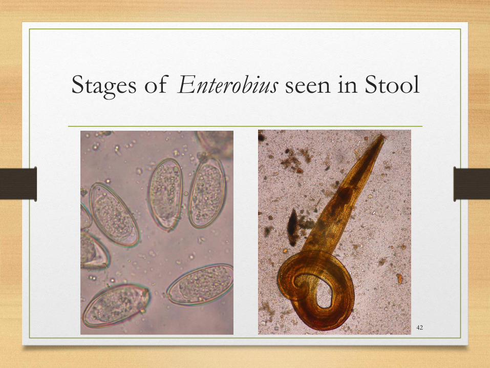
Stages of Enterobius seen in Stool
42
Enterobiasis Summary
• See worms in the appendix, look for cuticular darts
(alae)or eggs in the uterus for identification
• See cuticular darts (alae) on the cross section of the
worm, think Enterobius vermicularis
• See eggs in the uterus with one flattened and one
convex side, think Enterobius vermicularis
43
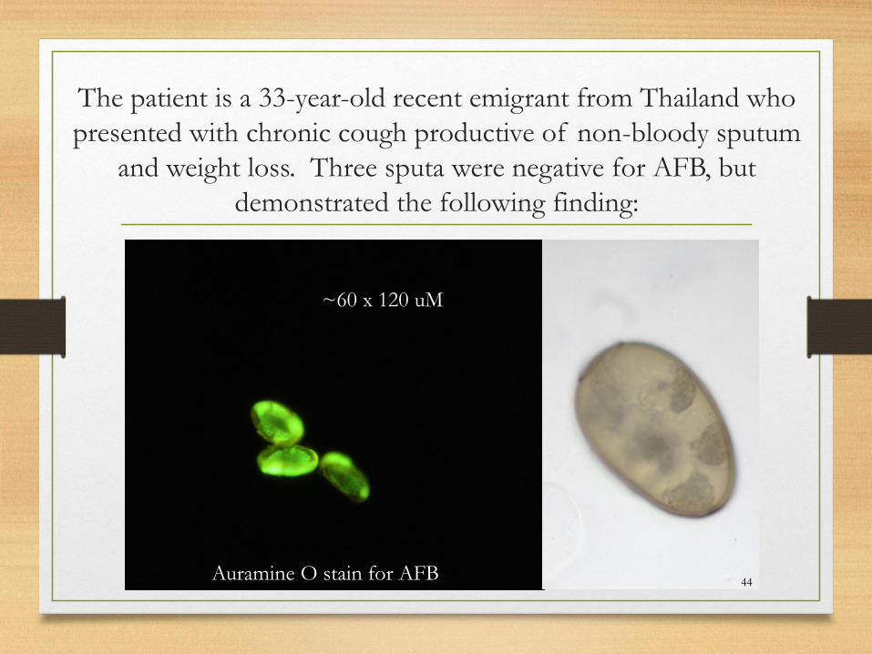
The patient is a 33-year-old recent emigrant from Thailand who
presented with chronic cough productive of non-bloody sputum
and weight loss. Three sputa were negative for AFB, but
demonstrated the following finding:
~60 x 120 uM
Auramine O stain for AFB44
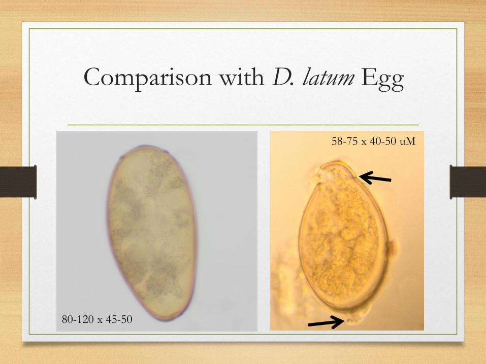
Comparison with D. latum Egg
45
58-75 x 40-50 uM
80-120 x 45-50
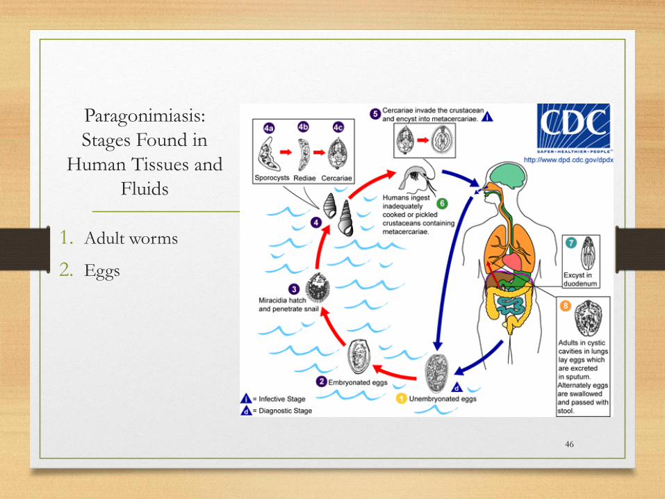
Paragonimiasis:
Stages Found in
Human Tissues and
Fluids
1. Adult worms
2. Eggs
46
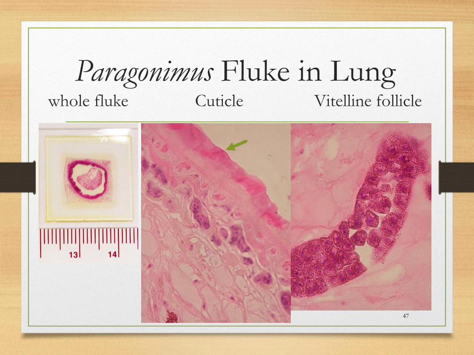
Paragonimus Fluke in Lungwhole fluke Cuticle Vitelline follicle
47
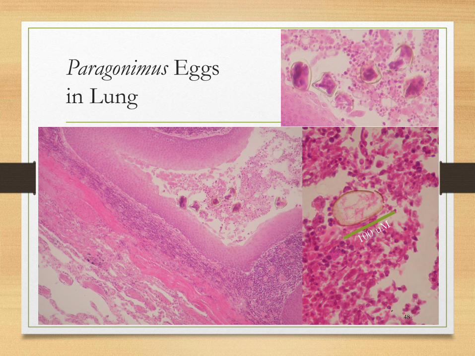
Paragonimus Eggs
in Lung
48
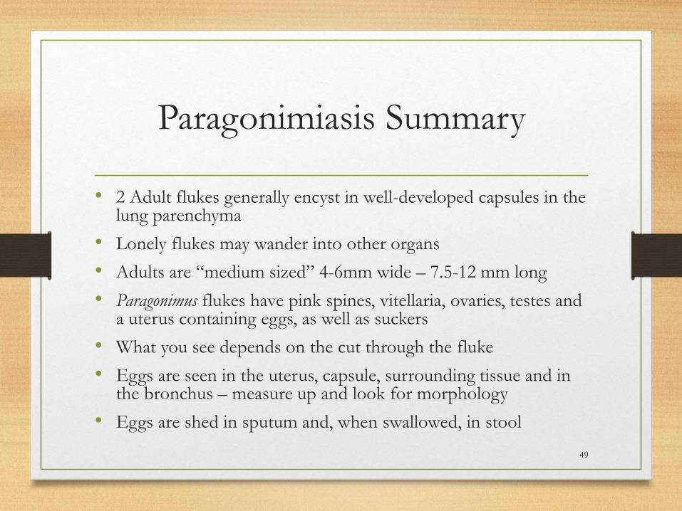
Paragonimiasis Summary
• 2 Adult flukes generally encyst in well-developed capsules in the lung parenchyma
• Lonely flukes may wander into other organs
• Adults are “medium sized” 4-6mm wide – 7.5-12 mm long
• Paragonimus flukes have pink spines, vitellaria, ovaries, testes and a uterus containing eggs, as well as suckers
• What you see depends on the cut through the fluke
• Eggs are seen in the uterus, capsule, surrounding tissue and in the bronchus – measure up and look for morphology
• Eggs are shed in sputum and, when swallowed, in stool
49
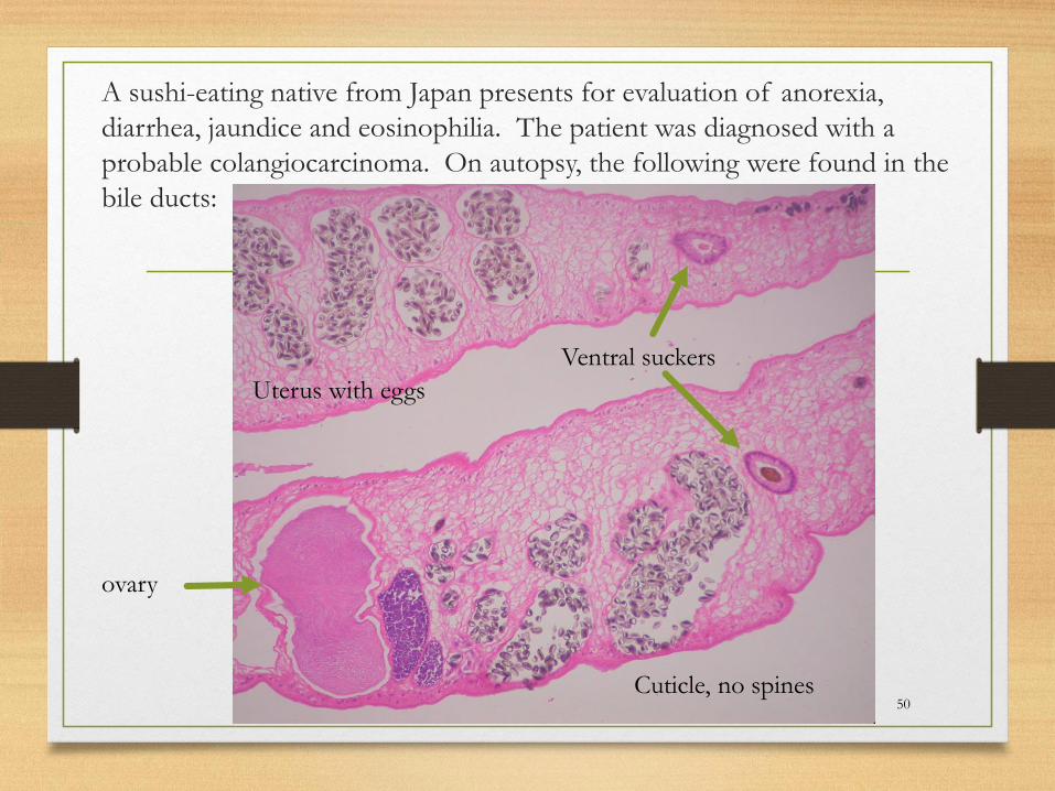
A sushi-eating native from Japan presents for evaluation of anorexia,
diarrhea, jaundice and eosinophilia. The patient was diagnosed with a
probable colangiocarcinoma. On autopsy, the following were found in the
bile ducts:
50
ovary
Uterus with eggs
Cuticle, no spines
Ventral suckers
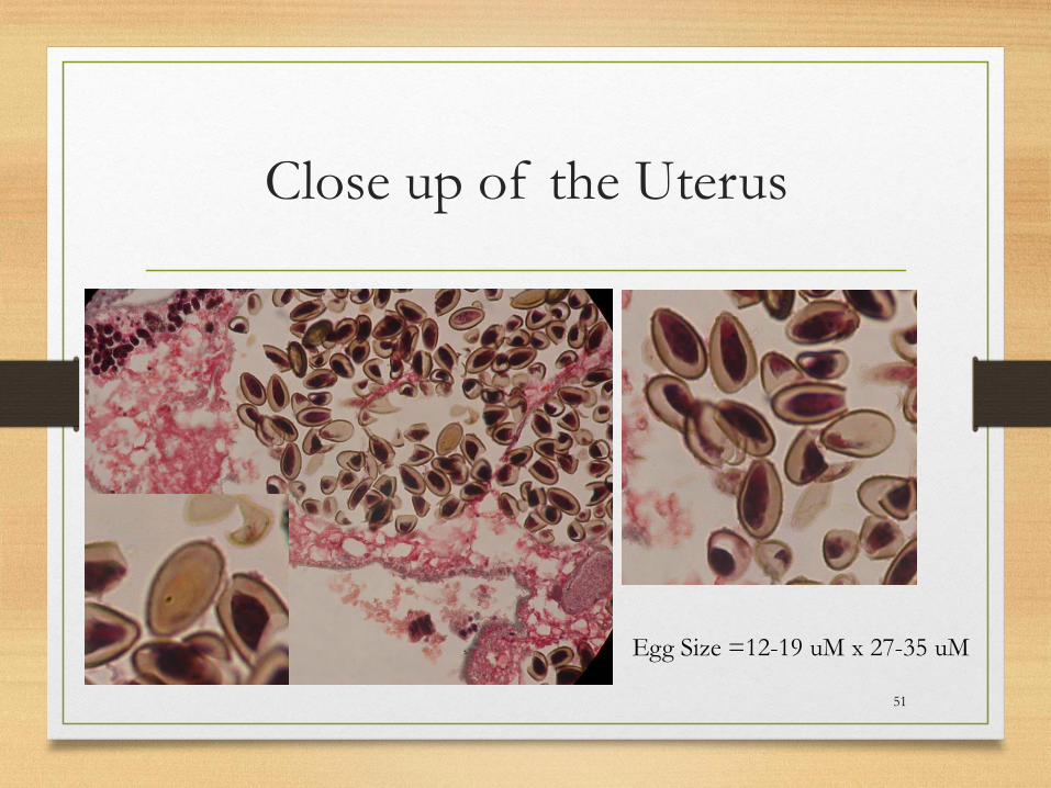
Close up of the Uterus
51
Egg Size =12-19 uM x 27-35 uM
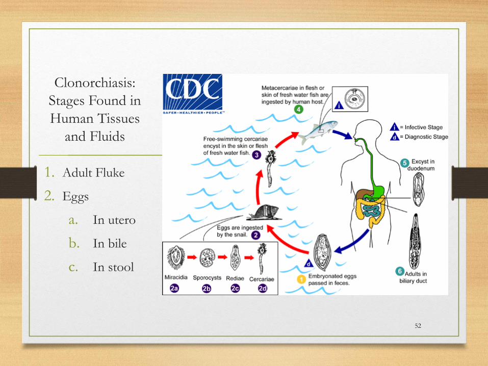
Clonorchiasis:
Stages Found in
Human Tissues
and Fluids
1. Adult Fluke
2. Eggs
a. In utero
b. In bile
c. In stool
52
53
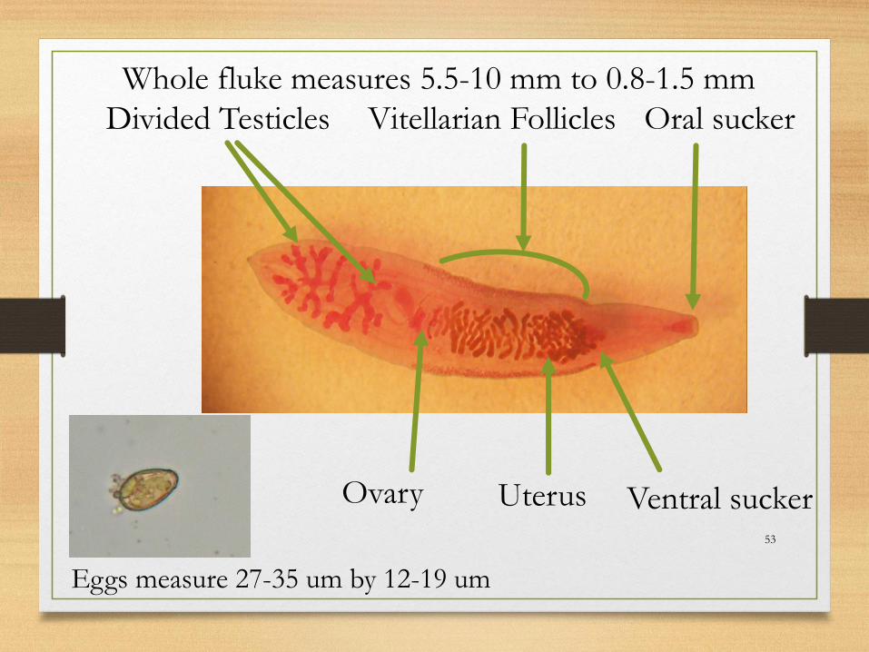
Divided Testicles Vitellarian Follicles
Ovary Uterus
Oral sucker
Ventral sucker
Whole fluke measures 5.5-10 mm to 0.8-1.5 mm
Eggs measure 27-35 um by 12-19 um
Clonorchiasis Summary
• Adult flukes generally live in liver bile ductules, rarely in the bile or pancreatic ducts
• Adults are “small” 5.5-10 mm long by 0.8-1.5 mm wide
• Flukes have vitellaria, ovaries, testes and a uterus containing eggs, and suckers
• Clonorhis has no spines in the cuticle
• What you see depends on the cut through the fluke
• Eggs are seen in the uterus– measure up and look for morphology
• Eggs are shed in stool
54

A hunter sacked a bear and then fed it to his extended family as a substitute for
hamburger meat. Two days after the picnic, family members began
complaining of abdominal pain and diarrhea that persisted for several days and
then passed. The next week after the meal, relatives developed fever, myalgia,
and periorbital edema. CBC demonstrated eosinophilia. Symptoms were most
severe in those who at the burgers cooked rare. Muscle biopsy taken at >6mos.
55
Stitchosome =
anterior, multi-
celled end of
the larva
containing the
embedded
esophagus50x

Trichinellosis:
Stages Found in
Human Tissues
and Fluids
1. Adult worms
2. Larvae
56
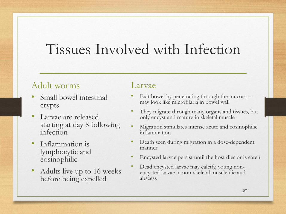
Tissues Involved with Infection
Adult worms
• Small bowel intestinal crypts
• Larvae are released starting at day 8 following infection
• Inflammation is lymphocytic and eosinophilic
• Adults live up to 16 weeks before being expelled
Larvae• Exit bowel by penetrating through the mucosa –
may look like microfilaria in bowel wall
• They migrate through many organs and tissues, but only encyst and mature in skeletal muscle
• Migration stimulates intense acute and eosinophilic inflammation
• Death seen during migration in a dose-dependent manner
• Encysted larvae persist until the host dies or is eaten
• Dead encysted larvae may calcify, young non-encysted larvae in non-skeletal muscle die and abscess
57

Trichinellosis Summary
• Striated (skeletal) muscle with encapsulated larvae –think Trichinella species
• Small bowel with small worms in crypts, but no eggs, think Trichinella species – look for the stitchosome too!
• Eosinophilia should make you think of any migrating helminth
• Small abscesses in multiple tissues other than striated muscle should make you think of trichinellosis
58
59
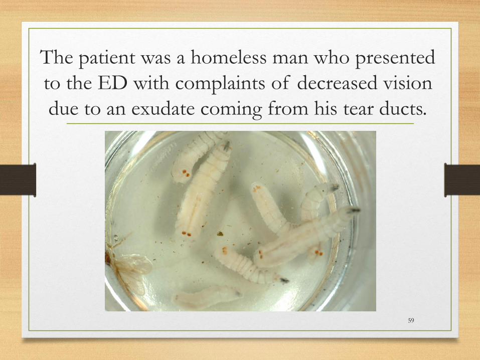
The patient was a homeless man who presented
to the ED with complaints of decreased vision
due to an exudate coming from his tear ducts.
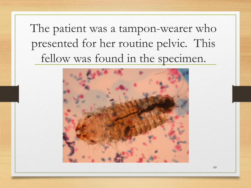
The patient was a tampon-wearer who
presented for her routine pelvic. This
fellow was found in the specimen.
60
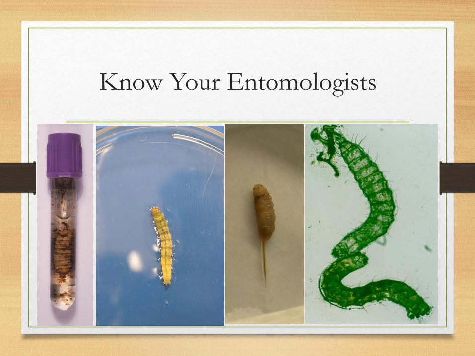
Know Your Entomologists
61
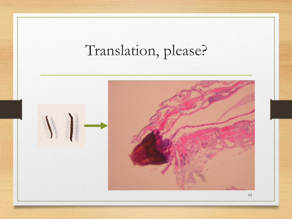
Translation, please?
62
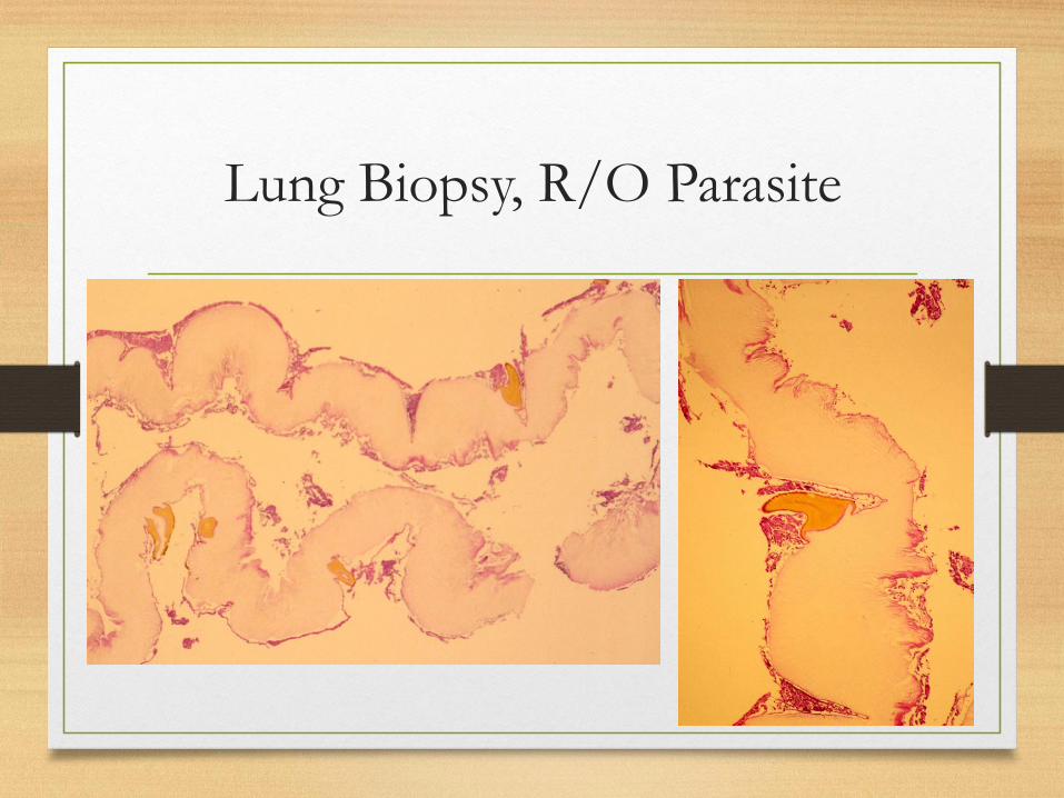
Lung Biopsy, R/O Parasite
63
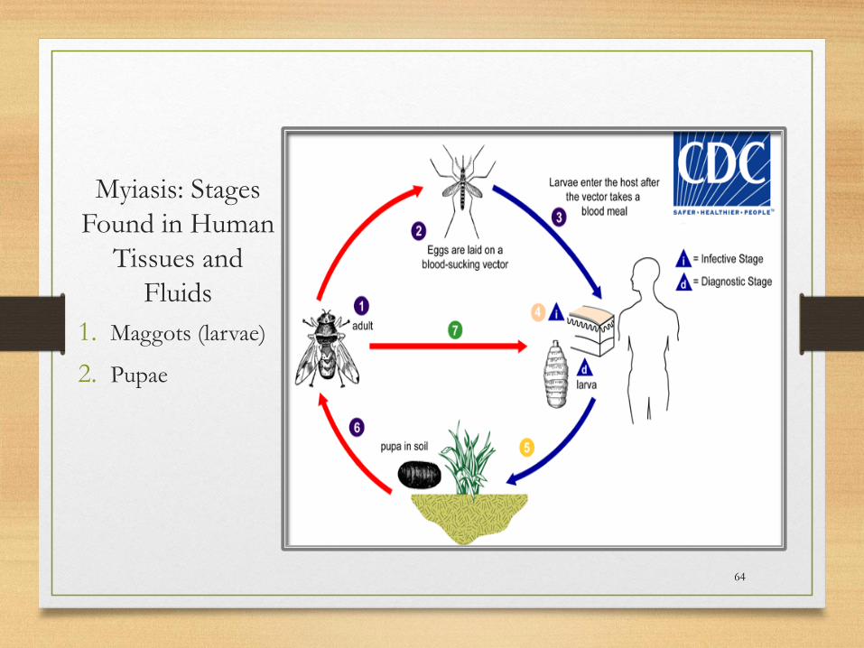
Myiasis: Stages
Found in Human
Tissues and
Fluids
1. Maggots (larvae)
2. Pupae
64
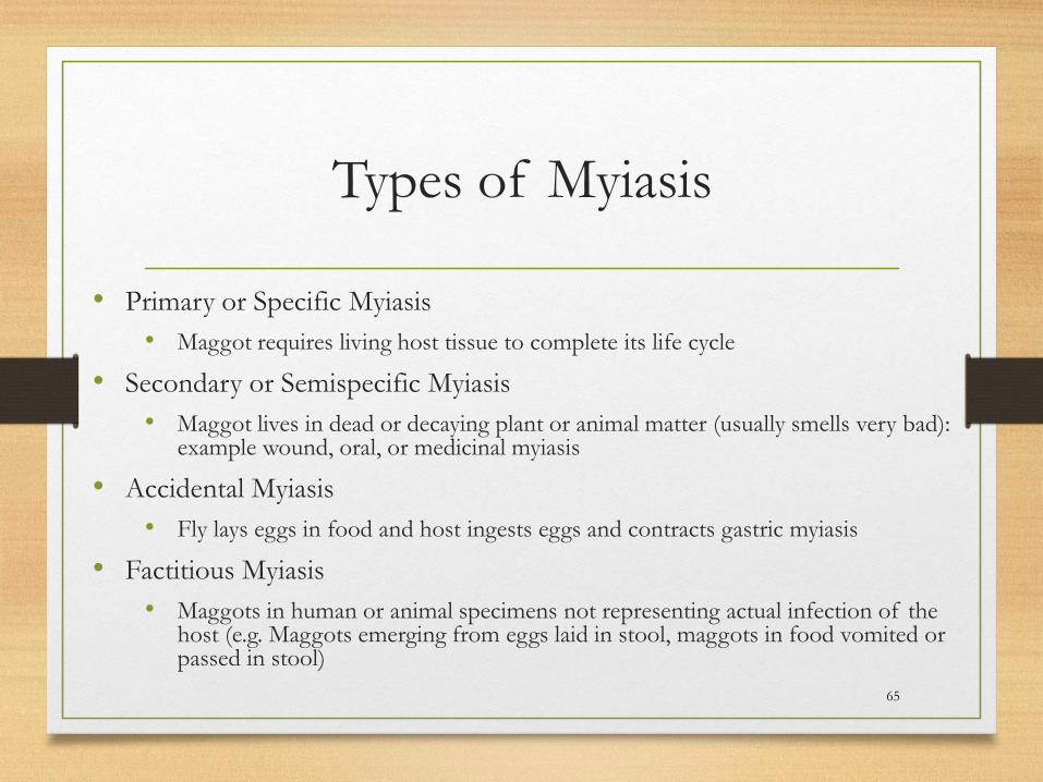
Types of Myiasis
• Primary or Specific Myiasis
• Maggot requires living host tissue to complete its life cycle
• Secondary or Semispecific Myiasis
• Maggot lives in dead or decaying plant or animal matter (usually smells very bad): example wound, oral, or medicinal myiasis
• Accidental Myiasis
• Fly lays eggs in food and host ingests eggs and contracts gastric myiasis
• Factitious Myiasis
• Maggots in human or animal specimens not representing actual infection of the host (e.g. Maggots emerging from eggs laid in stool, maggots in food vomited or passed in stool)
65
Myiasis Summary
• Entomologists are your friends as long as the maggot is intact
• Maggots have pigmented spines that can be seen coming from the larval cuticle- may be a clue to dx
• Not all “worms” seen in clinical specimens represent true parasitism
• Not all structures submitted as worms are indeed worms…..
66
67
Take Home Messages From these Cases
• Microbiologists can contribute to anatomic diagnoses
• Develop a differential based on tissue/location
• Develop a differential based on stages of the parasite
seen
• Measure up (eggs and worms alike)
• Eosinophilia does not accompany all parasite infection
• Knowing parasite life cycles can be useful
References
• Thomas Orihel and Lawrence Ash. 1995. Parasites
in Human Tissues. ASCP Press.
• Lawrence Ash and Thomas Orihel. 2007. Atlas of
Human Parasitology. ASCP Press.
• www.CDC.gov
68